Article

Low-Temperature Synthesis and Catalytic Activity of Cobalt Ferrite in Nitrous Oxide (N2O) Decomposition Reaction

Laboratory for Synthesis, Research and Testing of Catalytic and Adsorption Systems for Hydrocarbon Processing, Ivanovo State University of Chemistry and Technology, 153000 Ivanovo, Russia
*
Authors to whom correspondence should be addressed.
Submission received: 28 April 2021 / Revised: 15 July 2021 / Accepted: 18 July 2021 / Published: 22 July 2021
(This article belongs to the Special Issue New Horizons for Heterogeneous Catalysts)

Abstract

:
Cobalt ferrite (CoFe2O4) nanoparticles were synthesized and investigated as a catalyst in the reaction of nitrous oxide (N2O) decomposition. Cobalt ferrite was synthesized by solid–phase interaction at 1100 °C and by preliminary mechanochemical activation in a roller-ring vibrating mill at 400 °C. The nanoparticles were characterized by X-ray diffraction (XRD), synchronous thermal analysis (TG and DSC) and scanning electron microscopy (SEM). A low-temperature nitrogen adsorption/desorption test was used to evaluate the catalytic activity of the cobalt ferrite nanoparticles. Correlations between the structure and catalytic properties of the catalysts are reported. The highest catalytic activity of CoFe2O4 in the reaction of nitrous oxide decomposition was 98.1% at 475 °C for cobalt ferrite obtained by mechanochemical activation.

1. Introduction

Cobalt ferrite attracts the attention of many researchers for its unique properties that allow this compound to be used in various industries. Nanoparticles of cobalt ferrite are widely used as magnetic materials [1,2], biosensors [3], ferromagnetic fluids [4,5], catalysts, and photocatalysts [6,7].
It is known that materials used in different industries must have different properties. Ferrites, used as catalysts, should have a high surface area and a developed porous structure, as well as the necessary combination of structural and mechanical properties. These properties of the product mainly depend on the method of its synthesis. For the cobalt ferrite synthesis, various methods are used: solid-phase synthesis [8,9], co-precipitation [10], hydrothermal [11,12] and sol-gel synthesis [13,14], the Massart method [15], and microwave synthesis [16,17]. These methods have some drawbacks. Thus, solid-phase synthesis requires high temperatures and long calcination, which does not allow obtaining the product in a highly dispersed state. Methods based on obtaining ferrites from salt solutions are very laborious and multistage. They require strict control of the parameters of the process (temperature, pH, etc.), and there are large amounts of wastewater. Mechanochemical synthesis allows excluding most of these disadvantages. This simple and fast method of synthesis makes it possible to obtain a large amount of nanostructured powder [18,19]. There are some data about the mechanochemical synthesis of different ferrites [20,21,22,23,24]. Cobalt ferrite nanoparticles are proposed to be obtained by two-stage mechanochemical synthesis [24,25]: co-precipitation and subsequent mechanochemical grinding of co-precipitated precursors. Cu-Co ferrites are obtained by a similar method [25]. Cobalt ferrite supported on SiO2 [20] is proposed to be produced by a joint mechanical activation and further heat treatment of the 6Fe2O3-2Co3O4-Si system. CoFe2O4 is also obtained by mechanical grinding of previously prepared layered double cobalt hydroxycarbonate (CoFe2(OH)4(CO3)2·nH2O) [19]. Calcium hydroxide (Co(OH)2) and iron oxalate (FeC2O4·2H2O) are used as precursors for the mechanochemical synthesis of cobalt ferrite [23]. Analysis of the methods for obtaining highly dispersed cobalt ferrites under nonequilibrium conditions shows that the use of mechanochemical synthesis leads to the formation of crystalline cobalt ferrite.
As noted earlier, ferrites are widely used in catalysis. They are catalysts for a number of processes, such as the catalytic oxidation of chlorobenzene [6], the decomposition of cyclohexane [26], the dehydration of ethylbenzene [27], the oxidation of alkenes [28], etc. A promising direction of their use is the process of gas emissions neutralizing, since they can successfully replace the catalysts containing noble metals such as platinum, palladium, or gold [29].
These facts make cobalt ferrite a promising material that can be widely used. However, there is a lack of data on the effect of precursors on the formation of the crystalline phase of cobalt ferrite and its physicochemical and catalytic properties.
Therefore, the aim of the investigation was to study the system CoC2O4·2H2O-FeC2O4·2H2O during mechanical activation and heat treatment, as well as to identify the structural, physicochemical and catalytic properties of the cobalt ferrites in the N2O decomposition reaction.

2. Results

Low-temperature production of ferrites from Co and Fe metal oxalates. The calcination of a mixture of iron and cobalt oxalates subjected to mechanical activation at a temperature of 300 °C.
Since the temperature of CoFe2O4 formation is low, a developed porosity and a high specific surface area should be expected.
Cobalt ferrite exhibits high catalytic activity in the decomposition reaction of nitric oxide (I).

3. Discussion

3.1. Structural Characteristics of the Reagents

On the basis of X-ray phase analysis, it is found that the process of mechanical activation from 1 to 45 min is accompanied by a gradual decrease in the integral intensity of the reflections of the iron oxalate phase, which indicates its amorphization. In addition, the size of the iron oxalate crystallites decreases from 195 Ǻ (for the original FeC2O4·2H2O) to 88 Ǻ after 45 min of mechanical activation. On the contrary, the microstrain value increases from 0.1 to 0.82% (Table 1). It is necessary to note that the size of coherent scattering region (CSR) is smaller than the crystallite size, since it does not include the outer amorphized crystallite layers, and the microstrain value includes all lattice distortions caused by point (Frenkel and Schottky defects) and linear (edge and screw dislocations) and other types of defects [30]. Characteristic reflections of cobalt oxalate are not observed, this fact indicates its amorphous state. The formation of new phases at the stage of mechanical activation is not recorded.
The distributions of particle sizes of the initial components and products of mechanical activation are presented in Figure 1. The mixture subjected to mechanical activation consists mainly of particles of size up to 3 μm, from 3 to 25 μm and from 25 to 160 μm. The nature of the particle size distribution indicates that in the process of intensive mechanical actions, the minimum particle size is reached quickly, and the grinding process practically stops. The aspiration of the system to decrease the excess of free energy leads to the prevalence of secondary aggregation processes. Thus, the microstructure of the product is formed as a result of two opposite processes: reducing the size of individual particles during their destruction, and the formation of aggregates from these particles [31,32].

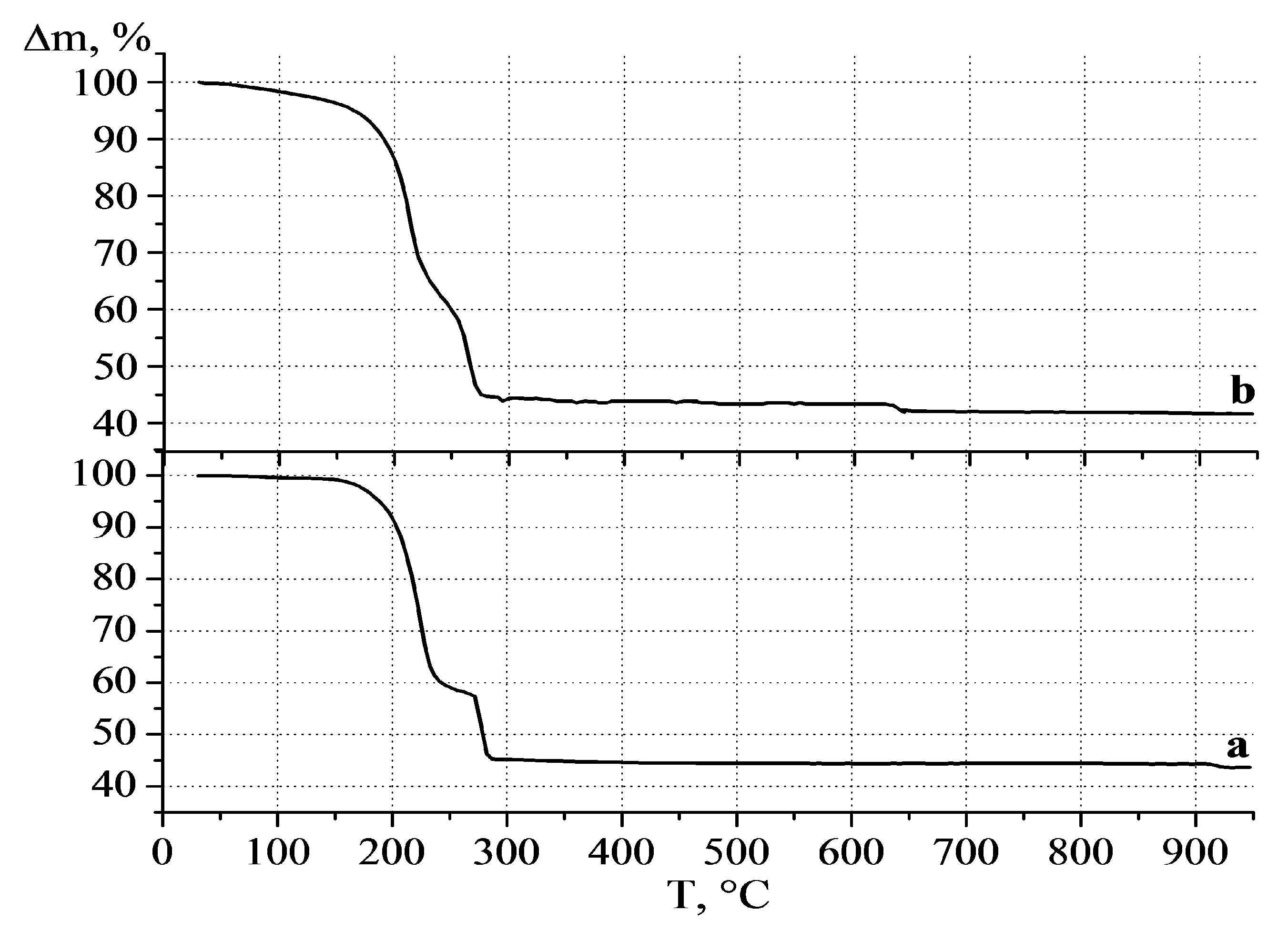
3.2. Samples Thermal Behavior

The calcination of a mixture of iron and cobalt oxalates subjected to mechanical activation at a temperature of 300 °C leads to the appearance of the γ-Fe2O3 phase (Figure 2 (a)). An increase in calcination temperature to 350 °C (Figure 2 (b)) does not lead to significant changes. However, the temperature increasing up to 400 °C leads to the appearance of a group of reflections which are characteristic for cobalt ferrite. Calcination of the samples at 450 °C leads to an increase in integral intensity of the CoFe2O4 reflections (Figure 2 (c)), so the ongoing crystallization processes are observed.
XRD patterns of the calcined samples prepared by solid-phase synthesis without preliminary mechanochemical treatment are shown in Figure 3 (a–d). Calcination at 300 °C leads to the decomposition of iron and cobalt oxalates and formation of amorphous mixture of iron and cobalt oxides, respectively (Figure 3 (a)). Heat treatment at 350 °C leads to the appearance of reflections characteristic for γ-Fe2O3, which indicates its crystallization (Figure 3 (b)). An increase in temperature to 450 °C leads to a transition of γ-Fe2O3 to α-Fe2O3 and the appearance of a crystalline phase of cobalt oxide Co3O4 (Figure 3 (c)). Further calcination at a temperature of 450–850 °C leads only to crystallization of iron and cobalt oxides (Figure 3 (d,e)). The formation of monophasic cobalt ferrite is observed only at a temperature of 1100 °C (Figure 3 (f)).
Calcination of the sample obtained by solid-phase synthesis without prior preliminary mechanochemical treatment is accompanied by six thermal effects (Figure 4 (a)). Zone I—endothermic effect, lying in the temperature range of 130–215 °C, is associated with the removal of crystallization water. Zones II and III—exothermic effects that lie in the temperature ranges of 215–255 °C and 265–295 °C. They are associated with the decomposition of iron and cobalt oxalates, as well as simultaneous carbon monoxide oxidation:
2FeC2O4(s) + 0.5O2 → Fe2O3(s) + 2CO+ 2CO2 (g) ↑
3CoC2O4(s) → Co3O4(s) + 4CO+ 2CO2 (g) ↑
Zone IV—exothermic effect, lying in the temperature range of 350–410 °C, is due to the transition of γ-Fe2O3 to α-Fe2O3. Zone V—endothermic effect, observed in the temperature range of 900–940 °C, is associated with decomposition of cobalt oxide Co3O4:
Co3O4 → 3CoO + 0.5O2
The TG curve of this sample (Figure 5 (a)) shows clearly pronounced step-wise mass losses due to the removal of crystallization water, decomposition of oxalates, and oxygen emissions from the Co3O4 structure.
The mechanochemical activation of the oxalates mixture leads to a change in the shape of the DSC curve (Figure 4 (b)). The thermal effects of the decomposition of iron oxalate (II) and cobalt (II) oxalates are shifted by 8 and 12 °C, respectively, towards lower temperatures. Gradual mass loss is also observed on the TG curve, but it has a flatter form. The IV thermal effect lying in the temperature range of 300–525 °C is caused by the ongoing crystallization of cobalt ferrite (CoFe2O4). The endothermic effect in the temperature range of 650–900 °C is due to the emission of oxygen from CoFe2O4 structure, as further indicated by the mass loss observed on the TG curve (Figure 5 (b)). Obviously, when mechanically activating, along with grinding and intensive mixing, there is also the accumulation of excess energy in the form of defects in the crystal structure. This helps to reduce the temperature of decomposition of the initial components and the temperature of formation of the product—cobalt ferrite.

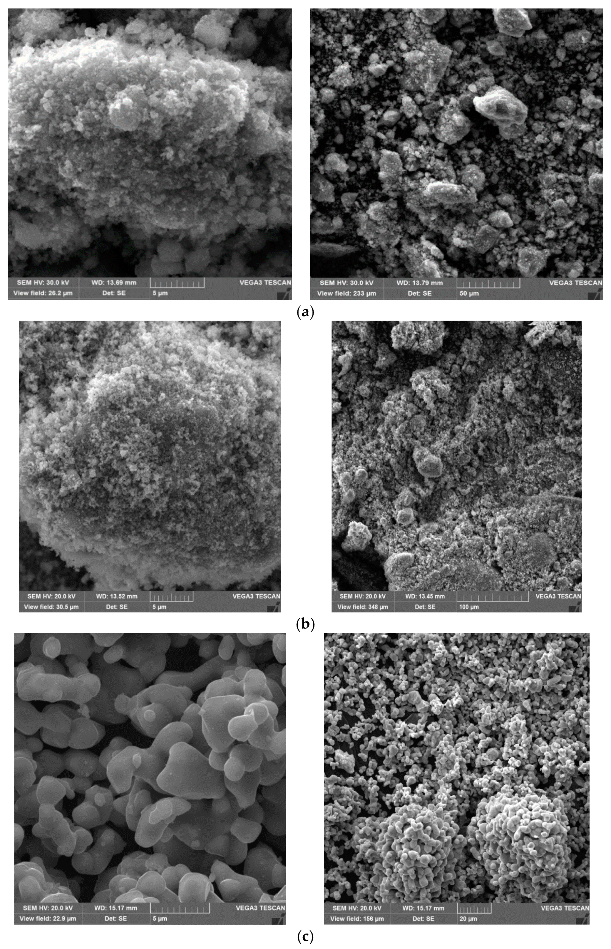
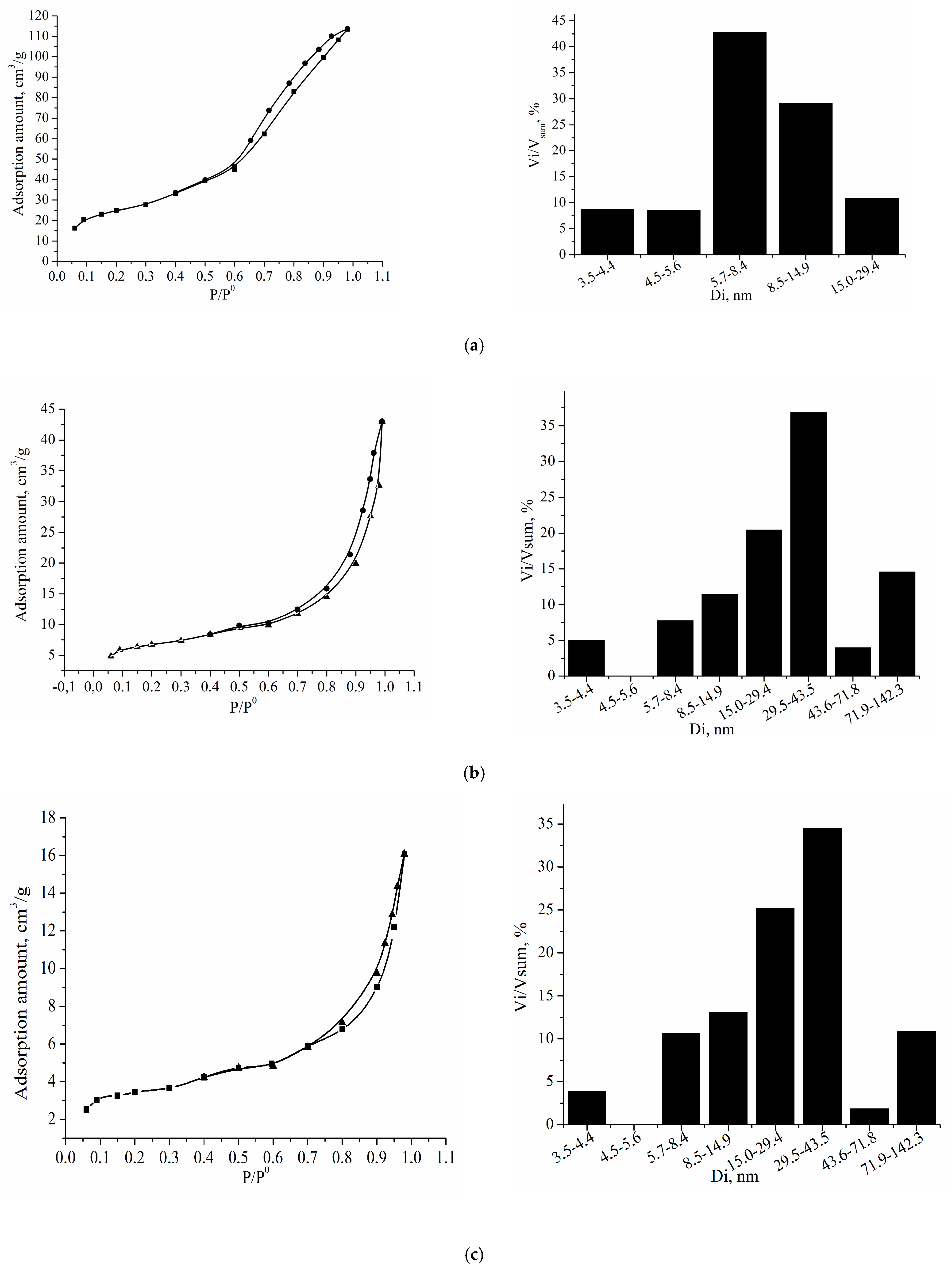
3.3. Morphology and Structural Characteristics

The synthesized cobalt ferrites obtained by the method of mechanochemical synthesis (Figure 6) are characterized by a hierarchical structure and consist of spherical particles with a size of 0.1 μm. Spherical nanocrystals agglomerate into larger porous particles of regular spherical shape with a size of 5–25 μm (Figure 6a).
An increase in the calcination temperature leads to an increase in the size of both the particles themselves and the aggregates they formed (Figure 6b). The cobalt ferrite obtained by the solid-phase synthesis without preliminary mechanochemical activation (Figure 6c) consists of spherical particles with pronounced morphology. Their size changes from 2.5 to 5 µm. A high heat treatment temperature (1100 °C) leads to sintering of the particles into larger aggregates (Figure 6c).
Table 2 summarizes the characteristics of cobalt ferrites obtained by mechanochemical and solid-phase methods.
Cobalt ferrite has a cubic crystal lattice with, according to the database [33], parameters a = b = c = 8.391 Ǻ. The calculated lattice parameters of cobalt ferrite obtained by solid-phase synthesis without preliminary mechanical activation at a temperature of 1100 °C coincide with the literature data (Table 2). Cobalt ferrite obtained by solid-phase synthesis with preliminary mechanical activation of the initial components has slightly distorted lattice parameters (Table 2). Obviously, mechanical activation leads to the formation of cobalt ferrite with a defective structure, as further indicated by calculation of root-mean-square microstrain, for which the value for samples obtained using mechanochemical synthesis is 2–3 times higher than that of the sample obtained by conventional solid-phase synthesis (Table 2).
The average crystallite size, determined from the values of the X-ray reflections broadening, varied in the range of 175 to 299 Ǻ depending on the calcination temperature of 300–450 °C. The data given in Table 2 indicate that with an increase in the synthesis temperature, the perfection of the crystal structure also increases, and the broadening of the diffraction lines of the samples synthesized at high temperatures relates mainly to the small size of the crystallites (about 20–30 nm).
The important characteristics of catalytic systems are specific surface area and porosity. An increase in the synthesis temperature leads to a decrease in these parameters (Table 2). The isotherms of nitrogen adsorption/desorption of samples obtained by mechanical activation are shown in Figure 7a–c. These isotherms can be attributed to type IV; such isotherms are characteristic of bodies that have predominantly transitional pores (mesopores) according to Dubinin’s classification; that is, pores with a diameter of tens to hundreds of angstroms [34]. A sample calcined at 350 °C has only mesopores with an average diameter of 3.5 to 29.4 nm. According to estimation, 71.92% of the total pores fraction has a diameter of 5.9 to 14.9 nm (Figure 7a).
With an increase in the calcinations temperature, a decrease in the specific surface area and the total pore volume is observed (Table 2), which is associated with the gradual sintering of the samples, as a result of which the particles become coarse and form larger and more tightly interconnected aggregates from them.
An increase in the calcination temperature to 400–450 °C leads to an increase in the content of larger mesopores and the appearance of macropores (Figure 7b,c). For example, a sample calcined at 400 °C has pores with a diameter from 15 to 43.6 nm. They make up 57.29% of the total fraction of pores (Figure 7b). With an increase in the calcination temperature, a decrease in the specific surface area and total pore volume are observed (Table 2). It is associated with the gradual sintering of the samples. As a result, the enlargement of particles takes place and the larger and tightly interconnected aggregates are formed. It is established that the sample obtained without the use of mechanochemical treatment has a very low specific surface area of 0.6 m2/g and is non-porous. This is confirmed by the SEM images of this sample, which show that the powder particles have a smooth surface and grow into dense aggregates that do not have pores (Figure 6c).

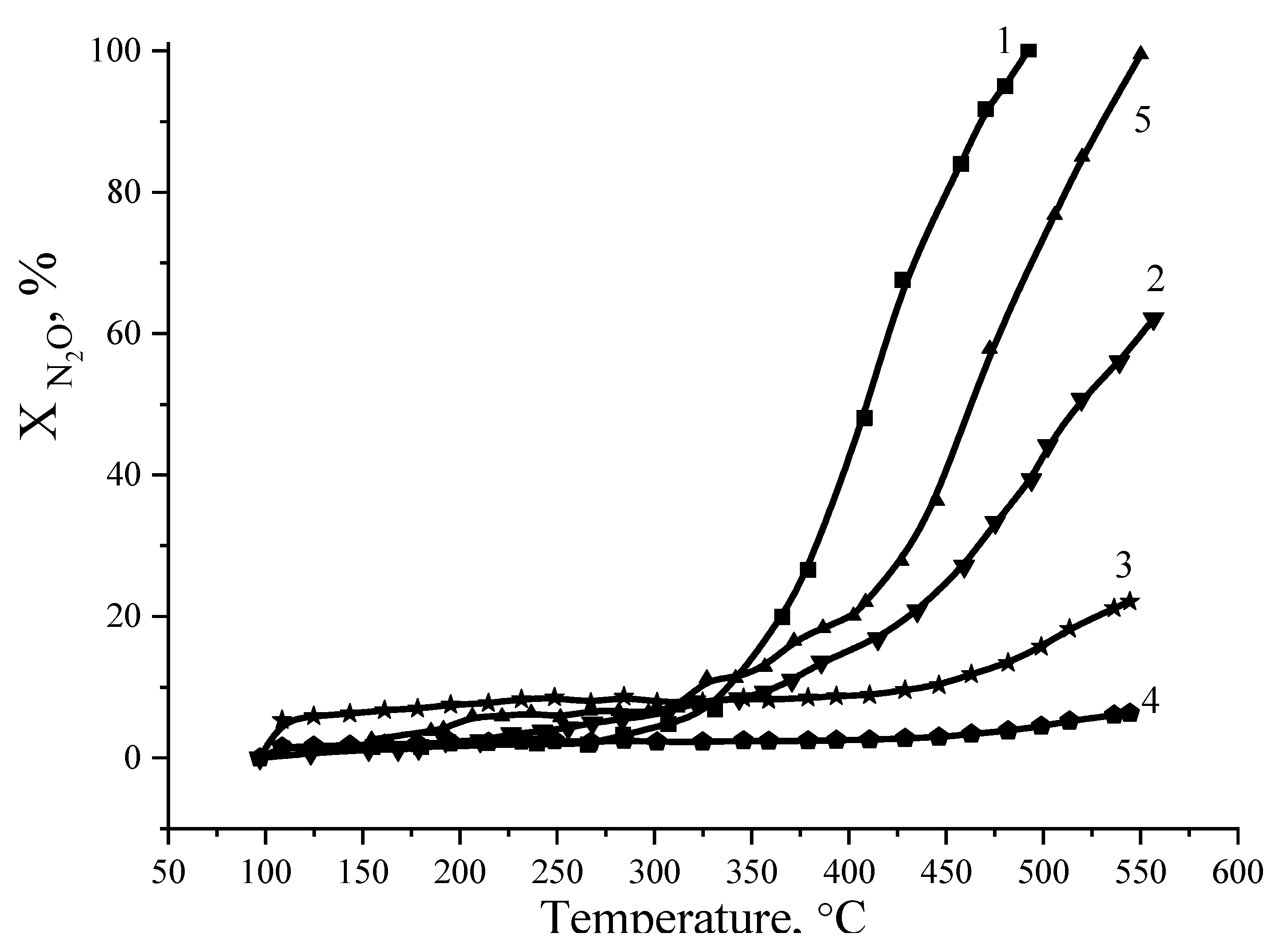
3.4. Catalytic Properties of Cobalt Ferrite

Under the best conditions, the activity of the catalyst depends on the number of active sites per unit mass of the active component. Mechanochemical synthesis provides quantitative yield in a relatively short reaction time, in contrast to solid–phase interaction. In the case of solid–phase interaction, the catalyst has a high temperature of synthesis 500 °C, hydration, sorption and low catalytic activity of N2O decomposition reaction. As a result, processes with mechanochemical activation are usually cost-effective and make it possible to obtain materials with high mechanical strength, a degree of homogeneity, and a low catalyst start-up temperature 250 °C.
Studies on the catalytic activity in the decomposition of nitrous oxide show a clear correlation between the specific surface of the samples and the degree of N2O decomposition (Figure 8). The maximum degree of decomposition is observed for the sample obtained by calcining the mechanically activated mixture at 350 °C. The degree of N2O decomposition at 475 °C is 98.10%. The catalytic activity of the sample calcined at 450 °C under the same conditions is significantly lower (the degree of N2O decomposition is only 12.87%) Thus, one can state that the observed dramatic activity decrease from the calcination temperature increase from 400 to 1000 °C was due to the decomposition of the spinel structure. In other words, it was due to the disappearance of the Co3+-Co2+ redox couple necessary for N2O decomposition. Further increase in the calcination temperature of the sample to 1100 °C leads to reduction of N2O decomposition up to 7.5% at 550 °C. It should be noted that monophase cobalt ferrite during solid-phase synthesis is formed at temperatures of 1000–1100 °C. Its activity (XN2O = 5.5% at temperature 550 °C) in the decomposition of nitrogen oxide (I) is close in value to the sample obtained by mechanochemical synthesis and further heat treatment at 1100 °C.
The mechanism of the reaction between N2O and the catalysts’ active centers is generally thought to be a charge transfer from the catalyst to the vacant orbitals of N2O, which destabilizes the N–O bond and leads to bond breaking [35,36,37,38]. Therefore, electron charge transfer from the metal ion to the N2O molecule is a crucial step for N2O decomposition [35,36,37,38]. Electron transfer occurs from a low oxidation state metal cation, which subsequently increases its oxidation state. The possibility of the ion reduction from a high to a low oxidation state is also important for the regeneration of the active centers. Hence, the activity of the Co3O4 oxide spinel is attributed to the coexistence of a Co2+ → Co3+ ion pair because of an easy one-electron transfer between these ions during N2O decomposition, as shown in the following mechanism [35,36,37,38]:
N2O(g) + Co2+ → N2O(ads.) … Co3+
O(ads.) … Co3+ → O2(g) + Co2+
As earlier studies of catalysts based on ferrites showed [39], the presence of oxygen, water, and nitrogen in the reaction gas mixture resulted in blocking of the catalyst active sites and a shift in the temperature of the onset of decomposition towards higher temperatures.
In the available publications, the authors test catalysts under different conditions (N2O content, gas space velocity, and others). All these parameters affect the catalytic activity. The AVC-10 alumina-vanadium catalyst used in large-scale nitric acid production (stage of gas purification from nitrogen oxides) was chosen as the object of comparison. It was found that the degree of decomposition of N2O increases with the temperature increase, and it is % at 550 °C. The specific surface area of the AVK-10 catalyst is 158 m2/g, and the best sample of cobalt ferrite is 78.9 m2/g. Obviously, during the decomposition of N2O, not only the size of the catalyst surface area has an influence, but also the valence of the active catalyst component. For vanadium oxide V2O5, the activity is much lower than for cobalt ferrite.
This can be explained by a change in the mechanism of the catalytic reaction, since vanadium has a higher valence.
In this case, decomposition occurs with isolated transition metal ions [V5+]:
N2O + [V5+] → N2 + [V5+]O
N2O + [V5+]O → N2 + O2 + [V5+]
It can be assumed that this catalyst also undergoes thermal decomposition due to the adsorption of N2O in the pores of the catalyst support—(Al2O3).

4. Materials and Methods

4.1. Raw Materials

All reagents were commercially available and were used without further purification.
Cobalt oxalate (CoC2O4·2H2O)—99.5 wt. %, (Fe2O3—0.25 wt. %, NiO—0.15 wt. %, and CuO—0.10 wt. %, as impurities). The particle size of cobalt oxalate is up to 25 μm (Figure 1 (a));
Iron oxalate (FeC2O4·2H2O)—99.0 wt. %, (Fe2O3—0.32 wt. %, CaO—0.25 wt. %, MgO—0.23 wt. % and K2O—0.2 wt. % as impurities). The particle size of iron oxalate is up to 100 μm (Figure 1 (b)).
The industrial catalyst for selective reduction of NOx AVC-10 is used as an object of comparison of catalytic properties. This type of catalyst is used in industrial units for the production of nitric acid at the stage of gas purification from nitrogen oxides.
The main characteristics of the catalyst:
  • The composition of the catalyst is Al2O3—98% wt., V2O5—10% wt.
  • The catalyst surface area is 158 m2/g.
  • Total pore volume—177, cm3/g.

4.2. Samples Preparation

The following methods were used for cobalt ferrite preparing: Solid-phase synthesis and joint mechanical activation.
Solid-phase synthesis. A mixture of cobalt oxalate and iron oxalate in a molar stoichiometric ratio was heat treated in a muffle furnace at the temperature range of 300–1100 °C for 4 h.
Joint mechanical activation. A mixture of cobalt oxalate and iron oxalate in a molar stoichiometric ratio was mechanochemically treated for 45 min and then heat treated at the temperature range of 300–450 °C. Mechanochemical treatment was carried out in a roller-ring vibrating mill VM-4. The diameter of grinding chamber, total volume of the chamber, oscillation frequency, amplitude, mass of grinding bodies, and mass of the loaded material were 98 mm, 0.302 L, 930 min−1, 10 mm, 1194 g, and 50 g, respectively.

4.3. Testing Procedures

Powder X-ray diffraction (XRD) spectroscopy. The patterns were recorded on DRON-3M (Russia) X-ray diffractometer. The CuKα radiation (λ = 0.15406 nm, Ni filter) was used with a power supply—40 kV and 20 mA, scanning rate—2 °/min, initial slit—2 mm, detector slit—0.25 mm.
Thermogravimetry (TG) and differential scanning calorimetry (DSC). The measurements were provided by the STA 449 F3 Netzsch (Germany) device under air atmosphere. The heating rate—5 °C/min.
The surface area was determined by the BET method for low-temperature adsorption-desorption of nitrogen. The measurements were provided by the Sorbi-MS (Russia) device. Before the test, the samples were dried in a nitrogen stream at a temperature of 250 °C for 60 min.
Scanning electron microscopy (SEM). Microphotographs were provided by the Vega 3 Tescan device (Czech Republic).
Catalytic activity. Samples were tested in a N2O decomposition test. The tests were carried out using the catalytic complex PKU-2VD (Russia), the volumetric gas velocity was 20,000 h−1. The composition of the reaction gas mixture was used according to the 1% mass. N2O and 99% mass. N2. The temperature in the reactor ranged from 100 to 550 °C. Gas chromatograph Krystallux 4000 M (Russia) with thermal conductivity detectors was used for analysis of the reaction products. Columns with NaX and HayeSep Q adsorbents were used to separate the gas mixture. The temperature in the columns was 65 °C.

4.4. Qualitativex-Rayanalysis

The interplanar distances (d), the dimensions of the coherent scattering region (CSR) were calculated from XRD data.
The identification of crystalline phases on the diffractograms was carried out by comparing the calculated interplanar distances with those given in the ASTM database. The interplanar distances were calculated according to the Bragg equation [40]:
d = λ/2sinΘ,
where λ is the wavelength(nm), Θ = Xc/2 is the diffraction angle, calculated as the gravity center position of reflex [41]:
X c = + I   ( ϑ ) d ϑ I m a x   и л и   X c = i = 1 N 1 ( I i + I i + 1 ) Δ ϑ 2 I m a x ,
where I is the intensity at the diffraction angle ϑ and Imax is the maximum intensity.
The dimension of coherent scattering region (CSR) was calculated using the modified Scherrer’s equation [42], after linearization:
βph cosΘ = λ/DSCR + 4ε sinΘ,
where DSCR is the dimension of the coherent scattering region, e is the value of the relative mean square microdeformation (MD), βph is the integral physical broadening of the X-ray sample profile, calculated using the Gaussian’s function [43]:
β s 2 = β p h 2 + β i n s t 2
where β s 2 is the integral broadening of the X-ray profile of the sample, β i n s t 2 is the instrumental broadening. Broadening of the X-ray profile of the etalon was used as instrumental broadening.
Integral broadening of X-ray profile was calculated by the equation [41]:
β = + I   ( ϑ ) ϑ d ϑ + I   ( ϑ ) d ϑ   и л и   β = i = 1 N 1 I i ϑ i i = 1 N I i ,
The interplanar distances and corresponding Miller’s indices were used to calculate the unit cell parameters. For cubic syngony [32]:
1 d 2 = h 2 + k 2 + l 2 a 2
where d is the interplanar distance, a, c are the unit cell parameters, h, k, l are the Miller’s indices.
Specific energy (E) was calculated according to the method proposed in [44]. The specific energy is only pure energy that is applied from the grinding media to the powder. This value does not include the loss of energy in the various mechanical components of the mill.

5. Conclusions

Thus, the preliminary mechanochemical treatment of the initial components makes it possible to decrease the temperature of the synthesis of monophasic cobalt ferrite three times compared with the solid–phase interaction of ones that have not been preactivated. Since the formation of new phases does not occur during mechanochemical activation, it is obvious that the main channel of relaxation of the supplied energy is the accumulation of defects in the structure of iron and cobalt oxalates, which in turn leads to the intensification of their decomposition processes.
The high homogeneity of the system subjected to preliminary mechanochemical activation contributes to a more complete and intensive interaction of the formed iron and cobalt oxides, which can also cause a decrease in the synthesis temperature. Cobalt ferrite obtained by mechanochemical synthesis has a more developed specific surface and a porous structure, which is explained by more “soft” conditions of its synthesis. Studies on the catalytic properties in the nitrous oxide decomposition show that cobalt ferrite exhibits catalytic activity in this reaction. The growth of catalytic activity is observed at 325 °C. The catalytic properties of cobalt ferrite are correlated with its surface area and porosity. The higher the porosity and specific surface area, the greater the degree of N2O decomposition.

Author Contributions

Conceptualization, A.A.I. and K.D.; methodology, J.S., R.R. and K.D.; validation, A.A.I., A.P.I. and N.G.; formal analysis, R.R., J.S., K.D. and A.A.I.; investigation, K.D. and J.S.; resources, K.D., A.A.I., R.R., J.S., A.P.I. and N.G.; data curation, A.A.I., R.R. and K.D.; writing—original draft preparation, K.D.; writing—review and editing, R.R., A.A.I., J.S., A.P.I. and N.G.; visualization, R.R.; supervision, N.G.; project administration, A.A.I., A.P.I. and R.R.; funding acquisition, N.G. All authors have read and agreed to the published version of the manuscript.

Funding

The work was funded by the project of the state assignment for the implementation of scientific research work (Topic No. FZZW 2020-0010).

Data Availability Statement

Data will be made available by authors on request.

Acknowledgments

When carrying out the research, the equipment of the Central Collective Use of the ISUCT was involved.

Conflicts of Interest

On behalf of all authors, the corresponding author states that there is no conflict of interest.

References

  1. Al Lehyani, S.H.A.; Hassan, R.A.; Alharbi, A.A.; Alomayri, T.; Alamri, H. Magnetic Hyperthermia using Cobalt Ferrite Nanoparticles: The Influence of Particle Size. Int. J. Adv. Technol. 2017, 8, 1–6. [Google Scholar] [CrossRef]
  2. López-Ortega, A.; Lottini, E.; Fernández, C.D.J.; Sangregorio, C. Exploring the Magnetic Properties of Cobalt-Ferrite Nanoparticles for the Development of a Rare-Earth-Free Permanent Magnet. Chem. Mater. 2015, 27, 4048–4056. [Google Scholar] [CrossRef]
  3. Kaçar, C.; Dalkiran, B.; Erden, P.E.; Kiliç, E. An amperometric hydrogen peroxide biosensor based on Co3O4 nanoparticles and multiwalled carbon nanotube modified glassy carbon electrode. Appl. Surf. Sci. 2014, 311, 139–146. [Google Scholar] [CrossRef]
  4. Tourinho, F.A.; Franck, R. Aqueous ferrofluids based on manganese and cobalt ferrites. J. Mater. Sci. 1990, 25, 3249–3254. [Google Scholar] [CrossRef]
  5. Cabuil, V.; Dupuis, V.; Talbot, D.; Neveu, S. Ionic magnetic fluid based on cobalt ferrite nanoparticles: Influence of hydrothermal treatment on the nanoparticle size. J. Magn. Magn. Mater. 2011, 323, 1238–1241. [Google Scholar] [CrossRef]
  6. Silva, J.B.; Diniz, C.F.; Lago, R.M.; Mohallem, N.D. Catalytic properties of nanocomposites based on cobalt ferrites dispersed in sol-gel silica. J. Non-Cryst. Solids 2004, 348, 201–204. [Google Scholar] [CrossRef]
  7. Abraham, A.G.; Manikandan, A.; Vadivel, S.; Jaganathan, S.K.; Baykal, A.; Renganathan, P.S. Enhanced magneto-optical and photo-catalytic properties of transition metal cobalt (Co2+ ions) doped spinel MgFe2O4 ferrite nanocomposites. J. Magn. Magn. Mater. 2018, 452, 380–388. [Google Scholar] [CrossRef]
  8. Rafferty, A.; Prescott, T.; Brabazon, D. Sintering behaviour of cobalt ferrite ceramic. Ceram. Int. 2008, 34, 15–21. [Google Scholar] [CrossRef] [Green Version]
  9. Khedr, M.; Omar, A.; Abdel-Moaty, S. Magnetic nanocomposites: Preparation and characterization of Co-ferrite nanoparticles. Colloids Surf. A Physicochem. Eng. Asp. 2006, 281, 8–14. [Google Scholar] [CrossRef]
  10. Kim, Y.I.; Kim, D.; Lee, C.S. Synthesis and characterization of CoFe2O4 magnetic nanoparticles prepared by temperature-controlled coprecipitation method. Phys. B Condens. Matter 2003, 337, 42–51. [Google Scholar] [CrossRef]
  11. Zhao, L.; Zhang, H.; Xing, Y.; Song, S.; Yu, S.; Shi, W.; Guo, X.; Yang, J.; Lei, Y.; Cao, F. Studies on the magnetism of cobalt ferrite nanocrystals synthesized by hydrothermal method. J. Solid State Chem. 2008, 181, 245–252. [Google Scholar] [CrossRef]
  12. Zhao, D.; Wu, X.; Guan, H.; Han, E. Study on supercritical hydrothermal synthesis of CoFe2O4 nanoparticles. J. Supercrit. Fluids 2007, 42, 226–233. [Google Scholar] [CrossRef]
  13. Meron, T.; Rosenberg, Y.; Lereah, Y.; Markovich, G. Synthesis and assembly of high-quality cobalt ferrite nanocrystals prepared by a modified sol–gel technique. J. Magn. Magn. Mater. 2005, 292, 11–16. [Google Scholar] [CrossRef]
  14. Gul, I.; Maqsood, A. Structural, magnetic and electrical properties of cobalt ferrites prepared by the sol–gel route. J. Alloy. Compd. 2008, 465, 227–231. [Google Scholar] [CrossRef]
  15. Massart, R. Magnetic Fluids and Process for Obtaining Them. U.S. Patent US4329241A, 11 May 1982. [Google Scholar]
  16. Bensebaa, F.; Zavaliche, F.; L’Ecuyer, P.; Cochrane, R.; Veres, T. Microwave synthesis and characterization of Co–ferrite nanoparticles. J. Colloid Interface Sci. 2004, 277, 104–110. [Google Scholar] [CrossRef] [PubMed]
  17. Ibrahim, A.M.; Mahmoud, M.M.; El-Latif, M.M. Microwave Synthesis of Cobalt-Ferrite Nano-Particles by Polyol Method. Process. Prop. Adv. Ceram. Compos. II 2010, 220, 17–26. [Google Scholar]
  18. Fecht, H.-J. Nanostructure formation by mechanical attrition. Nanostruct. Mater. 1995, 6, 33–42. [Google Scholar] [CrossRef]
  19. Manova, E.; Kunev, B.; Paneva, D.; Mitov, I.; Petrov, L.; Estournes, C.; D’Orléans, A.C.; Rehspringer, J.-L.; Kurmoo, M. Mechano-Synthesis, Characterization, and Magnetic Properties of Nanoparticles of Cobalt Ferrite, CoFe2O4. Chem. Mater. 2004, 16, 5689–5696. [Google Scholar] [CrossRef]
  20. Ennas, G.; Marongiu, G.; Marras, S.; Piccaluga, G. Mechanochemical route for the synthesis of cobalt ferrite–silica and iron–cobalt alloy–silica nanocomposites. J. Nanopart. Res. 2004, 6, 99–105. [Google Scholar] [CrossRef]
  21. Berchmans, L.J.; Karthikeyan, R.; Helan, M.; Berchmans, S.; Sepelak, V.; Becker, K.D. Mechanochemical Synthesis and Electrochemical Characterization of Nano Crystalline Calcium Ferrite. Catal. Lett. 2011, 141, 1451–1457. [Google Scholar] [CrossRef]
  22. Magaeva, A.A.; Naiden, E.P.; Terekhova, O.G.; Itin, V.I.; Verchekov, K.A.; Stadnichenko, A.I.; Boronin, A.I. Mechanochemical synthesis, phase composition, structural parameters, and magnetic properties of manganese ferrospinels. Nanotechnol. Russ. 2013, 8, 495–501. [Google Scholar] [CrossRef]
  23. Rumyantsev, R.; Il’In, A.A.; Denisova, K.O.; Il’In, A.P.; Volkova, A.V. Calcium Ferrite Structure Formation During Mechanochemical Interaction in the System FeC2O4∙2H2O–Ca(OH)2. Glas. Ceram. 2017, 73, 374–377. [Google Scholar] [CrossRef]
  24. Velinov, N.; Dimitrov, D.; Koleva, K.; Ivanov, K.; Mitov, I. Mechanochemical Synthesis and Characterization of Nanocrystalline Copper–Cobalt Ferrites. Acta Met. Sin. 2015, 28, 367–372. [Google Scholar] [CrossRef]
  25. Manova, E.; Paneva, D.; Kunev, B.; Estournès, C.; Rivière, E.; Tenchev, K.; Leaustic, A.; Mitov, I. Mechanochemical synthesis and characterization of nanodimensional iron–cobalt spinel oxides. J. Alloy. Compd. 2009, 485, 356–361. [Google Scholar] [CrossRef] [Green Version]
  26. Ramankutty, C.; Sugunan, S.; Thomas, B. Study of cyclohexanol decomposition reaction over the ferrospinels, A1−xCuxFe2O4 (A = Ni or Co and x = 0, 0.3, 0.5, 0.7 and 1), prepared by ‘soft’ chemical methods. J. Mol. Catal. A Chem. 2002, 187, 105–117. [Google Scholar] [CrossRef] [Green Version]
  27. Braga, T.P.; Sales, B.M.C.; Pinheiro, A.N.; Herrera, W.T.; Baggio-Saitovitch, E.; Valentini, A. Catalytic properties of cobalt and nickel ferrites dispersed in mesoporous silicon oxide for ethylbenzene dehydrogenation with CO2. Catal. Sci. Technol. 2011, 1, 1383–1392. [Google Scholar] [CrossRef]
  28. Kooti, M.; Afshari, M. Magnetic cobalt ferrite nanoparticles as an efficient catalyst for oxidation of alkenes. Sci. Iran. 2012, 19, 1991–1995. [Google Scholar] [CrossRef] [Green Version]
  29. Cota, H.M.; Katan, T.; Chin, M.; Schoenweis, F.J. Decomposition of dilute hydrogen peroxide in alkaline solutions. Nature 1964, 203, 1281. [Google Scholar] [CrossRef]
  30. Prokof’Ev, V.Y.; Gordina, N.; Efremov, A.M. Synthesis of type A zeolite from mechanoactivated metakaolin mixtures. J. Mater. Sci. 2013, 48, 6276–6285. [Google Scholar] [CrossRef]
  31. Rumyantsev, R.; Il’In, A.A.; Pazukhin, I.V. Influence of mechanical activation of molybdenum oxide on its catalytic activity in the reaction of the partial oxidation of methanol. Theor. Exp. Chem. 2011, 47, 41–44. [Google Scholar] [CrossRef]
  32. Sepelak, V.; Becker, K. Mechanochemistry: From Mechanical Degradation to Novel Materials Properties. J. Korean Ceram. Soc. 2012, 49, 19–28. [Google Scholar] [CrossRef] [Green Version]
  33. Swanson, H.E.; McMurdie, H.F.; Morris, M.C.; Evans, E.H.; Paretzkin, B. Standard X-ray Diffraction Powder Patterns; US Department of Commerce, National Bureau of Standard: Gaithersburg, MD, USA, 1953.
  34. Gregg, S.J.; Sing, K.S.W.; Salzberg, H.W. Adsorption Surface Area and Porosity. J. Electrochem. Soc. 1967, 114, 313. [Google Scholar] [CrossRef]
  35. Kapteijn, F.; Rodriguez-Mirasol, J.; Moulijn, J.A. Heterogeneous catalytic decomposition of nitrous oxide. Appl. Catal. B Environ. 1996, 9, 25–64. [Google Scholar] [CrossRef]
  36. Zeng, H.; Lin, J.; Teo, W.; Wu, J.; Tan, K. Monoclinic ZrO2 and its supported materials Co/Ni/ZrO2 for N2O decomposition. J. Mater. Res. 1995, 10, 545–552. [Google Scholar] [CrossRef]
  37. Abu-Zied, B.M. Nitrous Oxide Decomposition over Alkali-Promoted Magnesium Cobaltite Catalysts. Chin. J. Catal. 2011, 32, 264–272. [Google Scholar] [CrossRef]
  38. Abu-Zied, B.M.; Soliman, S.A. Nitrous Oxide Decomposition Over MCO3–Co3O4 (M = Ca, Sr, Ba) Catalysts. Catal. Lett. 2009, 132, 299–310. [Google Scholar] [CrossRef]
  39. Rumyantsev, R.N.; Ilyin, A.A.; Babichev, I.V.; Ilyin, A.P. Decomposition of nitric oxide (I) on the ferrite with different crystal structures. Sci. Isr. Technol. Advant. 2014, 16, 1–5. [Google Scholar]
  40. Jost, K. Röntgenbeugung in Kristallen; Akad. Vlg: Berlin, Germany, 1975; p. 404. [Google Scholar]
  41. Ludwig, G. Untersuchungsmethoden zur Charakterisierung Mechanisch Aktivierten Festkörpern; Közdok Pubk.: Budapest, Hungary, 1978; pp. 113–198. [Google Scholar]
  42. Giacovazzo, C.; Monaco, H.L.; Viterbo, D.; Scordari, F.; Gilli, G.; Zanotti, G.; Catti, M. Fundamentals of Crystallography; IUC-Oxford University Press: Oxford, UK, 1992. [Google Scholar]
  43. Chatfield, C.; Wruss, W.; Maly-Schreiber, M.; Ekström, T. The use of X-ray diffraction peak-broadening analysis to characterize ground Al2O3 powders. J. Mater. Sci. 1985, 20, 1266–1274. [Google Scholar]
  44. Heegn, H. On the connection between ultrafine grinding and mechanical activation of minerals. Aufbereit. Tech. 1989, 30, 635–642. [Google Scholar]
Figure 1. The particle size distribution of CoC2O4·2H2O (a), FeC2O4·2H2O (b) and their mixture (c) subjected to mechanical activation for 45 min.
Catalysts 11 00889 g001
Figure 2. XRD patterns of products of mechanochemical activation of FeC2O4·2H2O and CoC2O4·2H2O subjected to heat treatment at the temperatures, °C: a—300, b—350, c—450, d—500; 1—γ-Fe2O3; 2—CoFe2O4.
Catalysts 11 00889 g002
Figure 3. XRD patterns of the products of heat treatment of a mixture of FeC2O4·2H2O and CoC2O4·2H2O at the temperatures, °C: a—300, b—350, c—450, d—500, e—850, f—1100; 1—γ-Fe2O3; 2—α-Fe2O3; 3—Co3O4; 4—CoFe2O4.
Catalysts 11 00889 g003
Figure 4. DSC curves of a mixture of FeC2O4·2H2O and CoC2O4·2H2O without preliminary treatment (a) and after mechanical activation for 45 min (b).
Catalysts 11 00889 g004
Figure 5. TG curves of a mixture of FeC2O4·2H2O and CoC2O4·2H2O without preliminary treatment (a) and after mechanical activation for 45 min (b).
Catalysts 11 00889 g005
Figure 6. SEM images of cobalt ferrites obtained at the temperatures, °C (mechanochemical synthesis (a,b), solid phase synthesis (c)): (a)—350, (b)—450, (c)—1100.
Catalysts 11 00889 g006
Figure 7. Isotherms of nitrogen adsorption/desorption on cobalt ferrite samples and the corresponding pore size distributions. Heat treatment temperature, °C: (a)—300, (b)—400, (c)—450.
Catalysts 11 00889 g007
Figure 8. The effect of temperature on the degree of N2O decomposition. Calcination temperatures, °C: 1—350; 2—400; 3—450; 4—1100; 5—AVC-10.
Catalysts 11 00889 g008
Table 1. Changes in the size of crystallites and the microstrain value of iron oxalate.
Mechanical Activation Time, min
1153045
DSCR, Ǻ186177160108
ε, %0.260.410.650.82
E, kJ/g1.526.55380
Table 2. Characteristics of cobalt ferrites obtained by various methods.
Synthesis Method
MechanochemicalSolid-Phase
Heat treatment temperature, °C3504004501100
Heat treatment time, min300300300360
Phase compositionCoFe2O4
Lattice parameter a, Ǻ8288837883778390
Specific surface area SSSA, m2/g78.934.215.40.6
DCSR, Ǻ175264299310
Microstrain value ε, %0.380.300.210.10
Total pore volume, cm3/g0.1510.0630.023-
Publisher’s Note: MDPI stays neutral with regard to jurisdictional claims in published maps and institutional affiliations.

