Article

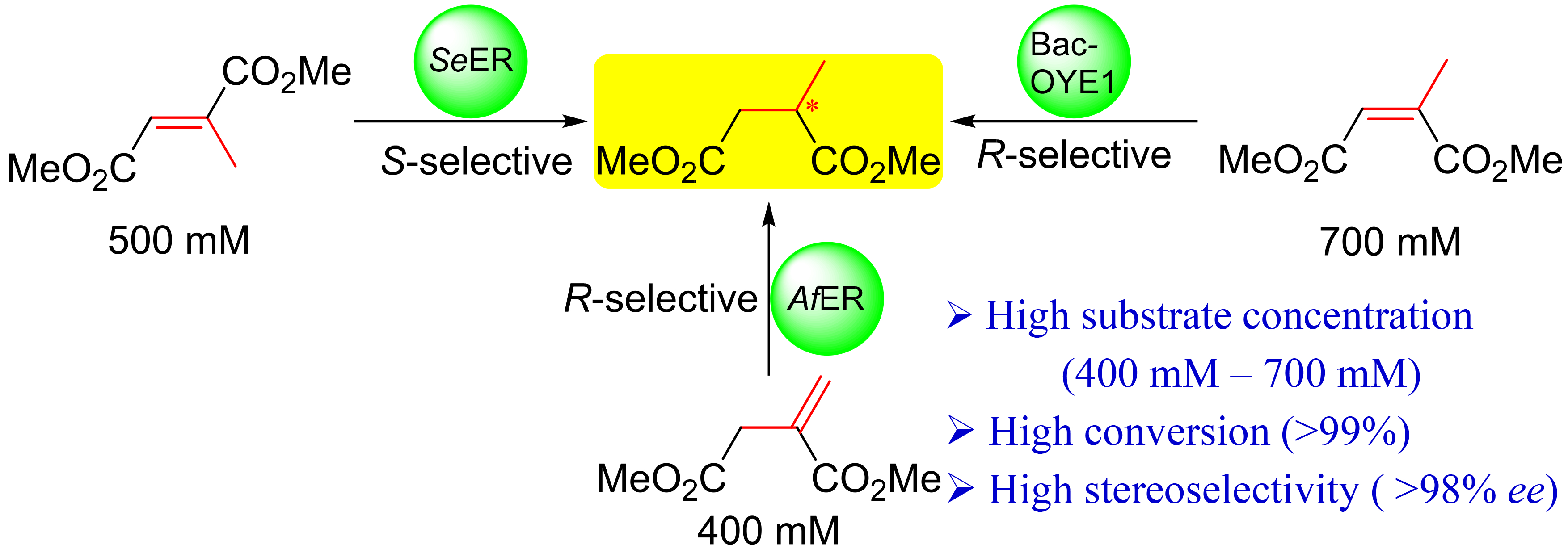
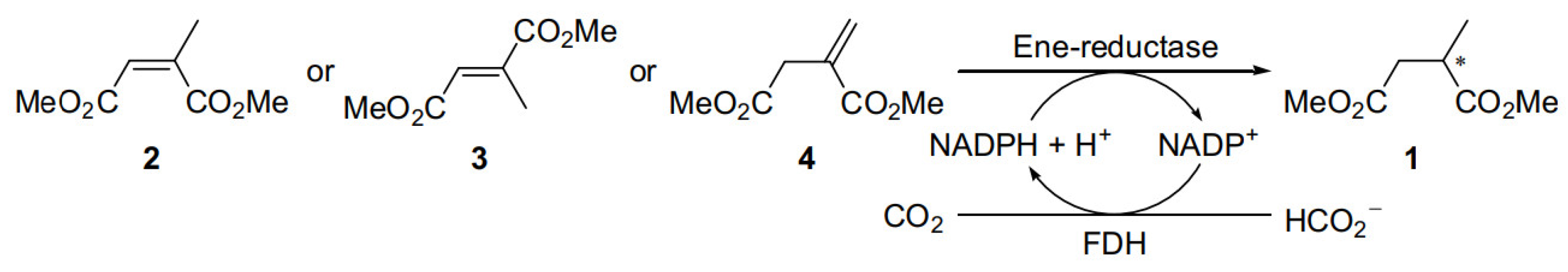
Asymmetric Synthesis of Both Enantiomers of Dimethyl 2-Methylsuccinate by the Ene-Reductase-Catalyzed Reduction at High Substrate Concentration

1
Key Laboratory of Industrial Fermentation Microbiology, Ministry of Education, College of Biotechnology, Tianjin University of Science & Technology, Tianjin 300457, China
2
National Center of Technology Innovation for Synthetic Biology, National Engineering Research Center of Industrial Enzymes and Tianjin Engineering Research Center of Biocatalytic Technology, Tianjin Institute of Industrial Biotechnology, Chinese Academy of Sciences, 32 West 7th Avenue, Tianjin Airport Economic Area, Tianjin 300308, China
*
Authors to whom correspondence should be addressed.
These authors contributed equally to this work.
Submission received: 19 August 2022 / Revised: 21 September 2022 / Accepted: 22 September 2022 / Published: 28 September 2022

Abstract

:
Chiral dimethyl 2-methylsuccinate (1) is a very important building block for the manufacturing of many active pharmaceutical ingredients and fine chemicals. The asymmetric reduction of C=C double bond of dimethyl citraconate (2), dimethyl mesaconate (3) or dimethyl itaconate (4) by ene-reductases (ERs) represents an attractive straightforward approach, but lack of high-performance ERs, especially (S)-selective ones, has limited implementing this method to prepare the optically pure dimethyl 2-methylsuccinate. Herein, three ERs (Bac-OYE1 from Bacillus sp., SeER from Saccharomyces eubayanus and AfER from Aspergillus flavus) with high substrate tolerance and stereoselectivity towards 2, 3 and 4 have been identified. Up to 500 mM of 3 was converted to (S)-dimethyl 2-methylsuccinate ((S)-1) by SeER in high yields (80%) and enantioselectivity (98% ee), and 700 mM of 2 and 400 mM of 4 were converted to (R)-1 by Bac-OYE1 and AfER, respectively, in high yields (86% and 77%) with excellent enantioselectivity (99% ee). The reductions of diethyl citraconate (5), diethyl mesaconate (6) and diethyl itaconate (7) were also tested with the three ERs. Although up to 500 mM of 5 was completely converted to (R)-diethyl 2-methylsuccinate ((R)-8) by Bac-OYE1 with excellent enantioselectivity (99% ee), the alcohol moiety of the esters had a great effect on the activity and enantioselectivity of ERs. This work provides an efficient methodology for the enantiocomplementary production of optically pure dimethyl 2-methylsuccinate from dimethyl itaconate and its isomers at high titer.

Graphical Abstract

1. Introduction

Chiral dimethyl 2-methylsuccinate (1) is exceptionally valuable as it is a very important building block for the manufacturing of many active pharmaceutical ingredients and fine chemicals. For example, it is an important chiral synthon of sacubitril, which was combined with valsartan for the treatment of heart failure [1]; aspernigrins C/D with anti-HIV-1 activities [2]; moiramide B with antibacterial activity against methicillin resistant staphylococcus aureus and a range of other antibiotic resistant human pathogens [3,4]; violaceimide A-E from the sponge-associate fungus Aspergillus violaceus [5]; and methyllycaconitine used as an antagonist at the α7 nicotinic acetylcholine receptor (Figure 1) [6]. It has also found wide application in complex convergent synthesis [7] or metal-organic frameworks [8] with enhanced material properties. In addition, it is often used as a reference for the determination of the absolute configuration of complex compounds [9]. Therefore, efficient enantioselective synthesis of chiral dimethyl 2-methylsuccinate (1) is attracting great interest from organic chemists in the academic and industrial community [10,11,12,13,14]. Among the various synthetic methods, the asymmetric reduction of C=C double bond of dimethyl citraconate (2), dimethyl mesaconate (3) or dimethyl itaconate (4) represents an attractive straightforward approach to access optically pure dimethyl 2-methylsuccinate (1). However, chemical hydrogenation methods suffer some shortcomings, including harsh reaction conditions, the use of high-pressure hydrogen, noble metal catalysts and expensive chiral ligands, and generally inadequate stereoselectivity [15].
As an alternative strategy, the asymmetric bioreduction of alkenes bearing an electron withdrawing group to chiral alkanes has received significant attention in recent years [16,17]. Ene-reductases (ERs), which perform C=C bond reduction employing NAD(P)H as hydride source via a flavin cofactor, are often highly chemo-, regio- and stereoselective [18,19,20,21]. There are some ERs from different sources [7,22], such as Kluyveromyces lactis [23], Yersinia bercovieri [23], Thermus scotoductus SA-01 [24], Saccharomyces carlsbergensis [25], Rattus norvegicus [26] and Pseudomonas putida [27], which have been used in the synthesis of chiral dimethyl 2-methylsuccinate (1), but the substrate concentration was not more than 10 mM in most cases. As yet, very rare examples of preparative-scale synthesis of dimethyl 2-methylsuccinate (1) via ER-mediated reduction have been reported [16]. Mangan et al. demonstrated that 223 mM (70 g, 1.99 L) of dimethyl citraconate (2) could be reduced to (R)-dimethyl-2-methylsuccinate ((R)-1) in 89% yield by ERED-04 cell paste in 59 h using a large amount of co-solvent toluene (28% v/v) [7]. Domínguez et al. reported that ENE-102 lyophilized powder mediated the asymmetric reduction of dimethyl itaconate (4) to give (R)-dimethyl-2-methylsuccinate ((R)-1) at high substrate concentration (730 mM) [28]. It should be noted that ERED-04 and ENE-102 were commercially purchased, and the unavailability of protein sequence information prevents improving their performance by protein engineering. This severely limits their applicability. In addition, the preparative scale synthesis of (S)-dimethyl-2-methylsuccinate ((S)-1) by this bioreduction at high substrate concentration has not been reported. In this study, through systematically exploring the activity of a collection of ERs, we successfully identified a group of high substrate-tolerant enzymes that could efficiently and enantiocomplementarily convert dimethyl citraconate (2), dimethyl mesaconate (3) or dimethyl itaconate (4) into the enantiomers of dimethyl 2-methylsuccinate (1) in high isolated yields with excellent optical purity. In particular, formate dehydrogenase (FDH) was used for co-factor regeneration using sodium formate as the hydrogen source, showing advantages in terms of atomic economy and environmental impact due to the by-products being CO2 (Figure 2).

2. Results and Discussion

A library of 81 ERs available in our group (Table S1 in the Supplementary Materials) were screened against dimethyl citraconate (2, 50 mM), dimethyl mesaconate (3, 50 mM) or dimethyl itaconate (4, 10 mM) using E. coli whole cells expressing ER genes. A total of 26 and 18 ERs catalyzed the asymmetric reduction of 2 and 4, respectively, to afford (R)-1 in 99% conversion with excellent enantioselectivity (>99% ee) (Table S3), while 17 out of 81 ERs were found to catalyze asymmetric reduction of 3 to furnish (S)-1 in excellent conversion (>99%) and enantioselectivity (>99% ee). Interestingly, NEM, X8-2, Y1-1ER, Y1-4ER, LrER, MsER, AaER and NcER1 showed complementary stereospecificity toward 3 with moderate to excellent enantioselectivities (70%–96% ee). To compare the catalytic performance of the best stereoselective enzymes, the asymmetric reductions of 2, 3 and 4 to (R/S)-1 were performed at different concentrations by employing 50 mg/mL wet E. coli cells of ERs in potassium phosphate buffer (100 mM, pH 7.0) containing 1.3 eq sodium formate, 2 U/mL LbFDH, 0.5 mg/mL NADP+ at 30 °C for 24 h. When the concentration was increased to 700 mM, only Bac-OYE1 from Bacillus sp. [29] show high activity toward 2 (>99% conversion (conv.)) (Tables S4 and S5). It is worth noting that the C=C bond isomerization of 2 to 3 was observed for some ERs according to GC analysis. This phenomenon was also discovered in the reduction of α-methylene-γ-butyrolactone catalyzed by ERs [30]. Bac-OYE1, YqjM from Bacillus subtilis [31], BzER from Bacillus zhangzhouensis and SeER from Saccharomyces eubayanus catalyzed the reduction of 3 at 300 mM concentration to give (S)-1 in >99% conversion, but only SeER showed excellent conversion (99%) and enantioselectivity (98% ee) when the concentration of 3 was increased to 500 mM (Table S6), which is the highest reaction concentration for the synthesis of (S)-1 reported so far. Asymmetric reduction of 4 at a concentration of 400 mM was achieved by AfER from Aspergillus flavus, affording (R)-1 in 99% conversion with 99% ee values (Table S7).
To explore the substrate specificity of the top five enzymes (Bac-OYE1, YqjM, BzER, AfER and SeER) with high activity and enantioselectivity, we tested their substrate tolerance and enantioselectivity towards 2, 3 and 4 (Table 1). They were all (S)-selective towards 3 and (R)-selective towards 2 and 4. Interestingly, up to 500 mM of 3 was almost completely converted to (S)-1 using wet cells of SeER, while the conversion of 2 and 4 at 50 mM was only 58% and 27%, respectively, indicating that SeER had an obvious preference for 3. The substrate tolerance of Bac-OYE1 and BzER towards 2 was significantly higher than that of 4, and that of AfER and YqjM towards 4 were significantly higher than other enzymes. Their specific activity towards 2, 3 and 4 was also tested using purified enzymes (Table 2). Bac-OYE1, SeER and AfER exhibited the highest activity towards 2, 3 and 4, respectively, which was consistent with the results of substrate tolerance assay. The specific activity of Bac-OYE1 towards 4 was higher than that of YqjM, but its substrate tolerance was lower than that of YqjM.
After evaluation of the enantioselectivity and relative activity, SeER, Bac-OYE1 and AfER were selected for further study. To investigate the effects of pH and temperature, activities of pure enzymes of SeER and AfER at different pH and temperature were measured. AfER exhibited optimum activity in the range of pH 7.0–8.5, but dropped off sharply between pH 8.5 and 9.0 or below 4.5 (Figure S2a). This was similar to the observation for Bac-OYE1 [32]. SeER had a broad pH range with optimum pH at pH 8.0 (Figure S3a). Bac-OYE1 showed higher activities in Tris–HCl buffer, while SeER and AfER showed higher activities in sodium phosphate buffer. Since the pH of the reactions will increase 0.5–1 during the reduction reaction, the preparative-scale reaction was carried out at slightly lower pH than the optimum one. The activities of SeER and AfER at the different temperatures were investigated (Figures S2b and S3b), it demonstrated that the optimum reaction temperature of SeER and AfER were 45 °C and 50 °C, respectively. The activity of SeER decreased significantly when the temperature was over 45 °C. To our delight, there was still 60% catalytic activity retained for AfER when the temperatures was increased to 80 °C. AfER and SeER could retain more than 80% after being incubated at 30 °C or 40 °C for 24 h, indicating that they were very stable below 40 °C. Interestingly, an increase in activity was observed at 40 °C within 4 h for AfER and 8 h for SeER. This may be due to the thermo-induced proper folding of the enzyme, leading to higher activity. However, the precise mechanism requires further studies.
The respective kinetic parameters of SeER, Bac-OYE1 and AfER towards 2, 3 and 4 were obtained by measuring the initial velocities of the enzymatic reaction at varied substrate concentrations and calculating with curve-fitting according to the Michaelis-Menten equation. The data of the catalytic rate (kcat) and catalytic efficiency (kcat/Km) are summarized in Table 3. SeER exhibited higher kcat/Km toward 3 than 2 and 4. Bac-OYE1 showed a 1.6-fold and 3.0-fold greater kcat/Km towards 2 than 3 and 4, respectively. AfER displayed an approximately 8-fold greater kcat/Km towards 4 than 2.
To further investigate the influence of the alcohol moiety of the ester functionalities on the activity and enantioselectivity of ERs, SeER, Bac-OYE1 and AfER were tested with diethyl citraconate (5), diethyl mesaconate (6) and diethyl itaconate (7) at a substrate concentration of 50 mM (Table S11). The three enzymes showed the same stereo-preference towards 2 and 5, and SeER displayed higher activity (>99% conversion) and enantioselectivity (97% ee) towards 5 than 2. Interestingly, all the enzymes showed relatively lower activity and enantioselectivity towards 6 and 7, demonstrating that the alcohol moiety of the esters has a great effect on the activity and enantioselectivity of ERs. Then, substrate tolerance of the three ERs towards 5 was also explored (Table S12), up to 500 mM 5 could be completely converted to (R)-diethyl 2-methylsuccinate ((R)-8) by Bac-OYE1 with excellent enantioselectivity (>99% ee), indicating that Bac-OYE1 has potential industrial application for the production of (R)-8.
Under the optimized conditions, preparative-scale asymmetric reductions of 2, 3 and 4 to (R)- or (S)-dimethyl 2-methylsuccinate (1) were carried out by using 50 g/L of wet cells of SeER, Bac-OYE1 or AfER, respectively (Table 4). The reactions were completed within 14–27 h. The reduction of 3 at 500 mM concentration by SeER generated (S)-1 in 80% isolated yield and 98% ee. (R)-1 was obtained from the reduction of 2 at 700 mM concentration by Bac-OYE1 or 4 at 400 mM concentration by AfER in 99% ee with the isolated yields of 86% and 77%, respectively.

3. Materials and Methods

3.1. Material

Materials used for culture media including peptone, yeast extract and agar were purchased from Becton, Dickinson and Company (BDX, Franklin Lake, NJ, USA). Formate dehydrogenase (LbFDH) [33] from Lactobacillus buchneri NRRL B-30929 was stored in our laboratory. Dimethyl citraconate (2), dimethyl itaconate (4), mesaconic acid, racemic dimethyl 2-methylsuccinate (1) and (R)-dimethyl 2-methylsuccinate ((R)-1) were purchased from Tokyo chemical industry Co. Ltd. (Tokyo, Japan), Aladdin Industrial Corporation and J&K Scientific (Guangdong, China). Citraconic acid and diethyl itaconate (7) were purchased from Shanghai Jizhi Biochemical Technology Co., Ltd (Shanghai, China) and McLean Biochemical Technology Co., Ltd (Shanghai, China). Dimethyl mesaconate was prepared from mesaconic acid according to the procedure published [34]. β-Nicotinamide adenine dinucleotide (NAD+) was purchased from Shanghai Yeasen Biotechnology Co., Ltd (Shanghai, China). β-Nicotinamide adenine dinucleotide 2′-phosphate reduced tetrasodium salt (NADPH), β-Nicotinamide adenine dinucleotide phosphate disodium salt (NADH) and β-Nicotinamide adenine dinucleotide phosphate disodium salt (NADP+) was purchased from Bontac Bio-Engineering (Shenzhen) Co., Ltd (Shenzhen, China). Mini Start syringe filter was purchased from Sartorius AG (Gottingen, Germany). Chromatographic silica gel (300-400 mesh) and chromatographic silica gel plate (GF254) were purchased from Yantai Xinnuo Chemical Co., Ltd (Yantai, China). CDCl3 containing 0.03% v/v tetramethylsilane (TMS) as an internal reference compound was purchased from Shanghai Acmec Biochemical Co., Ltd (Shanghai, China). All other chemicals were purchased from Sinopharm Chemical Reagent Co., Ltd (Shanghai, China). GC analysis was performed on an Agilent 7890A GC system (Agilent Technologies Co. Ltd., CA, USA). Columns include a 30 m CYCLOSIL-B column with 0.32 mm inner diameter and 0.25 μm film thickness (Agilent, Santa Clara, CA, USA). 1H and 13C NMR spectra were recorded on a Bruker Avance III 400 MHz NMR spectrometer (Bruker, Billerica, MA, USA). Determination of specific rotations was performed on an Anton Paar MCP 500 (Anton Paar, Graz, Austria) at 20 °C.

3.2. General Protein Expression Procedure

Flask scale gene expression was performed by the addition of a single colony of transformant from an agar plate into 20 mL Luria-Bertani (LB) medium (10 g/L tryptone, 10 g/L NaCl, 5 g/L yeast extract, pH 7.0) supplemented with ampicillin (100 μg/mL) or kanamycin (50 μg/mL) on a rotary shaker at 200 rpm and 37 °C overnight. The inoculum (1% v/v) was then used to inoculate 800 mL LB medium containing ampicillin (100 μg/mL) or kanamycin (50 μg/mL), and cultured under the same conditions. When the OD600 of the broth reached 0.6–0.8, isopropyl β-d-thiogalactoside (IPTG) was added to a final concentration of 0.1 mM and induction was performed at 25 °C for 12–14 h. The recombinant cells were then harvested by centrifugation (6000× g, 15 min) at 4 °C.

3.3. Purification of ERs

Cell pellets of ERs were resuspended to a concentration of 100 mg/mL in 100 mM pH 7.5 Tris-HCl buffer. The cell suspension was lysed using a high-pressure homogenizer. Following this, the cell debris was removed by centrifugation at 8000× g and 4 °C for 40 min, the supernatant was filtered through a 0.45 μm Mini Start syringe filter (Sartorius, Gottingen, Germany), and then purified by ÄKTA purifier (GE, Atlanta, GA, USA) using a Ni column. The column was conditioned with 0.1 M pH 7.5 Tris-HCl buffer containing 0.5 M NaCl and 0.02 M imidazole. After loading the supernatant into the column, the column was rinsed in three column volumes with 0.1 M pH 7.5 Tris-HCl buffer containing 0.5 M NaCl and 0.02 M imidazole. The protein was then eluted in 0.1 M pH 7.5 Tris-HCl buffer containing 0.5 M NaCl and 0.5 M imidazole. Fractions were then analyzed by SDS-PAGE (ThermoFisher, Shanghai, China). The protein was collected, then washed three times with the same volume of buffer through a 3000 kDa ultrafiltration tube for desalting. The protein concentration was determined by Pierce™ BCA Protein Assay Kit (Thermofisher, Shanghai, China) and finally we added glycerol with a final concentration of 10%, and stored at −80 °C.

3.4. General Procedure for the Preparation of Dimethyl Mesaconate (3), Diethyl Citraconate (5), Diethyl Mesaconate (6) and Diethyl 2-Methylsuccinate (8)

Dimethyl mesaconate (3), diethyl citraconate (5) and diethyl mesaconate (6) were prepared according to the procedure of reference [34]. To a solution of mesaconic acid (25.00 g, 192.3 mmol) in MeOH (150 mL) was added conc. H2SO4 (2 mL). The reaction mixture was refluxed and monitored by thin-layer chromatography [TLC, silica gel, methanol: dichloromethane: acetic acid = 15.0:84.5: 0.5, Rf = 0.40 for mesaconic acid; ethyl acetate: petroleum ether = 10:90, Rf = 0.63 for dimethyl mesaconate]. After completion of the reaction, it was concentrated in vacuo. Then, the reaction was diluted with water (100 mL) and extracted with ethyl acetate three times (3 × 100 mL). The combined organic layers were dried over sodium sulfate (Na2SO4) and the solvent was removed in vacuo. The crude residue was purified by silica gel column chromatography using ethyl acetate/petroleum ether 1:20 as eluent.
The dimethyl mesaconate (3) was obtained as colorless oil (28.53 g, 94% yield). 1H NMR (400 MHz, CDCl3) δ = 6.79 (s, 1 H), 3.79 (d, J = 14.79 Hz, 6 H), 2.30 (s, 3 H). 13C NMR (100 MHz, CDCl3) δ = 167.56, 166.29, 143.74, 126.47, 52.59, 51.69, 14.30 [35].
Following the same procedure, the diethyl citraconate (5) was obtained as colorless oil (6.30 g, 88% yield) from citraconic acid (5.00 g, 38.4 mmol). 1H NMR (400MHz, CDCl3) δ = 5.84 (q, J =1.59 Hz, 1 H), 4.28 (q, J = 7.09 Hz, 2 H), 4.18 (q, J = 7.21 Hz, 2 H), 1.99–2.13 (m, 3 H), 1.33 (t, J =7.15 Hz, 3 H), 1.24–1.30 (m, 3 H). 13C NMR (100 MHz, CDCl3) δ = 168.94, 164.92, 145.40, 120.91, 61.40, 60.70, 20.53, 14.11, 13.98.
Diethyl mesaconate (6) was obtained as colorless oil (6.10 g, 85% yield) from mesaconic acid (5.00 g, 38.4 mmol). 1H NMR (400 MHz, CDCl3) δ = 6.78 (q, J =1.51 Hz, 1 H), 4.20–4.28 (m, 4 H), 2.29 (d, J =1.59 Hz, 3 H), 1.32 (td, J =7.15, 4.16 Hz, 6 H). 13C NMR (100 MHz, CDCl3) δ = 167.16, 165.96, 143.76, 126.66, 61.57, 60.62, 14.26, 14.19, 14.13.
Diethyl 2-methylsuccinate (8) was obtained as colorless oil (6.10 g, 86% yield) from 2-methylsuccinic acid (5.00 g, 37.8 mmol). 1H NMR (400 MHz, CDCl3) δ = 4.11–4.17 (m, 4 H), 2.83–3.01 (m, 1 H), 2.65–2.78 (m, 1 H), 2.39 (dd, J =16.38, 6.11 Hz, 1 H), 1.15–1.31 (m, 9 H). 13C NMR (100 MHz, CDCl3) δ = 175.30, 171.88, 60.63, 60.55, 37.74, 35.88, 17.02, 14.19, 14.17.

3.5. Analytical Scale Reaction of ERs towards 2–7 with Wet Cells

1.0 mL reaction mixture contained the corresponding ester (50 mM for 2, 3, 5, 6, 7, or 10 mM for 4), 1.3 equivalent sodium formate, 2 U/mL LbFDH, 0.5 mM NADP+ (0.39 g/L) or NAD+ (0.33 g/L), and 50 mg wet cells of ERs and potassium phosphate buffer (100 mM, pH 7.0). DMSO (5% v/v) was added for 4 as the substrate. The reaction mixture was incubated at 30 °C with shaking at 200 rpm for 24 h, and then extracted with 1 mL ethyl acetate. The organic extract was dried over anhydrous sodium sulphate and analyzed by GC to measure the conversions and ee values of the product for the reaction of 2, 3 or 4. Then, we further increased the substrate concentration to 100–700 mM to investigate the substrate tolerance of enzymes with high activity and stereoselectivity based on the primary screening.
For the reaction of 5, 6 or 7, the conversions were determined directly by GC analysis, and the ee values of the products were determined by GC analysis of the dimethyl esters, which were obtained by the hydrolysis of diethyl ester products and esterification in methanol under the action of concentrated sulfuric acid. The general procedure was as follows. After the conversion were determined by GC, the organic solvent was removed in vacuo. The residues were dissolved in the mixture of 1 M NaOH (aq) and methanol (1.0 mL, 1:1) and stirred at 50 °C for 2 h to generate the corresponding acid. Then, the reaction mixture was quenched by 6 M HCl (100 μL) and dried in vacuo. The residues were dissolved in methanol (1 mL) containing concentrated sulfuric acid (30 μL) and refluxed for 5 h to prepare the corresponding dimethyl ester. After removal of the solvent in vacuo, the residues were diluted with ethyl acetate (1.0 mL) and washed with saturated sodium bicarbonate solution (0.5 mL). The organic extract was dried over anhydrous sodium sulphate and analyzed by GC to determine the ee values of the product.

3.6. Enzyme Activity Assay

The activity was measured photometrically by monitoring the decrease of NADPH at 340 nm (ε = 6220 L/mol/cm) including an oxygen consuming system [31,36]. Assay conditions (200 μL): Tris-HCl buffer (100 mM, pH 8.0), substrate (50–150 mM), glucose (50 mM), glucose oxidase (20 U); NADPH (0.5 mM, 0.42 g/L), 10 μL pure enzyme (1.92 g/L Bac-OYE1, 4.20 g/L YqjM, 15.36 g/L BzER, 11.94 g/L AfER or 23.55 g/L SeER). Every measurement was conducted in triplicate and corrected by a blank not containing any substrate.

3.7. Effect of pH and Temperature on Purified ERs

Sodium citrate (100 mM, pH 4.5 or 5.5), potassium phosphate buffer (100 mM, pH 5.5, 6.5, 7.0, 7.5 or 8.0), Tris-HCl buffer (100 mM, pH 8.0, 8.5 or 9.0) and glycine-NaOH buffer (100 mM, pH 9.0 or 10.0) were used to determine the optimum pH for pure enzyme. The effect of temperature on the activity of purified ERs were determined by assaying the activity of purified ERs over the range of 30–80 °C. The thermal stability of the enzyme was determined by incubating purified ERs in 1 mL potassium phosphate buffer (100 mM, pH 7.0) at various temperatures (30, 40 and 50 °C). Samples were withdrawn at a regular intervals and residual activity was measured under standard assay conditions (by using the above-described oxygen consuming system). Graphs were drawn using Origin 9.6.4 (OriginLab, Northampton, MA, USA).

3.8. Determination of Kinetic Parameters

The kinetic parameters of ERs for 2, 3 and 4 were determined by measuring the oxidation of NADPH in a range of substrate concentration from 0.1 to 150 mM at 25 °C and the measurements were performed in 96-wells plate as follows: glucose (50 mM), glucose oxidase (20 U), NADPH (0.5 mM, 0.42 g/L), 10 μL pure enzyme (1.92 g/L Bac-OYE1, 4.20 g/L YqjM, 15.36 g/L BzER, 11.94 g/L AfER or 23.55 g/L SeER) in the corresponding optimum pH buffer (100 mM, potassium phosphate buffer (pH 7.0 or 7.5), Tris-HCl buffer (pH 8.0 or 8.5), 200μL). Every measurement was conducted in triplicate and corrected by a blank not containing any substrate. Protein concentrations to assign specific activities were derived as described before. Methanol (10% v/v) was added when measuring 3, and DMSO (5% v/v) were added when measuring 4. The Km and Vmax values were obtained from non-linear regression of Michaelis–Menten plots using SigmaPlot 12.0 (Systat, IL, USA).

3.9. Preparative Scale Synthesis of Dimethyl 2-Methylsuccinate (1)

3.9.1. (R)-Dimethyl 2-Methylsuccinate ((R)-1) Using Bac-OYE1

To a 200 mL conical flask, dimethyl citraconate (2) (11.07 g, 700 mM), NADP+ (0.5 mM, 0.39 g/L), sodium formate (910 mM), Bac-OYE1 wet cells (5 g, 50 g/L) and LbFDH (2 U/mL) in Tris-HCl (100 mM, pH 8.0) were added. The total volume was 100 mL, and the reaction was shaken at 37 °C for 20 h. The reaction mixture was adjusted to pH 8.0 with 1 M HCl at intervals, and monitored by GC. After complete conversion, the reaction was quenched using 1 M HCl. The reaction mixture was extracted three times with equal volumes of petroleum ether and ethyl acetate (v/v = 4/1), and dried over anhydrous Na2SO4. The product was obtained by removal of the solvent under reduced pressure and characterized by 1H NMR and 13C NMR. The product was obtained as colorless oil (9.63 g, 86% yield, 99% ee). 1H NMR (400 MHz, CDCl3) δ = 3.69 (d, J = 6.97 Hz, 6 H), 2.86–2.99 (m, 1 H), 2.68–2.82 (m, 1 H), 2.41 (dd, J = 16.50, 5.99 Hz, 1 H), 1.23 (d, J = 7.09 Hz, 3 H). 13C NMR (100 MHz, CDCl3) δ = 175.74, 172.31, 51.96, 51.75, 37.43, 35.72, 17.04. [α]D20 = 5.280 (c = 1.0, CHCl3, for (R)-1 with ee = 99%) [lit. [35] [α]D20 = 5.8 (c = 1.0, CHCl3) with 98% ee for (R)].

3.9.2. (S)-Dimethyl 2-Methylsuccinate ((S)-1) Using SeER

To a 200 mL conical flask, dimethyl mesaconate (3) (3.95 g, 500 mM), NADP+ (0.5 mM, 0.39 g/L), sodium formate (650 mM), SeER wet cells (2.5 g, 50 g/L) and LbFDH (2 U/mL) in potassium phosphate (100 mM, pH 7.0) were added. The total volume was 50 mL, and the reaction was shaken at 37 °C when added. The reaction mixture was adjusted to pH 7.0 with 1 M HCl at intervals, and monitored by TLC and GC. After complete conversion, the reaction was quenched using 1 M HCl. The reaction mixture was extracted three times with equal volumes of petroleum ether and ethyl acetate (v/v = 4/1), and dried over anhydrous Na2SO4. The product was obtained by removal of the solvent under reduced pressure and characterized by 1H NMR and 13C NMR. The product was obtained as colorless oil (3.22 g, 80% yield, 98% ee). 1H NMR (400 MHz, CDCl3) δ = 3.69 (d, J = 6.97 Hz, 6 H), 2.84–3.01 (m, 1 H), 2.69–2.81 (m, 1 H), 2.41 (dd, J = 16.50, 5.99 Hz, 1 H), 1.23 (d, J = 7.21 Hz, 3 H). 13C NMR (100 MHz, CDCl3) δ = 175.74, 172.32, 51.96, 51.75, 37.43, 35.73, 17.04. [α]D20 = −5.560 (c = 1.0, CHCl3, for (S)-1 with ee = 98%) [lit. [37] [α]D20 = −3 (c = 1.0, CHCl3) with 90% ee for (S)].

3.9.3. (R)-Dimethyl 2-Methylsuccinate ((R)-1) Using AfER

To a 200 mL conical flask, dimethyl itaconate (4) (3.16 g, 400 mM), NADP+ (0.5 mM, 0.39 g/L), DMSO (1.6% v/v), sodium formate (520 mM), AfER wet cells (2.5 g, 50 g/L) and LbFDH (2 U/mL) in potassium phosphate (100 mM, pH 7.0) were added. The total volume was 50 mL, and the reaction was shaken at 30 °C for 27 h. The reaction mixture was adjusted to pH 7.0 with 1 M HCl at intervals, and monitored by TLC and GC. After complete conversion, the reaction was quenched using 1 M HCl. The reaction mixture was extracted three times with equal volumes of petroleum ether and ethyl acetate (v/v = 4/1), and dried over anhydrous Na2SO4. The product was obtained by removal of the solvent under reduced pressure and characterized by 1H NMR and 13C NMR. The product was obtained as colorless oil (2.45 g, 77%, yield, 99% ee). 1H NMR (400 MHz, CDCl3) δ = 3.69 (d, J = 6.97 Hz, 6 H), 2.86–3.02 (m, 1 H), 2.68–2.81 (m, 1 H), 2.41 (dd, J = 16.50, 5.99 Hz, 1 H), 1.23 (d, J = 7.09 Hz, 3 H). 13C NMR (100 MHz, CDCl3) δ = 175.73, 172.32, 51.96, 51.75, 37.43, 35.73, 17.04. [α]D20 = 5.670 (c = 1.0, CHCl3, for (R)-1 with ee = 99%) [lit. [35] [α]D20 = 5.8 (c = 1.0, CHCl3) with 98% ee for (R)].

4. Conclusions

In summary, through evaluation of a large collection of ERs, three ERs with high substrate tolerance, and high enantioselectivity towards 2, 3 and 4 have been identified and applied for the synthesis of optically pure dimethyl 2-methylsuccinate. In particular, SeER efficiently converted 3 into (S)-dimethyl 2-methylsuccinate ((S)-1) in high yields (80%) and enantioselectivity (98% ee), the first example of the preparative-scale synthesis of (S)-dimethyl 2-methylsuccinate ((S)-1) via ER-catalyzed reduction. In addition, Bac-OYE1 or AfER catalyzed the reduction of 2 or 4 at high substrate concentration (700 mM or 400 mM, respectively) affording (R)-dimethyl 2-methylsuccinate ((R)-1) in high yields and enantioselectivity. Meanwhile, diethyl citraconate (5, 500 mM) could also be completely converted to (R)-diethyl 2-methylsuccinate ((R)-8) by Bac-OYE1 with excellent enantioselectivity (99% ee). These results lay a foundation for the development of a commercially feasible biotransformation process for the efficient production of optically pure dimethyl 2-methylsuccinate from dimethyl itaconate, its isomers or a mixture of these isomers. Further protein engineering of these enzymes in our laboratory is underway to shed light on the substrate-binding and stereoselective mechanisms, and to obtain mutants of enzyme with higher activity and enantioselectivity for industrial application.

Supplementary Materials

The following supporting information can be downloaded at: https://0-www-mdpi-com.brum.beds.ac.uk/article/10.3390/catal12101133/s1, Table S1: Ene reductases (ERs) used in this study; Table S2: GC conditions for ERs-catalyzed reduction; Table S3: Screening results of substrates 2, 3 and 4 for ERs library; Table S4: Analytical scale reaction results of ERs towards 2 with 500 mM substrate concentration; Table S5: Reaction results of Bac-OYE1 and BzER with increasing substrate concentration; Table S6: Conversion and ee for screening with increasing substrate concentration of 3; Table S7: Conversion and ee for screening with increasing substrate concentration of 4; Table S8: Specific activity of ERs towards 2; Table S9: Specific activity of ERs towards 3; Table S10: Specific activity of ERs towards 4; Table S11: Asymmetric reduction of 5, 6 and 7 catalyzed by 3 ERs; Table S12: Analytical scale reaction results of Bac-OYE1, AfER and SeER towards 5 with different concentration; Figure S1: SDS-PAGE analysis of the selected ERs recombinantly expressed in E. coli BL21 (DE3); Figure S2: Effects of pH and temperature on the activity and stability of purified AfER; Figure S3: Effects of pH and temperature on the activity and stability of purified SeER; Figure S4: Non-linear regression analysis of ERs towards 2, 3 and 4; Figure S5: 1H-NMR (400 MHz) spectra of substrate dimethyl mesaconate (3) in CDCl3; Figure S6: 13C-NMR (100 MHz) spectra of substrate methyl dimethyl mesaconate (3) in CDCl3; Figure S7: GC chromatograms of substrate dimethyl citraconate (a), dimethyl mesaconate (b) and dimethyl itaconate (c); Figure S8: GC chromatograms of racemic dimethyl 2-methylsuccinate (a), (R)-dimethyl 2-methylsuccinate (b) and products of Bac-OYE1 (c). Figure S9: 1H-NMR (400 MHz) spectra of biocatalytic products catalyzed by Bac-OYE1 in CDCl3; Figure S10: 13C-NMR (100 MHz) spectra of biocatalytic products catalyzed by Bac-OYE1 in CDCl3; Figure S11: GC chromatograms of racemic dimethyl 2-methylsuccinate (a), (R)-dimethyl 2-methylsuccinate (b) and products of SeER (c); Figure S12: 1H-NMR (400 MHz) spectra of biocatalytic products catalyzed by SeER in CDCl3; Figure S13: 13C-NMR (100 MHz) spectra of biocatalytic products catalyzed by SeER in CDCl3; Figure S14: GC chromatograms of racemic dimethyl 2-methylsuccinate (a), (R)-dimethyl 2-methylsuccinate (b) and products of AfER (c); Figure S15: 1H-NMR (400 MHz) spectra of biocatalytic products catalyzed by AfER in CDCl3; Figure S16: 13C-NMR (100 MHz) spectra of biocatalytic products catalyzed by AfER in CDCl3; Figure S17: 1H-NMR (400 MHz) spectra of substrate diethyl citraconate in CDCl3; Figure S18: 13C-NMR (100 MHz) spectra of substrate diethyl citraconate in CDCl3; Figure S19: 1H-NMR (400 MHz) spectra of substrate diethyl mesaconate in CDCl3; Figure S20: 13C-NMR (100 MHz) spectra of substrate diethyl mesaconate in CDCl3; Figure S21: 1H-NMR (400 MHz) spectra of products diethyl methylsuccinate in CDCl3; Figure S22: 13C-NMR (100 MHz) spectra of products diethyl methylsuccinate in CDCl3; Figure S23: GC chromatograms of substrate diethyl citraconate; Figure S24: GC chromatograms of substrate diethyl mesaconate; Figure S25: GC chromatograms of substrate diethyl itaconate; Figure S26: GC chromatograms of racemic diethyl 2-methylsuccinate; Figure S27: GC chromatograms of the analytical reaction product of Bac-OYE1 towards diethyl citraconate; Figure S28: GC chromatograms of the analytical reaction product of AfER towards diethyl mesaconate; Figure S29: GC chromatograms of the analytical reaction product of AfER towards diethyl itaconate; Figure S30. GC chromatograms of racemic dimethyl 2-methylsuccinate (a), analytical reaction products of Bac-OYE1 (b) and AfER (c) towards diethyl citraconate after hydrolysis and esterification to the corresponding dimethyl esters; Figure S31: GC chromatograms of racemic dimethyl 2-methylsuccinate (a), analytical reaction products of AfER towards diethyl mesaconate after hydrolysis and esterification to the corresponding dimethyl esters (b); References [29,31,38,39,40,41,42,43,44,45,46,47,48] are cited in the supplementary materials.

Author Contributions

Conceptualization, J.L. (Jiacheng Li) and J.L. (Jianjiong Li); methodology, J.L. (Jianjiong Li); software, J.L. (Jiacheng Li) and J.F; formal analysis and investigation, J.L. (Jiacheng Li), J.L. (Jianjiong Li) and P.Y.; resources, Y.C.; data curation, J.L. (Jiacheng Li) and J.F.; writing—original draft preparation, J.L. (Jiacheng Li) and J.L. (Jianjiong Li); writing—review and editing, P.Y., Q.W. and D.Z.; supervision and project administration, M.W., P.Y., Q.W. and D.Z.; funding acquisition, P.Y. and D.Z. All authors have read and agreed to the published version of the manuscript.

Funding

This research was funded by the National Key R&D Program of China (No. 2021YFC2102000), National Natural Science Foundation of China (No. 92056101 and No. 22177130) and Tianjin Synthetic Biotechnology Innovation Capacity Improvement Project (TSBICIP-KJGG-009).

Data Availability Statement

Data is contained within the article or Supplementary Materials.

Conflicts of Interest

The authors declare no conflict of interest.

References

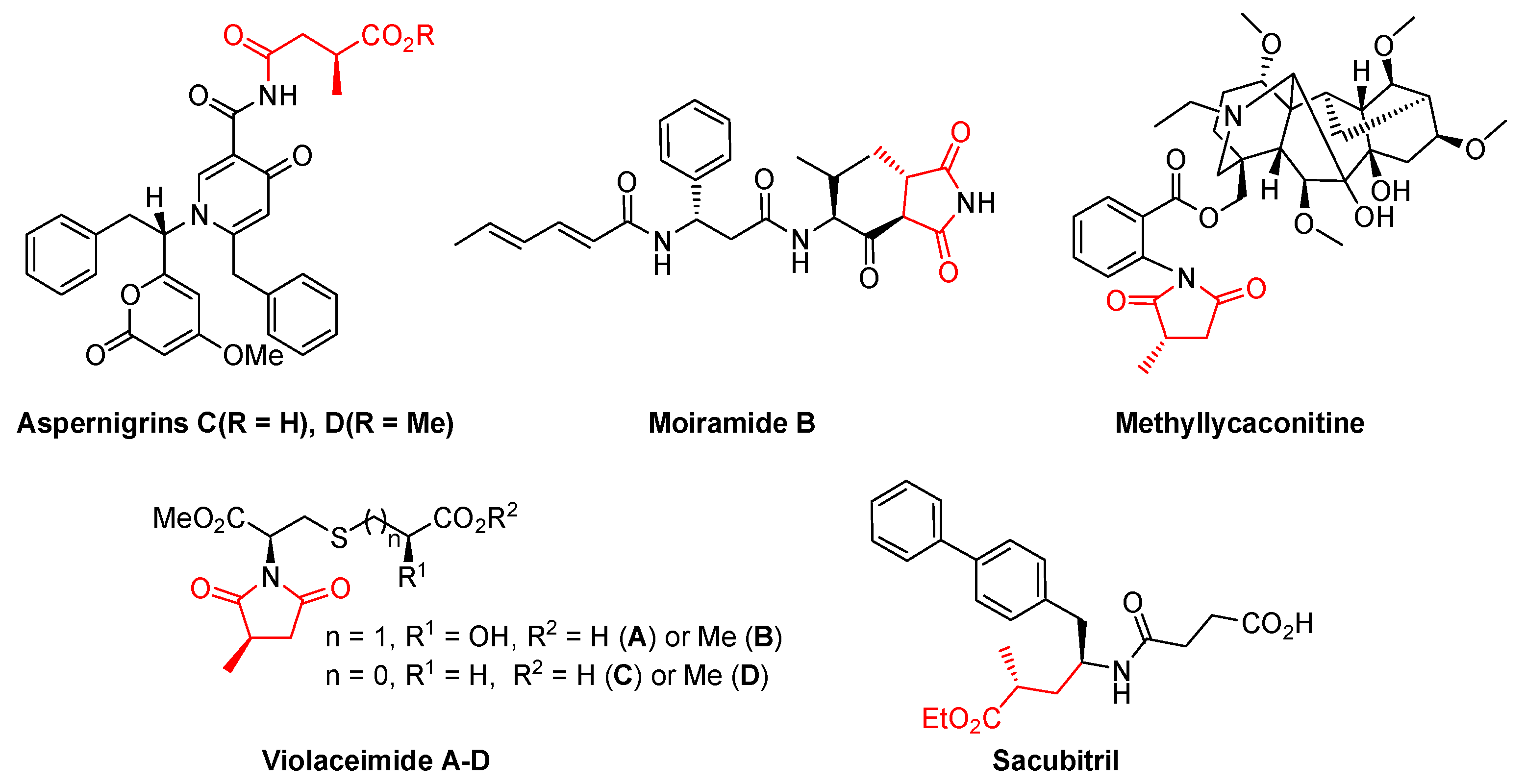
  1. Novick, S.J.; Dellas, N.; Garcia, R.; Ching, C.; Bautista, A.; Homan, D.; Alvizo, O.; Entwistle, D.; Kleinbeck, F.; Schlama, T.; et al. Engineering an Amine Transaminase for the Efficient Production of a Chiral Sacubitril Precursor. ACS Catal. 2021, 11, 3762–3770. [Google Scholar] [CrossRef]
  2. Zhou, X.; Fang, W.; Tan, S.; Lin, X.; Xun, T.; Yang, B.; Liu, S.; Liu, Y. Aspernigrins with anti-HIV-1 activities from the marine-derived fungus Aspergillus niger SCSIO Jcsw6F30. Bioorg. Med. Chem. Lett. 2016, 26, 361–365. [Google Scholar] [CrossRef] [PubMed]
  3. Dixon, D.J.; Davies, S.G. Asymmetric synthesis of moiramide B. Chem. Commun. 1996, 1797–1798. [Google Scholar] [CrossRef]
  4. Pohlmann, J.; Lampe, T.; Shimada, M.; Nell, P.G.; Pernerstorfer, J.; Svenstrup, N.; Brunner, N.A.; Schiffer, G.; Freiberg, C. Pyrrolidinedione derivatives as antibacterial agents with a novel mode of action. Bioorg. Med. Chem. Lett. 2005, 15, 1189–1192. [Google Scholar] [CrossRef]
  5. Yin, J.; Zhang, C.; Huang, J.; Zhang, J.; Liu, D.; Huang, J.; Proksch, P.; Lin, W. Violaceimides A–E, sulfur-containing metabolites from a sponge-associated fungus Aspergillus violaceus. Tetrahedron Lett. 2018, 59, 3157–3160. [Google Scholar] [CrossRef]
  6. Ismail, K.A.; Bergmeier, S.C. Structure–activity studies with ring E analogues of methyllycaconitine. Synthesis and evaluation of enantiopure isomers of selective antagonist at the α3 nicotinic receptor. Eur. J. Med. Chem. 2002, 37, 469–474. [Google Scholar] [CrossRef]
  7. Mangan, D.; Miskelly, I.; Moody, T.S. A Three-Enzyme System Involving an Ene-Reductase for Generating Valuable Chiral Building Blocks. Adv. Synth. Catal. 2012, 354, 2185–2190. [Google Scholar] [CrossRef]
  8. Jacobsen, J.; Achenbach, B.; Reinsch, H.; Smolders, S.; Lange, F.D.; Friedrichs, G.; Vos, D.; Stock, N. The first water-based synthesis of Ce(Ⅳ)-MOFs with saturated chiral and achiral C4-dicarboxylate linkers. Dalton Trans. 2019, 48, 8433–8441. [Google Scholar] [CrossRef] [PubMed]
  9. Alonso, B.; Ocejo, M.; Carrillo, L.; Vicario, J.L.; Reyes, E.; Uria, U. Using heteroaryl-lithium reagents as hydroxycarbonyl anion equivalents in conjugate addition reactions with (S,S)-(+)-pseudoephedrine as chiral auxiliary; enantioselective synthesis of 3-substituted pyrrolidines. J. Org. Chem. 2013, 78, 614–627. [Google Scholar] [CrossRef]
  10. Kovalenko, V.N.; Kulinkovich, O.G. The resolution of trans-2,2-dichloro-3-methylcyclopropanecarboxylic acid via crystallization of its salts with (+)- and (−)-α-phenylethylamine, and the transformation of the resulting enantiomers into (R)- and (S)-dimethyl 2-methylsuccinates. Tetrahedron Asymmetry 2011, 22, 26–30. [Google Scholar] [CrossRef]
  11. Pongrácz, P.; Bartal, B.; Kollár, L.; Mika, L.T. Rhodium-catalyzed hydroformylation in γ-valerolactone as a biomass-derived solvent. J. Organomet. Chem. 2017, 847, 140–145. [Google Scholar] [CrossRef]
  12. Frank, D.J.; Franzke, A.; Pfaltz, A. Asymmetric hydrogenation using rhodium complexes generated from mixtures of monodentate neutral and anionic phosphorus ligands. Chem. Eur. J. 2013, 19, 2405–2415. [Google Scholar] [CrossRef] [PubMed]
  13. Vaghi, L.; Cirilli, R.; Pierini, M.; Rizzo, S.; Terraneo, G.; Benincori, T. PHANE-TetraPHOS, the First D2 Symmetric Chiral Tetraphosphane. Synthesis, Metal Complexation, and Application in Homogeneous Stereoselective Hydrogenation. Eur. J. Org. Chem. 2021, 2021, 2367–2374. [Google Scholar] [CrossRef]
  14. Sen, A.; Kumar, R.; Pandey, S.; Vipin, K.R.; Kumar, P.; Kumar, V.; Chikkali, S.H. Mechanistically Guided One Pot Synthesis of Phosphine-Phosphite and Its Implication in Asymmetric Hydrogenation. Eur. J. Org. Chem. 2022, 2022, e202101447. [Google Scholar] [CrossRef]
  15. Parmeggiani, F.; Brenna, E.; Colombo, D.; Gatti, F.G.; Tentori, F.; Tessaro, D. “A Study in Yellow”: Investigations in the Stereoselectivity of Ene-Reductases. ChemBioChem 2022, 23, e202100445. [Google Scholar] [CrossRef]
  16. Hollmann, F.; Opperman, D.J.; Paul, C.E. Biocatalytic Reduction Reactions from a Chemist’s Perspective. Angew. Chem. Int. Ed. 2021, 60, 5644–5665. [Google Scholar] [CrossRef]
  17. Hollmann, F.; Arends, I.W.C.E.; Holtmann, D. Enzymatic reductions for the chemist. Green Chem. 2011, 13, 2285–2313. [Google Scholar] [CrossRef]
  18. Winkler, C.K.; Faber, K.; Hall, M. Biocatalytic reduction of activated C=C-bonds and beyond: Emerging trends. Curr. Opin. Chem. Biol. 2018, 43, 97–105. [Google Scholar] [CrossRef]
  19. Stuermer, R.; Hauer, B.; Hall, M.; Fabe, K. Asymmetric bioreduction of activated C=C bonds using enoate reductases from the old yellow enzyme family. Curr. Opin. Chem. Biol. 2007, 11, 203–213. [Google Scholar] [CrossRef]
  20. Williams, R.E.; Bruce, N.C. ‘New uses for an Old Enzyme’ the Old Yellow Enzyme family of flavoenzymes. Microbiology 2002, 148, 1607–1614. [Google Scholar] [CrossRef] [Green Version]
  21. Toogood, H.S.; Scrutton, N.S. Discovery, Characterisation, Engineering and Applications of Ene Reductases for Industrial Biocatalysis. ACS Catal. 2018, 8, 3532–3549. [Google Scholar] [CrossRef] [PubMed]
  22. Brenna, E.; Gatti, F.G.; Manfredi, A.; Monti, D.; Parmeggiania, F. Steric Effects on the Stereochemistry of Old Yellow Enzyme-Mediated Reductions of Unsaturated Diesters: Flipping of the Substrate within the Enzyme Active Site Induced by Structural Modifications. Adv. Synth. Catal. 2012, 354, 2859–2864. [Google Scholar] [CrossRef]
  23. Yanto, Y.; Winkler, C.K.; Lohr, S.; Hall, M.; Faber, K.; Bommarius, A.S. Asymmetric Bioreduction of Alkenes Using Ene Reductases YersER and KYE1 and Effects of Organic Solvents. Org. Lett. 2011, 13, 2540–2543. [Google Scholar] [CrossRef]
  24. Nett, N.; Duewel, S.; Schmermund, L.; Benary, G.E.; Ranaghan, K.; Mulholland, A.; Opperman, D.J.; Hoebenreich, S. A robust and stereocomplementary panel of ene-reductase variants for gram-scale asymmetric hydrogenation. Mol. Catal. 2021, 502, 111404. [Google Scholar] [CrossRef]
  25. Iqbal, N.; Rudroff, F.; Brige, A.; Beeumen, J.V.; Mihovilovic, M.D. Asymmetric bioreduction of activated carbon-carbon double bonds using Shewanella yellow enzyme (SYE-4) as novel enoate reductase. Tetrahedron 2012, 68, 7619–7623. [Google Scholar] [CrossRef] [PubMed]
  26. Durchschein, K.; Wallner, S.; Macheroux, P.; Schwab, W.; Winkler, T.; Kreis, W.; Faber, K. Nicotinamide-Dependent Ene Reductases as Alternative Biocatalysts for the Reduction of Activated Alkenes. Eur. J. Org. Chem. 2012, 2012, 4963–4968. [Google Scholar] [CrossRef]
  27. Mueller, N.J.; Stueckler, C.; Hauer, B.; Baudendistel, N.; Housden, H.; Bruce, N.C.; Faber, K. The Substrate Spectra of Pentaerythritol Tetranitrate Reductase, Morphinone Reductase,N-Ethylmaleimide Reductase and Estrogen-Binding Protein in the Asymmetric Bioreduction of Activated Alkenes. Adv. Synth. Catal. 2010, 352, 387–394. [Google Scholar] [CrossRef]
  28. Domínguez, B.; Schell, U.; Bisagni, S.; Kalthoff, T. Reduction of Activated Carbon-Carbon Double Bonds using Highly Active and Enantioselective Double Bond Reductases. Johnson Matthey Technol. Rev. 2016, 60, 243–249. [Google Scholar] [CrossRef]
  29. Zhang, H.; Gao, X.; Ren, J.; Feng, J.; Zhang, T.; Wu, Q.; Zhu, D. Enzymatic hydrogenation of diverse activated alkenes. Identification of two Bacillus old yellow enzymes with broad substrate profiles. J. Mol. Catal. B Enzym. 2014, 105, 118–125. [Google Scholar] [CrossRef]
  30. Durchschein, K.; Wallner, S.; Macheroux, P.; Schwab, W.; Winkler, T.; Kreis, W.; Faber, K. Unusual C=C bond isomerization of an α, β-unsaturated γ-butyrolactone catalysed by flavoproteins from the old yellow enzyme family. ChemBioChem 2012, 13, 2346–2351. [Google Scholar] [CrossRef] [Green Version]
  31. Rüthlein, E.; Classen, T.; Dobnikar, L.; Schölzel, M.; Pietruszka, J. Finding the Selectivity Switch A Rational Approach towards Stereocomplementary Variants of the Ene Reductase YqjM. Adv.Synth. Catal. 2015, 357, 1775–1786. [Google Scholar] [CrossRef]
  32. Schmida, R.; Antoulas, S.; Ruttimannh, A.; Schmida, M.; Vecchia, M.; Weiserb, H. Synthesis of All Four Stereoisomers of (E)-Vitamin K, (Phylloquinone), Analysis of Their Diastereoisomeric and Enantiomeric Purities and Determination of Their Biopotencies. Helv. Chim. Acta 1990, 73, 1276–1299. [Google Scholar] [CrossRef]
  33. Alpdagtas, S.; Sevil, Y.; Handan, A.K.; Liu, S.; Binay, B. Discovery of an acidic, thermostable and highly NADP+ dependent formate dehydrogenase from Lactobacillus buchneri NRRL B-30929. Biotechnol. Lett. 2018, 40, 1135–1147. [Google Scholar] [CrossRef]
  34. Klimovica, K.; Grigorjeva, L.; Maleckis, A.; Popelis, J.; Jirgensons, A. C-Quaternary Vinylglycinols by Metal-Catalyzed Cyclization of Allylic Bistrichloroacetimidates. Synlett 2011, 2011, 2849–2851. [Google Scholar]
  35. Bernasconi, M.; Muller, M.A.; Pfaltz, A. Asymmetric hydrogenation of maleic acid diesters and anhydrides. Angew. Chem. Int. Ed. 2014, 126, 5489–5492. [Google Scholar] [CrossRef]
  36. Muler, A.; Sturmer, R.; Hauer, B.; Rosche, B. Stereospecific alkyne reduction: Novel activity of old yellow enzymes. Angew. Chem. Int. Ed. 2007, 46, 3316–3318. [Google Scholar] [CrossRef] [PubMed]
  37. Yu, L.; Wang, Z.; Wu, J.; Tu, S.; Ding, K. Directed orthogonal self-assembly of homochiral coordination polymers for heterogeneous enantioselective hydrogenation. Angew. Chem. Int. Ed. 2010, 122, 3709–3712. [Google Scholar] [CrossRef]
  38. Li, M.; Cui, Y.; Xu, Z.; Chen, X.; Feng, J.; Wang, M.; Yao, P.; Wu, Q.; Zhu, D. Asymmetric synthesis of N-substitutedγ-amino esters and γ-lactams containing α,γ-stereogenic centers via a stereoselective enzymatic cascade. Adv. Synth. Catal. 2022, 364, 372–379. [Google Scholar] [CrossRef]
  39. Padhi, S.K.; Bougioukou, D.J.; Stewart, J.D. Site-saturation mutagenesis of tryptophan 116 of saccharomyces pastorianus old yellow enzyme uncovers stereocomplementary variants. J. Am. Chem. Soc. 2009, 131, 3271–3280. [Google Scholar] [CrossRef]
  40. Hall, M.; Stueckler, C.; Hauer, B.; Stuermer, R.; Friedrich, T.; Breuer, M.; Kroutil, W. Asymmetric bioreduction of activated C=C bonds using zymomonas mobilis ncr enoate reductase and old yellow enzymes OYE 1–3 from yeasts. Eur. J. Org. Chem. 2008, 2008, 1511–1516. [Google Scholar] [CrossRef]
  41. Cheallaigh, A.N.; Mansell, D.J.; Toogood, H.S.; Tait, S.; Lygidakis, A.; Gardiner, J.M. Chemoenzymatic synthesis of the intermediates in the peppermint monoterpenoid biosynthetic pathway. J. Nat. Prod. 2018, 81, 1546–1552. [Google Scholar] [CrossRef] [PubMed]
  42. Spiegelhauer, O.; Mende, S.; Dickert, F.; Knauer, S.H.; Ullmann, G.M.; Dobbek, H. Cysteine as a modulator residue in the active site of xenobiotic reductase a: A structural, thermodynamic and kinetic study. J. Mol. Biol. 2010, 398, 66–82. [Google Scholar] [CrossRef] [PubMed]
  43. Walton, A.Z.; Sullivan, B.; Patterson-Orazem, A.C.; Stewart, J.D. Residues controlling facial selectivity in an alkene reductase and semirational alterations to create stereocomplementary variants. ACS Catal. 2014, 4, 2307–2318. [Google Scholar] [CrossRef] [PubMed]
  44. Breithaupt, C.; Strassner, J.; Breitinger, U.; Huber, R.; Macheroux, P.; Schaller, A.; Clausen, T. X-Ray structure of 12-oxophytodienoate reductase 1 provides structural insight into substrate binding and specificity within the family of oYE. Structure 2001, 9, 419–429. [Google Scholar] [CrossRef]
  45. Miura, K.; Tomioka, Y.; Suzuki, H.; Yonezawa, M.; Hishinuma, T.; Mizugaki, M. Molecular cloning of the nemA gene encoding N-ethylmaleimide reductase from Escherichia coli. Biol. Pharm. Bull. 1997, 20, 110–112. [Google Scholar] [CrossRef]
  46. French, C.E.; Bruce, N.C. Purification and characterization of morphinone reductase from Pseudomonas putida M10. Biochem. J. 1994, 301, 97–103. [Google Scholar] [CrossRef]
  47. Gao, X.; Ren, J.; Wu, Q.; Zhu, D. Biochemical characterization and substrate profiling of a new NADH-dependent enoate reductase from Lactobacillus casei. Enzyme Microb. Tech. 2012, 51, 26–34. [Google Scholar] [CrossRef]
  48. Sheng, X.; Yan, M.; Xu, L.; Wei, M. Identification and characterization of a novel Old Yellow Enzyme from Bacillus subtilis str.168. J. Mol. Cataly. B Enzym. 2016, 130, 18–24. [Google Scholar] [CrossRef]
Figure 1. Examples of bioactive natural products and drugs derived from chiral dimethyl 2-methylsuccinate (1).
Catalysts 12 01133 g001
Figure 2. The synthetic strategy towards chiral dimethyl 2-methylsuccinate (1) in this study. Asterisk (*) indicates a chiral center.
Catalysts 12 01133 g002
Table 1. Asymmetric reduction of 2, 3 and 4 catalyzed by 5 ERs 1.
ERs234
Conc.
(mM)
Conv.
(%) 2
ee
(%) 2
Conc.
(mM)
Conv.
(%) 2
ee
(%) 2
Conc.
(mM)
Conv.
(%) 2
ee
(%) 2
Bac-OYE1700>9999R300>9999S509799R
YqjM5007599R300>9999S4009999R
BzER500>9999R300>9999S30>9999R
AfER5006299R300>9999S400>9999R
SeER505854R5009998S502799R
1 Reaction conditions: substrate (2, 3 or 4) at varied concentration in potassium phosphate buffer (1 mL, 100 mM, pH 7.0), 1.3 eq sodium formate, 2 U/mL LbFDH, 0.5 mM NADP+ (0.39 g/L), and 50 mg wet cells at 30 °C for 24 h. 2 The conversion and ee values were determined by GC analysis, the absolute configuration of products was assigned by comparison their retention times with standard sample (R)-1, which appears as superscript font.
Table 2. Specific activity of ERs towards 2, 3 and 4 1.
EnzymeSpecific Activity (U/mg)
234
Bac-OYE12.170.360.95
YqjM1.060.050.61
BzER0.550.100.53
AfER0.250.141.43
SeER0.030.380.02
1 Assay conditions: 50–150 mM substrate, 50 mM glucose, 20 U glucose oxidase; 0.5 mM NADPH (0.42 g/L), 10 μL pure enzyme (1.92 g/L Bac-OYE1, 4.20 g/L YqjM, 15.36 g/L BzER, 11.94 g/L AfER or 23.55 g/L SeER) in 200 μL Tris-HCl buffer (100 mM, pH 8.0).
Table 3. Kinetic parameters towards 2, 3 and 4 substrates for ERs 1.
EnzymeCatalysts 12 01133 i001Catalysts 12 01133 i002Catalysts 12 01133 i003
Km
(mM)
kcat
(s−1)
kcat/Km
(mM−1
min−1)
Km
(mM)
kcat
(s−1)
kcat/Km
(mM−1·
min−1)
Km
(mM)
kcat
(s−1)
kcat/Km
(mM−1·
min−1)
Bac-OYE144.51.572.126.0 0.13 1.30 63.40.750.71
YqjM34.40.350.61- 2- 2- 276.40.330.26
BzER30.60.270.535.50.080.8769.70.380.33
AfER82.80.260.193.6 0.10 1.67 42.51.101.55
SeER- 2- 2- 210.00.301.80- 2- 2- 2
1 Assay conditions: 0.1 to 150 mM substrate, 50 mM glucose, 20 U glucose oxidase, 0.5 mM NADPH (0.42 g/L), 10 μL pure enzyme (1.92 g/L Bac-OYE1, 4.20 g/L YqjM, 15.36 g/L BzER, 11.94 g/L AfER or 23.55 g/L SeER), 200 μL potassium phosphate buffer (pH 7.0 or 7.5) or Tris-HCl buffer (pH 8.0 or 8.5) in 96-wells plate at 25 °C. Methanol (10% v/v) were added when measuring 3. DMSO (5% v/v) were added when measuring 4. 2 Below detection limit.
Table 4. Preparative-scale synthesis of (R/S)-1 1.
Sub.Prod.Conc.Vol.
(mL)
ERsConv.
(%) 2
Yield
(%) 3
ee
(%) 2
3(S)-1500 mM
(3.95 g, 79.1 g/L)
50SeER>998098
2(R)-1700 mM
(11.07 g, 110.7 g/L)
100Bac-OYE1>998699
4400 mM
(3.16 g, 63.3 g/L)
50AfER>997799
1 Reaction conditions: substrate (500 mM 3, 700 mM 2, or 400 mM 4), 1.3 eq. sodium formate, 2 U/mL LbFDH, 0.5 mM NADP+ (0.39 g/L), 50 g/L wet cells (SeER in 50 mL potassium phosphate buffer (100 mM, pH 7.0) for 3, Bac-OYE1 in 100 mL Tris-HCl (100 mM, pH 8.0) for 2, or AfER in 50 mL potassium phosphate buffer (100 mM, pH 7.0) with 1.6% (v/v) DMSO for 4), 37 °C to 2 for 14 h and 3 for 20 h, or 30 °C to 4 for 27 h, the reaction mixture was adjusted to the initial pH with 1 M HCl at intervals. 2 The conversion and ee values were determined by GC analysis. 3 isolated yield.
Publisher’s Note: MDPI stays neutral with regard to jurisdictional claims in published maps and institutional affiliations.

