Article

Stable Nickel-Based Metal–Organic Framework Containing Thiophene/Diimidazole Units for Effective Near-Infrared Photothermal Conversion

1
School of Materials Science and Engineering, Zhengzhou University, Zhengzhou 450001, China
2
Henan Province Function-Oriented Porous Materials Key Laboratory, College of Chemistry and Chemical Engineering, Luoyang Normal University, Luoyang 471934, China
*
Author to whom correspondence should be addressed.
Submission received: 17 June 2022 / Revised: 8 July 2022 / Accepted: 9 July 2022 / Published: 13 July 2022
(This article belongs to the Special Issue MOFs Catalyst for Energy-Related Reactions)

Abstract

:
Herein, a new Ni-based metal–organic framework (MOF, 1) bearing highly structural stability is synthesized by the reaction of utilizing a rigid and functionalized linker, 2,6-bis(pyridin-4-yl)-1,7-dihydrobenzo[1,2-d:4,5-d′]diimidazole (BBI4PY), in combination with Ni(NO3)2·6H2O and dibenzo[b,d]thiophene-3,7-dicarboxylic acid 5,5-dioxide (L1) under solvothermal conditions. The crystal structure of complex 1 is determined by single-crystal X-ray diffraction and is demonstrated to be a two-dimensional layered structure. In addition, PXRD, IR, TGA and UV/Vis-NIR spectra are also tested carefully to explore the solid structure of this complex. Remarkably, although no significant accumulation effect could be observed between the two-dimensional layers, a stacking interaction between DMF solvent molecules and ligand L1 could be found, which might promote non-radiative transitions and trigger obvious near-infrared photothermal conversion. Under 660 nm laser (0.6 W cm−2) illumination, the temperature of complex 1 increased rapidly from room temperature to 45.2 °C, with good thermal stability and cycle durability. Its photothermal conversion efficiency could reach 10.75%. This work provides an efficient way for assessing the promise of materials in the field of photothermal therapy.

Graphical Abstract

1. Introduction

Photothermal therapy has great potential for cancer therapy due to the effective and highly selective killing of diseased lesions under the irradiation of light, preferably near-infrared (NIR) light [1,2,3]. The photothermal agents (PTAs) for converting NIR light to heat are either purely inorganic materials or purely organic materials exemplified by conjugated polymers and molecular dyes [4,5,6,7]. However, the disadvantages of the poor photostability and biodegradation of these materials hinder their further application [8,9,10]. As a new type of crystalline porous inorganic−organic hybrid material, metal–organic frameworks (MOFs) have aroused widespread concern among scientists due to their large specific surface area, highly tunable structures, modular porosity, and structural stability [11,12,13,14,15,16,17]. Therefore, various MOF structures, such as MOF-5, MOF-74, ZIF-8, and UTSA-74, have been constructed successfully through various methods; their properties, such as gas adsorption, catalysis, sensors, photoelectric performance, and photothermal effect, have also been explored [18,19,20,21,22,23,24,25]. Several hundred articles about the performance exploration of MOF structures have been reported successfully [26,27,28,29,30]. These studies have also prompted more chemists to design and synthesize additional MOFs with better structures and performance, which will drive the rapid development of and offer a great leap-forward for MOF materials.
In the process of designing functionalized MOF structures bearing photothermal effects, the selection of both metal ions and organic ligands is extremely important. The utilization of metal ions and ligand linkers plays a vital role in the structural construction and performance implementation of MOF structures. For example, Chen reported a ruthenium metal−organic framework; after TCNQ loading, the complex exhibited an enhanced photothermal property with a photothermal conversion efficiency of 29.1% [31]. In addition, various MOF materials bearing near-infrared photothermal conversion performance have been reported [32,33,34,35,36]. For example, a group of researchers synthesized a new Zn-MOF containing an electron donor, tetrathiafulvalene (TTF), and an electron acceptor, naphthalene diimide. Interestingly, the material exhibited wide absorption peaks in the near-infrared region and displayed efficient near-infrared photothermal conversion performance (from room temperature to 250 °C) under 808 nm laser (0.4 W cm–2) illumination [37]. From previous reports, it can be found that the π-π stacking interaction plays an important role in structural generation and photothermal triggering. Thus, the selection of appropriate ligand components to construct an effective stacking interaction is critical for the resulting photothermal effect.
Based on this, a new pillar ligand, 2, 6-bis (pyridin-4-yl)-1, 7- dihydrobenzo[1,2-d:4,5-d′]-diimidazole (BBI4PY) with a benz-bis(imidazole) moiety, is selected. Moreover, this new linker has multiple hydrogen bonding and Lewis basic interaction sites, as shown in Scheme 1. Importantly, BBI4PY has an obvious conjugate center, which could cause an effective stacking interaction. The choice of this ligand is of great significance for the synthesis of the MOF. Herein, through the solvothermal method based on two organic ligands, 2,6-bis(pyridin-4-yl)-1,7-dihydrobenzo[1,2-d:4,5-d′]diimidazole (BBI4PY) and dibenzo[b,d]thiophene-3,7-dicarboxylic acid 5,5-dioxide (L1), a new Ni-based complex is obtained and its synthesis and crystal structure are reported. Its near-infrared photothermal conversion properties are investigated in detail.

2. Results

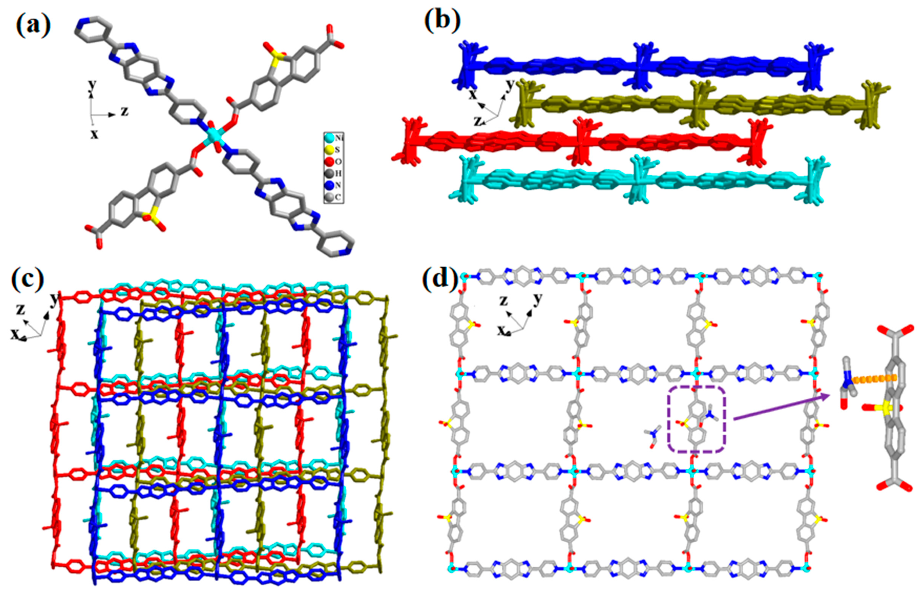
2.1. The Structure of Complex 1

The crystal structure was demonstrated by single-crystal X-ray diffraction analysis, reflecting that complex 1 crystallized in the triclinic P-1 space group (Table 1). Observing the single-crystal structure, the asymmetric unit contained one crystallography-independent Ni (II) metal center, 0.5 ligand L1, 0.5 ligand BBI4PY, and two coordinated water molecules (Figure 1a). The nickel cations were coordinated by six atoms, including two carboxyl oxygen atoms of L12− ligands, two pyridine nitrogen atoms from the neutral BBI4PY ligand, and two oxygen atoms from two H2O molecules, respectively. In addition, each L12− ligand could achieve coordination with two nickel cations and two carboxyl oxygen atoms. Each BBI4PY ligand connected to two nickel cations, showing a bidentate coordination mode. Finally, a series of rectangular windows (19.18 × 15.32 Å2) were formed from four nickel cations, two BBI4PY ligands, and two L12− ligands (Figure 1b,c). These rectangular windows resulted in the formation of a two-dimensional layered structure. The Ni−O bond lengths were 2.082(5), 2.068(5), 2.083(4), and 1.987(5) Å. At the same time, the lengths of the two Ni−N bonds were 2.109(5) and 2.124(5) Å (Table 2, Figure 1d). In addition, the separation between the two-dimensional layered structures was 7.5 Å, which was long enough that no interactions between the two layered structures could be detected. However, obvious π-π stacking interactions between free DMF molecules and ligands L1 or BBI4PY were found, the distances between which were 3.42 and 3.56 Å, respectively. The stacking interactions might have caused an effective photothermal effect, and the loss and addition of DMF solvent molecules might have led to the disappearance and reappearance of a photothermal effect.

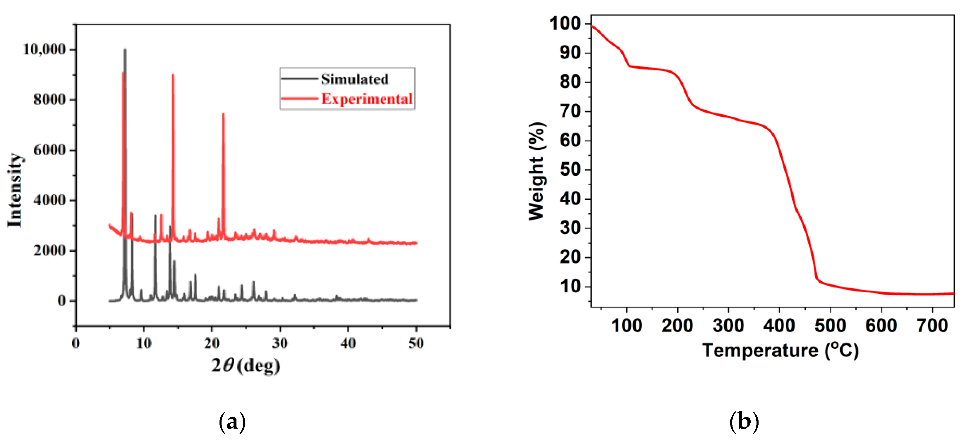
2.2. The PXRD and TGA Exploration of Complex 1

To determine the phase purity of synthesized powder complex 1, PXRD measurements were performed carefully. The simulated and experimental PXRD patterns of complex 1 (the latter obtained at room temperature) are shown in Figure 2a. All main diffractive peaks of the powder sample were nearly consistent with the simulated pattern produced from the single-crystal diffraction data, reflecting the good phase purity of the solid-state complex. Moreover, the discrepancy in reflection intensities between the simulated and experimental patterns might be attributed to variations in the preferred orientation of the solid samples. Both the shift between two-dimensional layered structures, due to weak interaction, and the loss of solvent molecules might result in the discrepancy between Yexperimental and Ysimulate. Nevertheless, the good match between the main peaks (X = 6.9, 8.3, 11.6, 14.5, 21.0 and 21.7) proved the stability of the major structure.
The TG analysis curve of complex 1 is displayed in Figure 2b; the weight loss was 15.09% for complex 1 in the range of 0–100 °C, which corresponded to the loss of 2.0 free water and DMF molecules (calcd 6.12% for complex 1). From 100 to 180 °C, no losses were observed. Then, a weight loss of 10.22% was observed in the range of 100–288 °C, corresponding to the loss of two coordinated water molecules (calcd 10.40% for complex 1). After that, the successive losses from 258 to 500 °C corresponded to the collapse of the skeleton and the decomposition of the organic ligand.

2.3. The IR and UV/Vis-NIR Spectra of Complex 1

In addition, the IR spectrum of complex 1, BBI4PY, and L1 are clearly shown in Figure 3. The IR spectrum of complex 1 exhibited a strong band at 1174 cm−1, owing to the S=O stretching vibrations, similar to the peak position in the infrared spectrum of L1. Strong absorptions at 1663 cm−1 and 1248 cm−1 occurred due to the stretching of the C=O and C-O bonds from the carboxylate group of L12−, and they all moved to lower wave numbers in different degrees, as compared to L1, possibly due to the coordination of the metal center and the π- π stacking interactions. The C-H deformation vibration absorption peaks on the pyridine group were 1451 cm−1. Furthermore, the characteristic absorption peak of pyridine in the IR spectrum of BBI4PY was 1440 cm−1. Moreover, the analysis of the IR spectrum also showed strong bands at 1615 cm−1 and 3175 cm−1, attributed to the stretching vibration of the C=N and N-H bonds of the imidazole group. We can unambiguously see that the strong peaks at 1618 cm−1 and 3373 cm−1 are assigned to the C=N and N-H bonds of the imidazole group on the IR spectrum of BBI4PY [38,39,40,41,42,43,44].
Moreover, the solid-state UV/Vis-NIR spectra of ligands L1, BBI4PY, and complex 1 were studied at room temperature. As shown in Figure 3b, there was a broad absorption peak from 600 to 800 nm for complex 1 (Abs: 1.152 at 660 nm), displaying strong absorption in the NIR region. This illustrates the existence of strong stacking interactions in complex 1, which is consistent with the analysis of the crystal structure. However, for the free ligands L1 and BBI4PY, the adsorption peaks were very weak and the Abs values were 0.536 and 0.157 at 660 nm.

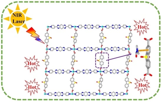
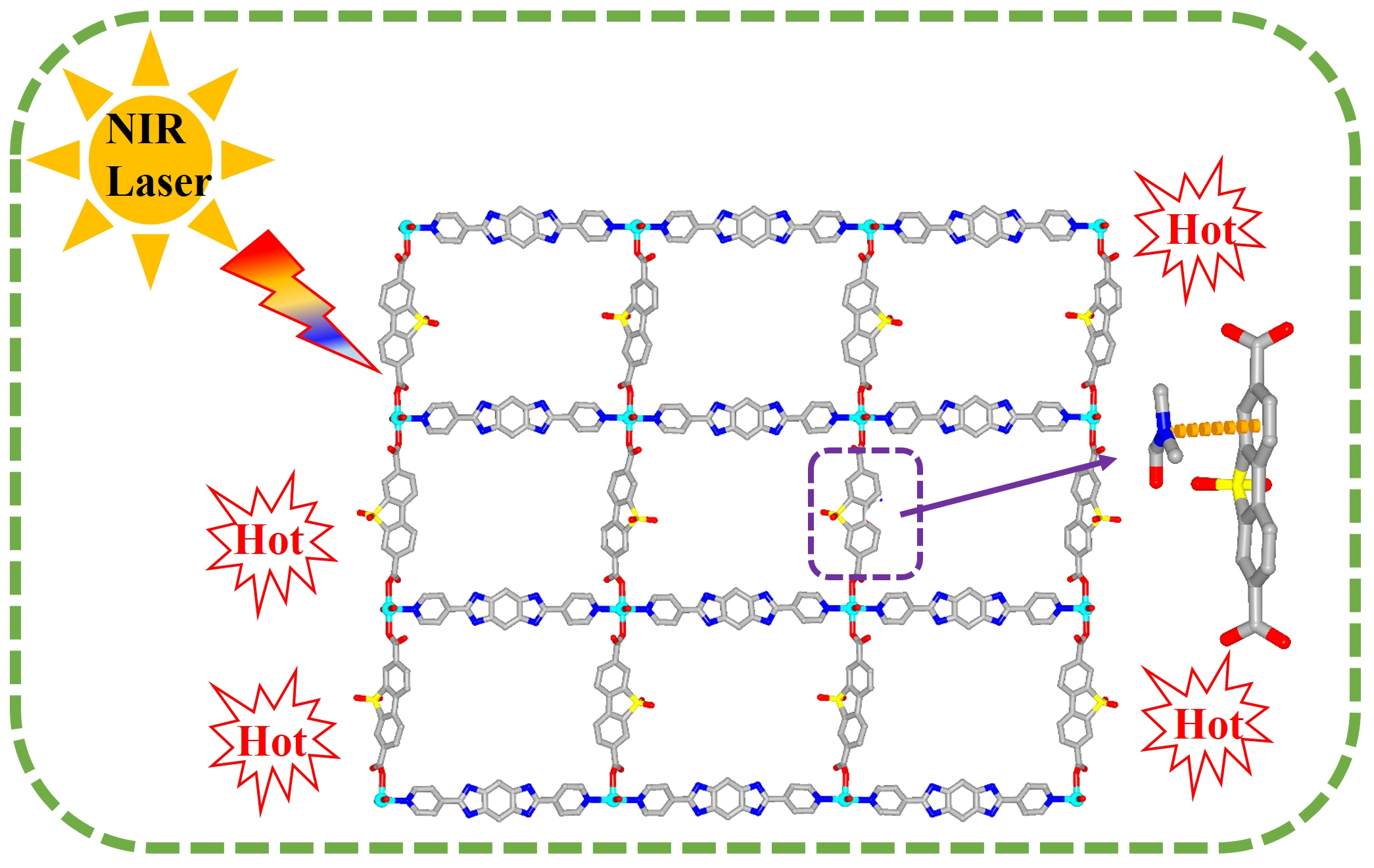
2.4. The Near-Infrared Photothermal Conversion Studies

Previous reports have displayed that π-π stacking interactions could induce active nonradiative pathways and the inhibition of the radiative transition process, causing photothermal (PT) conversion [45,46,47,48,49]. Meanwhile, based on the strong absorption of complex 1 in the NIR region, the dark color in the solid state (Figure 4a), and the TGA tests of crystal 1, it has already been demonstrated that the structural stability of complex 1 could be maintained up to 200 °C. Therefore, this tells us that the temperature change from the photothermal effect could not cause structural changes (Figure 2b). Therefore, the near-infrared photothermal conversion properties of complex 1 were explored in detail under the infrared light irradiation of 660 nm. As a comparison, the near-infrared photothermal conversion experiments of the two ligands, L1 and BBI4PY, were carried out under the same conditions.
Therefore, the crystalline complex 1 was irradiated under 0.6 W/cm2 laser irradiation at 660 nm in a solid state. The temperature change in crystalline complex 1 reached 19.1 °C (from 26.3 to 45.4 °C) (Figure 4b). This is a viable temperature change. When the laser irradiation of 660 nm was removed, a rapid cooling process was observed clearly by the infrared imaging camera. Based on the reduced temperature data, the near-infrared photothermal efficiency of complex 1 was calculated to be 10.75%, according to the calculation method described below (Equations (1)–(4)), showing good near-infrared photothermal performance of this Ni−MOF, 1. Meanwhile, the temperature changes of ligands L1 and BBI4PY under laser irradiation at 660 nm for 3 h were also tested. The results showed that although the temperature change of L1 was very weak and could be ignored, the temperature increase in ligand BBI4PY was 5.3 °C (from 22.0 to 27.3 °C), possibly due to the formation of self-accumulation between two BBI4PY ligands (Figure 5a). In addition, the cycle performance of photothermal efficiency for complex 1 was explored by continuous 0.6 W/cm2 laser irradiation at 660 nm four times, as well as the addition of DMF solvents (Figure 5b). The temperature change in solid crystalline 1 could still reach 45.2 °C every time, clearly reflecting high stability and photothermal efficiency in the cycle performance. The absence of fluorescence in the crystal indicates that the photothermal conversion effect was realized by non-radiative transition. It also reflects that stacking interactions play an important role in the stability and photothermal properties of crystal 1. According to the above experimental data, we can infer a rule of experience: the photothermal conversion efficiency is relevant to π-π stacking interactions.
η = hS (ΔTsample-ΔTsolvent)/I(1−10−A)
hS =∑mCp/τs
τs = −t/lnθ
θ = (Tamb − T)/(Tamb − Tmax)
In Equations (1)–(4) above, h is the heat transfer coefficient, S is the surface area of the container, τs is the sample system time constant, m is the mass of the products, Cp is the specific heat capacity of the solvent, and the value of τs was obtained from the fitting linear of these solutions (Figure 6).

3. Experiment

3.1. Materials and Methods

The selected reagents were analytic-grade and acquired through commercial sources. They were utilized without further purification. Elemental analyses for C and H were carried out on a Perkin-Elmer 240 elemental analyzer. The FT-IR spectra were tested from KBr pellets in a particular range from 4000 to 400 cm−1 on a Bruker VECTOR 22 spectrometer. The UV/Vis-NIR spectra were tested in a particular range from 600 to 800 nm on a UV-3100 spectrophotometer. Thermal analysis measurements were executed on an SDT 2960 thermal analyzer from 25 °C to 800 °C, based on a heating rate of 20 K min–1 under nitrogen flow. Powder X-ray diffraction (PXRD) data were collected smoothly on a Rigaku D/Max-2500PC diffractometer with Cu Kα radiation (λ = 1.5406 Å) over the 2θ range of 5−50° with a scan speed of 5°/min at 25 °C.
Solid-state UV-Vis-NIR spectra were measured on a UV-3100 spectrophotometer. BaSO4 was used as the reference. Spectra are reported as the Kubelka–Munk transform, where F(R) = (1 − R) 2/2R (R is the diffuse reflectance of the sample as compared to BaSO4).
The single-crystal X-ray diffraction analysis of complex 1 was carried out on a Rigaku Saturn 724 CCD diffractomer (Mo-Kα, λ = 0.71073 Å) at room temperature. The structure was solved by Direct Methods with SHELXS-97 and refined by the full-matrix least-squares method on F2 with anisotropic displacement parameters for all non-H atoms (SHELXL-97). An empirical absorption correction was applied by the SADABS program [50,51]. The hydrogen atoms were assigned with common isotropic displacement factors and included in the final refinement by the use of geometrical restrains. The crystallographic data and selected bond lengths and angles for complex 1 are listed in Table 1 and Table 2. Crystallographic data for the structural analysis have been deposited with the Cambridge Crystallographic Data Center, and CCDC No. for compound 1 contains the supplementary crystallographic data for this paper. These data can be obtained free of charge from The Cambridge Crystallographic Data Centre via http://www.ccdc.cam.ac.uk/Community/Requestastructure (accessed on 16 June 2022).

3.2. Synthesis of Complex 1

A mixture of Ni(NO3)2·6H2O (0.2 mmol, 0.058 g), 2,6-bis(pyridin-4-yl)-1,7- dihydrobenzo[1,2-d:4,5-d′]diimidazole (BBI4PY) (0.2 mmol, 0.0624 g), dibenzo[b, d]thiophene-3,7-dicarboxylic acid 5,5-dioxide (L1) (0.2 mmol, 0.061 g) and 4 mL of DMF and 2 mL of H2O was stirred for 10 min. The mixture was then transferred and sealed into a Teflon reactor (23 mL) and heated at 115 °C for 48 h. After that, the mixture was cooled to room temperature. Blue block crystals of complex 1 were washed by ethanol (3 × 10 mL) and dried in air at room temperature (yield: 65% based on Ni). Anal. Calc. (%) for C76H72N16Ni2O20S2: C 53.35, H 4.24, N 13.10; found (%): C 53.31, H 4.26, N 13.12. IR (KBr, cm−1): 3175.10(w), 1663.20(w), 1615.60(s), 1451.44(s), 1375.92(m), 1296.31(m), 1174.01(m), 1019.70(w), 963.06(w), 840.77(s), 780.33(s), 699.59(s), 599.85(s), 578.11(s)

3.3. X-ray Crystallography

Single-crystal X-ray diffraction data for complex 1 were acquired at 298 K on an Oxford Diffraction SuperNova area-detector diffractometer by utilizing mirror optics monochromated MoK α radiation (λ = 0.71073 Å). CrysAlisPro [50] was utilized for the crystal data collection, general data reduction, and further empirical absorption correction. The crystal structure of complex 1 was solved by SHELXS-2014 and least-squares refined with SHELXL-2014 [51]. The crystal refinement parameters are provided in Table 1. A solvent mask was calculated and 188 electrons were found in a volume of 1026\%A^3^ in 1 void per unit cell. This is consistent with the presence of 2[C3H7NO] per asymmetric unit, which accounts for 160 electrons per unit cell.

4. Conclusions

In summary, we synthesized a new Ni-based MOF complex by a solvothermal method based on a decarboxylic acid ligand and a bidentate pyridine ligand. Single-crystal X-ray diffraction analysis determined the compound to be a two-dimensional layered structure. A series of structural characterization from PXRD, IR, TGA and UV-vis-NIR spectra showed good structural stability and a wide range of near infrared absorption. In addition, obvious π-π stacking interactions could be found, which promoted non-radiative transitions and triggered the effective photothermal conversion effect. The near-infrared photothermal exploration showed a temperature variation of 19.1 °C and the near-infrared photothermal efficiency reached 10.75%, demonstrating good near-infrared photothermal performance. This research encourages us to synthesize and explore more photothermal conversion materials.

Author Contributions

X.P. and L.D., synthesis of the title complex, formal analysis, IR-related experiment; X.P., writing—review and editing; T.Z. and T.C., formal analysis and photo-thermal exploration experiment; F.R., UV/Vis-NIR absorption spectra experiment; S.L.: supervision, writing—review and editing. All authors have read and agreed to the published version of the manuscript.

Funding

This work was supported by the National Natural Science Foundation of China (Grant Nos. 52003253 and 22101108), Natural Science Foundation of Henan Province (No. 212300410209), and the Shanghai Science and Technology Committee (19DZ2270100).

Conflicts of Interest

The authors declare no conflict of interest.

References

  1. Lyu, Y.; Fang, Y.; Miao, Q.Q.; Zhen, X.; Ding, D.; Pu, K.Y. Intraparticle molecular orbital engineering of semiconducting polymer nanoparticles as amplified theranostics for in vivo photoacoustic imaging and photothermal therapy. ACS Nano 2016, 10, 4472–4481. [Google Scholar] [CrossRef]
  2. Pu, K.; Shuhendler, A.J.; Jokerst, J.V.; Mei, J.G.; Gambhir, S.S.; Bao, Z.N.; Rao, J.H. Semiconducting polymer nanoparticles as photoacoustic molecular imaging probes in living mice. Nat. Nano 2014, 9, 233–239. [Google Scholar] [CrossRef] [Green Version]
  3. Chen, Q.; Liang, C.; Sun, X.Q.; Chen, J.W.; Yang, Z.J.; Zhao, H.; Feng, L.Z.; Liu, Z. H2O2-responsive liposomal nanoprobe for photoacoustic inflammation imaging and tumor theranostics via in vivo chromogenic assay. Proc. Natl. Acad. Sci. USA 2017, 114, 5343–5348. [Google Scholar] [CrossRef] [Green Version]
  4. Lovell, J.F.; Jin, C.S.; Huynh, E.; Jin, H.L.; Kim, C.; Rubinstein, J.L.; Chan, W.C.W.; Cao, W.G.; Wang, L.V.; Zheng, G. Porphysome nanovesicles generated by porphyrin bilayers for use as multimodal biophotonic contrast agents. Nat. Mater. 2011, 10, 324–332. [Google Scholar] [CrossRef] [Green Version]
  5. Chen, W.R.; Adams, R.L.; Higgins, A.K.; Bartels, K.E.; Nordquist, R.E. Photothermal effects on murine mammary tumors using indocyanine green and an 808-nm diode laser: An in vivo efficacy study. Cancer Lett. 1996, 98, 169–173. [Google Scholar] [CrossRef]
  6. Yang, J.; Choi, J.; Bang, D.; Kim, E.; Lim, E.K.; Park, H.; Suh, J.S.; Lee, K.; Yoo, K.H.; Kim, E.K.; et al. Convertible organic nanoparticles for near-Infrared photothermal ablation of cancer cells. Angew. Chem. Int. Ed. 2011, 50, 441–444. [Google Scholar] [CrossRef]
  7. Zhang, J.Z.; Sun, D.D.; Zhang, B.; Sun, Q.Q.; Zhang, Y.; Liu, S.R.; Wang, Y.M.; Liu, C.T.; Chen, J.Z.; Chen, J.B.; et al. Intrinsic carbon nanotube liquid crystalline elastomer photoactuators for high-definition biomechanics. Mater. Horiz. 2022, 9, 1045–1056. [Google Scholar] [CrossRef]
  8. Wang, Y.; Zhu, W.G.; Du, W.N.; Liu, X.F.; Zhang, X.T.; Dong, H.L.; Hu, W.P. Cocrystals strategy towards materials for near-infrared photothermal conversion and imaging. Angew. Chem. Int. Ed. 2018, 57, 3963–3967. [Google Scholar] [CrossRef]
  9. Song, Q.; Jiao, Y.; Wang, Z.Q.; Zhang, X. Tuning the energy gap by supramolecular approaches: Towards near-infrared organic assemblies and materials. Small 2016, 12, 24–31. [Google Scholar] [CrossRef]
  10. Qian, G.; Wang, Z.Y. Near-infrared organic compounds and emerging applications. Chem. Asian J. 2010, 5, 1006–1029. [Google Scholar] [CrossRef]
  11. Li, T.T.; Dang, L.L.; Zhao, C.C.; Lv, Z.Y.; Yang, X.G.; Zhao, Y.; Zhang, S.H. A self-sensitized Co (II)-MOF for efficient visible-light-driven hydrogen evolution without additional cocatalysts. J. Solid State Chem. 2021, 304, 122609–122614. [Google Scholar] [CrossRef]
  12. Qin, J.H.; Xu, P.; Huang, Y.D.; Xiao, L.Y.; Lu, W.W.; Yang, X.G.; Ma, L.F.; Zang, S.S. High loading of Mn(II)-metalated porphyrin in MOF for photocatalytic CO2 reduction in gas−solid condition. Chem. Commun. 2021, 57, 8468–8471. [Google Scholar] [CrossRef]
  13. Huang, Y.B.; Liang, J.; Wang, X.S.; Cao, R. Multifunctional metal-organic framework catalysts: Synergistic catalysis and tandem reactions. Chem. Soc. Rev. 2017, 46, 126–157. [Google Scholar] [CrossRef]
  14. Chang, X.H.; Qin, W.J.; Zhang, X.Y.; Jin, X.; Yang, X.G.; Dou, C.X.; Ma, L.F. Angle-Dependent polarized emission and photoelectron performance of dye-encapsulated metal-organic framework. Inorg. Chem. 2021, 60, 10109–10113. [Google Scholar] [CrossRef]
  15. Yang, X.G.; Zhai, Z.M.; Lu, X.M.; Qin, J.H.; Li, F.F.; Ma, L.F. Hexanuclear Zn(II)-Induced dense π-Stacking in a metal–organic framework featuring Long-Lasting room temperature phosphorescence. Inorg. Chem. 2020, 59, 10395–10399. [Google Scholar] [CrossRef]
  16. Chang, X.H.; Ling, X.L.; Lu, X.M.; Yang, X.G.; Li, F.F.; Guo, Y.M. Near-infrared phosphorescence emission of three-fold interpenetrated MOF based on 1,4-bis(imidazole-1-ylmethyl)benzene: Syntheses, structure and photoelectron performance. J. Solid State Chem. 2020, 292, 121694. [Google Scholar] [CrossRef]
  17. Zeng, L.; Guo, X.Y.; He, C.; Duan, C.Y. Metal-organic frameworks: Versatile materials for heterogeneous photocatalysis. ACS Catal. 2016, 6, 7935–7947. [Google Scholar] [CrossRef]
  18. Luo, F.; Yang, C.S.; Dang, L.L.; Krishna, R.; Zhou, W.; Wu, H.; Dong, X.L.; Han, Y.; Hu, T.L.; Keeffe, M.Z.; et al. UTSA-74: A MOF-74 isomer with two accessible binding sites per metal center for highly selective gas separation. J. Am. Chem. Soc. 2016, 138, 5678–5684. [Google Scholar] [CrossRef]
  19. Huang, J.; Li, Y.; Huang, R.K.; He, C.T.; Gong, L.; Hu, Q.; Wang, L.S.; Xu, Y.T.; Tian, X.Y.; Liu, S.Y.; et al. Electrochemical exfoliation of pillared-layer metal−organic framework to boost the oxygen evolution reaction. Angew. Chem. 2018, 130, 4722–4726. [Google Scholar] [CrossRef]
  20. Böhme, U.; Barth, B.; Paula, C.; Kuhnt, A.; Schwieger, W.; Mundstock, A.; Caro, J.; Hartmann, M. Ethene/ethane and propene/propane separation via the olefifin and paraffifin selective metal–organic framework adsorbents CPO-27 and ZIF- 8. Langmuir 2013, 29, 8592–8600. [Google Scholar] [CrossRef]
  21. Gao, Z.; Yu, Z.W.; Liu, F.Q.; Yu, Y.; Su, X.M.; Wang, L.; Xu, Z.Z.; Yang, Y.L.; Wu, G.R.; Feng, X.F.; et al. Ultralow content iron-decorated Ni-MOF-74 fabricated by a metal-organic framework surface reaction for efficient electrocatalytic water oxidation. Inorg. Chem. 2019, 58, 11500–11507. [Google Scholar] [CrossRef]
  22. Li, H.L.; Eddaoudi, M.; O’Keeffe, M.; Yaghi, O.M. Design and synthesis of an exceptionally stable and highly porous metal-organic framework. Nature 1999, 402, 276–279. [Google Scholar] [CrossRef] [Green Version]
  23. Zhao, Y.; Wang, Y.J.; Wang, N.; Zheng, P.; Fu, H.R.; Han, M.L.; Ma, L.F.; Wang, L.Y. Tetraphenylethylene-Decorated Metal−Organic Frameworks as Energy-Transfer Platform for the Detection of Nitro-Antibiotics and White-Light Emission. Inorg. Chem. 2019, 58, 12700–12706. [Google Scholar] [CrossRef]
  24. Dang, L.L.; Li, T.T.; Cui, Z.; Sui, D.; Ma, L.F.; Jin, G.X. Selective construction and stability studies of a molecular trefoil knot and Solomon link. Dalton Trans. 2021, 50, 16984–16989. [Google Scholar] [CrossRef]
  25. Yang, X.G.; Liu, X.Y.; Zhai, Z.M.; Qin, J.H.; Chang, X.H.; Han, M.L.; Li, F.F.; Ma, L.F. π-Type halogen bonding enhanced long-last room temperature phosphorescence of Zn(II) coordination polymers for photoelectron response applications. Inorg. Chem. Front. 2020, 7, 2224–2230. [Google Scholar] [CrossRef]
  26. Dang, L.L.; Li, T.T.; Zhao, C.C.; Zhang, T.T.; Ye, X.Y.; Sun, X.T.; Wang, H.R.; Ma, L.F. Supramolecular Rh6 catalytic system promoting directed [4+4] cycloaddition reaction of anthracene under UV irradiation. J. Solid State Chem. 2022, 306, 122785–122792. [Google Scholar] [CrossRef]
  27. Qin, J.H.; Huang, Y.D.; Zhao, Y.; Yang, X.G.; Li, F.F.; Wang, C.; Ma, L.F. Highly dense packing of chromophoric linkers achievable in a Pyrene-Based Metal–Organic framework for photoelectric response. Inorg. Chem. 2019, 58, 15013–15016. [Google Scholar] [CrossRef]
  28. Wang, H.R.; Yang, X.G.; Qin, J.H.; Ma, L.F. Long-lived room temperature phosphorescence of organic–inorganic hybrid systems. Inorg. Chem. Front. 2021, 8, 1942–1950. [Google Scholar] [CrossRef]
  29. Wang, H.; Meng, W.; Wu, J.; Ding, J.; Hou, H.; Fan, Y. Crystalline central-metal transformation in metal-organic frameworks. Coord. Chem. Rev. 2016, 307, 130–146. [Google Scholar] [CrossRef]
  30. Zhang, T.; Cao, J.W.; Jiang, X.; Chen, J.; Chen, T.W.; Chen, K.J. Band gap modulation enabled by TCNQ loading in a Ru-based metal–organic framework for enhanced near-infrared absorption and photothermal conversion. Cryst. Growth Des. 2021, 21, 729–734. [Google Scholar] [CrossRef]
  31. Yan, T.; Li, Y.Y.; Su, J.; Wang, H.Y.; Zuo, J.L. Charge transfer metal-organic framework containing redox-active TTF/NDI units for highly efficient near-infrared photothermal conversion. Chem. Eur. J. 2021, 27, 11050–11055. [Google Scholar] [CrossRef]
  32. Lü, B.Z.; Chen, Y.F.; Li, P.Y.; Wang, B.; Müllen, K.; Yin, M.Z. Stable radical anions generated from a porous perylenediimide metal-organic framework for boosting near-infrared photothermal conversion. Nat. Commun. 2019, 10, 767. [Google Scholar] [CrossRef]
  33. Liu, Y.G.; Liu, G.L.; Peng, T.; Gu, C.; Gu, C.; Li, J.J.; Liu, X.Q.; Sun, L.B. Near-infrared light triggered release of ethane from a photothermal metal-organic framework. Chem. Eng. J. 2021, 420, 130490. [Google Scholar] [CrossRef]
  34. Dang, L.L.; Li, T.T.; Zhang, T.T.; Zhao, Y.; Chen, T.; Gao, X.; Ma, L.F.; Jin, G.X. Highly selective synthesis and near-infrared photothermal conversion of metalla-Borromean ring and [2]catenane assemblies. Chem. Sci. 2022, 13, 5130–5140. [Google Scholar] [CrossRef]
  35. Wang, Y.F.; Li, S.H.; Ma, L.F.; Geng, J.L.; Wang, L.Y. Syntheses, crystal structures, and magnetic studies of two cobalt(II) coordination polymers based on concurrent ligand extension. Inorg. Chem. Commun. 2015, 62, 42–46. [Google Scholar] [CrossRef]
  36. Yan, T.; Li, Y.Y.; Gu, Q.Y.; Li, J.; Su, J.; Wang, H.Y.; Zuo, J.L. A Tetrathiafulvalene/Naphthalene Diimide-Containing Metal–Organic Framework with fsc Topology for Highly Efficient Near-Infrared Photothermal Conversion. Inorg. Chem. 2022, 61, 3078–3085. [Google Scholar] [CrossRef]
  37. Wu, X.; Fu, H.; Han, M.; Zhou, L.; Ma, L. Tetraphenylethylene Immobilized Metal–Organic Frameworks: Highly Sensitive Fluorescent Sensor for the Detection of Cr2O72– and Nitroaromatic Explosives. Cryst. Growth Des. 2017, 17, 6041–6048. [Google Scholar] [CrossRef]
  38. Chang, X.H.; Qin, J.H.; Han, M.L.; Ma, L.F.; Wang, L.Y. Exploring the structural diversities and magnetic properties of copper(II) and manganese(II) complexes based on 5-methoxyisophthalate and flexible bis(imidazole) ligands. CrystEngComm 2014, 16, 870–882. [Google Scholar] [CrossRef]
  39. Zhou, Z.; Wang, Y.; Peng, F.; Meng, F.; Zha, J.J.; Ma, L.; Du, Y.H.; Peng, N.; Ma, L.F.; Zhang, Q.H.; et al. Intercalation-activated layered MoO3 nanobelts as biodegradable nanozymes for tumor-specific photo-enhanced catalytic therapy. Angew. Chem. Int. Ed. 2022, 61, e202115939. [Google Scholar]
  40. Yang, X.G.; Zhai, Z.M.; Lu, X.M.; Ma, L.F.; Yan, D.P. Fast crystallization-deposition of orderly molecule level heterojunction thin films showing tunable up-conversion and ultrahigh photoelectric response. ACS Cent. Sci. 2020, 6, 1169–1178. [Google Scholar] [CrossRef]
  41. Yang, X.G.; Qin, J.H.; Huang, Y.D.; Zhai, Z.M.; Ma, L.F.; Yan, D.P. Highly enhanced UV-vis-NIR light harvesting and photoelectric conversion of pyrene MOF by encapsulation of D-π-A cyanine dye. J. Mater. Chem. C 2020, 8, 17169–17175. [Google Scholar] [CrossRef]
  42. Dang, L.L.; Chen, T.; Zhang, T.T.; Li, T.T.; Song, J.L.; Zhang, K.J.; Ma, L.F. Size-Induced Highly Selective Synthesis of Organometallic Rectangular Macrocycles and Heterometallic Cage Based on Half-Sandwich Rhodium Building Block. Molecules 2022, 27, 3756. [Google Scholar] [CrossRef]
  43. Xue, X.; Wang, H.; Han, Y.; Hou, H. Photoswitchable nonlinear optical properties of metal complexes. Dalton Trans. 2018, 47, 13–22. [Google Scholar] [CrossRef]
  44. Qin, J.H.; Zhang, H.; Sun, P.; Huang, Y.D.; Shen, Q.; Yang, X.G.; Ma, L.F. Ionic liquid induced highly dense assembly of porphyrin in MOF nanosheets for photodynamic therapy. Dalton Trans. 2020, 49, 17772–17778. [Google Scholar] [CrossRef]
  45. Gao, X.; Cui, Z.; Shen, Y.R.; Liu, D.; Lin, Y.J.; Jin, G.X. Synthesis and Near-Infrared photothermal conversion of discrete supramolecular topologies featuring Half-Sandwich [Cp*Rh] units. J. Am. Chem. Soc. 2021, 143, 17833–17842. [Google Scholar] [CrossRef]
  46. Lu, Y.; Liu, D.; Lin, Y.J.; Jin, G.X. A Hierarchical Assembly Strategy for Near-Infrared Photothermal Conversion: Unconventional Heterogeneous Metalla[2]catenanes. Chem. Sci. 2020, 11, 11509–11513. [Google Scholar] [CrossRef]
  47. Zhao, Y.; Yang, X.G.; Lu, X.M.; Yang, C.D.; Fan, N.N.; Yang, Z.T.; Wang, L.Y.; Ma, L.F. {Zn6} Cluster Based Metal–Organic Framework with Enhanced Room-Temperature Phosphorescence and Optoelectronic Performances. Inorg. Chem. 2019, 58, 6215–6221. [Google Scholar] [CrossRef]
  48. Zhang, T.T.; Chen, T.; Dang, L.L.; Li, T.T.; Sun, K.X.; Gao, Y.J.; Ma, L.F.; Li, D.S. Self-assembly and near-infrared photothermal conversion research of molecular figure-of-eight. J. Solid State Chem. 2022, 313, 123320–123328. [Google Scholar] [CrossRef]
  49. Dang, L.L.; Zhang, T.T.; Li, T.T.; Chen, T.; Zhao, Y.; Zhao, C.C.; Ma, L.F. Stable Zinc-Based Metal-Organic Framework Photocatalyst for Effective Visible-Light-Driven Hydrogen Production. Molecules 2022, 27, 1917. [Google Scholar] [CrossRef]
  50. Sheldrick, G.M. A short history of SHELX. Acta Crystallogr. 2008, A64, 112–122. [Google Scholar] [CrossRef] [Green Version]
  51. Sheldrick, G.M. Phase annealing in SHELX-90: Direct methods for larger structures. Acta Crystallogr. Sect. A Found. Crystallogr. 1990, 46, 467–473. [Google Scholar] [CrossRef]
Scheme 1. Chemical structures of L1 and BBI4PY in this work.
Catalysts 12 00777 sch001
Figure 1. (a) Single-crystal X-ray structure of complex 1. Representation of the Ni(II) coordination environments and ligands’ coordination modes in complex 1. (b) Side view of the stacking framework of complex 1. (c) View of the stacking framework of complex 1. (d) View of the 2D framework of complex 1; insert shows the π-π stacking interactions between the DMF molecule and L1 ligand.
Catalysts 12 00777 g001
Figure 2. (a) PXRD patterns of simulated (black) and the as-synthesized samples (red) of complex 1. (b) Thermogravimetric analysis curve of complex 1. Weight losses were 15.09% for complex 1 in the range of 0–100 °C, which corresponded to the loss of 2.0 free water and DMF molecules (calcd 6.12% for complex 1).
Catalysts 12 00777 g002
Figure 3. (a) The IR spectrum of complex 1. (b) The IR spectrum of BBI4PY. (c) The IR spectrum of L1. (d) The absorption of complex 1 in the near-infrared region of the topology at 660 nm.
Catalysts 12 00777 g003
Figure 4. (a) The different colors of complex 1, ligands L1, and BBI4PY. (b) Photothermal conversion images of complex 1, ligands L1, and BBI4PY. (c) Diagram of photothermal conversion measurement.
Catalysts 12 00777 g004
Figure 5. (a) Photothermal conversion curve of complex 1. (b) Photothermal conversion cycle experiments: solid state of complex 1.
Catalysts 12 00777 g005
Figure 6. Fitting linear of lnθ−t in the solid state of complex 1 under the laser irradiation of 0.6 W/cm2.
Catalysts 12 00777 g006
Table 1. Crystallographic data and experimental details for complex 1.
Complex1
Empirical formula C76H72N16Ni2O20S2
Formula weight 1711.03
Temperature/K 293(2)
Crystal system triclinic
Space group P-1
a/Å 11.6770(5)
b/Å 18.4276(15)
c/Å 21.3747(11)
Volume/Å3 4144.5(5)
Z2
ρcalc/g cm–3 1.371
µ/mm–1 0.583
F(000)/e1776.0
2θ range for data collection/° 6.708 to 50.052
Reflections collected 27223
Independent reflections 14551 [Rint = 0.0757, Rsigma = 0.1491]
Data/restraints/parameters 14551/12/980
Goodness-of-fit on F2 1.002
Δρfin (max/min), e Å–31.27/–0.63
Final R indexes [I ≥ 2σ (I)] R1 = 0.0905, wR2 = 0.1580
Final R indexes [all data] R1 = 0.1581, wR2 = 0.1893
R = [Σ||F0|–|Fc||/Σ|F0|], RW = ΣW [|F02–Fc2|2W(|Fw|2)2]1/2.
Table 2. Selected bond lengths (Å) for complex 1.
Ni1O1 12.082(5)Ni1O10 32.083(4)
Ni1O4 22.068(5)Ni1O16 41.987(5)
Ni1N82.124(5)
Ni1N92.109(5)
1 2−x,2−y,1−z; 2 1/2+x,7/4−y,5/4−z; 3 7/4−x,1/2+y,5/4−z; 4 9/4−x,7/4−y,−1/2+z.
Publisher’s Note: MDPI stays neutral with regard to jurisdictional claims in published maps and institutional affiliations.

