Article

Dinuclear Silver(I) Nitrate Complexes with Bridging Bisphosphinomethanes: Argentophilicity and Luminescence

by
Kristina F. Baranova
1,2,
Aleksei A. Titov
1,*,
Oleg A. Filippov
1,3,
Alexander F. Smol’yakov
1,
Alexey A. Averin
4 and
Elena S. Shubina
1,*
1
A. N. Nesmeyanov Institute of Organoelement Compounds, Russian Academy of Sciences, Vavilov Str., 28, 119991 Moscow, Russia
2
Faculty of Chemistry, Lomonosov Moscow State University, 1-3 Leninskie Gory, 119991 Moscow, Russia
3
Shemyakin-Ovchinnikov Institute of Bioorganic Chemistry, Russian Academy of Sciences, Miklukho-Maklaya St., 16/10, 117997 Moscow, Russia
4
A. N. Frumkin Institute of Physical Chemistry and Electrochemistry, Russian Academy of Sciences, Leninsky prosp. 31/4, 199071 Moscow, Russia
*
Authors to whom correspondence should be addressed.
Submission received: 3 July 2020 / Revised: 24 September 2020 / Accepted: 25 September 2020 / Published: 29 September 2020

Abstract

:
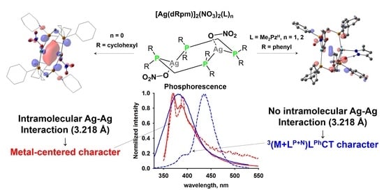
Two silver nitrate complexes with bisphosphines were obtained and characterized: [Ag(dcypm)]2(NO3)2 (1; dcypm = bis(dicyclohexylphosphino)methane) and [Ag(dppm)]2(Me2PzH)n(NO3)2 (n = 1, 2a; n = 2, 2b; dppm = bis(diphenylphosphino)methane, Me2PzH = 3,5-dimethylpyrazole). The steric repulsions of bulky cyclohexyl substituents prevent additional ligand coordination to the silver atoms in 1. Compounds obtained feature the bimetallic eight-member cyclic core [AgPCP]2. The intramolecular argenthophilic interaction (d(Ag···Ag) = 2.981 Å) was observed in complex 1. In contrast, the coordination of pyrazole led to the elongation of Ag···Ag distance to 3.218(1) Å in 2a and 3.520 Å in 2b. Complexes 1 and 2a possess phosphorescence both in the solution and solid state. Time-dependent density-functional theory (TD-DFT) calculations demonstrate the origin of their different emission profile. In the case of 1, upon excitation, the electron leaves the Ag–P bonding orbital and locates on the intramolecular Ag···Ag bond (metal-centered character). Complex 2a at room temperature exhibits a phosphorescence originating from the 3(M + LP+N)LPhCT state. At 77 K, the photoluminescence spectrum of complex 2a shows two bands of two different characters: 3(M + LP+N)LPhCT and 3LCPh transitions. The contribution of Ag atoms to the excited state in both complexes 2a and 2b decreased relative to 1 in agreement with the structural changes caused by pyrazole coordination.

Graphical Abstract

1. Introduction

The non-covalent metal–metal interactions play a significant role in the emission [1,2] or catalytic [3,4,5] properties of the d10 metal complexes. Dinuclear or trinuclear coinage metal complexes with bridging bis- or trisphosphine ligands represent a unique class of molecules occupying a position between mononuclear complexes and nanoparticles. They have remarkable chemical and physicochemical properties due to the presence of metal–metal bonds of various energy [6]. For example, di- and tetranuclear silver nitrates with bis(diphenylphosphino)methane are known since 1983 [7]. The metal-centered (MC) nature of emission is well known for coinage metal complexes with bridging phosphines. Already in 1989, Che et al. showed on an example of gold dimer [Au2(dppm)2]2+ that shortened Au–Au distances participate in electron transitions [8]. Then, on the example of silver dinuclear complexes with bisphosphines [Ag(dcypm)]2X2 (X = PF6, and CF3SO3, or CN; dcypm = bis(dicyclohexylphosphino)methane) [9,10] and [Ag(dmb)]2X2 complexes (X = Cl, Br, I; dmb = 1,8-diisocyano-ρ-menthane) [11], it has been shown that UV–Vis (Ultraviolet–visible) absorption band of complexes to d–p transition from intramolecular argentophilic interaction. The metallophilic nature of this band corresponding to the stretching Ag–Ag vibration (80 cm−1) was proven by Raman spectroscopy with excitation at the UV maximum [9]. The same behavior has also been observed for metallothioneins [11,12] and nanoclusters [13,14]. The interaction between two coinage metal ions has been investigated by spectral as well as computational methods [15,16]. Recently, using the example of Au/Ag di- and trinuclear complexes with diphosphanyl NHC ligands, this phenomenon was explained experimentally and theoretically [17].
Surprisingly, despite the wide structural diversity of coinage metals and bisphosphine complexes, there is practically no information about silver(I) nitrate complexes. Herein, we present the synthesis, structures, photophysical properties of dinuclear silver(I) bisphosphines [Ag(dcypm)]2(NO3)2 (1) (dcypm = bis(dicyclohexylphosphino)methane) and [Ag(dppm)]2(Me2PzH)n(NO3)2 (n = 1, 2a; n = 2, 2b) (dppm = diphenylphosphino)methane) with 3,5-dimethylpyrazole (Scheme 1). The influence of Ag···Ag distances or the ligand at silver atoms on the emission properties will be discussed involving the theoretical interpretation.

2. Materials and Methods

1H and 31P{1H} nuclear magnetic resonance (NMR) measurements were carried out on a Bruker Avance 400 spectrometer. Infrared (IR) spectra were collected on a Shimadzu IR Prestige-21 FT-IR (Fourier-transform infrared spectroscopy) spectrometer using KBr pellets. The photoluminescence spectra and lifetime measurements of the phosphors were recorded at 77 K and 298 K on a Fluorolog-3 spectrofluorometer system (HORIBA Jobin-Yvon, the excitation source was a 450 W xenon lamp with Czerny–Turner double monochromators, and the registration channels were a R928 photomultiplier, while a 150 W pulsed xenon lamp was used for lifetime measurements). The powders for these measurements were packed in quartz capillaries. The phosphorescence quenching curves were analyzed using the FluoroEssence™ software for calculation of the phosphorescence lifetime values. The UV–Vis spectra were measured on a Cary50 spectrometer.
X-ray diffraction study. Single-crystal X-ray diffraction experiments for complexes 1, 2a, and 2a/2b were carried out with a Bruker SMART APEX II diffractometer. The structures were solved by the direct method and refined in anisotropic approximation for non-hydrogen atoms. Hydrogen atoms of methyl, methylene, and aromatic fragments were calculated according to those idealized geometries and refined with constraints applied to C–H and N–H bond lengths and equivalent displacement parameters (Ueq(H) = 1.2Ueq(X), where X is the central atom of the XH₂ group; and Ueq(H) = 1.5Ueq(Y), Y—central atom of YH₃ group). All structures were solved with the ShelXT program [18] and refined with the ShelXL program [19]. Molecular graphics were drawn using the OLEX2 program [20]. The cyclohexyl fragment in complex 1, Me2PzH ligand in complex 2a, and in the second molecule in co-crystal 2a/2b were disordered, and the occupancy of positions of disordered levels was 0.5. Nitrate anions in the second molecule (2a) in co-crystal 2a/2b were disordered, and the occupancy of positions of disordered levels were 0.3 and 0.7. CCDC 2013947, 2013948, and 2033764 contained the supplementary crystallographic data for complexes 1, 2a, and 2a/2b. These data can be obtained free of charge from the Cambridge Crystallographic Data Center via https://www.ccdc.cam.ac.uk/structures. Crystal data and structure refinement parameters are presented in Table 1.
Computational details. Full geometry optimizations of complexes 1 and 2 were performed using the PBE0 functional [21] and SVP (split valence polarization) basis set [22,23] without any symmetry restrictions using Gaussian09 software [24]. Optimized geometries of 1, 2a, and 2b reasonably reproduced the X-ray geometry (Figure S14 in the Supplementary Materials). The equilibrium geometry for 1 and 2b had a Ci symmetry point group (as it was found by X-ray analysis), leading to the symmetry forbidden electronic transitions, so only transitions of Au symmetry were analyzed. For complex 2a, the second pyrazole molecule was left, but rotated to be in the Me2PzH ligand position of a neighbor complex in the crystal. Herewith, that molecule remained non-bonded by the Ag–N bond, but its presence helps to keep the complex similar to the X-ray characterized one. Vertical singlet and triplet excitations were calculated by TD-DFT on the same level of theory. Analysis of the excited states was done with the Multiwfn program [25]. The similarity of the excited states was analyzed by the approach suggested by Chen [26].
s H / E = 1 i | a i b i | 2
where ai and bi are contributions of the i atom to the hole (sH) or electron (sE) in the states a and b.
Synthesis. All operations were performed with Schlenk techniques under a dry argon atmosphere. Commercially available solvents and ligands were used without additional purification.
[Ag(dcypm)]2(NO3)2 (1). Mixture of AgNO3 (50 mg, 0.294 mmol) and bis(dicyclohexylphosphino)methane (120 mg, 0.294 mmol) was stirred in 5 mL of CH2Cl2 at room temperature for 3 h. A white solid was afforded upon precipitation by n-hexane. The precipitate formed was filtered, washed with 10 mL of acetone, and dried in vacuum. Yield 91%. NMR (CD2Cl2, ppm) 1H: δ = 1.19–2.13 ppm (m, 88 HCy and 4 CH2), 31P{1H}: δ = 28.8 (m, 4P). Anal. calcd/found for C50H92Ag2N2O6P4: C, 51.91/52.01; H, 8.02/8.08; N, 2.42/2.40.
[Ag(dppm)]2(Me2PzH)(NO3)2 (2a). The suspension of 50 mg of AgNO3 (0.294 mmol) and 17.3 mg of 3,5-dimethylpyrazole (0.180 mmol) was stirred in 10 mL of acetone for the one hour until a slurry precipitate was formed, and then the solution of dppm (113 mg, 0.294 mmol) in 3 mL of acetone was added. The reaction mixture was stirred overnight at room temperature. The precipitate formed was filtered off and washed with 10 mL of CH2Cl2. The solution obtained was added to 20 mL of hexane, and the mixture was kept in the refrigerator at ca. 5 °C for one hour. The precipitate formed was filtered off, washed with hexane, and both solids were combined and dried under reduced pressure. Complex 2a was obtained by slow crystallization at room temperature from boiling CH3CN solution of small portions of these solids. Yield 68%. NMR (CD3CN, ppm) 1H: δ = 2.07 ppm (s, 6H, MePz), 3.71 ppm (m, 4H, CH2dppm), 5.74 ppm (s, 1H, CHPz), 7.20–7.44 (m, 40H, Phdppm), 31P{1H}: δ = 7.5 (m, 4P); IR (KBr, cm−1): ν 3201 (νNH), 3138, 3050, 2922, 2876 (νCH), 1580. Anal. calcd/found for C55H54Ag2N4O6P4: C, 54.75/54.07; H, 4.51/4.24; N, 4.64/4.60.

3. Results and Discussion

3.1. Synthetic Procedures

Complex 1 was obtained by the interaction of AgNO3 with bis(dicyclohexylphosphino)methane (dcypm) in dichloromethane (DCM) and subsequent precipitation with hexane. Moreover, the addition of dcypm to the solution of the complex [Ag(Me2PzH)]NO3 generated in situ only led to complex 1 with high yield. This suggests that the steric effect of bulk cyclohexyl substituents makes impossible the pyrazole coordination to the silver atoms. In contrast, mixing AgNO3 with 3,5-dimethylpyrazole and less sterically demanding bis(diphenylphosphino)methane (dppm) in acetone led to the formation of the precipitate. The elemental analysis and 1H NMR spectrum demonstrated that the composition of the bulk solids corresponded to the complex [Ag2(Me2PzH)(dppm)]2(NO3)2 (2b) containing two pyrazole molecules per [Ag(dppm)]2(NO3)2 moiety (Figures S19 and S20). Unfortunately, reprecipitation or crystallization of this complex at low temperatures led to the partial loss of pyrazole molecules, resulting in a mixture of complexes [Ag(dppm)]2(Me2PzH)(NO3)2 (2a) [Ag2(Me2PzH)(dppm)]2(NO3)2 (2b). In a pure form, only complex 2a could be obtained by slow crystallization from boiling acetonitrile solution at room temperature. It should be noted that the elimination of the second 3,5-dimethylpyrazole molecule was not observed.

3.2. Crystal Structure of Complexes

The structures of compounds obtained were proved by single-crystal X-ray diffraction. The crystallographic data and structure refinement details are summarized in Table 1, and the relevant bond distances and angles are collected in Table 2.
Colorless crystals of complex 1 was obtained by slow evaporation of their DCM/hexane (v/v = 2:1) solutions at ca. 5 °C. Complex 1 features a chair form of the eight-member cyclic [AgPCP]2 core (Figure 1), in which silver atom is coordinated to four atoms (two Pdppm (2.396(2) and 2.429(2) Å), ONitrate (2.558(9) Å) and Ag (2.9812(7) Å), forming the distorted triangular pyramidal or tetrahedron environment (Figure 2). The angles P1-Ag1-P2, P1-Ag1-O1, P1-Ag1-Ag1, O1-Ag1-Ag1 were 152.22(6)°, 119.1(2)°, 94.21(4)°, and 74.4(2)°, respectively. There were only several examples of the similar [Ag(dcypm)]22+ core with PF6, CF3SO3 [9], and CN [10] counter ions. Observed Ag–Ag bond length (2.9812(7) Å) was in the range of shared argentophilic interactions [27,28,29] being non-significantly longer than that present in the literature (av. 2.93 Å). The central core [MPCP]2 was similar for dinuclear silver bisphosphines, but additional κ1 coordination of the nitrate anion led to a significant distortion of the central cycle. Silver atoms as well as CH2 fragments lie out the P4 plane for 0.557 and 0.607 Å, respectively. For example, the same parameters for complex with CN were 0.415 and 0.575 Å [10]. These structural peculiarities reflect the influence of the nitrate anion on the electronic properties of complexes (vide infra). The Ag–P bond lengths (2.396(2) and 2.429(2) Å) were typical for the silver bis-phosphines.
Supramolecular packing of 1 was realized via the network of weak CCy-H···ONitrate interactions of neighboring molecules (2.316 Å, O-H-C = 156.1°; Figure 2) [30,31].
In contrast, in the case of interaction of the silver nitrate with dppm ligand and 3,5-dimethylpyrazole, complexes of different compositions were observed. As demonstrated above, the interaction led to the formation of the precipitate. Analysis of this solid indicates that it mainly contained one Ag atom per one dppm ligand and one Me2PzH molecule. However, varying the crystallization conditions allowed for two different types of complexes. The solubility of the solids obtained was very low, and crystallization was performed from significantly diluted solutions (mM concentrations). The most stable one was complex 2a containing one pyrazole molecule, crystals that could be obtained at different temperatures (−10, 0, or 25 °C) and from different solvents (MeOH, CH3CN, and DCM/hexane mixture). Slow crystallization from the boiling CH3CN at room temperature was the best condition for obtaining pure complex 2a. Complex 2b was obtained only as a co-crystal with 2a, representing two independent molecules with one and two Me2PzH molecules. This sample was crystallized in an NMR tube after a 1H NMR experiment in CD2Cl2, which eventually showed the 1/1 ratio of Me2PzH/dppm in solution. These data demonstrate that crystallization from a diluted solution led to the elimination of one pyrazole from the complex [Ag(dppm)Me2PzH]2(NO3)2 (2b), resulting in a more stable [Ag(dppm)]2(Me2PzH)(NO3)2 (2a) complex.
Complex 2a crystallizes in a chair form of the central [AgPCP]2 cycle (Figure 3). There was only one molecule of pyrazole coordinated with one of the silver atoms in the crystal. The complex in the crystal was in the superposition of two possible isomers in which the pyrazole ligand coordinates by nitrogen to both silver atoms (occupation 0.5). This behavior leads to the presence of a pseudo inversion center at the center of the Ag–Ag distance. Interaction with Me2PzH ligand led to the pentacoordinate environment distorted trigonal bipyramidal of one silver atom (two Pdppm (2.403(2) and 2.442(2) Å), NPz (2.446(6) Å), ONitrate (2.554(6) Å), and Ag (3.218(1) Å) (Figure 3). The angles P1-Ag1-P2, P1-Ag1-O1, and P1-Ag1-N1 were 151.61(8)°, 126.5(1)°, and 98.6(2)°, respectively. The presence of the 3,5-dimethylpyrazole ligand led to the κ1 coordination of the nitrate to silver atoms in contrast to the known [Ag(dppm)]2(NO3)2 complex [7,32], which possesses the κ2 coordination mode of the nitrate anion. Moreover, the coordination of pyrazole led to the elongation of the Ag–Ag distance to 3.218 Å, which was significantly longer than that for [Ag(dppm)]2(NO3)2 (3.090–3.110 Å) [7,32]. The Ag–Ag distance was in the range of closed-shell metallophilic interactions [27]. Silver atoms as well as CH2 fragments, were located on both sides of the P4 plane lying out at 0.594 and 0.720 Å, respectively, which determines the chair-configuration of the central M2P4 core. It should be noted that Tiekink [32] also reported the same configuration for [Ag(dppm)(NO3)]2 with a more distorted central core in contrast to the boat form obtained by Bau [7]. The Ag–P bonds lengths in 2 (2.403(2) and 2.442(2) Å) are typical for the silver bis-phosphines complexes. The Ag–NPz bond length (2.446(4) Å) is typical for the silver phosphine complexes with donor aromatic nitrogen ligands [33,34]. The complex was stabilized via O1Nitrate∙∙∙H–NPz intramolecular hydrogen bond (1.702 Å, O-H-N = 165.9°) [30,31].
Complex 2b was obtained only in the form of a co-crystal of two molecules of general motif [Ag(dppm)]2(NO3)2 with one and two pyrazoles, respectively (Figure 4). The part that contained one molecule of pyrazole was generally identical to compound 2a with non-significant differences in bond lengths and angles. The second part was a dinuclear complex 2b with two molecules of 3,5-dimethylpyrazole coordinated to both silver atoms. Complex 2b also crystallized in a chair form of the central [AgPCP]2 cycle. Moreover, there was the inversion center of this fragment in the middle of the Ag–Ag distance. The second pyrazole molecule’s coordination led to the significant elongation of the Ag–Ag distance (3.520 Å). As a result, the presence of one or two coordinated ligands to the silver atoms in [Ag(dppm)]2 cores led to the elongation of Ag–Ag distances and the absence of the shared intramolecular argenthophilic interactions. The Ag–P (2.446(1) and 2.422(1) Å) bond lengths were similar to those observed in complexes 1 and 2a. The interaction of silver atoms with pyrazoles in 2b was non-significantly stronger than that for 2a in accordance with the correlation of the shortened Ag–NPz bond lengths (2.446(1) Å for 2a and 2.381(4) Å for 2b). In contrast, the coordination of addition pyrazole led to the significant elongation of the Ag–ONitrate bonds (2.554(6) Å for 2a and 2.674 (3) Å for 2b).

3.3. Photophysical Properties

The only pure compounds 1 and 2a, in contrast to 2b, were isolated. All experimental studies (UV–Vis and photoluminescence) were performed for these complexes. The theoretical investigations (TD-DFT) were performed for all possible types of complexes (1, 2a, and 2b).
The TD-DFT calculations were performed for complexes 1 and 2b keeping their local Ci symmetry. The presence of the inversion center led to the appearance of the symmetry forbidden Ag, and the symmetry allowed Au transitions. This is why only symmetry allowed Au transitions are discussed further. The lowest excitations of 1 were S1 and S2 with energies of 4.18 and 5.01 eV (296 and 247 nm; Table S1), respectively. The S1 had a pronounced LCNO3 character with a negligible impact of Ag atoms (Figure S1, Table S1). The oscillator strength (f) value of 0.0002 demonstrates that this excitation was ineffective (dark state). In contrast, the second singlet transition (5.01 eV, f = 0.1853) had a metal-centered (MC) nature (Figure 5). Upon this excitation, the electron leaves the Ag–P σ bonding orbital (impacts of Ag—40% and P—36%, Table S1) and locates on the intramolecular Ag–Ag bond, which is generated by the empty 5pz orbitals of the silver cations. The vertical excitation energies for the three lowest triplets were 3.70, 4.30, and 4.60 eV (334, 287, and 269 nm), respectively. The first two triplets were of a LCNO3 nature, and the impact of metals and dpcym ligands appeared in the T3 state. The T3 had an excellent match to the S2 state; their similarity was sH = 0.91 and sE = 0.95. Along with the oscillator strengths, this indicates a high probability for the excitation of 1 directly to S2 state, S2 → T3 interconversion, followed by the triplet emission from the T3 state, which describes the emission observed (vide infra, Figure S13).
The excitation pattern of the complex [Ag(Me2PzH)(dppm)]2(NO3)2 (2b) differed from that observed for [Ag(dcypm)]2(NO3)2 (1). The first singlet (4.16 eV, 298 nm, f = 0.2354; Figure 6) possessed a comparable impact to the hole from metals (35%) and phosphorus atoms (from dppm, 33%) with some contribution from 10% dimethylpyrazole (Figure S2, Table S2). However, the excited electron was located mainly on the organic part of the dppm ligand (64%), indicating the charge transfer character of this transition. Therefore, it could be ascribed to (Ag+ + Pdppm + NPyrazole) → (Ph + CH2)dppm charge transfer. The electron density at the Ag···Ag intramolecular contact was still present in the excited S1 state. Still, the contribution of Ag atoms to the excited state dropped by three times compared to the S2 of complex 1, and as a result, it could be observed only at the 0.03 a.u. isosurface (Figure S2). These data indicate the role of the dimethylpyrazole ligand in the metal-induced electronic transitions. The four lowest triplet states for 2b were the 3LC states of Ph substituents in dppm ligands, while T5 was the 3LCNO3 state (Figure S2). The T6 of MLCT nature is the state coupled with the S1 excitation, having high similarity (sH = 0.91 and sE = 0.90) to the S0 → T6 transition energy 4.02 eV (308 nm) (Figure 6). Similar behavior was observed in the case of dinuclear silver pyrazolates with PCy3 and PPh3 ligands [35].
The TD-DFT analysis of [Ag(dppm)]2(Me2PzH)(NO3)2 (2a) excitations led to nearly the same pattern, except the disappearance of symmetry forbidden states, which led to doubling of the number of corresponding electronic transitions. At that, the S1 and S2 states both possess mixed MLCT (Ag+ + Pdppm + NPyrazole) → (Ph + CH2)dppm + LCNO3 character with close excitation energies and oscillator strengths (4.11 eV, 301 nm, f = 0.0994 for S1 and 4.17 eV, 297 nm, f = 0.1605 for S2) as a result of mixing with symmetry forbidden AG 1LCNO3 state upon loss of symmetry (Figure 7, Table S3). Despite this mixing, the simulated excitation spectra were the same for 2a and 2b (Tables S2 and S3, Figure S3). For triplet transitions due to the unsymmetrical structure, the eight lowest triplets were 3LC states of Ph substituents in dppm ligands, and T9–T10 are 3LCNO3 states, while T11 and T12 were MLCT states. Due to the mixing of singlet states with LCNO3 transition, the discussed singlet and triplet states had a low similarity. Still, if we ignore the NO3 group’s contributions, the similarity for S2-T11 was sH = 0.83, sE = 0.79 and for S2–T12 it was sH = 0.84, sE = 0.75. Based on these, practically the same conclusion can be made as for 2b. The lowest singlet excitation should lead to the high energy triplets T11–T12 that is likely to interconvert to the lower 3LC states of dppm’ Ph substituents, which in turn has a trend to non-emissive relaxation at room temperature [35,36,37], but could be stabilized at 77 K.
The UV−Vis spectra of 1 and 2a were measured in CH2Cl2 (Figure 8). Complex 1 demonstrated an intense absorbance band at 267 nm (ε = 13350 cm−1 M). This band can be attributed to the transition with a metal-centered (MC) character originating from intramolecular Ag(I)–Ag(I) interactions [9]. The presence of the nitrate anion in complex 1 resulted in the non-intensive tail around 300 nm, which correlated with the absorption spectrum of the aqueous solution of silver nitrate.
UV–Vis spectrum of compound 2a showed a broad, intense band at 288 nm (ε = 12800 cm−1 M−1) with a long tail at ca. 335 nm, which can be assigned to the charge-transfer (MLCT or LLCT) transitions. Additionally, in the high energy region (<280 nm), weakly resolved bands with the contribution from several bands of close energies were observed. This band could be assigned only to the transitions of the π → π* character within the Ph substituents in bisphosphine and pyrazole ligands.
The emission spectra of complexes 1 and 2a were studied in CH2Cl2 solution and the solid state (Figure 9). The room temperature (RT) emission spectrum of complex 1 in the solution displayed two bands at 370 and 390 nm, respectively, which can be assigned to the emission from the electronically excited states of MC nature. The emission bands of 1 in the solid state were in the same position. Temperature decrease led to the non-significant redistribution of the solid sample band’s intensities. The triplet state decays also demonstrate this mixed emissive behavior. The phosphorescence decay of 1 both in the solution and in solid state at RT and 77 K can only be fitted with bi-exponential function. In the CH2Cl2 solution at RT, lifetimes were 1 and 10 µs. In the solid state lifetimes, τ was practically independent of temperature being ca. 2 and 16 µs. These data demonstrate that complex 1 possessed the same photoluminescence both in solution and solid state. Complex 2a in the acetonitrile solution possessed phosphorescence bands at 365 and 410 nm (for both τ = 8.7 µs). In the solid state at RT, complex 2a showed an unstructured band at 380 nm of CT nature (τ = 9 µs). Interestingly, at 77 K, the presence of a new intense band at 440 nm was observed (τ = 37 µs). The position of the band observed at RT shifted from 380 to 390 nm at 77 K, but the lifetime of this emission was not dependent on the temperature (8.9 µs). The results of calculations (vide supra) showed that the T1–T8 states were of LC nature within Ph substituents. As we have shown for structurally similar silver pyrazolate/dppm complexes, these states are not emissive due to the intersection of their energy surface with the ground state, which allows effective non-radiative relaxation [35,36,37]. The emission observed at 298 K can be ascribed to the system relaxation from T11–12 (3.98–4.21 eV) and higher states of ((M + LP+N)LdppmCT) nature. The temperature decrease led to the stabilization of triplet states of the LCPh nature and these channels possess a main influence on the emission of 2a observed at 77 K.

4. Conclusions

The reaction of silver nitrate with bisphosphines in the presence of 3,5-dimethylpyrazole gave new examples of dinuclear silver(I) complexes of different composition [Ag(dcypm)]2(NO3)2 (1), [Ag(dppm)]2(NO3)2(Me2PzH) (2a), and [Ag(Me2PzH)(dppm)]2(NO3)2 (2a). This suggests that the steric repulsion of bulky cyclohexyl substituents does not allow coordination of additional pyrazole ligand to the silver atoms in [Ag(dcypm)]2(NO3)2. Complex 2a was obtained by the crystallization of 2b from a diluted solution in accordance with low solubility accompanied by the elimination of one pyrazole molecule. For all complexes, non-typical κ1 coordination of the nitrate anion was observed, which led to a significant distortion of the central eight-member cycle [AgPCP]2 in comparison to related silver bisphosphine complexes. Complexes possessed phosphorescence both in the solution and in the solid-state. The intramolecular argenthophilic interaction observed in complex 1 (d(Ag∙∙∙Ag) = 2.981 Å) introduces the main excitation channel in which an electron leaves the Ag–P bond orbital and locates on the intramolecular Ag–Ag bond (metal-centered character). The coordination of dimethylpyrazole to Ag(I) in complex 2a led to the elongation of the Ag∙∙∙Ag distance (3.218 Å). Complex 2a exhibited a phosphorescence originating from the charge-transfer from the 3(M + LP+N)LPhCT state at room temperature. At 77 K, the emission was split into two components: 3(M + LP+N)LPhCT and 3LCPh transitions. According to the TD-DFT calculations, the coordination of dimethylpyrazole to the metal atom mainly affects the energy of transition.

Supplementary Materials

The following are available online at https://0-www-mdpi-com.brum.beds.ac.uk/2073-4352/10/10/881/s1, Figure S1: Natural transition orbital’s of 1 as the isosurface at 0.05 a.u.; Table S1: Computed characteristics of the excited states of 1. For analysis of fragment impacts (in %) to the ground state (hole) and excited state (electron), complex was divided to fragments of all Ag atoms, all P atoms, nitrate anions, and all atoms of dcypm ligand except P.; Figure S2: Natural transition orbital’s of 2 as the isosurface at 0.05 a.u. except for S1 and T6; Table S2: Computed characteristics of the Au symmetry excited states of 2. For analysis of fragment impacts (in %) to the ground state (hole) and excited state (electron), the complex was divided to fragments of all Ag atoms, all P atoms, nitrate anions, pyrazolates, and all atoms of dppm ligand except P.; Figure S3: Natural transition orbital’s of 2a as the isosurface at 0.05 a.u., Table S3. Computed characteristics of the AU symmetry excited states of 2a; Figure S4: Phosphorescence decay by delay of complex 1 at RT in CH2Cl2 solution (left) and solid state (right); Figure S5: Phosphorescence decay by delay of complex 1 in the solid state at 77 K; Figure S6. Phosphorescence decay by delay of complex 2a at RT in CH2Cl2 solution (left) and solid state (right); Figure S7: Phosphorescence decay by delay of complex 2a in the solid state at 77 K (measured at 390 nm); Figure S8: Phosphorescence decay by delay of complex 2a in the solid state at 77 K (measured at 440 nm); Figure S9: The 1H NMR spectrum of complex 1 in CD2Cl2; Figure S10: The 31P{1H} NMR spectrum of complex 1 in CD2Cl2; Figure S11: The 1H NMR spectrum of complex 2a in CD3CN; Figure S13: Schematic Jablonski diagram for 1, 2a, 2b. Presumably active states indicated by solid lines; Figure S14: Comparison of X-ray and DFT geometry of Ag2P4(NO3)2 core for 1, 2a and 2b. Atoms of X-ray structure is shaded, DFT structure is colored. Ag—cyan, P—orange, C—gray, O—red, N—blue; Figure S15: Excitation spectra of 1 in CH2Cl2 solution (left, em. on 370 and 390), and in the solid state (right, em. 370); Figure S16: Excitation spectra of 2a in CH2Cl2 solution (left, em. on 410) and in the solid state (right, em. on 380); Figure S17: IR spectrum of complex 2a in KBr pellets; Figure S18: The simulated electronic spectra of 1, 2a, and 2b. The 200 lowest energy singlet transitions were considered, and full width at half maximum was set to 40; Figure S19: 1H NMR spectrum of the precipitate obtained in the case of interaction silver nitrate with dppm and Me2PzH, demonstrating Ag/(Me2PzH)/dppm = 1/1/1 ratio, CD2Cl2; Figure S20: 1H NMR spectrum of the precipitate obtained in the case of interaction silver nitrate with dppm and Me2PzH, demonstrating Ag/(Me2PzH)/dppm = 1/1/1 ratio, CD3OD.

Author Contributions

Synthetic experiments, K.F.B.; Experiments and writing original draft, A.A.T.; Calculations and visualizations, O.A.F.; XRD studies, A.F.S.; Photoluminescence, A.A.A.; Writing review & editing, A.A.T., O.A.F., and E.S.S. All authors have read and agreed to the published version of the manuscript.

Funding

This work was financially supported by the Russian Foundation for Basic Research (RFBR grant no. 18-33-20060).

Acknowledgments

NMR and XRD measurements were performed using the equipment of the Center for Molecular Composition Studies at the INEOS RAS and were supported by the Ministry of Science and Higher Education of the Russian Federation. Photophysical properties were measured using the equipment of the Center of Physical Methods of Investigation at the IPCE RAS.

Conflicts of Interest

The authors declare that there is no conflicts of interest.

References

  1. Yam, V.W.; Au, V.K.; Leung, S.Y. Light-Emitting Self-Assembled Materials Based on d(8) and d(10) Transition Metal Complexes. Chem. Rev. 2015, 115, 7589–7728. [Google Scholar] [CrossRef]
  2. Schmidbaur, H.; Schier, A. Argentophilic interactions. Angew. Chem. Int. Ed. 2015, 54, 746–784. [Google Scholar] [CrossRef]
  3. Dolan, N.S.; Scamp, R.J.; Yang, T.; Berry, J.F.; Schomaker, J.M. Catalyst-Controlled and Tunable, Chemoselective Silver-Catalyzed Intermolecular Nitrene Transfer: Experimental and Computational Studies. J. Am. Chem. Soc. 2016, 138, 14658–14667. [Google Scholar] [CrossRef]
  4. Alderson, J.M.; Corbin, J.R.; Schomaker, J.M. Tunable, Chemo- and Site-Selective Nitrene Transfer Reactions through the Rational Design of Silver(I) Catalysts. Acc. Chem. Res. 2017, 50, 2147–2158. [Google Scholar] [CrossRef]
  5. Mak, C.L.; Bostick, B.C.; Yassin, N.M.; Campbell, M.G. Argentophilic Interactions in Solution: An EXAFS Study of Silver(I) Nitrene Transfer Catalysts. Inorg. Chem. 2018, 57, 5720–5722. [Google Scholar] [CrossRef]
  6. Grachova, E.V. Design of Supramolecular Cluster Compounds of Copper Subgroup Metals Based on Polydentate Phosphine Ligands. Russ. J. Gen. Chem. 2019, 89, 1102–1114. [Google Scholar] [CrossRef]
  7. Ho, D.M.; Bau, R. Preparation and structural characterization of [Ag2(dpm)2(NO3)2] and [Ag4(dpm)4(NO3)2]2+[PF6]2−: Conformational flexibility in the M2P4 core structure of bis(diphenylphosphino)methane complexes. Inorg. Chem. 1983, 22, 4073–4079. [Google Scholar] [CrossRef]
  8. Che, C.-M.; Kwong, H.-L.; Yam, V.W.-W.; Cho, K.-C. Spectroscopic properties and redox chemistry of the phosphorescent excited state of [Au2(dppm)2]2+[dppm = bis(diphenylphosphino)methane]. J. Chem. Soc. Chem. Commun. 1989, 885–886. [Google Scholar] [CrossRef]
  9. Che, C.-M.; Tse, M.-C.; Chan, M.C.W.; Cheung, K.-K.; Phillips, D.L.; Leung, K.-H. Spectroscopic Evidence for Argentophilicity in Structurally Characterized Luminescent Binuclear Silver(I) Complexes. J. Am. Chem. Soc. 2000, 122, 2464–2468. [Google Scholar] [CrossRef]
  10. Lin, Y.Y.; Lai, S.W.; Che, C.M.; Fu, W.F.; Zhou, Z.Y.; Zhu, N. Structural variations and spectroscopic properties of luminescent mono- and multinuclear silver(I) and copper(I) complexes bearing phosphine and cyanide ligands. Inorg. Chem. 2005, 44, 1511–1524. [Google Scholar] [CrossRef]
  11. Piché, D.; Harvey, P.D. The lowest energy excited states of the binuclear silver(I) halide complexes, Ag2(dmb)2X2. Metal-centered or charge transfer states? Canad. J. Chem. 1994, 72, 705–713. [Google Scholar] [CrossRef]
  12. Chan, J. Studies of metal binding reactions in metallothioneins by spectroscopic, molecular biology, and molecular modeling techniques. Coord. Chem. Rev. 2002, 233-234, 319–339. [Google Scholar] [CrossRef]
  13. Chen, Y.; Yang, T.; Pan, H.; Yuan, Y.; Chen, L.; Liu, M.; Zhang, K.; Zhang, S.; Wu, P.; Xu, J. Photoemission mechanism of water-soluble silver nanoclusters: Ligand-to-metal-metal charge transfer vs strong coupling between surface plasmon and emitters. J. Am. Chem. Soc. 2014, 136, 1686–1689. [Google Scholar] [CrossRef]
  14. Yang, T.; Dai, S.; Tan, H.; Zong, Y.; Liu, Y.; Chen, J.; Zhang, K.; Wu, P.; Zhang, S.; Xu, J.; et al. Mechanism of Photoluminescence in Ag Nanoclusters: Metal-Centered Emission versus Synergistic Effect in Ligand-Centered Emission. J. Phys. Chem. C 2019, 123, 18638–18645. [Google Scholar] [CrossRef]
  15. Blake, A.J.; Donamaria, R.; Lippolis, V.; Lopez-de-Luzuriaga, J.M.; Manso, E.; Monge, M.; Olmos, M.E. Influence of crown thioether ligands in the structures and of perhalophenyl groups in the optical properties of complexes with argentoaurophilic interactions. Inorg. Chem. 2014, 53, 10471–10484. [Google Scholar] [CrossRef]
  16. Donamaria, R.; Lippolis, V.; Lopez-de-Luzuriaga, J.M.; Monge, M.; Nieddu, M.; Olmos, M.E. Influence of the Number of Metallophilic Interactions and Structures on the Optical Properties of Heterometallic Au/Ag Complexes with Mixed-Donor Macrocyclic Ligands. Inorg. Chem. 2018, 57, 11099–11112. [Google Scholar] [CrossRef]
  17. Ai, P.; Mauro, M.; Gourlaouen, C.; Carrara, S.; De Cola, L.; Tobon, Y.; Giovanella, U.; Botta, C.; Danopoulos, A.A.; Braunstein, P. Bonding, Luminescence, Metallophilicity in Linear Au3 and Au2Ag Chains Stabilized by Rigid Diphosphanyl NHC Ligands. Inorg. Chem. 2016, 55, 8527–8542. [Google Scholar] [CrossRef]
  18. Sheldrick, G. SHELXT—Integrated space-group and crystal-structure determination. Acta Crystallogr. Sect. A. 2015, 71, 3–8. [Google Scholar] [CrossRef] [Green Version]
  19. Sheldrick, G. Crystal structure refinement with SHELXL. Acta Crystallogr. Sect. C. 2015, 71, 3–8. [Google Scholar] [CrossRef]
  20. Dolomanov, O.V.; Bourhis, L.J.; Gildea, R.J.; Howard, J.A.K.; Puschmann, H. OLEX2: A complete structure solution, refinement and analysis program. J. Appl. Crystallogr. 2009, 42, 339–341. [Google Scholar] [CrossRef]
  21. Adamo, C.; Barone, V. Toward reliable density functional methods without adjustable parameters: The PBE0 model. J. Chem. Phys. 1999, 110, 6158–6170. [Google Scholar] [CrossRef]
  22. Andrae, D.; Häußermann, U.; Dolg, M.; Stoll, H.; Preuß, H. Energy-adjustedab initio pseudopotentials for the second and third row transition elements. Theor. Chim. Acta 1990, 77, 123–141. [Google Scholar] [CrossRef]
  23. Weigend, F.; Ahlrichs, R. Balanced basis sets of split valence, triple zeta valence and quadruple zeta valence quality for H to Rn: Design and assessment of accuracy. Phys. Chem. Chem. Phys. 2005, 7, 3297–3305. [Google Scholar] [CrossRef]
  24. Frisch, M.J.; Trucks, G.W.; Schlegel, H.B.; Scuseria, G.E.; Robb, M.A.; Cheeseman, J.R.; Scalmani, G.; Barone, V.; Mennucci, B.; Petersson, G.A.; et al. Gaussian 09, Revision B.01; Gaussian Inc.: Wallingford, CT, USA, 2009. [Google Scholar]
  25. Lu, T.; Chen, F. Multiwfn: A multifunctional wavefunction analyzer. J. Comput. Chem. 2012, 33, 580–592. [Google Scholar] [CrossRef]
  26. Chen, R.; Tang, Y.; Wan, Y.; Chen, T.; Zheng, C.; Qi, Y.; Cheng, Y.; Huang, W. Promoting Singlet/triplet Exciton Transformation in Organic Optoelectronic Molecules: Role of Excited State Transition Configuration. Sci. Rep. 2017, 7, 6225. [Google Scholar] [CrossRef]
  27. Das, S.; Sharma, S.; Singh, H.B.; Butcher, R.J. Metallophilic Mercuraazamacrocycles Derived from Bis{6-formyl-(2,3,4-trimethoxy)phenyl}mercury: Reactivity with d10 and d8 Metal Ions. Eur. J. Inorg. Chem. 2018, 2018, 4093–4105. [Google Scholar] [CrossRef]
  28. Titov, A.A.; Filippov, O.A.; Smol’yakov, A.F.; Averin, A.A.; Shubina, E.S. Synthesis, structures and luminescence of multinuclear silver(I) pyrazolate adducts with 1,10-phenanthroline derivatives. Dalton Trans. 2019, 48, 8410–8417. [Google Scholar] [CrossRef]
  29. Emashova, S.K.; Titov, A.A.; Filippov, O.A.; Smol’yakov, A.F.; Titova, E.M.; Epstein, L.M.; Shubina, E.S. Luminescent AgI Complexes with 2,2′-Bipyridine Derivatives Featuring [Ag-(CF3)2Pyrazolate]4 Units. Eur. J. Inorg. Chem. 2019, 2019, 4855–4861. [Google Scholar] [CrossRef]
  30. Desiraju, G.R. Hydrogen bridges in crystal engineering: Interactions without borders. Acc. Chem. Res. 2002, 35, 565–573. [Google Scholar] [CrossRef]
  31. Steiner, T. The Hydrogen Bond in the Solid State. Angew. Chem. Int. Ed. 2002, 41, 48–76. [Google Scholar] [CrossRef]
  32. Tiekink, E.R.T. Bis-μ-[methylenebis(diphenylphosphine)]-dinitratodisilver(I) dichloroform solvate. Acta Crystallogr. Sect. C. 1990, 46, 235–238. [Google Scholar] [CrossRef]
  33. Cui, Y.-Z.; Yuan, Y.; Li, Z.-F.; Liu, M.; Jin, Q.-H.; Jiang, N.; Cui, L.-N.; Gao, S. From ring, chain to network: Synthesis, characterization, luminescent properties of silver(I) complexes constructed by diphosphine ligands and various N-donor ligands. Polyhedron 2016, 112, 118–129. [Google Scholar] [CrossRef]
  34. Qiu, Q.-M.; Huang, X.; Zhao, Y.-H.; Liu, M.; Jin, Q.-H.; Li, Z.-F.; Zhang, Z.-W.; Zhang, C.-L.; Meng, Q.-X. Synthesis, structure and terahertz spectra of six Ag(I) complexes of bis(diphenylphosphino)methane with 4,4′-bipyridine and its derivations. Polyhedron 2014, 83, 16–23. [Google Scholar] [CrossRef]
  35. Titov, A.A.; Filippov, O.A.; Smol’yakov, A.F.; Baranova, K.F.; Titova, E.M.; Averin, A.A.; Shubina, E.S. Dinuclear Cu(I) and Ag(I) Pyrazolates Supported with Tertiary Phosphines: Synthesis, Structures, and Photophysical Properties. Eur. J. Inorg. Chem. 2019, 2019, 821–827. [Google Scholar] [CrossRef]
  36. Titov, A.A.; Filippov, O.A.; Smol’yakov, A.F.; Averin, A.A.; Shubina, E.S. Copper(I) complex with BINAP and 3,5-dimethylpyrazole: Synthesis and photoluminescent properties. Mendeleev Commun. 2019, 29, 570–572. [Google Scholar] [CrossRef]
  37. Titov, A.A.; Filippov, O.A.; Smol’yakov, A.F.; Godovikov, I.A.; Shakirova, J.R.; Tunik, S.P.; Podkorytov, I.S.; Shubina, E.S. Luminescent Complexes of the Trinuclear Silver(I) and Copper(I) Pyrazolates Supported with Bis(diphenylphosphino)methane. Inorg. Chem. 2019, 58, 8645–8656. [Google Scholar] [CrossRef]
Scheme 1. Representation of central [MPCP]2 core for the complexes studied (R = Cy (1), Ph (2)).
Crystals 10 00881 sch001
Figure 1. X-ray diffraction (XRD) structure of complex 1 (carbon and hydrogen atoms are shown as sticks).
Crystals 10 00881 g001
Figure 2. Supramolecular packing of 1.
Crystals 10 00881 g002
Figure 3. XRD structure of the complex 2 (carbon and hydrogen atoms are shown as sticks).
Crystals 10 00881 g003
Figure 4. XRD structure of the co-crystal 2a/2b and dichloromethane (DCM) molecule (carbon, hydrogen, and chlorine atoms are shown as sticks).
Crystals 10 00881 g004
Figure 5. Highest occupied natural transition orbital (HONTO) and lowest unoccupied natural transition orbital (LUNTO) for the S0 → S2 (top) and S0 → T3 (bottom) transition in complex 1 as the isosurface at 0.05 a.u.
Crystals 10 00881 g005
Figure 6. HONTO and LUNTO for the S0 → S1 and S0 → T6 in complex 2b as the isosurface at 0.05 au.
Crystals 10 00881 g006
Figure 7. HONTO and LUNTO for the S0 → S2 and S0 → T11 in complex 2a as the isosurface at 0.05 au.
Crystals 10 00881 g007
Figure 8. Absorption spectra of complexes 1 (blue) and 2a (red) in CH2Cl2 solution.
Crystals 10 00881 g008
Figure 9. Normalized emission (λexc = 320 nm) spectra of complexes 1 (solid line) and 2a (dashed line) in the solution (black) and solid state at 298 K (red) and 77 K (blue).
Crystals 10 00881 g009
Table 1. Crystallographic data for complexes 1, 2a, and 2a/2b.
12a2a/2b
Empirical FormulaC₂₅H₄₆AgNO₃P₂C₅₅H₅₃Ag₂N₄O₆P₄C117H116Ag4Cl₂N₅O₆P₄
Formula weight578.441205.632675.24
DiffractometerBruker SMART APEX CCDBruker SMART APEX CCDBruker SMART APEX CCD
Scan modeϕ and ω scansϕ and ω scansω and ϕ scans
Anode [Wavelength, Å]MoKα [0.71073] sealed tubeMoKα [0.71073] sealed tubeMoKα [0.71073] sealed tube
Crystal Dimensions, mm0.1 × 0.15 × 0.360.08 × 0.11 × 0.150.08 × 0.12 × 0.44
Crystal colorcolorless colorless colorless
Crystal systemtetragonalmonoclinictriclinic
a, Å29.8637(11)10.940(2)10.9934(13)
b, Å29.8637(11)19.805(4)11.9540(14)
c, Å12.6441(5)11.719(2)22.858(3)
α, deg909077.727(2)
β, deg9091.087(4)82.796(2)
γ, deg909085.688(2)
Volume, ų11276.5(9)2538.5(9)2908.5(6)
Density, g cm⁻³1.3631.5771.527
Temperature, K120120120
Tmin/Tmax0.7056/0.74610.6339/0.74600.6141/0.7459
μ, mm⁻¹0.8530.9530.930
Space groupI4₁/aP2₁/nP1
Z1622
F(000)486412261360
Reflections collected545872361137353
Independent reflections5546499516342
Reflections (I > 2σ (I))457635459421
Parameters234308740
Rint0.03500.10530.0860
2θmin–2θmax2.728–51.9984.040–52.0003.670–59.410
wR₂ (all reflections)0.20320.15710.1221
R₁ (I > σ (I))0.07170.08660.0561
GOF1.0271.2200.972
ρminmₐₓ, eÅ⁻³−1.042/1.733−1.338/0.927−1.011/1.385
Table 2. Selected geometric parameters (bond lengths in Å, angles in degrees) for complexes 1 and 2a.
Bonds LengthsAngles
1212
Ag1-P12.396(2)Ag1-P12.403(2)Ag1-Ag1-P282.90(4)Ag1-Ag1-P281.59(5)
Ag1-O12.557(9)Ag1-O12.554(6)Ag1-P1-C1111.1(2)Ag1-O1-N3129.8(5)
Ag1-Ag12.9810(7)Ag1-N12.443(6)Ag1-P1-C2115.5(2)Ag1-N1-N2124.7(5)
Ag1-P22.428(2)Ag1-Ag13.218(1)Ag1-P1-C8113.2(3)P1-Ag1-O1126.5(1)
P1-C11.838(8)Ag1-P22.442(2)Ag1-O1-N1106.9(5)P1-Ag1-N198.6(2)
P1-C21.850(6)P1-C41.846(8)P1-Ag1-O1119.1(2)P1-Ag1-Ag188.90(5)
P1-C81.85(1)P1-C171.801(8)P1-Ag1-Ag194.21(4)P1-Ag1-P2151.61(8)
P2-C11.827(7)P1-C231.828(8)P1-C1-P2114.4(3)O1-Ag1-N184.1(2)
P2-C171.84(1)P2-C41.822(8)P1-Ag1-P2152.22(6)O1-Ag1-Ag191.5(1)
P2-C231.844(6)P2-C51.827(8)O1-Ag1-Ag174.4(2)O1-Ag1-P280.7(1)
O1-N11.27(1)P2-C111.804(8)O1-Ag1-P286.9(2)N1-Ag1-Ag1172.5(2)
O2-N11.244(8)P2-Ag12.442(2) N1-Ag1-P291.7(2)
O3-N11.12 (1)O1-N31.25(1) N2-N1-C1101.8(6)
O2-N31.24(1)
O3-N31.24(1)
N1-N21.31(1)
N1-C11.50(1)
N1-C21.58(1)
N2-C31.34(1)
N2-C21.02(1)