Screw-Dislocation-Driven Hierarchical Superstructures of Ag-Ag2O-AgO Nanoparticles


1
College of Chemistry, Chemical Engineering and Materials Science, Soochow University, Suzhou 215123, China
2
Testing & Analysis Center, Soochow University, 199 RenAi Road, Suzhou 215123, China
*
Author to whom correspondence should be addressed.
Crystals 2020, 10(12), 1084; https://0-doi-org.brum.beds.ac.uk/10.3390/cryst10121084
Submission received: 27 October 2020
/
Revised: 23 November 2020
/
Accepted: 25 November 2020
/
Published: 27 November 2020
(This article belongs to the Section Hybrid and Composite Crystalline Materials)
Abstract
:Constructing multi-dimensional hierarchical superstructures has been, for a longtime, regarded as a promising strategy for modifying the physiochemical properties of nanomaterials. Guided by this rule, this work reports the synthesis of hierarchical superstructures of Ag-Ag2O-AgO nanoparticles (HSANs) using a convenient and surfactant-less photochemical method under 254 nm UV-irradiation. The formation of the HSANs superstructures is dominated by screw-dislocation-driven growth mechanism at low supersaturation condition. The structural evolution of the HSANs superstructures has been systematically investigated. The average size of the HSANs superstructures increased with prolonged 254 nm UV-irradiation. The step density on the superstructure surfaces also increased along with the 254 nm UV-irradiation time.

1. Introduction
Many researches reveal that there are three classical crystal growth modes: screw-dislocation-driven (SDD) spiral growth [1,2], layer-by-layer (LBL) growth [3], and diffusion-limited dendritic growth [4]. The crystal growth behaviors are related to supersaturation of the system on the base of the classical crystal growth theory. Supersaturation is referred as where c and c0 represent the precursor concentration and the equilibrium concentration, respectively. At low supersaturations, anisotropic crystal growth is dominated by the SDD growth mode. LBL growth mode takes over the crystal growth process at intermediate supersaturations. When there is a high supersaturation, the crystal growth mode is governed by diffusion-limited dendritic growth. Recent studies have showed that classical crystal growth theory could also be applied to the growth of nanomaterials [2]. The SDD growth mode is known as the Burton–Cabrera–Frank (BCF) theory [5,6,7]. Screw dislocation defects can promote the growth of bulk crystals under low supersaturation conditions. When the screw dislocation line intersects with the crystal surface, step edges will be produced. These step edges will spread in a self-permanent growth spiral. Dependent on this mode, step edges are continuously formed where atoms can be added into the crystalline phase under growth. Interestingly, because of no nucleation of new crystal steps, there is no energy barrier need to be overcome. Thus, the crystals can keep growing even under low supersaturation conditions. To date, tremendous efforts have been dedicated to prepare nanomaterials driven by SDD growth mechanism, such as nanorods [8], nanowires [9,10,11,12], nanotubes [9,11,13], and nanoplates [14,15]. The anisotropic nanostructures have been used for energy conversion, thermoelectric properties, and electronic sensor devices [16,17,18]. Different types of dislocation spiral growth behaviors have been observed in detail [14]. The growth mechanism of nanowires has been extensively studied [19]. While Stephen A. Morin and co-workers [13] observed that, when the dislocation strain energy was enough to overcome the surface energy needed to form new inner surface, a new inner surface would appear to form a hollow tubes. Theoretically, crystals with dislocation defects carry the dislocation strain energy. Here, the strain energy is related to the magnitude of the Burgers vector (b) and is quadratically dependent on the value of b. Thus, as b increases, strain energy progressively exceeds the energy needed to form new surface, as a result, solid nanowires becomes hollow nanotubes. According to the literature [2,13], b is related to one-dimensional (1D) nanomaterials driven by SDD spiral growth. The 1D nanomaterials have the “Eshelby twist”, which could cause the axial growth of 1D nanomaterials. The Eshelby twist (α) is predicted as α = b/πR2, where R is the radius of the cylinders with axial dislocations. For 1D nanomaterials, the magnitude of α can be measured directly through TEM to determine the value of b. In these studies, maintaining constant low supersaturation is critical. While for LBL growth, the nucleation of new atomic layers is especially needed to create step edges where more new atoms can be added to the edges. To intentionally regulate and control the appropriate supersaturation of the system, continuous flow reactors have been widely adopted [9,13]. However, the continuous flow reactors are complex. Exploring a simple and cost-effective photochemical method to control the system with a low supersaturation is increasingly important. Screw-dislocation-driven formation of Ag-Ag2O-AgO nanomaterials is rarely reported. In recent years, the research of Ag2O as an effective visible light photocatalyst has become a hot topic [20,21]. Moreover, Ag2O is prepared by the typical precipitation reaction of silver nitrate and sodium hydroxide [20].
In this study, we report a facile surfactant-less photochemical method for the preparation of hierarchical superstructures of Ag-Ag2O-AgO nanoparticles (HSANs) under 254 nm UV-irradiation. This kind of HSAN is crystallized through a screw-dislocation-driven growth mechanism at low supersaturation conditions. The effects of UV irradiation time on the HSANs’ morphology have been systematically investigated. Interestingly, we can control the size and morphology of HSANs simply by adjusting reaction parameters.
2. Experimental Section
2.1. Chemicals and Synthesis
Silver nitrate, AgNO3 (≥99.8%, AR), was purchased from Shanghai Lingfeng Chemical Reagent Co. Silicon wafer was obtained from Suzhou Ruicai Semiconductor Co. Ltd, Suzhou, China. All of the reagents were used directly, without further purification. The deionized (DI) water was purified using the Laboratory Water Purification System (Shanghai Hitech Instruments Co, Shanghai, China).
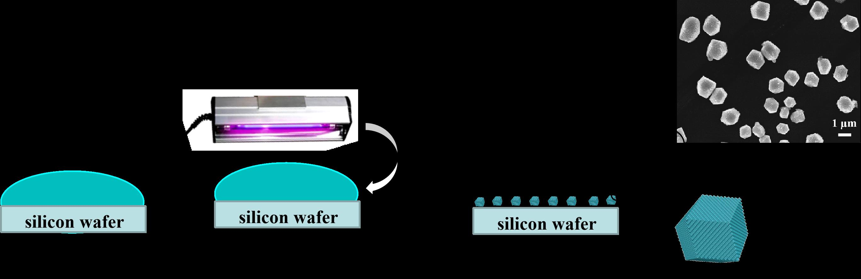
In a typical synthesis process, small silicon wafers (7 mm × 7 mm) served as substrates for collecting the HSANs superstructures grown from solution phase. Prior to each sample preparation, the silicon wafers were carefully cleaned using the following processes: firstly, the silicon wafers were soaked in Aqua regia solution for 4 h and piranha solution for 1 h, followed by sonication in the distilled water for 30 min, and finally dried by N2. In the next step, as shown in Scheme 1, 80 μL of 5 M AgNO3 aqueous solution was dropped on the surface of silicon wafers. The AgNO3 solution supported on the silicon wafers was irradiated by 254 nm UV under the ambient conditions in winter (T = 10.1 °C, and the relative humidity RH% = 60%). After the 254 nm UV-irradiation, the samples were thoroughly rinsed with plenty of deionized water and then stored in desiccators for the following characterizations.
AgNO3 was used as the precursor of Ag-Ag2O-AgO. The morphology of the HSANs was controlled by the UV irradiation time. The reduction of AgNO3 to metallic Ag0 by γ-irradiation and ultra-violet irradiation has been known in 1964 [22]. The photolysis of AgNO3 aqueous solution under 254 nm UV irradiation revealed that the electrons were transferred from the water molecules to Ag+ [23]. Then, the generated H2O+ decomposes into H+ and OH radicals immediately, where Ag, H+, OH radicals coexisted in the water cages each of which is consisted of about 10 water molecules. Finally, the water cage decomposed and the silver atoms are released out of the water cage. Simply as following:
Generally, ultraviolet with high-energy photons can dissociate oxygen and wat molecules into reactive oxygen species (e.g., O(1D), O(3P), and O3) and hydroxyl radicals (OH), which can readily oxidize Ag to Ag2O. In addition, the O2 dissolved in silver nitrate solution can be converted into O3 under 254 nm UV-irradiation [24,25,26]. Then, silver is oxidized to Ag2O and Ag2O is further oxidized into AgO by O3 [27]. Simply as following:
Eventually, the final product includes Ag, Ag2O and AgO, while Ag2O is the main product.
2.2. Characterizations
The morphology and structure of the as-synthesized samples were investigated by field-emission scanning electron microscopy (FESEM). The SEM images and energy-dispersive X-ray spectroscopy (EDX) were recorded on a Hitachi S-4700 (Hitachi, Japan), operating at 15 kV under high vacuum. X-ray diffraction (XRD) spectra were obtained using a desktop diffractometer (D2 PHASER, Bruker, Germany) with monochromatic Cu Kα radiation (λ = 1.54056 Å) operated at 30 kV and 40 mA. The size and morphology of samples were recorded using Transmission Electron Microscope (TEM, Tecnai G2 F20, FEI) operating at the accelerating voltage of 200 kV. The Raman spectra were recorded at room temperature in the reflection configuration on a micro-Raman spectroscope (Horiba HR800, Jobin Yvon, France) coupled with a confocal microscope (Olympus, LMPlanFI, Japan). A He-Ne laser (λ = 632.8 nm) was employed as the excitation source. The X-ray photoelectron spectra (XPS) were collected using an ESCALab220i-XL electron spectrometer equipped with 300 W Al Kα radiation from VG Scientific.
3. Results and Discussions
As shown in Figure 1, the XRD reflections at 2θ of 33.0°, 38.1°, 55.2°, 65.8°, and 69.1° can be attributed to the (111), (200), (220), (311), and (222) crystallographic planes of primitive cubic silver oxide (Ag2O) (JCPDS No. 75-1532), respectively. Besides, the XRD reflection at 2θ of 33.9° is associated with the forbidden 1/3 (422) reflection of silver and the reflection at 2θ of 38.1° and 44.5° can be attributed to the (111) and (200) of face-centered cubic silver (JCPDS No. 87-0719). In addition, the XRD reflections at 2θ of 32.4°, 34.2°, 37.2°, 39.5°, 42.2°, 47.5°, and 56.8° can be ascribed to the (11), (002), (111), (02), (102), (12), and (13) crystallographic planes of monoclinic AgO (JCPDS No. 74-1750). Therefore, the final product includes Ag, Ag2O, and AgO. There are two reasons for partial oxidation of Ag, one of which is that the Ag is oxidized by ∙OH radicals to form the Ag2O, the second is that Ag2O is oxidized into AgO by the O3 in the solution.
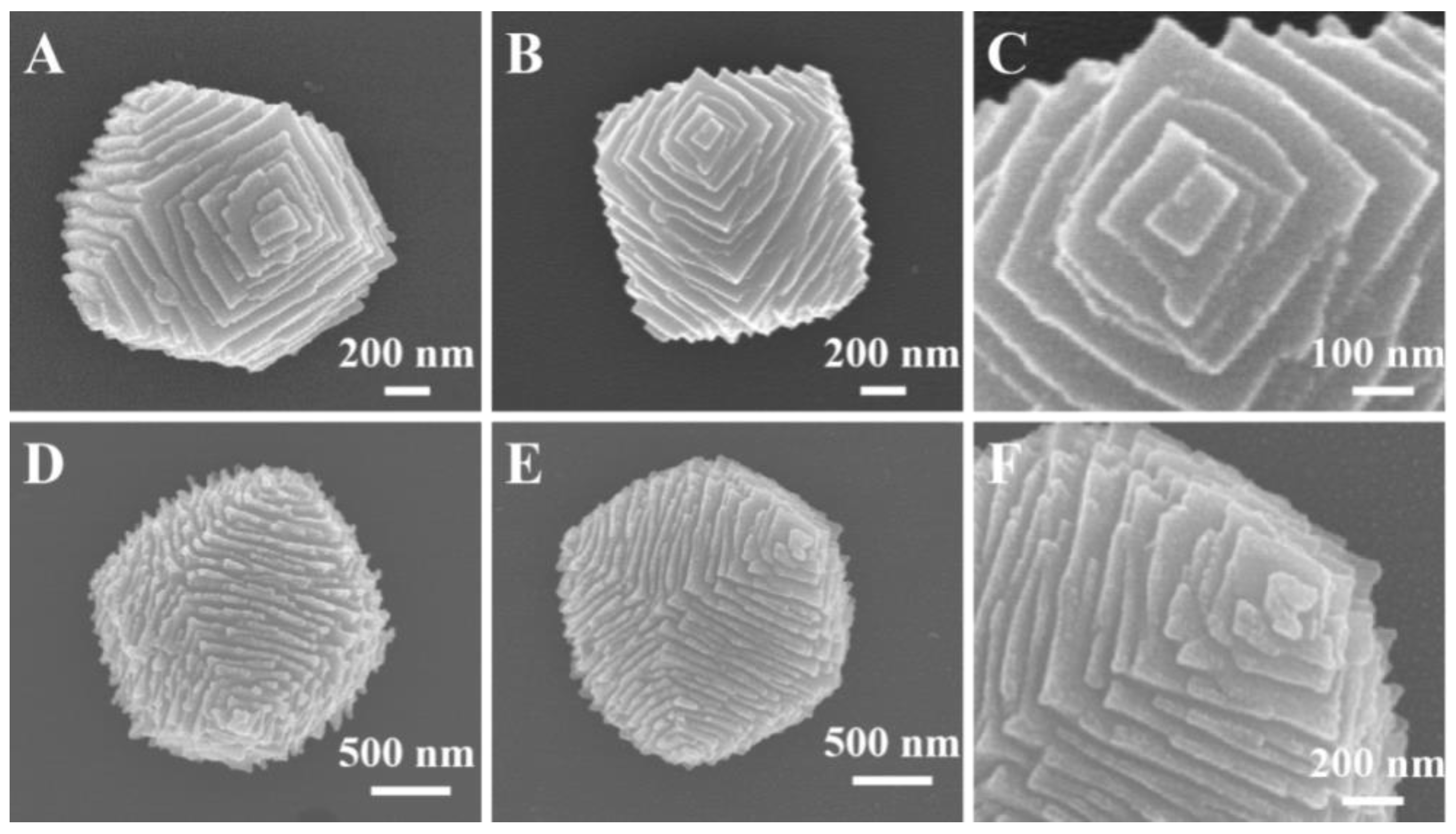
The HSANs were deposited on silicon wafer surface through a facile and low cost photochemical method. First, the low supersaturation conditions in the system means that the synthesized samples further crystallized into HSANs through a screw-dislocation-driven growth mechanism [2]. Figure 2 shows representative SEM images of HSANs, which clearly demonstrated its surface morphology from different perspective. At the same time, Figure 2 shows the growth hillocks with steps and terrace width. The growth hillocks with steps is the dislocation step edges. Through Figure 2C,F, the dislocation core was at the center of the growth hillocks. Thus, the crystallization of HSANs is dominated by screw-dislocation-driven growth mechanism. We guess that this novel morphology originate from a cube, where the center of each square faces of cube generate an emanating spiral resulting the appearance of growth hillocks. Here, each growth hillock is surrounded by four triangular faces. At first, the faces of two different triangles have a certain angle, then the angle turn into 180° gradually and the two triangles mix together into a rhombus with further extension of UV irradiation. According to Figure 2C,F, it is worth noting that the HSANs with particular and novel morphology are built from assemblies of nanometer-scale particles. Figure 2D,E shows the growth hillocks with more steps and small terrace width. Obviously, the slopes of the growth hillock are related to the step height (h) and terrace width (λ). Morin et al. [15] have reported that the slopes of the growth hillock (p) is inversely proportional to the terrace width, and is proportional to the step height (P = h/λ). Moreover, terrace width is inversely proportional to supersaturation of the system based on BCF theory [5,6,7]. Here, it is easy to find that the slopes of Figure 2A–C is smaller than that of Figure 2D–F, so supersaturation of Figure 2A–C is lower than Figure 2D–F. The EDX spectrum of the sample obtained at 30 min UV irradiation time showed the existence of Ag and O elements in the samples. Meanwhile, the existence of Si comes from silicon wafer, Au comes from spray gold, and C comes from atmosphere.
Figure 3 shows the low and high magnification SEM images of HSANs prepared from a 5 M AgNO3 aqueous solution under 60 min UV-irradiation. Due to the absence of surfactants, the size of superstructures in the same area is not uniform (see Figure 3A,B). Through low magnification SEM images, we find that particles were monodisperse and polyhedral, such as rhombic dodecahedra. Figure 3C,D clearly shows the surface morphology with hierarchical step edges of the superstructures. We propose that the sizes of HSANs are determined by the size of the initial cubes. If the cube is large, HSANs will be large.
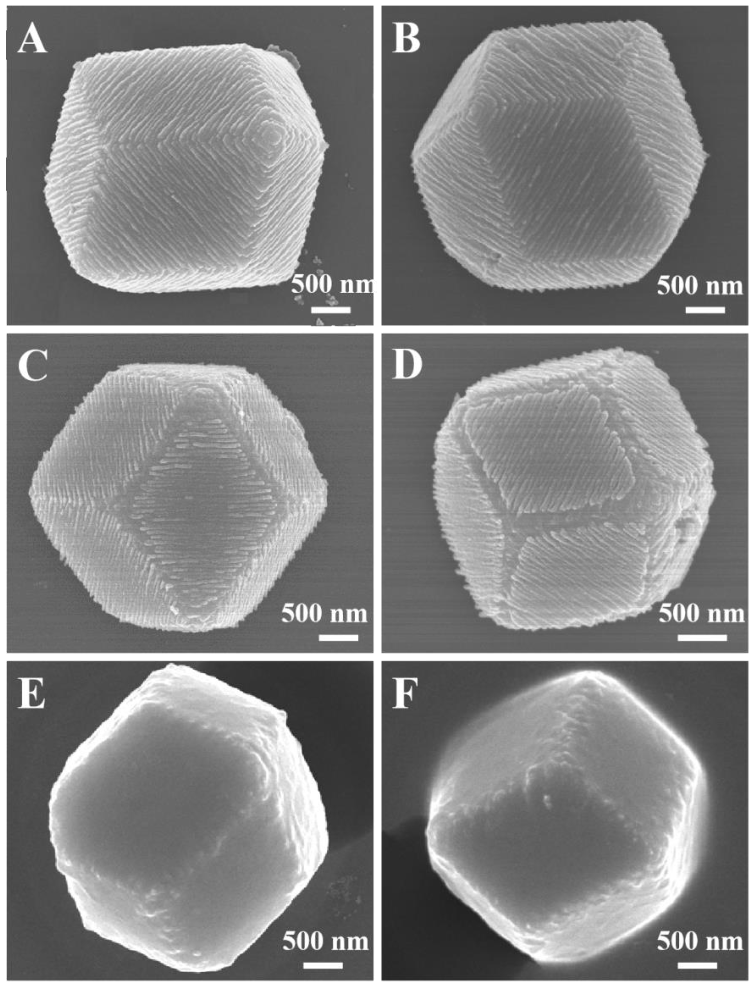
When the UV irradiation time increased to 60 min with 5 M AgNO3 aqueous solution, as shown in Figure 3 and Figure 4A,B, hierarchical superstructures, such as rhombic dodecahedra, generated. For the HSANs, when its structure converted from hierarchical step-edges-like superstructures to rhombic dodecahedra, its size is self-limited because of its structural self-confinement. We also proposed that the surface of the HSANs transformed from hierarchical into relatively smooth and its size did not change at the same time. In our work, HSANs, such as rhombic dodecahedra, have twelve apices which each apex are surrounded by three rhombi. Here, eight apices are from the apices of cube and another six apices are from the center of the hillocks in hierarchical step-edges-like HSANs. Rhombic dodecahedra HSANs have twelve rhombi and each rhombus is from two triangles in hierarchical step-edges-like superstructure of Ag-Ag2O-AgO nanoparticles. Figure 4C,D shows the SEM images of particles synthesized by a 5 M AgNO3 aqueous solution with 70 min UV irradiation time. Interestingly, nanoparticles on the surface of the rhombic dodecahedra HSANs begin to fall off along the direction of the edges. The fallen nanoparticles dispersed in AgNO3 aqueous solution and were rinsed by plenty of deionized water. Finally, the surface of HSANs became smooth (see Figure 4E,F).
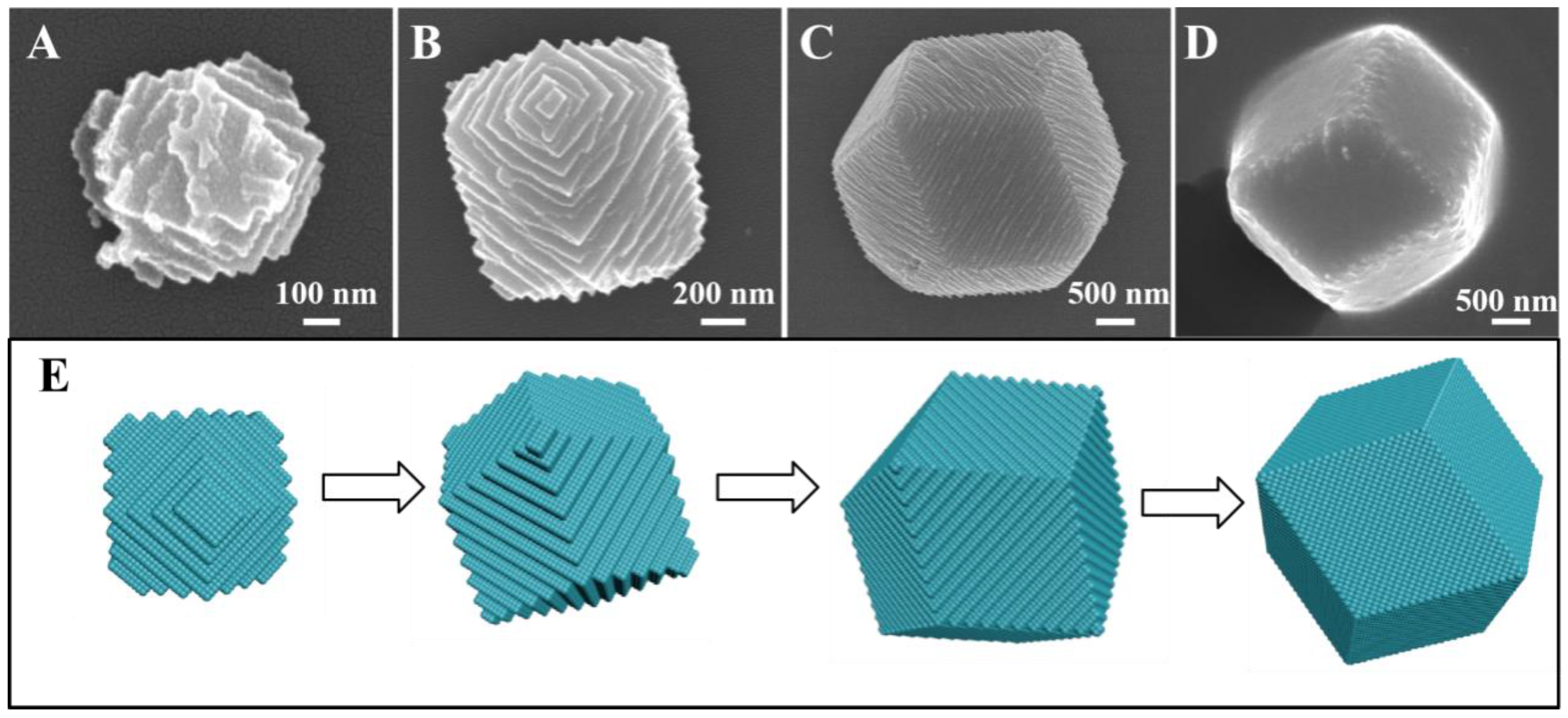
To further investigate the growth process of the HSANs, we adjust the reaction parameter. Unsurprisingly, we find the ideal morphological changes by tuning the reaction time and the result is in line with our conjecture. Figure 5 shows the structural evolution of HSANs. Obviously, it was observed that the number of steps transformed from less to more, and the step sizes were converted from large to small relatively. We propose that the HSANs are nanometers-scale at the initial reaction time with 20 min using 5 M AgNO3 aqueous solution (Figure 5A). After reaction for 30 min, the morphology of the superstructures was regular (Figure 5B). At 60 min UV-irradiation, we yielded the large superstructures with the most step edges and its topography turned into rhombic dodecahedra-like regular structure (Figure 5C). With further extension of UV irradiation from 60 min to 95 min, the products, such as rhombic dodecahedra, gradually turned from hierarchical into relatively smooth without the change of size (Figure 5D). Our novel morphology can be well explained by the BCF theory [5,6,7]. The step edges became more and more and new product atom can be added because there is no need to overcome the energy barrier to form new steps [2]. Thus, the superstructures have little steps and large terrace width and the slopes of growth hillocks are small at this condition (see Figure 5A). With increase of supersaturation, the slopes of growth hillocks become larger (see Figure 5B,C). Obviously, the order of the degree of supersaturation (σ) in Figure 5 is: σA < σB < σC, and the slopes of growth hillocks is: pA < pB <pC. The supersaturation of the system plays a vital role in the surface morphology of HSANs. The whole structural evolution of HSANs is illustrated schematically in Figure 5E. In short, our novel morphology originates from a cube, where the growth hillocks generate. Then the edges of the hillocks become more and more and the slopes of growth hillocks become larger. At a particular time, new crystal steps are no longer formed, as a result, the surface of the superstructures become smooth. Finally, HSANs evolve into polyhedral, such as rhombic dodecahedra. In our system, supersaturation is low, so the crystallization of superstructures is dominated by screw-dislocation-driven growth mechanism. As schematic diagram shown, supersaturation is becoming larger in this reaction procedure and is of great significance for the whole structural evolution. The samples with little steps are difficult to be observed, so we propose that HSANs grown fast at first, then we could obtain plenty of samples with more steps, which means slower growth rate.
The samples were further characterized using TEM. Figure 6A,B shows typical TEM images of HSANs prepared from 30 min and 60 min UV-irradiation. The size of the superstructures is smaller compared with Figure 6B. As depicted in Figure 6B, the surface of the sample is smooth and its size is larger according to Figure 6A. The selected area electron diffraction (SAED) pattern, which was taken from near the edge of Figure 6B, is shown in Figure 6C. The electron diffraction rings could be corresponded with lattice parameter of both Ag and Ag2O. The SAED pattern resolved the 1/3 {422}, {200}, and {220} reflections of fcc silver, with measured d-spacings of 2.49, 2.01, and 1.49 Å, respectively. Besides, the SAED pattern also resolved the {111} and {123} reflections of primitive cubic Ag2O, with the measured d-spacing of 2.75 and 1.27 Å, respectively, which confirm the truth of partial oxidation of Ag. Figure 6D–F depicts the high-resolution TEM image of the superstructures acquired near the edge of Figure 6B, which clearly resolves the simultaneous presence of Ag and Ag2O phase. The lattice fringes with a measured d-spacing of 2.35 Å agree with the (111) reflection of fcc silver. Besides, the lattice fringes with a measured d-spacing of 2.75 Å indexed to the (111) lattice of primitive cubic Ag2O. Figure 6F discloses the lattice fringes with a d-spacing of 2.49 Å, which can be ascribed to the forbidden reflection of 1/3 {422} [28], in agreement with the XRD pattern and SAED pattern. Figure 6D indicates the {111} twin planes in the Ag particles. Because the amount of AgO is too small, we have not obtained the corresponding data. Moreover, the HSANs synthesized through 60 min UV-irradiation were also characterized by normal Raman. As shown in Figure 7, the broad band at 490 cm–1 can be ascribed to Ag2O [27,29]. Raman peaks at 219, 300, and 429 cm–1 can be attributed to AgO [27,29].
For studying the influence of UV radiation on the morphology, the samples through by natural supersaturation for 60 min were also obtained. As seen in Figure 8, it is revealed that no hierarchical superstructures of Ag-Ag2O-AgO nanoparticles are prepared without the UV irradiation, suggesting the determined influence of UV radiation on the formation of hierarchical nanostructure.
The elemental composition and chemical states of the samples were analyzed by X-ray photoelectron spectroscopy (XPS). Figure 9A shows the full-range XPS spectrum of the HSANs, revealing only the elemental peaks of Ag, O, and C. The C 1s peak comes from the adventitious carbon species, including the surface-adsorbed CO2 molecules [24]. The high-resolution XPS spectrum of Ag 3d5/2 and Ag 3d3/2 is given in Figure 9B. The binding energy of Ag 3d5/2 photoelectrons is 367.9 eV. According to the literature [27], generally speaking, the binding energy of the Ag 3d5/2 photoelectrons shifts to the lower energy region upon the oxidation of Ag. The binding energy of Ag 3d5/2 photoelectrons for Ag, Ag2O, and AgO is 368.2, 367.8, and 367.4 eV, respectively [27]. In our case, the binding energy of the Ag 3d5/2 photoelectrons is very close to the reported values for Ag2O [30,31,32]. It suggests that the silver exists mainly in one valence state (Ag+) in the hierarchical superstructures, which is consistent with the XRD results (see Figure 1). Figure 9C shows the high-resolution XPS spectrum for the O 1s photoelectrons. More detailed information about the elemental compositions and chemical states are elucidated in the high-resolution XPS spectrum of the O 1s photoelectrons. The asymmetric O 1s peak can be deconvoluted into four symmetric Gaussian–Lorentzian peaks, suggesting four different chemical states of O element in the hierarchical superstructures. The peak at 531.4 eV can be assigned to the surface –OH groups or the surface-adsorbed water molecules [33]. The broad peak at 530.8 eV indicates the presence of surface-adsorbed CO2 molecules [34,35,36]. The peak at 529.2 eV and 528.6 eV is associated with the lattice oxygen in Ag2O and AgO [27], respectively. The XPS results indicated that the co-existence of both Ag2O and AgO, in good agreement with the XRD results.
4. Conclusions
In conclusion, the HSANs nanocrystals were prepared through a simple and surfactant-less photochemical method. This kind of superstructures was obtained from nanometer-scale particles-mediated crystallization processes, with the assistance of the H2O-to-Ag+ electron transfers accompanied by the formation of metallic Ag particles. It was proposed that the formation of silver oxide particles is attributed to the oxidation of metallic Ag particles by the air under ambient conditions, intermediate OH radicals, and the ozone (O3) generated under UV254 irradiation. The crystallization process for the formation of HSANs was governed by a screw-dislocation-driven crystallization mechanism under low-supersaturation conditions. The structural evolutions of the HSANs have been systematically exploited. The average size of the superstructures increased along with the time of UV254 irradiation. The step density on the surfaces of the HSANs increased as well with the prolonged UV254 irradiation time. When the time for UV254 irradiation increased to 95 min, the surfaces of the HSANs converted from hierarchical to relatively smooth. This work proposed a promising strategy for the synthesis of HSANs nanocrystals, which would be guided for the fabrication of advanced nanocrystals various applications in wide areas in the near future.
Author Contributions
H.Y., W.T.C., and J.R.Z. conceived and designed the experiments; H.Y. and W.C. performed the experiments; H.Y., W.C., and J.R.Z. analyzed the data; J.Z. contributed analytical tools; H.Y. and J.H.W. wrote the paper. All authors have read and agreed to the published version of the manuscript.
Funding
We are thankful for the financial support from the National Natural Science Foundation of China (Project 21373145), the College of Chemistry, Chemical Engineering and Materials Science, Suzhou University, China, the Priority Academic Program Development of Jiangsu Higher Education Institutions (PAPD), and the project of scientific and technologic infrastructure of Suzhou (SZS201708).
Conflicts of Interest
The authors declare no conflict of interest.
References
- Yang, S.K.; Li, M.Y.; Zhu, X.; Xu, G.Q.; Wu, J.H. Photochemical Synthesis of Hierarchical Multiple-Growth-Hillock Superstructures of Silver Particles on ZnO. J. Phys. Chem. C 2015, 119, 14312–14318. [Google Scholar]
- Meng, F.; Morin, S.A.; Forticaux, A.; Jin, S. Screw Dislocation Driven Growth of Nanomaterials. Acc. Chem. Res. 2013, 46, 1616–1626. [Google Scholar] [CrossRef] [PubMed]
- Li, M.Y.; Mao, Y.Q.; Yang, S.K.; Dai, T.T.; Yang, H.; Feng, F.; Wu, T.; Chen, M.; Xu, G.Q.; Wu, J.H. Out-of-Substrate Ag-Ag2O Nanoplates: Surfactantless Photochemical Synthesis, Structural Evolution, and Mechanistic Study. ACS Omega 2016, 1, 696–705. [Google Scholar] [CrossRef] [PubMed] [Green Version]
- Xu, H.; Shang, H.Y.; Wang, C.; Du, Y.K. Ultrafine Pt-Based Nanowires for Advanced Catalysis. Adv. Funct. 2020, 30, 2000793. [Google Scholar] [CrossRef]
- Frank, F.C. The influence of dislocations on crystal growth. Discuss. Faraday Soc. 1949, 5, 48–54. [Google Scholar] [CrossRef]
- Burton, W.K.; Cabrera, N.; Frank, F.C. The growth of crystals and the equilibrium structure of their surfaces. Philos. Trans. R. Soc. Lond. Ser. A Math. Phys. Sci. 1951, 243, 299–358. [Google Scholar]
- Burton, W.K.; Cabrera, N.; Frank, F.C. Role Of Dislocations in Crystal Growth. Nature 1949, 163, 398–399. [Google Scholar] [CrossRef]
- Liang, H.; Meng, F.; Lamb, B.K.; Ding, Q.; Li, L.; Wang, Z.; Jin, S. Solution Growth of Screw Dislocation Driven α-GaOOH Nanorod Arrays and Their Conversion to Porous ZnGa2O4 Nanotubes. Chem. Mater. 2017, 29, 7278–7287. [Google Scholar] [CrossRef]
- Jin, S.; Bierman, M.J.; Morin, S.A. A New Twist on Nanowire Formation: Screw-Dislocation-Driven Growth of Nanowires and Nanotubes. J. Phys. Chem. Lett. 2010, 1, 1472–1480. [Google Scholar] [CrossRef]
- Morin, S.A.; Jin, S. Screw dislocation-driven epitaxial solution growth of ZnO nanowires seeded by dislocations in GaN substrates. Nano Lett. 2010, 10, 3459–3463. [Google Scholar] [CrossRef]
- Meng, F.; Jin, S. The solution growth of copper nanowires and nanotubes is driven by screw dislocations. Nano Lett. 2012, 12, 234–239. [Google Scholar] [CrossRef] [PubMed]
- Lau, Y.K.A.; Chernak, D.J.; Bierman, M.J.; Jin, S. Formation of PbS Nanowire Pine Trees Driven by Screw Dislocations. J. Am. Chem. Soc. 2009, 131, 16461–16471. [Google Scholar] [CrossRef] [PubMed]
- Morin, S.A.; Bierman, M.J.; Tong, J.; Jin, S. Mechanism and Kinetics of Spontaneous Nanotube Growth Driven by Screw Dislocation. Science 2010, 328, 476–480. [Google Scholar] [CrossRef] [PubMed]
- Shearer, M.J.; Samad, L.; Zhang, Y.; Zhao, Y.; Puretzky, A.; Eliceiri, K.W.; Wright, J.C.; Hamers, R.J.; Jin, S. Complex and Noncentrosymmetric Stacking of Layered Metal Dichalcogenide Materials Created by Screw Dislocations. J. Am. Chem. Soc. 2017, 139, 3496–3504. [Google Scholar] [CrossRef]
- Morin, S.A.; Forticaux, A.; Bierman, M.J.; Jin, S. Screw dislocation-driven growth of two-dimensional nanoplates. Nano Lett. 2011, 11, 4449–4455. [Google Scholar] [CrossRef]
- Zou, K.; Zhang, X.H.; Duan, X.F.; Meng, X.M.; Wu, S.K. Seed-mediated synthesis of silver nanostructures and polymer/silver nanocables by UV irradiation. J. Cryst. Growth 2004, 273, 285–291. [Google Scholar] [CrossRef]
- Langille, M.R.; Perspnick, M.L.; Mirkin, C.A. Plasmon-Mediated Syntheses of Metallic Nanostructures. Angew. Chem. Int. Ed. 2013, 52, 13910–13940. [Google Scholar] [CrossRef]
- Dasgupta, N.P.; Sun, J.; Liu, C.; Brittman, S.; Andrews, S.C.; Lim, J.; Gao, H.; Yan, R.; Yang, P. 25th anniversary article: Semiconductor nanowires—Synthesis, characterization, and applications. Adv. Mater. 2014, 26, 2137–2184. [Google Scholar] [CrossRef]
- Morales, A.M.; Lieber, C.M. A Laser Ablation Method for the Synthesis of Crystalline Semiconductor Nanowires. Science 1998, 279, 208–211. [Google Scholar] [CrossRef]
- Wang, X.F.; Li, S.F.; Yu, H.G.; Yu, J.G.; Liu, S.W. Ag2O as a New Visible-Light photoachtungtrenung catalyst: Self-Stability and High photoachtungtrenung catalytic Activity. Chem. Eur. J. 2011, 17, 7777–7780. [Google Scholar] [CrossRef]
- Wang, G.; Ma, X.C.; Huang, B.B.; Cheng, H.F.; Wang, Z.Y.; Zhan, J.; Qin, X.Y.; Zhang, X.Y.; Dai, Y. Controlled synthesis of Ag2O microcrystals with facet-dependent photocatalytic activities. J. Mater. Chem. C 2012, 22, 21189. [Google Scholar] [CrossRef]
- Mahlman, H.A.; Willmarth, T.E. Radiolytic and Photolytic Reduction of Aqueous Silver Nitrate Solutions. Nature 1964, 202, 590–591. [Google Scholar] [CrossRef]
- Hada, H.; Yonezawa, Y.; Yoshida, A.; Kurakake, A. Photoreduction of Silver Ion in Aqueous and Alcoholic Solutions. JPC 1976, 80, 2728–2731. [Google Scholar] [CrossRef]
- Yu, K.-P.; Lee, G.W.M. Decomposition of gas-phase toluene by the combination of ozone and photocatalytic oxidation process (TiO2/UV, TiO2/UV/O3, and UV/O3). Appl. Catal. B Environ. 2007, 75, 29–38. [Google Scholar] [CrossRef]
- Kim, J.; Zhang, P.; Li, J.; Wang, J.; Fu, P. Photocatalytic degradation of gaseous toluene and ozone under UV254+185 nm irradiation using a Pd-deposited TiO2 film. Chem. Eng. J. 2014, 252, 337–345. [Google Scholar] [CrossRef]
- Fu, P.; Zhang, P. Characterization of Pt-TiO2 film used in three formaldehyde photocatalytic degradation systems: UV254 nm, O3+UV254 nm and UV254+185 nm via X-ray photoelectron spectroscopy. Chin. J. Catal. 2014, 35, 210–218. [Google Scholar] [CrossRef]
- Waterhouse, G.I.N.; Bowmaker, G.A.; Metson, J.B. Oxidation of a polycrystalline silver foil by reaction with ozone. Appl. Surf. Sci. 2001, 183, 191–204. [Google Scholar] [CrossRef]
- Xiong, Y.; Siekkinen, A.R.; Wang, J.; Yin, Y.; Kim, M.J.; Xia, Y. Synthesis of silver nanoplates at high yields by slowing down the polyol reduction of silver nitrate with polyacrylamide. J. Mater. Chem. 2007, 17, 2600–2602. [Google Scholar] [CrossRef]
- Waterhouse, G.I.N.; Bowmaker, G.A.; Metson, J.B. The thermal decomposition of silver (I, III) oxide: A combined XRD, FT-IR and Raman spectroscopic study. Phys. Chem. Chem. Phys. 2001, 3, 3838–3845. [Google Scholar] [CrossRef]
- Murray, B.J.; Li, O.; Newberg, J.T.; Menke, E.J.; Hemminger, J.C.; Penner, R.M. Shape-and Size-Selective Electrochemical Synthesis of Dispersed Silver(I) Oxide Colloids. Nano Lett. 2005, 5, 2319–2324. [Google Scholar] [CrossRef]
- Hammond, J.S.; Gaarenstroom, S.W.; Winograd, N. X-Ray Photoelectron Spectroscopic Studies of Cadmium- and Silver-Oxygen Surfaces. Anal. Chem. 1975, 47, 2193–2199. [Google Scholar] [CrossRef]
- Jeong, N.C.; Prasittichai, C.; Hupp, J.T. Photocurrent enhancement by surface plasmon resonance of silver nanoparticles in highly porous dye-sensitized solar cells. Langmuir 2011, 27, 14609–14614. [Google Scholar] [CrossRef] [PubMed]
- Chakraborty, A.K.; Kebede, M.A. Preparation and characterization of WO3/Bi3O4Cl nanocomposite and its photocatalytic behavior under visible light irradiation. React. Kinet. Mech. Catal. 2012, 106, 83–98. [Google Scholar] [CrossRef]
- Bukhtiyarov, V.I.; Kondratenko, V.A.; Boronin, A.I. Features of the interaction of a CO + O2, mixture with silver under high pressure. Surf. Sci. Lett. 1993, 293, L826–L829. [Google Scholar]
- Xu, H.; Shang, H.Y.; Jin, L.J.; Chen, C.Y.; Wang, C.; Du, Y. Boosting electrocatalytic oxygen evolution over Prussian blue analog/transition metal dichalcogenide nanoboxes by photo-induced electron transfer. J. Mater. Chem. A 2019, 7, 26905–26910. [Google Scholar] [CrossRef]
- Xu, H.; Shang, H.Y.; Wang, C.; Jin, L.J.; Chen, C.Y.; Wang, C.Y.; Du, Y.K. Three-dimensional open CoMoOx/CoMoSx/CoSx nanobox electrocatalysts for efficient oxygen evolution reaction. Appl. Catal. B Environ. 2020, 265, 118605. [Google Scholar] [CrossRef]
Scheme 1.
Schematic diagram illustrating the synthesis process of screw-dislocation-driven hierarchical superstructures of Ag-Ag2O-AgO nanoparticles (HSANs).
Scheme 1.
Schematic diagram illustrating the synthesis process of screw-dislocation-driven hierarchical superstructures of Ag-Ag2O-AgO nanoparticles (HSANs).

Figure 1.
XRD pattern of the HSANs obtained at 60 min UV irradiation time. Concentration of AgNO3 precursor: 5 M.
Figure 1.
XRD pattern of the HSANs obtained at 60 min UV irradiation time. Concentration of AgNO3 precursor: 5 M.

Figure 2.
Representative SEM images of HSANs synthesized by a 5 M AgNO3 aqueous solution with (A–C) 30 min and (D–F) 40 min UV irradiation time.
Figure 2.
Representative SEM images of HSANs synthesized by a 5 M AgNO3 aqueous solution with (A–C) 30 min and (D–F) 40 min UV irradiation time.

Figure 3.
(A,B) Low and (C,D) high magnification SEM images of HSANs, such as rhombic dodecahedra, obtained at 60 min UV irradiation. Concentration of AgNO3 precursor: 5 M.
Figure 3.
(A,B) Low and (C,D) high magnification SEM images of HSANs, such as rhombic dodecahedra, obtained at 60 min UV irradiation. Concentration of AgNO3 precursor: 5 M.

Figure 4.
Typical SEM images of HSANs, such as rhombic dodecahedra, grown from a 5 M AgNO3 aqueous solution with different irradiation time: (A,B) 60 min, (C,D) 70 min, (E,F) 95 min.
Figure 4.
Typical SEM images of HSANs, such as rhombic dodecahedra, grown from a 5 M AgNO3 aqueous solution with different irradiation time: (A,B) 60 min, (C,D) 70 min, (E,F) 95 min.

Figure 5.
SEM images of HSANs prepared from (A) 20 min; (B) 30 min; (C) 60 min; (D) 95 min UV-irradiation. (E) Concentration of AgNO3 precursor: 5 M.
Figure 5.
SEM images of HSANs prepared from (A) 20 min; (B) 30 min; (C) 60 min; (D) 95 min UV-irradiation. (E) Concentration of AgNO3 precursor: 5 M.

Figure 6.
(A) TEM image of the HSANs prepared from 30 min UV-irradiation; (B) TEM image of HSANs prepared from 60 min UV-irradiation like rhombic dodecahedra; (C) selected area electron diffraction (SAED) pattern and (D–F) HRTEM images obtained near the edges of (B).
Figure 6.
(A) TEM image of the HSANs prepared from 30 min UV-irradiation; (B) TEM image of HSANs prepared from 60 min UV-irradiation like rhombic dodecahedra; (C) selected area electron diffraction (SAED) pattern and (D–F) HRTEM images obtained near the edges of (B).

Figure 7.
Raman spectra for the HSANs synthesized through 60 min UV-irradiation. Concentration of AgNO3 precursor: 5 M.
Figure 7.
Raman spectra for the HSANs synthesized through 60 min UV-irradiation. Concentration of AgNO3 precursor: 5 M.

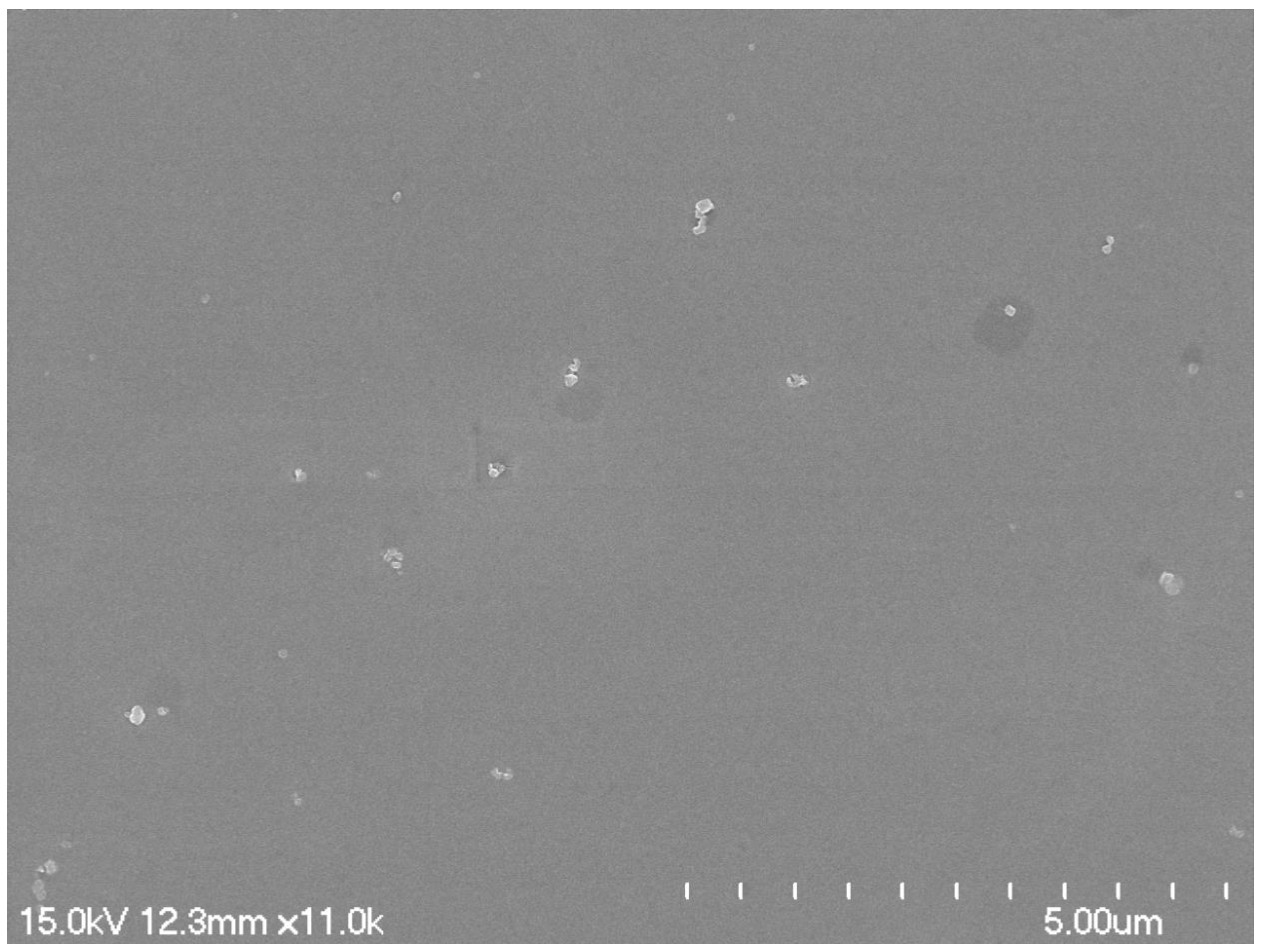
Figure 8.
The representative SEM image of the product obtained by using natural evaporation for 60 min.
Figure 8.
The representative SEM image of the product obtained by using natural evaporation for 60 min.

Figure 9.
(A) XPS survey scan of HSANs obtained at 60 min UV irradiation time; high-resolution spectrum of (B) Ag 3d and (C) O 1s.
Figure 9.
(A) XPS survey scan of HSANs obtained at 60 min UV irradiation time; high-resolution spectrum of (B) Ag 3d and (C) O 1s.

Publisher’s Note: MDPI stays neutral with regard to jurisdictional claims in published maps and institutional affiliations. |
© 2020 by the authors. Licensee MDPI, Basel, Switzerland. This article is an open access article distributed under the terms and conditions of the Creative Commons Attribution (CC BY) license (http://creativecommons.org/licenses/by/4.0/).
Share and Cite
MDPI and ACS Style
Yang, H.; Zhang, J.R.; Cao, W.; Zhen, J.; Wu, J.H. Screw-Dislocation-Driven Hierarchical Superstructures of Ag-Ag2O-AgO Nanoparticles. Crystals 2020, 10, 1084. https://0-doi-org.brum.beds.ac.uk/10.3390/cryst10121084
AMA Style
Yang H, Zhang JR, Cao W, Zhen J, Wu JH. Screw-Dislocation-Driven Hierarchical Superstructures of Ag-Ag2O-AgO Nanoparticles. Crystals. 2020; 10(12):1084. https://0-doi-org.brum.beds.ac.uk/10.3390/cryst10121084
Chicago/Turabian StyleYang, Hua, Jing Ru Zhang, Wentao Cao, Jin Zhen, and Ji Hong Wu. 2020. "Screw-Dislocation-Driven Hierarchical Superstructures of Ag-Ag2O-AgO Nanoparticles" Crystals 10, no. 12: 1084. https://0-doi-org.brum.beds.ac.uk/10.3390/cryst10121084
Note that from the first issue of 2016, this journal uses article numbers instead of page numbers. See further details here.