
Green Synthesis of Ge1−xSnx Alloy Nanoparticles for Optoelectronic Applications


Department of Mechanical and Materials Engineering, Florida International University, Miami, FL 33174, USA
*
Author to whom correspondence should be addressed.
Crystals 2021, 11(10), 1216; https://0-doi-org.brum.beds.ac.uk/10.3390/cryst11101216
Submission received: 18 September 2021
/
Revised: 1 October 2021
/
Accepted: 5 October 2021
/
Published: 8 October 2021
(This article belongs to the Special Issue Feature Papers on "Hybrid and Composite Crystalline Materials" 2021-2022)
Abstract
:Compositionally controlled, light-emitting, group IV semiconductor nanomaterials have potential to enable on-chip data communications and infrared (IR) imaging devices compatible with the complementary metal−oxide−semiconductor (CMOS) technology. The recent demonstration of a direct band gap laser in Ge-Sn alloys opens avenues to the expansion of Si-photonics. Ge-Sn alloys showed improved effective carrier mobility as well as direct band gap behavior at Sn composition above 6–11%. In this work, Ge1−xSnx alloy nanoparticles with varying Sn compositions from x = 0.124 to 0.178 were prepared via colloidal synthesis using sodium borohydride (NaBH4), a mild and non-hazardous reducing reagent. Successful removal of the synthesized long-alkyl-chain ligands present on nanoparticles’ surfaces, along with the passivation of the Ge-Sn nanoparticle surface, was achieved using aqueous (NH4)2S. The highly reactive surface of the nanoparticles prior to ligand exchange often leads to the formation of germanium oxide (GeO2). This work demonstrates that the (NH4)2S further acts as an etching reagent to remove the oxide layer from the particles’ surfaces. The compositional control and long-term stability will enable the future use of these easily prepared Ge1−xSnx nanoalloys in optoelectronic devices.
1. Introduction
Germanium-tin (Ge-Sn) is a complementary metal–oxide–semiconductor (CMOS) compatible group IV semiconductor material that has attracted great attention over the past two decades owing to its compatibility with Si and its great potential for use in optoelectronic integration circuits (OEIC) [1]. When the Sn content exceeds x = 0.06–0.11, the material becomes a direct bandgap semiconductor, offering a paradigm shift for Si-photonics toward the monolithic integration of light emitters [2,3]. The favorable optical properties originate in a modified band structure, changing the bandgap from an indirect to a direct one. The first demonstration of such a direct band gap laser has recently been accomplished for Ge-Sn [2]. Ge-Sn has been also reported to have a higher effective hole mobility than Ge due to the decrease in light hole effective mass of Ge-Sn, with the incorporation of Sn in the case of p-channel metal–oxide–semiconductor field effect transistors (p-MOSFETs) [4]. The bandgap of Ge1−xSnx can be tuned from 0.6 to 0 eV by varying the Sn content, thus making this alloy suitable for use in near-infrared and mid-infrared detectors [3].
However, the high crystallization temperature of Ge (above 300 °C) and large lattice mismatch (∼14%) with Sn, as well as the low equilibrium solubility (<1%) of Sn in Ge, make it difficult to synthesize compositionally uniform Ge1−xSnx alloys [5]. Recently, various Ge-Sn materials, including alloy nanocrystals [6], quantum dots [7], nanowires [8], and core/shell particles [9] have been reported. Different strategies such as pulsed laser melting [10], epitaxial growth [11], microwave synthesis [12] and solution-based methods [13] have been investigated to produce direct gap group IV semiconductors. Of all the strategies, colloidal synthesis of high-quality Ge1−xSnx without phase segregation of Sn is a desirable route that would enable inexpensive fabrication, processing, and device assembly [14].
In this work, we report the colloidal synthesis, characterization, and physical properties of a series of Ge1−xSnx alloy nanoparticles with Sn compositions ranging from x = 0.124 to 0.178, which resulted from the intended doping concentrations of x = 0.12, 0.15, 0.18. This range of concentrations was selected to obtain the direct band gap semiconductor behavior for the Ge1−xSnx alloy, provided that the transition from an indirect to direct band gap occurred when the doping concentration of Sn exceeded x = 0.06–0.11. We were able to obtain Ge1−xSnx nanoparticles with x = 0.124, 0.151 and 0.178. For doping concentrations exceeding x = 0.20, we observed the segregation of Sn. The nanoparticles were obtained by using NaBH4 as a reducing agent, which is mild, safer to use and easier to handle than stronger, more highly reactive and pyrophoric agents such as n-BuLi and LiAlH4. Oleylamine surface ligands that remained on the particle’s surface upon synthesis were exchanged with short sulfide ligands by a solution-phase ligand-exchange approach using aqueous (NH4)2S [15]. It was found that the (NH4)2S not only effectively passivated the surface of Ge-Sn from oxidation but the process also successfully removed the undesired surface oxides. The synthesis and ligand exchange processes are shown in Scheme 1.
2. Materials and Methods
2.1. Materials
Tin dichloride (98%) was purchased from Sigma Aldrich (Saint Louis, MO, USA) and germanium diiodide was purchased from Gelest Inc. (Morrisville, PA, USA) and stored under argon. Oleylamine (70%) and sodium borohydride (NaBH4) solution (2.0 M in triethyleneglycol dimethyl ether) were purchased from Sigma Aldrich (Saint Louis, MO, USA). ACS-grade solvents, methanol, chloroform, and toluene, were purchased from Fisher Scientific (Waltham, MA, USA) and used without further purification. (NH4)2S (20% solution in water diluted to 10% before use) was purchased from Fisher Scientific (Waltham, MA, USA). OLA was dried by heating at 120 °C under vacuum for 1 h prior to storage under argon.
2.2. Characterization
The crystal structure and purity of the prepared alloy nanoparticles were determined by X-ray powder diffraction (XRD) using a Rigaku Miniflex 600 X-Ray diffractometer (Tokio, Japan) (Cu Kα radiation, λ = 1.5405 Å). Crystallite sizes were estimated by applying the Scherrer formula to the (111), (220), and (311) reflections of cubic Ge. To confirm product purity by Raman spectroscopy, Raman spectra were obtained with a WITec Alpha 300 Raman microscope (Ulm, (Bavaria), Germany) equipped with a 532 nm laser. The morphology and size of the synthesized alloy nanoparticles were determined by transmission electron microscopy (TEM) imaging, using a Talos F200X (FEI, Hillsboro, OR, USA), and scanning electron microscopy (SEM) imaging, with a JEOL 6330F (Peabody, MA, USA). Elemental distribution of the alloy nanoparticles was determined with the energy dispersive spectroscopy (EDS) feature of the JEOL 6330F SEM and Talos F200X TEM. The nanoparticles absorption spectra were collected using a Shimadzu UV-3600 Plus Ultraviolet–Visible–Near-InfraRed (UV–Vis–NIR) spectrophotometer (Shimadzu, Kyoto, Japan).
2.3. Preparation of Ge-Sn Nanoparticles
An amount of 10 mL of oleylamine was added to a 100 mL two-necked round-bottomed flask and degassed at 120 °C for at least 1 hour. After cooling to room temperature, it was flushed with argon and the flask was transferred to the glovebox. Predetermined amounts of GeI2 and SnCl2 for each experimental setting (Table 1) were added to the flask and the entire setup was sealed and carefully transferred to the Schlenk line.
The reaction mixture was further degassed at 120 °C and flushed with argon. The process was repeated three times. The temperature was raised to 220 °C and 0.5 mL of NaBH4 (2.0 M in triethyleneglycol dimethylether) was added. The color of the reaction mixture immediately turned brown upon NaBH4 addition. The reaction temperature was further raised to 300 °C and kept at this temperature for 60 min followed by cooling of the reaction mixture with compressed air.
After cooling to room temperature, 10 mL of toluene and 70 mL of methanol were added to the flask to precipitate the nanoparticles. The mixture was transferred to a 50 mL centrifuge tube and centrifuged at 8000 rpm for 10 min to obtain the Ge-Sn alloy nanoparticles. The supernatant was discarded and the solid product—a black color pellet—was further dispersed in 10 mL toluene and reprecipitated by adding 30 mL methanol. This process was repeated twice. The final product was collected by centrifugation and dried in a vacuum oven.
2.4. Ligand Exchange and Surface Oxide Removal
A suspension (5 mL) of Ge-Sn nanoparticles in chloroform (5 mg/mL) was mixed with 5 mL of aqueous (NH4)2S solution (10%) in a vial. The mixture was shaken vigorously for 2 min and allowed to settle until the chloroform and aqueous (NH4)2S were phase-separated. The Ge-Sn nanoparticles completely transferred from the chloroform phase to the aqueous (NH4)2S phase. An amount of 10 mL ethanol was added to the vial, and the mixture was transferred to a centrifuge tube and centrifuged for 4 min at 6000 RPM. The supernatant was discarded, and the precipitate was washed with a mixture of ethanol: chloroform (1:1, 20 mL) twice. The product collected after centrifugation was dried overnight in vacuum oven for further use.
3. Results and Discussion
3.1. Synthesis
Phase-pure Ge1−xSnx nanoparticles with compositions in the range of x = 0.124−0.178 were successfully produced by the co-reduction of GeI2 and SnCl2. The synthesized nanoparticles were found to be very stable even for months after synthesis. No sign of degradation or surface oxidation was observed even three months after the synthesis while stored under ambient conditions (Figure S1, Supplementary Materials). Reactions with higher Sn/Ge ratios produced the alloy nanoparticles with a lower percentage of Sn incorporation along with the formation of β-Sn impurities (Figure S2, Supplementary Materials).
All of these reactions were very sensitive and a rigorous approach is required to exclude even traces of moisture and air from the system. Even after taking caution, minor peaks of GeO2 were observed due to exposure to air during workup under ambient conditions.
3.2. XRD and Raman Analysis
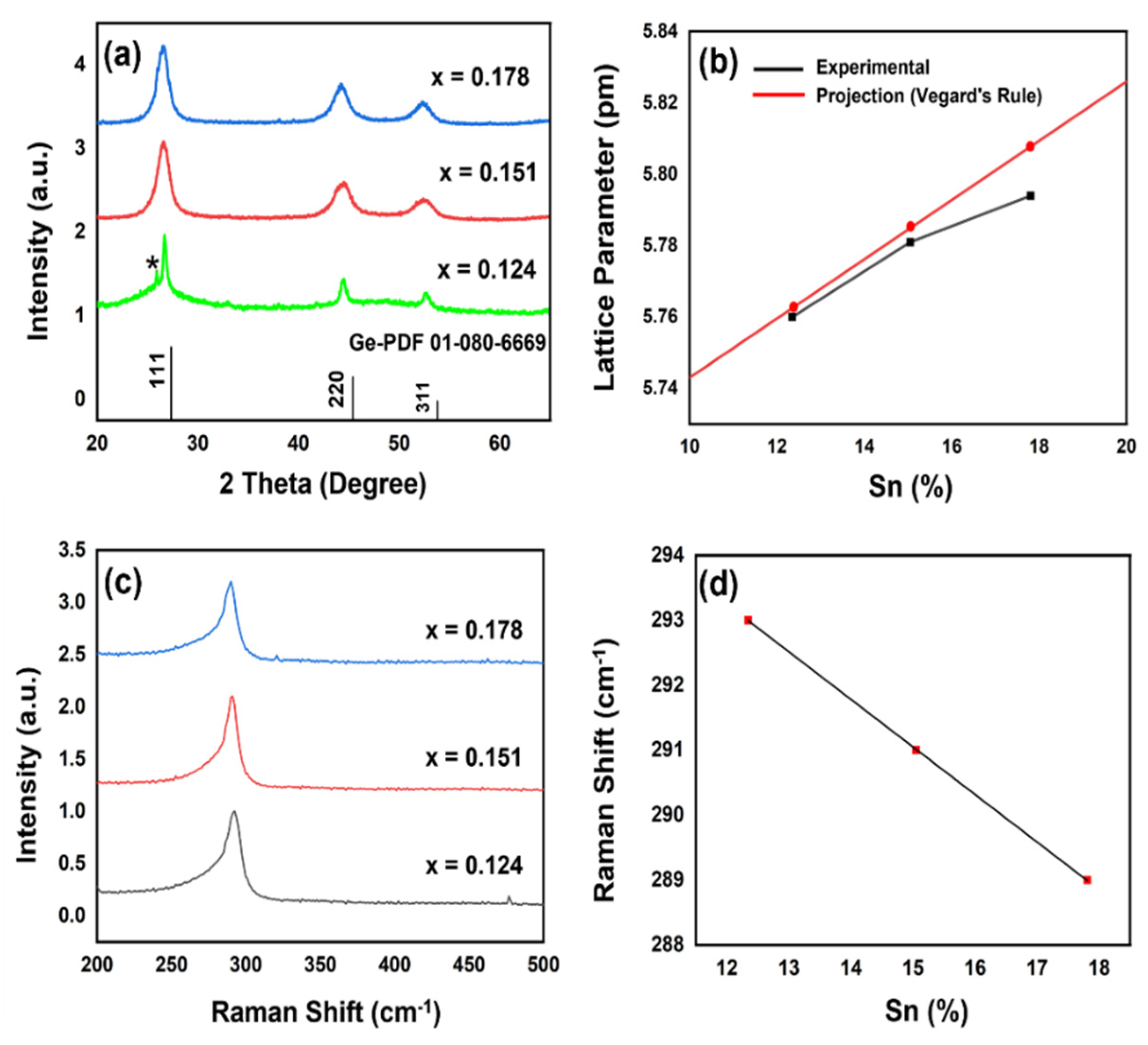
With an increasing Sn concentration, the powder X-ray diffraction (XRD) patterns shift to lower 2θ angles owing to the expansion of the cubic Ge structure by α-Sn, suggesting the incorporation of Sn into Ge (Vegard’s Law) [16]. The major diffraction peaks were indexed to the (111), (220), and (311) planes of diamond-cubic Ge with F3m space group (ICDD: 01-080-6669). No diffraction peaks corresponding to α-Sn, or β-Sn (tetragonal Sn) impurity phases were detected. The peak observed near 26° 2θ, attributable to GeO2 (Figure 1a) could be ascribed to ambient isolation and purification of nanoparticles, as weakly bound surfactant ligands can be lost via excessive washing and centrifugation, thus exposing the nanoparticles surface [17]. The lattice constants for cubic Ge and α-Sn are 5.66 and 6.49 Å, respectively. In contrast, the lattice constant values in Ge1−xSnx nanoparticles vary from 5.76 Å for x = 0.124 to 5.794 Å for x = 0.178 (Table S1, Supplementary Materials). The values of lattice constants indicate a near linear expansion of the cubic Ge structure with increasing Sn. This experimentally observed trend of increasing lattice constant values was found to be in agreement with Vegard’s rule (Figure 1b) [16], which states that the lattice parameter of the alloys varies linearly with the change in the concentration of the components.
Bulk Ge exhibits a Raman peak at 300 cm−1 that corresponds to the longitudinal optical (LO) phonon mode of Ge–Ge bonds [18]. As heavier Sn atoms are incorporated into the Ge crystal, a systematic red shift of the Ge–Ge phonon mode (288−282 cm−1 for x = 0.124−0.178) was observed as a result of the longer Ge–Sn bonds and the heavier Sn atoms [19]. The observed red shift with increasing Sn content is consistent with the weakening (or lengthening) of the Ge–Ge bond and lattice constants computed from Vegard’s law (Figure 1c).
3.3. SEM and TEM
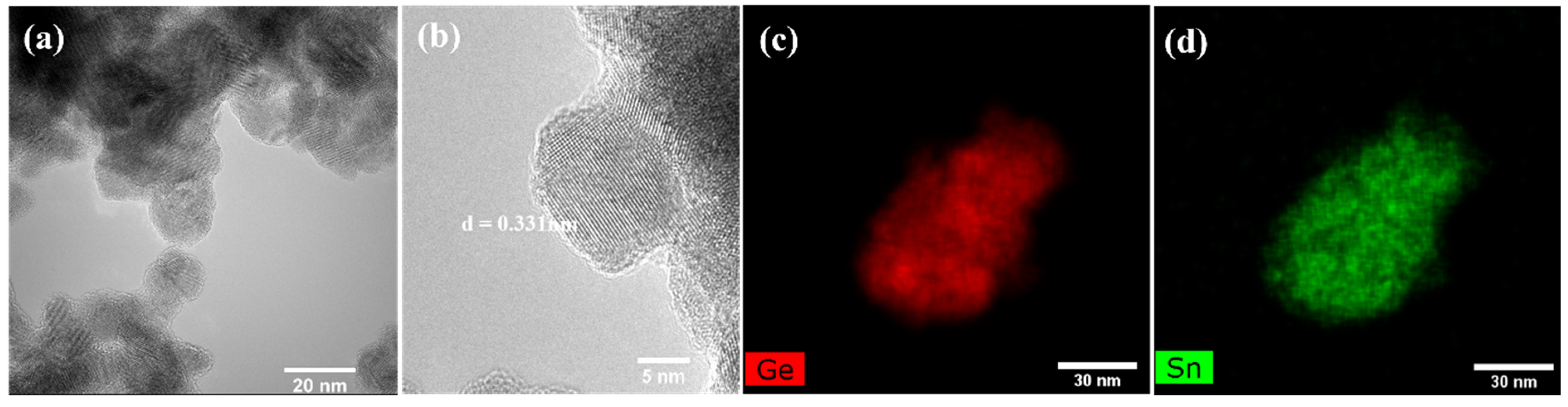
In conjunction with the XRD and Raman analysis, SEM and TEM have been used to confirm the formation, compositional uniformity, and homogeneity of Ge-Sn nanoparticles. The TEM image shows that the as-synthesized nanoparticles are irregular in shape and polydisperse (Figure 2a). The high-resolution TEM image (HRTEM) indicates the lattice spacing of 3.3 Å, which corresponds to the expanded (111) plane of diamond cubic Ge1−xSnx and is slightly larger than that of pure Ge (3.26 Å) [20], further confirming the expansion of cubic Ge lattice (Figure 2b). The TEM-EDS and SEM-EDS elemental mapping profile shows that the Sn and Ge are uniformly distributed over the entire nanoparticle, confirming it as a homogeneous, solid solution and free of segregated Sn species. EDX data of Ge1−xSnx nanocrystals show good agreement with the elemental composition calculated by Vegard’s law.
3.4. Optical Properties and Ligand Exchange
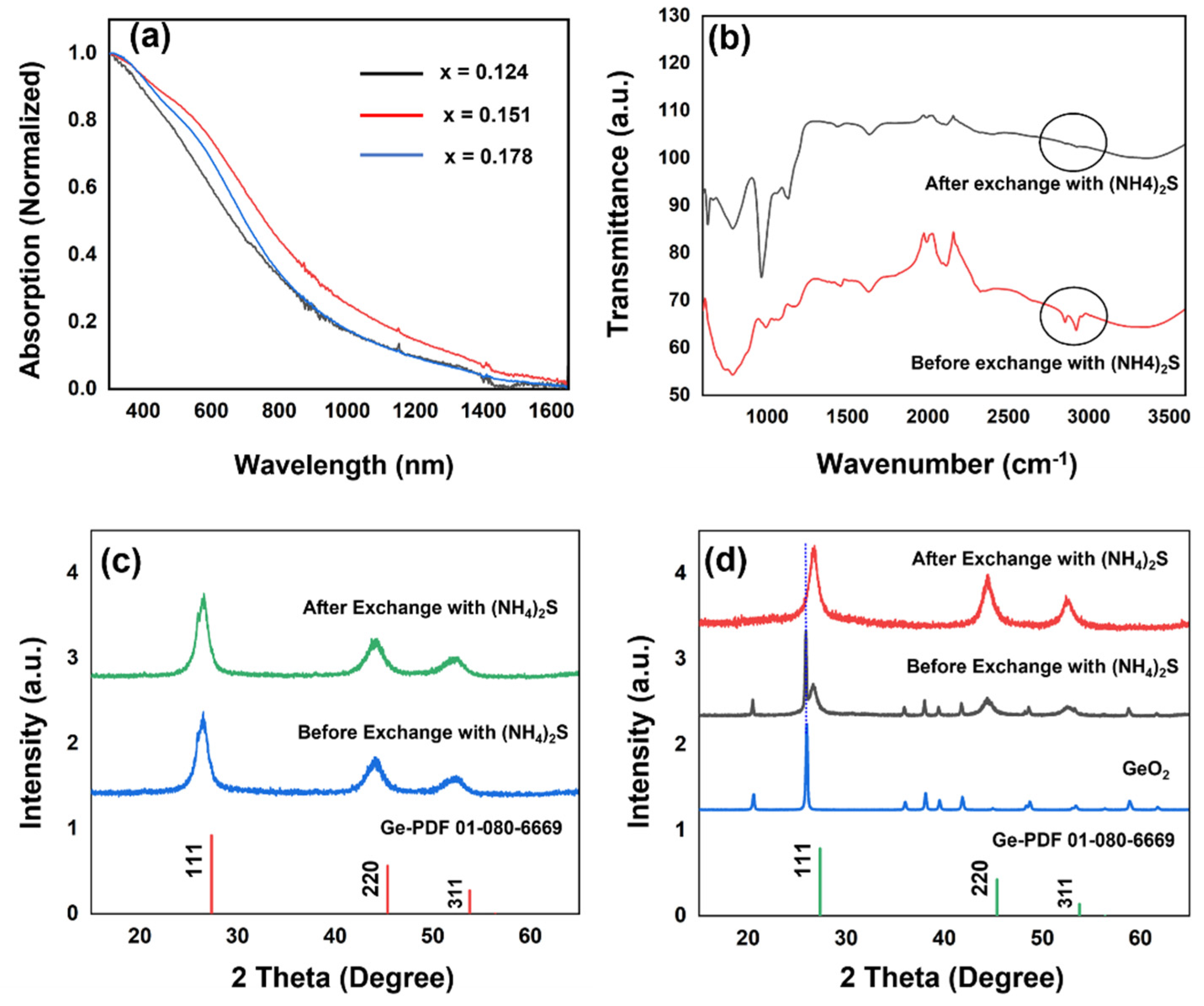
The solution-based UV–Vis–NIR spectra of the samples dispersed in chloroform was featureless across the whole range of wavelengths (Figure 3a). The incorporation of Sn significantly red-shifts the absorption onsets into the near-IR (NIR) region, further expanding its optical window for application in NIR optoelectronic devices.
The as-synthesized nanoparticles carry surface ligands originated from the synthesis. Various methods for replacing the long-alkyl-chain solvent/stabilizer used in synthesis have been reported [15,21,22,23,24]. Such methods include the replacement of oleylamine with much shorter atomic S2− ligands by a solution-phase ligand-exchange process, accomplished by vigorously shaking a mixture of nanoparticles suspended in an adequate solvent (chloroform used herein) and aqueous (NH4)2S. The removal of the surface ligands was qualitatively validated by the loss of nanoparticle dispersibility in chloroform and further confirmed by FT-IR analysis, which shows the disappearance of bands centered around 2800–3000 cm−1 ascribed to C–H stretching vibrations of oleylamine (Figure 3b). The XRD pattern of the sample in Figure 3c confirms the phase purity of Ge1−xSnx nanoparticles after the ligand exchange. According to the literature, (NH4)2S treatment creates a Ge1−xSnx–S monolayer passivating the dangling surface bonds of both Ge and Sn, and thus protects Ge1−xSnx surfaces from oxidation [25].
The treatment with aqueous (NH4)2S not only successfully exchanged the surface ligands but also removed the native surface oxide, which could cause poor chemical stability, high surface trap state density, and unfavorable electrical properties. The powder XRD analysis of Ge1−xSnx samples containing the GeO2 impurity shows the complete elimination of the corresponding oxide peaks after treatment with aqueous (NH4)2S (Figure 3d). It is anticipated that the removal of oxide impurities from the surface of Ge1−xSnx alloy nanoparticles will further improve their charge, career mobility and performance in electronic devices.
4. Conclusions
The successful synthesis of uniform Ge1−xSnx alloy nanoparticles with Sn concentrations varying from x = 0.124 to 0.178 using the mild reducing reagent NaBH4 has been achieved, proving to be a facile, ecofriendly, and economically viable route for producing Ge-Sn nanoparticles. Using a solution-phase ligand-exchange approach, the long-chain oleylamine ligands were replaced by short inorganic sulfide-capping groups using aqueous (NH4)2S. The surface-passivating effect of (NH4)2S suppresses the formation of germanium oxide while preventing any segregation of Sn atoms, as showed by the long-term stability of the nanomaterial. (NH4)2S not only successfully removes the organic ligands but also etches any undesired surface oxide that occasionally forms during synthesis.
Supplementary Materials
The following are available online at https://0-www-mdpi-com.brum.beds.ac.uk/article/10.3390/cryst11101216/s1, Table S1: Crystallite size and lattice parameter of Ge1−xSnx alloy nanoparticles, Figure S1: Powder XRD patterns of Ge1−xSnx nanoparticles after months of storage under ambient conditions, Figure S2: Powder XRD patterns of Ge1−xSnx nanoparticles with higher composition of Sn (20–30%) showing peaks of segregated Sn impurities, Figure S3: (a–c) SEM images of Ge1−xSnx nanoparticles (x = 0.15). (d,e) Ge and Sn SEM−EDX element mapping from the image c, showing that Ge and Sn are homogeneously dispersed through the alloy nanoparticles, Figure S4: (a–c) SEM images of Ge1−xSnx nanoparticles (x = 0.178). (d,e) Ge and Sn SEM−EDX element mapping from the image c, showing that Ge and Sn are homogeneously dispersed through the alloy nanoparticles, Figure S5: Representative SEM-EDS spectrum of Ge0.85Sn0.15 nanoparticles.
Author Contributions
The individual authors’ contributions are as follows: conceptualization of the synthetic pathway, D.R.R. and C.-Y.L.; methodology for the synthetic approach, G.S.A. and M.L.; validation of data and processes, D.R.R. and C.-Y.L.; formal analysis of the research results, G.S.A.; investigation, G.S.A. and M.L.; resources provided for the project, D.R.R. and C.-Y.L.; data curation, G.S.A.; writing—original draft preparation, G.S.A.; writing—review and editing, D.R.R. and C.-Y.L.; supervision, D.R.R. and C.-Y.L.; project administration, D.R.R. and C.-Y.L.; funding acquisition, D.R.R. and C.-Y.L. All authors have read and agreed to the published version of the manuscript.
Funding
This research was funded by the US Department of Defense, Office of Naval Research (ONR), Award number N00014-20-1-2539, the US National Aeronautics and Space Administration (NASA) Award number 80NSSC10M0201, and the US National Science Foundation, Award number NSF1924412.
Institutional Review Board Statement
Not applicable.
Informed Consent Statement
Not applicable.
Data Availability Statement
Data and methods used in this work are presented in sufficient detail in the paper so that other researchers can replicate the work. Raw data are available from the corresponding author upon request.
Conflicts of Interest
The authors declare no conflict of interest. The funders had no role in the design of the study; in the collection, analyses, or interpretation of data; in the writing of the manuscript, or in the decision to publish the results.
References
- Ruddy, D.; Johnson, J.; Smith, E.R.; Neale, N. Size and Bandgap Control in the Solution-Phase Synthesis of Near-Infrared-Emitting Germanium Nanocrystals. ACS Nano 2010, 4, 7459–7466. [Google Scholar] [CrossRef] [PubMed]
- Geiger, R.; Zabel, T.; Sigg, H. Group Iv Direct Band Gap Photonics: Methods, Challenges, and Opportunities. Front. Mater. 2015, 2, 52. [Google Scholar] [CrossRef] [Green Version]
- Suyog, G.; Chen, R.; Huang, Y.-C.; Yihwan, K.; Sanchez, E.; Harris, J.S.; Saraswat, K.C. Highly Selective Dry Etching of Germanium over Germanium–Tin (Ge1–XSnX): A Novel Route for Ge1–XSnX Nanostructure Fabrication. Nano Lett. 2013, 13, 3783–3790. [Google Scholar]
- Wang, L.; Su, S.; Wang, W.; Gong, X.; Yang, Y.; Guo, P.; Zhang, G.; Xue, C.; Cheng, B.; Han, G.; et al. Strained germanium–tin (GeSn) p-channel metal-oxide-semiconductor field-effect-transistors (p-MOSFETs) with ammonium sulfide passivation. Solid-State Electron. 2013, 83, 66–70. [Google Scholar] [CrossRef]
- Kittel, C. Introduction to Solid State Physics; John Wiley & Sons Inc.: New York, NY, USA, 2005. [Google Scholar]
- Esteves, R.J.A.; Hafiz, S.; Demchenko, D.O.; Özgür, Ü.; Arachchige, I.U. Ultra-Small Ge1–XSnX Quantum Dots with Visible Photoluminescence. Chem. Commun. 2016, 52, 11665–11668. [Google Scholar] [CrossRef]
- Venkatesham, T.; Nakagawara, T.A.; Demchenko, D.O.; Özgür, Ü.; Arachchige, I.U. Ge1–XSnX Alloy Quantum Dots with Composition-Tunable Energy Gaps and near-Infrared Photoluminescence. Nanoscale 2018, 10, 20296–20305. [Google Scholar]
- Barth, S.; Seifner, M.S.; Bernardi, J. Microwave-Assisted Solution–Liquid–Solid Growth of Ge1–XSnX Nanowires with High Tin Content. Chem. Commun. 2015, 51, 12282–12285. [Google Scholar] [CrossRef]
- Meng, A.C.; Fenrich, C.; Braun, M.; McVittie, J.P.; Marshall, A.F.; Harris, J.S.; McIntyre, P.C. Core-Shell Germanium/Germanium–Tin Nanowires Exhibiting Room-Temperature Direct- and Indirect-Gap Photoluminescence. Nano Lett. 2016, 16, 7521–7529. [Google Scholar] [CrossRef]
- Gao, K.; Prucnal, S.; Huebner, R.; Baehtz, C.; Skorupa, I.; Wang, Y.; Helm, M.; Zhou, S.; Skorupa, W. Ge1–XSnX alloys synthesized by ion implantation and pulsed laser melting. Appl. Phys. Lett. 2014, 105, 42107. [Google Scholar] [CrossRef]
- Kormoš, L.; Kratzer, M.; Kostecki, K.; Oehme, M.; Šikola, T.; Kasper, E.; Schulze, J.; Teichert, C. Surface analysis of epitaxially grown GeSn alloys with Sn contents between 15% and 18%. Surf. Interface Anal. 2016, 49, 297–302. [Google Scholar] [CrossRef]
- Newton, K.A.; Sully, H.R.; Bridges, F.; Carter, S.A.; Kauzlarich, S.M. Structural Characterization of Oleylamine- and Dodecanethiol-Capped Ge1–xSnx Alloy Nanocrystals. J. Phys. Chem. C 2021, 125, 6401–6417. [Google Scholar] [CrossRef]
- Lu, X.; Korgel, A.B.A.; Johnston, K.P. High Yield of Germanium Nanocrystals Synthesized from Germanium Diiodide in Solution. Chem. Mater. 2005, 17, 6479–6485. [Google Scholar] [CrossRef]
- Esteves, R.J.A.; Ho, M.Q.; Arachchige, I.U. Nanocrystalline Group Iv Alloy Semiconductors: Synthesis and Characterization of Ge1–XSnX Quantum Dots for Tunable Bandgaps. Chem. Mater. 2015, 27, 1559–1568. [Google Scholar] [CrossRef]
- Nag, A.; Kovalenko, M.V.; Lee, J.-S.; Liu, W.; Spokoyny, B.; Talapin, D.V. Metal-Free Inorganic Ligands for Colloidal Nanocrystals: S2–, Hs–, Se2–, HSe–, Te2–, HTe–, TeS32–, OH–, and NH2– as Surface Ligands. J. Am. Chem. Soc. 2011, 133, 10612–10620. [Google Scholar] [CrossRef]
- Chizmeshya, A.V.G.; Bauer, M.R.; Kouvetakis, J. Experimental and Theoretical Study of Deviations from Vegard′s Law in the SnxGe1−x System. ChemInform 2003, 34. [Google Scholar] [CrossRef]
- Lee, D.C.; Pietryga, J.M.; Robel, I.; Werder, D.J.; Schaller, R.D.; Klimov, V. Colloidal Synthesis of Infrared-Emitting Germanium Nanocrystals. J. Am. Chem. Soc. 2009, 131, 3436–3437. [Google Scholar] [CrossRef] [PubMed]
- Volodin, V.A.; Marin, D.; Sachkov, V.; Gorokhov, E.B.; Rinnert, H.; Vergnat, M. Applying an improved phonon confinement model to the analysis of Raman spectra of germanium nanocrystals. J. Exp. Theor. Phys. 2014, 118, 65–71. [Google Scholar] [CrossRef]
- Boote, B.W.; Men, L.; Andaraarachchi, H.P.; Bhattacharjee, U.; Petrich, J.W.; Vela, J.; Smith, E.A. Germanium–Tin/Cadmium Sulfide Core/Shell Nanocrystals with Enhanced Near-Infrared Photoluminescence. Chem. Mater. 2017, 29, 6012–6021. [Google Scholar] [CrossRef] [Green Version]
- Yang, Q.; Zhao, X.; Wu, X.; Li, M.; Di, Q.; Fan, X.; Zhu, J.; Song, X.; Li, Q.; Quan, Z. Facile Synthesis of Uniform Ge1–XSnX Alloy Nanocrystals with Tunable Bandgap. Chem. Mater. 2019, 31, 2248–2252. [Google Scholar] [CrossRef]
- Chen, C.-C.; Stone, K.; Lai, C.-Y.; Dobson, K.; Radu, D. Sulvanite (Cu3VS4) nanocrystals for printable thin film photovoltaics. Mater. Lett. 2018, 211, 179–182. [Google Scholar] [CrossRef]
- Liu, M.; Lai, C.-Y.; Chang, C.-Y.; Radu, D.R. Solution-Based Synthesis of Sulvanite Cu3TaS4 and Cu3TaSe4 Nanocrystals. Crystals 2021, 11, 51. [Google Scholar] [CrossRef]
- Liu, M.; Lai, C.-Y.; Selopal, G.S.; Radu, D.R. Synthesis and optoelectronic properties of Cu3VSe4 nanocrystals. PLoS ONE 2020, 15, e0232184. [Google Scholar] [CrossRef] [PubMed]
- Liu, M.; Radu, D.; Selopal, G.; Bachu, S.; Lai, C.-Y. Stand-Alone CuFeSe2 (Eskebornite) Nanosheets for Photothermal Cancer Therapy. Nanomaterials 2021, 11, 2008. [Google Scholar] [CrossRef] [PubMed]
- Lei, D.; Wang, W.; Zhang, Z.; Pan, J.; Gong, X.; Liang, G.; Tok, E.-S.; Yeo, Y.-C. Ge0.83Sn0.17 p-channel metal-oxide-semiconductor field-effect transistors: Impact of sulfur passivation on gate stack quality. J. Appl. Phys. 2016, 119, 24502. [Google Scholar] [CrossRef] [Green Version]
Scheme 1.
An illustration of the synthesis and ligand exchange process of Ge1−xSnx alloy nanoparticles.
Scheme 1.
An illustration of the synthesis and ligand exchange process of Ge1−xSnx alloy nanoparticles.

Figure 1.
(a) Powder XRD patterns of Ge1−xSnx nanoparticles. (b) A plot illustrating the lattice parameters based on XRD patterns (Black) and projection of theoretical lattice parameters calculated using Vegard’s law (Red). * GeO2 impurity peak. (c) Raman spectra of the Ge1−xSnx nanoparticles. (d) A plot illustrating the systematic red-shifting of the Ge–Ge optical phonon mode with increasing Sn composition.

Figure 2.
(a) HRTEM images of Ge1−xSnx nanoparticles (x = 0.151). (b) HRTEM image of Ge1−xSnx nanoparticles (x = 0.151), showing lattice fringes that correspond to the (111) plane of cubic Ge. (c) and (d) TEM/EDS elemental maps of Ge and Sn, respectively.
Figure 2.
(a) HRTEM images of Ge1−xSnx nanoparticles (x = 0.151). (b) HRTEM image of Ge1−xSnx nanoparticles (x = 0.151), showing lattice fringes that correspond to the (111) plane of cubic Ge. (c) and (d) TEM/EDS elemental maps of Ge and Sn, respectively.

Figure 3.
(a) UV–Vis spectrum of the prepared Ge1−xSnx nanoparticles. (b) FT-IR spectra of Ge1−xSnx nanoparticles sample before and after ligand exchange. (c) and (d) Powder XRD patterns of Ge1−xSnx nanoparticles sample before and after ligand exchange.
Figure 3.
(a) UV–Vis spectrum of the prepared Ge1−xSnx nanoparticles. (b) FT-IR spectra of Ge1−xSnx nanoparticles sample before and after ligand exchange. (c) and (d) Powder XRD patterns of Ge1−xSnx nanoparticles sample before and after ligand exchange.


Table 1.
Amounts of GeI2 and SnCl2 for the synthesis of Ge1−xSnx nanoparticles.
| Targeted Composition | GeI2 Amount | SnCl2 Amount | ||
|---|---|---|---|---|
| mg | mmol | mg | mmol | |
| Ge0.88Sn0.12 | 172.3 | 0.528 | 13.6 | 0.072 |
| Ge0.85Sn0.15 | 166.5 | 0.510 | 17.1 | 0.090 |
| Ge0.82Sn0.18 | 160.6 | 0.492 | 20.5 | 0.108 |
Publisher’s Note: MDPI stays neutral with regard to jurisdictional claims in published maps and institutional affiliations. |
© 2021 by the authors. Licensee MDPI, Basel, Switzerland. This article is an open access article distributed under the terms and conditions of the Creative Commons Attribution (CC BY) license (https://creativecommons.org/licenses/by/4.0/).
Share and Cite
MDPI and ACS Style
Attar, G.S.; Liu, M.; Lai, C.-Y.; Radu, D.R. Green Synthesis of Ge1−xSnx Alloy Nanoparticles for Optoelectronic Applications. Crystals 2021, 11, 1216. https://0-doi-org.brum.beds.ac.uk/10.3390/cryst11101216
AMA Style
Attar GS, Liu M, Lai C-Y, Radu DR. Green Synthesis of Ge1−xSnx Alloy Nanoparticles for Optoelectronic Applications. Crystals. 2021; 11(10):1216. https://0-doi-org.brum.beds.ac.uk/10.3390/cryst11101216
Chicago/Turabian StyleAttar, Gopal Singh, Mimi Liu, Cheng-Yu Lai, and Daniela R. Radu. 2021. "Green Synthesis of Ge1−xSnx Alloy Nanoparticles for Optoelectronic Applications" Crystals 11, no. 10: 1216. https://0-doi-org.brum.beds.ac.uk/10.3390/cryst11101216
Note that from the first issue of 2016, this journal uses article numbers instead of page numbers. See further details here.