Article

Synthesis, Cytotoxic Activity, Crystal Structure, DFT Studies and Molecular Docking of 3-Amino-1-(2,5-dichlorophenyl)-8-methoxy-1H-benzo[f]chromene-2-carbonitrile

by
Menna El Gaafary
1,2,
Tatiana Syrovets
2,
Hany M. Mohamed
3,4,
Ahmed A. Elhenawy
4,5,
Ahmed M. El-Agrody
4,*,
Abd El-Galil E. Amr
6,7,*,
Hazem A. Ghabbour
8 and
Abdulrahman A. Almehizia
6
1
Department of Pharmacognosy, College of Pharmacy, Cairo University, Cairo 11562, Egypt
2
Institute of Pharmacology of Natural Products and Clinical Pharmacology, Ulm University, 89081 Ulm, Germany
3
Chemistry Department, Faculty of Science, Jazan University, Jazan 45142, Saudi Arabia
4
Chemistry Department, Faculty of Science, Al-Azhar University, Nasr City, Cairo 11884, Egypt
5
Chemistry Department, Faculty of Science and Art, AlBaha University, Mukhwah, Al Bahah 65731, Saudi Arabia
6
Pharmaceutical Chemistry Department, Drug Exploration and Development Chair (DEDC), College of Pharmacy, King Saud University, Riyadh 11451, Saudi Arabia
7
Applied Organic Chemistry Department, National Research Centre, Dokki, Giza 12622, Egypt
8
Department of Medicinal Chemistry, Faculty of Pharmacy, University of Mansoura, Mansoura 35516, Egypt
*
Authors to whom correspondence should be addressed.
Submission received: 27 January 2021 / Revised: 8 February 2021 / Accepted: 11 February 2021 / Published: 13 February 2021

Abstract

:
The target compound 3-amino-1-(2,5-d ichlorophenyl)-8-methoxy-1H-benzo[f]-chromene-2-carbonitrile (4) was synthesized via a reaction of 6-methoxynaphthalen-2-ol (1), 2,5-dichlorobenzaldehyde (2), and malononitrile (3) in ethanolic piperidine solution under microwave irradiation. The newly synthesized β-enaminonitrile was characterized by FT-IR, 1H NMR, 13C NMR, mass spectroscopy, elemental analysis and X-ray diffraction data. Its cytotoxic activity was evaluated against three different human cancer cell lines MDA-MB-231, A549, and MIA PaCa-2 in comparison to the positive controls etoposide and camptothecin employing the XTT cell viability assay. The analysis of the Hirshfeld surface was utilized to visualize the reliability of the crystal package. The obtained results confirmed that the tested molecule revealed promising cytotoxic activities against the three cancer cell lines. Furthermore, theoretical calculations (DFT) were carried out with the Becke3-Lee-Yang-parr (B3LYP) level using 6-311++G(d,p) basis. The optimization geometry for molecular structures was in agreement with the X-ray structure data. The HOMO-LUMO energy gap of the studied system was discussed. The intermolecular-interactions were studied through analysis of the topological-electron-density(r) using the QTAIM and NCI methods. The novel compound exhibited favorable ADMET properties and its molecular modeling analysis showed strong interaction with DNA methyltransferase 1.

1. Introduction

Chromene and benzochromene derivatives represent an important class of oxygen-containing heterocyclic compounds. They exhibit a wide range of biological activities, such as antimicrobial [1,2,3,4], anti-influenza virus [5] anti-proliferative [6,7,8], anti-inflammatory, analgesic, and selective COX-2 inhibitory activities [9] and effects. Also, the small molecules of benzochromene derivatives apply their anticancer activities by binding to DNA, thereby damaging DNA structure, changing the replication of double helix DNA, hindering the division of cancer cells, and binding to DNA through covalent and/or non-covalent interactions [8,10]. In addition, 4H-benzo[h]chromene derivatives are a very successful choice in the treatment of several human diseases. For instance, some derivatives of 2-amino-4-aryl-4H-benzo[h]chromene-3-carbonitrile and 2-amino- 4-aryl-6-methoxy- 4H-benzo[h]chromene-3-carbonitrile act as potential tumor vascular-disrupting agents and induce cell cycle arrest at G2/M and apoptosis [11,12], thus they have emerged as a new class of cytotoxic agents [13,14,15,16,17,18,19,20,21]. In addition, a range of studies has revealed the potential anticancer effect of 1H-benzo[f]chromene derivatives [22,23,24,25,26,27]. Some 3-amino-1-aryl-1H-benzo[f]chromene-2-carbonitrile derivatives exhibit c-Src kinase inhibitory and antiproliferative activities [22] and show a high potency for the inhibition of hAChE [23], while inducing cell cycle arrest and apoptosis in human cancer cells via dual inhibition of topoisomerase I and II [26,27]. In continuation of our previous work in benzochromenes chemistry [2,3,4,22,26,27] and the study of crystal structure for benzochromene derivatives [2,28,29,30,31,32,33,34], we report the synthesis, NMR spectra, crystal structure, theoretical calculations (DFT), molecular modeling analyses, and cytotoxic activity of 3-amino-1-(2,5-dichlorophenyl)-8-1H-benzo[f]chromene-2-carbonitrile.

2. Results and Discussion
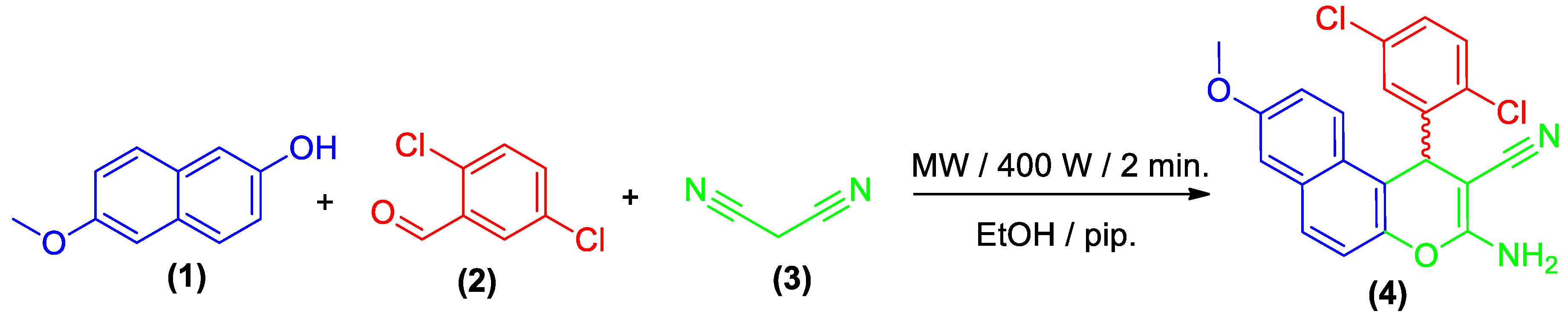
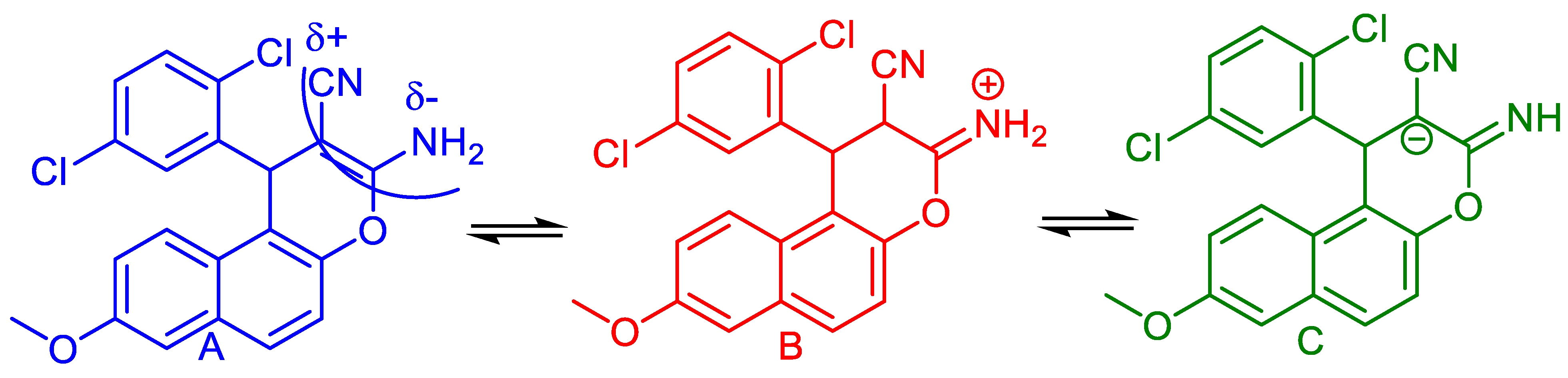
2.1. Chemistry

The preparation of the heterocyclic derivative is cited in (Scheme 1). Multi-component reaction of 6-methoxynaphthalen-2-ol (1) with 2,5-dichlorobenzaldehyde (2) and malononitrile (3) in ethanolic piperidine (25 mL/0.5 mL) solution under microwave irradiation for 2 min at 140 °C provide 3-amino-1-(2,5-dichlorophenyl)- 8-methoxy-1H-benzo[f]chromene-2-carbonitrile (4). The maximum power of microwave irradiation was optimized by repeating the reaction at different powers (200, 300, and 400 W) and for periods of time (1, 1.5, and 2 min). The optimum condition was obtained by using microwave irradiation at 400 W and a reaction time of 2 min, which gave the highest yield of the desired compound. It is also important to note that the 1-position of compound 4 is a chiral center and the reaction was controlled using the TLC technique. The optical activity of the target compound 4 was measured using a Carl Zeiss polarimeter. The results indicated that compound 4 has zero rotation (i.e., it is optically inactive) and thus it is in the form of a racemic (±) mixture [27], as illustrated in (Scheme 1).

2.2. Spectroscopic Data

The structure was studied using spectral data. The IR spectrum of compound 4 confirmed the presence of the characteristic NH2 absorption bands at υ 3450, 3337, 3213 cm−1, and CN absorption band at υ 21 cm−1. On the other hand, the 1H NMR spectrum of 4 showed the singlet signals of the amino, methine, and methoxy protons at δ 7.07, δ 5.74, and δ 3.83 ppm, respectively. The 13 C NMR spectrum of 4 showed signals resonating at δ 55.21 and δ 36.23 ppm which were attributable to the methoxy and methine carbons. Furthermore, the mass spectrum, elemental analysis, and the single crystal X-ray analysis gave an absolute confirmation for structure 4.

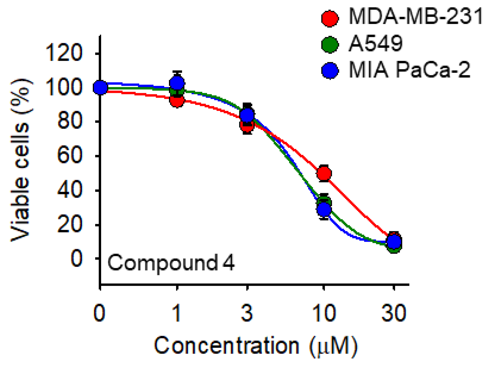
2.3. Cell Viability Assay
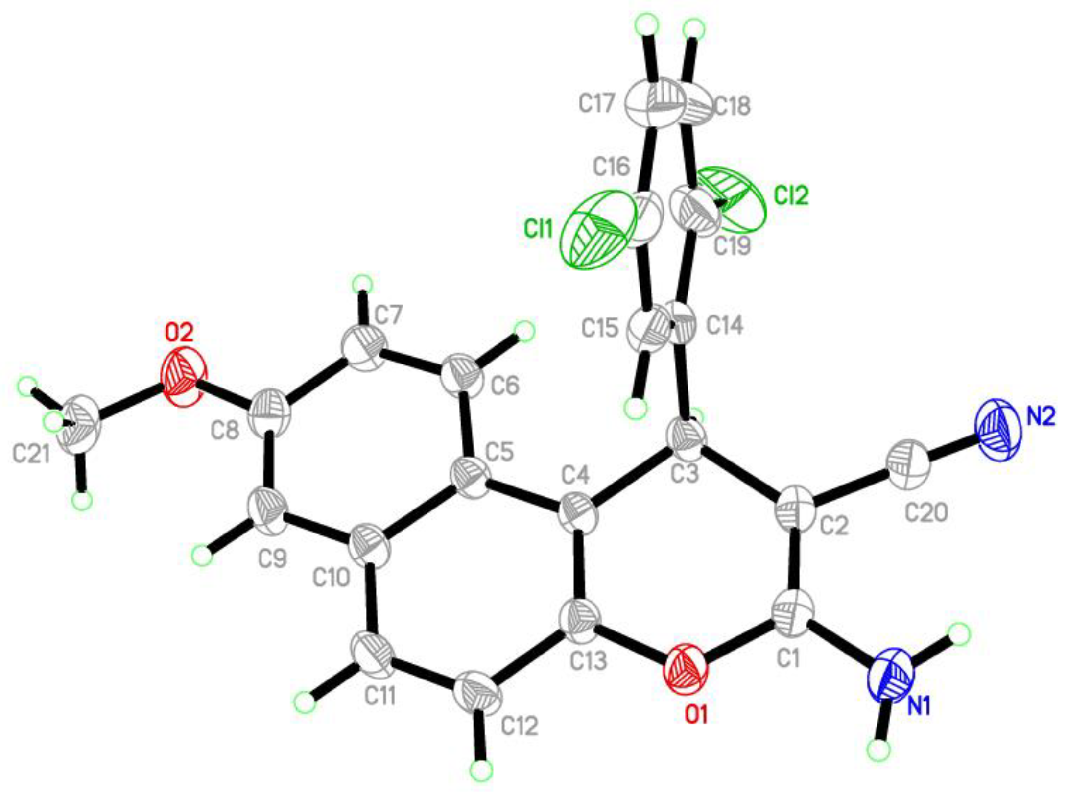
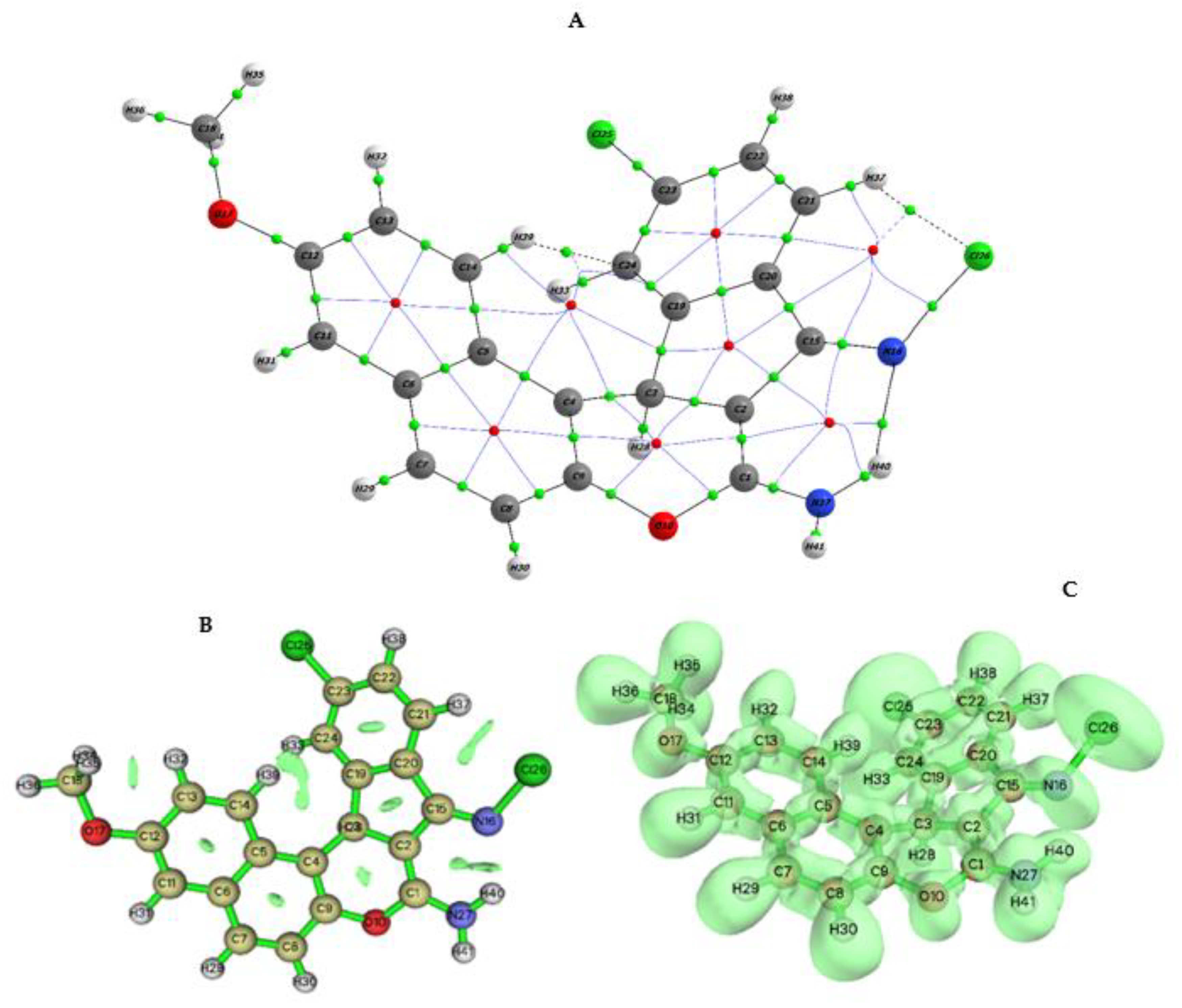
Cytotoxic activity of the newly synthesized 3-amino-1-(2,5-dichlorophenyl)-8- methoxy-1H-benzo[f]chromene-2-carbonitrile (compound 4) was examined in three human cancer cell lines, namely, the triple negative breast cancer (TNBC) MDA-MB-231, the non-small cell lung cancer (NSCLC) A549, and the pancreatic adenocarcinoma MIA PaCa-2 using the sodium 3´-[1-(phenylaminocarbonyl)-3,4-tetrazolium]-bis (4-methoxy-6- nitro)-benzene sulfonic acid hydrate (XTT) colorimetric assay [35,36]. Camptothecin and etoposide were included in the experiments as reference cytotoxic drugs. The results were shown in Table 1 and Figure 1.
Table 1 indicates that the target compound 4 displayed a promising cytotoxic activity against the tested cancer cell lines. Investigations of the cytotoxic activity against MDA-MB-231, A549, and MIA PaCa-2 indicated that compound 4 has IC50 values of 10.7 µM, 7.7 µM, and 7.3 µM, respectively. Etoposide, a known chemotherapy medication, which is used for the treatment of a number of types of cancer, did not show any sign of toxicity at the used concentrations range. On the other hand, camptothecin, the known antitumor agent, showed IC50 values of 22.4 µM, 20.0 µM, and 0.9 µM, for MDA-MB-231, A549, and MIA PaCa-2, respectively. Finally, grafting a lipophilic electron withdrawing substituent (chlorine atoms) in 2,5-postion of the phenyl ring at 1-postion of the 1H-benzo[f]chromene moiety could be beneficial.

2.4. Crystal Data

In the compound 4, C21H14Cl2N2O2, the crystallographic data and refinement information are summarized in Table 2. The selected bond lengths and bond angles are listed in Table 3. The asymmetric unit contains one independent molecule as shown in Figure 2. All bond lengths and angles are in normal ranges [37], the dichlorophenyl moiety makes a plane which is perpendicular with the plane of 1H-benzo[f]chromene moiety, with the dihedral angle equal to 89.33°. In the crystal packing, Figure 3, the molecules are linked via three intermolecular hydrogen bonds along the c axis Table 4. Generally, the dichlorophenyl and 1H-benzo[f]chromene rings are connected by C3-C14 bond. The mean of plane passing through the 1H-benzo[f]chromene forms an 89.33 angle with the dichlorophenyl. This indicates the appearance of some twisting between two of the fragments (Figure 1). The cyano and amino groups located in the plane with a 1H-benzo[f]chromene fragment form strong molecular intra H-bond interaction with a length of 0.85 Å, and dihedral bond of 165, as seen in Table 4. The bond lengths for the C–O, C–N, and C–Cl fell within the ranges of 1.357(3)–1.395(3), 1.336(4), and 1.734(3)–1.740(3) Å, respectively. Additionally, the length of CN is 1.140(4) Å due to formation of the strong H-bonds with the neighbor molecule. The three of 4 particles were packed in the three dimensions in the asymmetric unit and showed a intermolecular H-bond via CN• • • HN with a distance of 2.60 Å, as shown in Figure 3. These three molecules were structurally identical and were further linked together by two intermolecular H-bonds as N–H• • •OCH3 (2.52 Å) and one CN• • •H via C7 in 1H-benzo[f]chromene (with a contact distance of 2.60 Ǻ). This is of interest when using two H-bonds via Cl• • •H N and Cl• • •OCH3. The later H-bonds are responsible in the crystal packing shown in Figure 3 and Table 4.

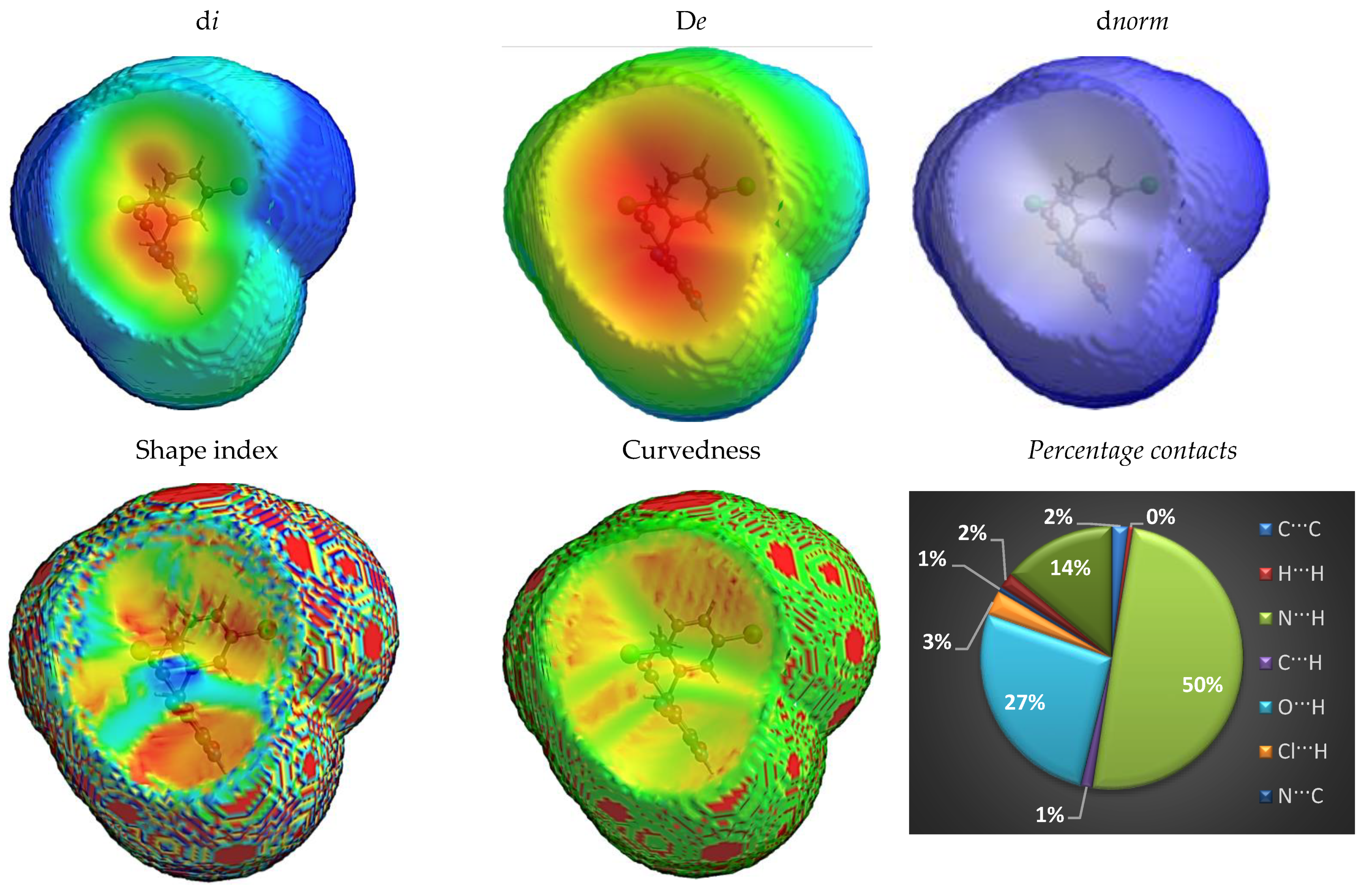
Fingerprint Plots for the Hirshfeld Surface

Hirshfeld surface analysis (HFS) is a fingerprint plot. It is a powerful map for exploring the sites that have intermolecular non-bonding interactions between the adjacent molecular groups in crystal packing [38]. The HFSs for the compound 4 have been figured out via a “d norm” descriptor, which clarifies two aspects: (i) “de” is the distance between any surface-point adjacent to the interior-atom, (ii) “di” illustrates the distance between the surface-point closest to the external atom. That is represented equationally as (dnorm = (dirvdWi) /rvdWi + (dervdWe)/rvdWe) based on the Van der Waals ratio of the atom, which is also another important parameter for representing HFS [39]. The rvdWi and rvdWe refer to Van der Waals’ radii of the adjacent two interior and exterior atoms via the molecular surface. The +Ve / -Ve dnorm values are relating to shorter / longer than rvdW for the intermolecular contacts, respectively.
Crystal explorer [40] was applied to get a 3D-map and 2D-histogram (de Vs di) of the HFS for 4 molecules in Figure 4 (see also Figure S4 and Table S1 as shown in the Supplementary Materials).
The molecular HFS was plotted over the dnorm, di,de, shape-index, and curvedness in value ranges (−0.9779 to 1.02128) Å, (2.0284 to 5.3629) Å, (−1.9848 to −1.0000) Å, and (−4.2169 to 4.000), respectively, and illustrated in Figure S4 (Supplementary Materials). The HFSs for the compound 4 depicted the halogen-halogen interactions (as red spick zone; dnorm), in Figure 4. The dnorm fingerprint map represents the presences of the strong H-bond interactions as (N⋅⋅⋅H/H⋅⋅⋅N) and (N–H···O) between the amino group with methoxy and the cyano group. These H-bond interactions were marked by bright red as little spikes located on the concave side of the di fingerprint map shown in Figure 4. The (N⋅⋅⋅H/H⋅⋅⋅N) and O⋅⋅⋅H intermolecular interactions contribute to the crystal packing by 50% and 27%, respectively (Figure S4; Supplementary Materials). From the analysis, the concave areas in curvedness and shape-index in Figure 4 can determine the presence of little stacking ππ interactions. The most sensitive indicator to any variation in morphology of the structure is the shape index. The red triangles (concave zone) located at the upper plane of a particle indicate dichlorophenyl outside the surface, while the blue triangles representing the benzo[f]chromene ring are located in internal surface. Thus, an insignificant contribution-percentage for CC (2 %) is located by HFS in Figure 4. The data were obtained from the shape index in agreement with a 2D plot-fingerprint (Figure S4; Supplementary Materials). The curvedness map was used to measure the surface shape for the particles. The surface with flat sites refers to minimum values for curvedness, whereas the sharpness curvature zones reflected the large curvedness values. Consequently, splitting the surface into two patches, which tend to the interactions, occurs between adjacent particles. The large flat areas which are outlined by red mean areas lacking stacking π···π interactions with a contribution% = 2% [39].

2.5. Molecular DFT Studies

2.5.1. Geometry Optimization

All computational simulations for compound 4 have been calculated in DFT with a B3LYP/6-311++G(d,p) basis set. Optimization of the structural geometry for ligand was compared via the X-ray model structure shown in Table S2, Figure S5 (Supplementary Materials). For the obtained data in Table S2, one can reveal good agreement between the calculated optimization parameters.

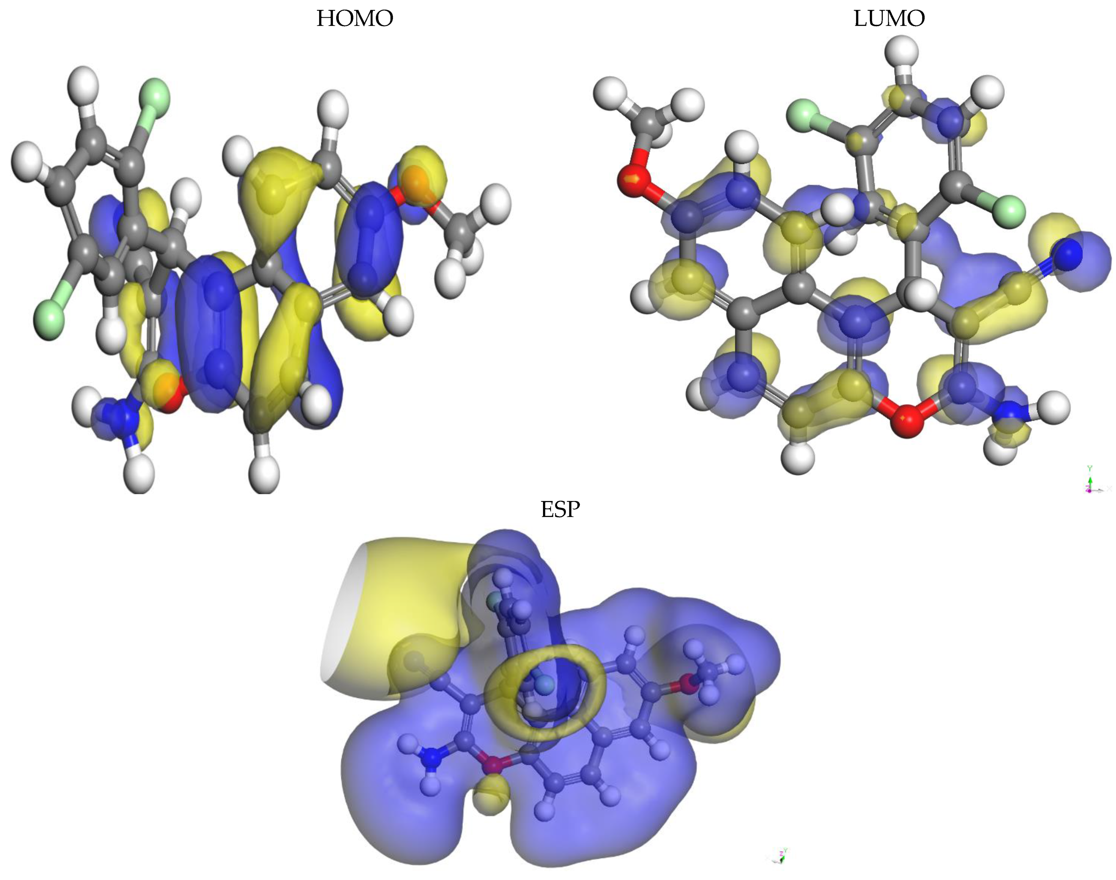
2.5.2. Analysis of Frontier Molecular Orbital

Frontier molecular orbitals (FMOs) through two types of HOMO (donating electron) and LUMO (accepting electron) are crucial orbitals for a molecule. These orbitals were able to display the binding manner of a biomolecule with a receptor. The FMOs gap was characterized by the chemical reactivity and kinetic stability of the molecule [41,42,43]. The molecule 4 has EHOMO (−0.218Hatree = −5.947 eV) with ELUMO(−0.08780 Hatree = −2.389 eV) which reflects the strength ability of the molecule for donating electron to biological media [41]. HOMO and LUMO areas had capped 1H-benzo[f]chromene-2-carbonitrile, as shown in Figure 5. These data elucidate the intramolecular charge transfer through which HOMO→ LUMO takes place.

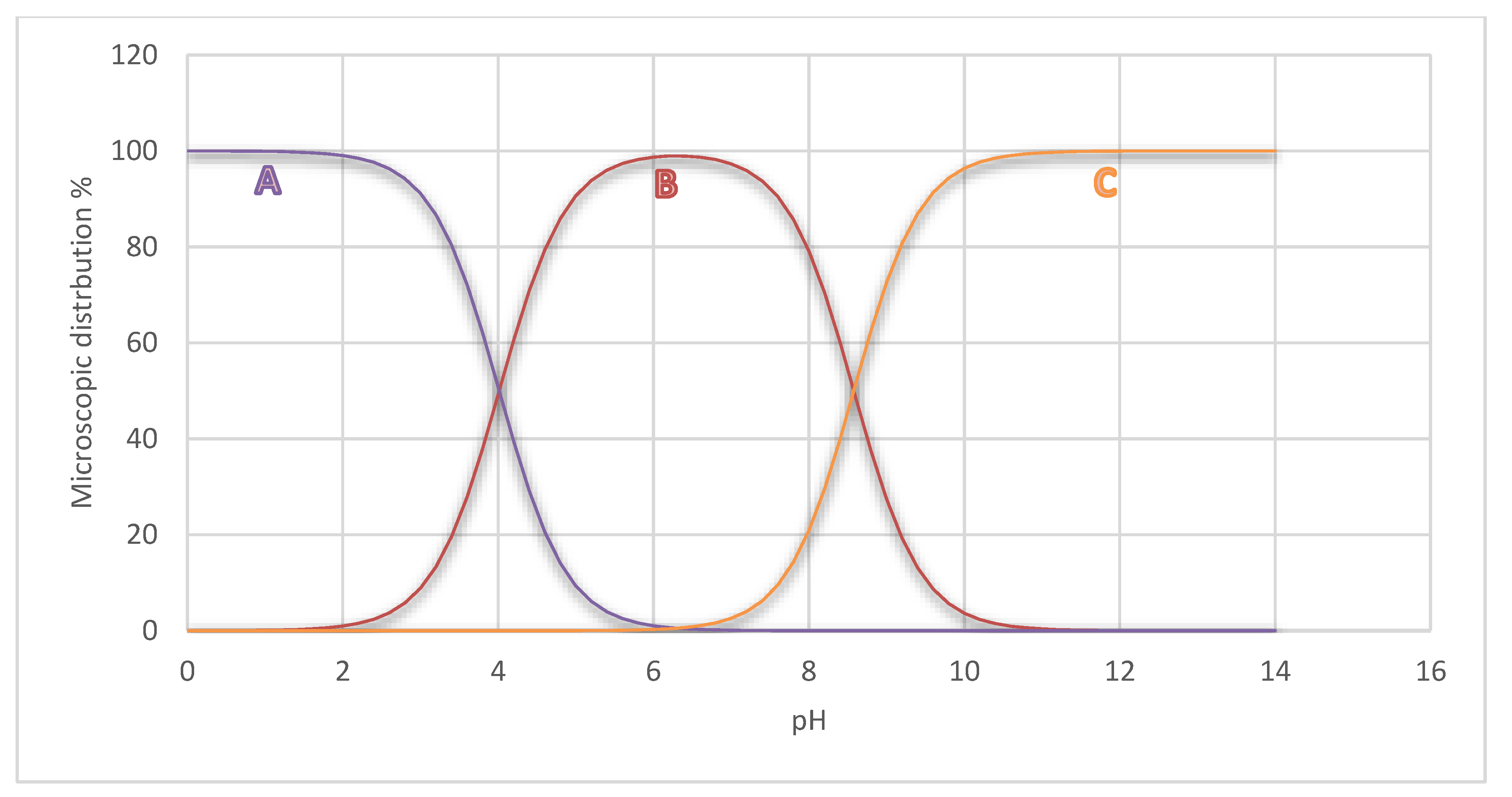
2.5.3. Chemical Reactivity

Chemical reactivity reflects the modification possibility for the molecular system during chemical and biochemical reactions. The increased energy of the FMOs (E-FMOs) has been linked to the soft nucleophiles and the hard electrophiles. ΔG was used as a measure of the stability index. The molecule with a small ΔG exhibits high polarizability and softness, hence raising the chemical reactivity and nucleophilicity (easy donation of an electron to an acceptor of biological media), with vice versa. Furthermore, stabilization interaction is conversely related to ΔG [43]. Thus, we studied the electrophiles and nucleophiles depending on softness and hardness concepts. The stability and reactivity of the molecular system were predicted by global reactivity parameters such as “S” softness (it measures stability of molecules and is directly proportional with chemical reactivity) [44]. “η” hardness (reciprocal of softness), µ (chemical potential), χ (electronegativity) [41], μ (donating power of electron), μ+ (catching electron efficiency), and ωi (Electrophilicity index) estimated reducing energy, which resulted from the maximal electrons current transfer between the donor and the acceptor [45]. These restrictions were demonstrated in both terms I (ionization potential) and A (electron affinity) [45]. The charge transfer has been examined through the reactivity index term, which measured stabilization energy for the media during chemical reactions when it is gaining a maximum amount of the electrons (ΔNmax) from the environment. The variation in binding energy at the ground state is computed using the maximal (ΔEmax) and minimal (ΔEmin) electrons transfer terms shown in Table 5.
The obtained data showed that the low ΔG value promoted increasing most chemical descriptors (S, χ, ωi; µ+, µ, µ, ΔNmax, ΔEmin, and ΔEmax), as seen in Table 5. These results reflect the higher reactivity for compound 4 against biological media. The compound 4 transfers an amount of electrons (ΔNmax = −1.71) with significant stability (ΔEmax = −4.68 and ΔEmin = −2.34) when interacting with a bioreceptor.
Local descriptors describe the variation in the electronic system that is represented as: F “Fukui function”. The more reactive center in a chemical species has a high value of F κ + due to the deformation of the electronic density at a specific site toward accepting or donating the electron [46,47]. These parameters were calculated as reported previously [48]. The highest F κ value has been over C18 and C24 (Table S4; Supplementary Materials). Thus, these atoms are the most sensitive regions against electrophilic attack. In case of the F κ + , negative charge has been transferred in between C18 and C17 in the same ring.

2.5.4. Analysis of the MEP Surface

The molecular electrostatic potential (MEP) map was initially performed to investigate the sensitivity of molecular sites toward electrophilic and nucleophilic attacks [47,49,50,51,52]. MEP figures the electrons distribution on the reactive molecular sites, predicting the interaction manner of the molecule as well as physiochemical descriptors relationships [53,54]. Furthermore, MEP introduces a balance between both repulsive interaction of the nuclei (positive charge as nucleophilic reactivity in blue color) and attractive interaction of the electrons (negative charge as electrophilic potency in yellow and red colors). The gray color is visualized as an intermediate potential value. The variation in colors on MEPs exhibits the difference in electrostatic potential values, which increases in the order gray < yellow < blue, as shown in Figure 5. The highest negative area covered the N atom for CN group. The electron density covering O of OCH3 group was higher than those covering O in the chromene ring seen in Figure 5. That reflects the high sensitivity for these sites to electrostatic attacks with the receptor. The maximum positive region shielded most rings. These regions provided one with knowledge about which molecular sites can form intermolecular interactions. Furthermore, raising the blue region may be explained by a high electrophilic potency, which is responsible for recognizing this compound against a binding site for the receptor through electrostatic interactions between the substrate and the receptor.

2.5.5. QTAIM, NCI and ELF Approaches

The AIM ”atoms in molecule theory” was utilized routinely to assess H-bond formation [55]. QTAIM “Bader’s quantum theory of atoms” was applied using AIMAll [56], to detect the contribution of the intramolecular-forces of the molecular conformation inside the studied systems. The topological analysis of the electron density ρ(r) for compound 4 was performed based on Bond-Critical-Points (BCPs, as green dots) and Ring-Critical-Points (RCPs, red dots) represented in Figure 6A. The weak intramolecular interaction was identified at C21–H37⋯O26 and C14–H39⋯O24 with the calculated Hbond energies varying from −0.08to −0.05 kcal mol−1, respectively. The QTAIM analysis is not sufficient for examining the supposed weak “NCI” non-covalent-interactions, especially involving intramolecular hydrogen bonds [57]. Thus, the NCI approach [58,59] was additionally applied using Multiwfn [60]. The reduced-density-gradient “s(r)” isosurface was examined at a gradient surface corresponding to s = 0.1 a.u., then the corresponding plots were generated in Figure 6B. The weak attractive NCIs were indicated between both phenyl rings as represented by Broad multiform domains, whereas small domains correspond to the strongest hydrogen bonds identified in the studied compounds. Delocalized electrons of aromatic groups are represented by the egg-shape domains, as shown in Figure 6B. Furthermore, we decided to study the electronic structure of compound 4 through utilizing the topological analysis of the electron localization function “ELF” based on the valence basin populations and plotted in Figure 6C. ELF topological analysis shows the atomic basin population changes along the backbone of compound to stabilize the crystal package.
The reduced density gradient (RDG) was plotted versus the electron density multiplied by the sign of the second Hessian eigenvalue (Figure S6; Supplementary Materials). The domains-shape is correlated with the interaction-power. The Broad disk-shaped domains indicate weak attractive (λ2 < 0) and repulsive (λ2 > 0) interactions. Gradient surfaces correspond to s = 0.1 a.u. The observed attractive and repulsive contacts are in equilibrium, allowing two aromatic fragments to maintain the defined mutual orientation.

2.5.6. NMR Spectra

The absolute isotropic chemical shielding was calculated for 4 using the same quantum level. Then relative chemical shifts were investigated [61] using the corresponding TMS shielding (σcal). The chemical shifts were calculated numerically by δcal = σcal (TMS) − σcal. The obtained experimental values are provided in Table S5 (Supplementary Materials). The protons for aromatic rings resonated in the range 6.8–7.73 ppm for chlorophenyl and 6.86–7.46 for a naphthyl ring. The outside H atom of th chormene ring showed a simulated chemical shift at s 5.32 ppm value. The H-atoms for the CH3 group at C17 resonated theoretically at 3.84, 3.47, and 3.55 ppm. As expected, 13C NMR chemical shifts of the entire phenyl carbon atoms were greater than 100 ppm. The predicted shifts were in the range of 110.78–141.54 for naphthalen and 122.12–137.45 for chlorophenyl rings. The predicted chemical shift for theOCH3 was 51.84 ppm. The cyno group simulated a chemical shift at 104.25 ppm due to the adjecent nitrogen atom. The C1 & C2 in the chormen ring neighboring cyano and ClPhe groups predicated a chemical shift at 43.21 and 62.68 ppm.

2.5.7. ADMET and Bioactivity Profile in Silico

Oral-bioavailability plays a key role in drug design and discovery therapeutic activity of the molecule through the determination of Absorption, Distribution, Metabolism, Excretion, and Toxicity (ADMET) for the effective pharmacophoric sites in enhancing the antimicrobial or other biological activity [62]. The ADMET was measured for compound 4, camptothecin, and etoposide as standard drugs based on the POM analysis. The POM is a new and dynamic bioinformatics-platform [63] used to predict the pharmacophore sites with antitumor/antimicrobial efficiency. The POM analysis postulated that the compounds which have different charges (δ—δ+) between two-heteroatoms in the same dipolar pharmacophoric site might inhibit tumor or bacterial growth over fungal strains [63,64,65]. For antifungal efficiency, the molecules should possess the same type of charge (δ—δ) in a pharmacophore site [54,55].
The tautomer A for the tested compound 4 has an ideal antitumor pharmacophore site ( NH   δ CN   δ + ) . The microspecies-distribution (%) for compound 4 in Figure 7 represents the neutral A, protonated B, and protonated C forms as illustrated in Scheme 2. Those forms are responsible for the regeneration of antitumor pharmacophore sites with higher IC50 than lower reference drugs against all tested cells.

2.6. Pharmacokinetic-Descriptors (Table S2, Figure S5; Supplementary Materials)

The ADMET predicted profile data of compound 4 shown in (Table S3; Supplementary Materials) and reference drugs have fulfilled Lipinski rules [66]. Lipophilicity character (clog P) < 5.0 for this compound showed a good permeability and absorption across the cell membrane [60]. TPSA is another important criterion in drug-likeness that systematized the membrane-permeability by referring to the polar atoms of the biomolecule. TPSA for compound 4 is lower than 140Å2, while reference drugs showed higher than ideal TPSA values [61]. That indicates that the tested compound is not passively absorbed with good absorption properties. Also, the drug likeness values were in the required limits. The hydrophilic-hydrophobic equilibrium state is important for the molecule solubility in aqueous media (blood, cytoplasm, etc.) and its distribution in membranes. Compared to the optimum value of clog P ≈ 3 [62], compounds 4 exhibited nearly optimum values (clog P = 3.37) compared to etoposide and camptothecin with clog P = 1.339 and 2.08, respectively, as seen in Table S3.

Bioactivity and Toxicity Risk Prediction

The bioactivity score (absorption, distribution, excretion, and metabolism) is a critical measurement used to predict the potency of a biomolecule. The bioactivity scores were calculated by comparing structural known drug fragments with the tested compound [63]. The obtained data showed that the tested compound have promising bioactivity scores via models such as “GPCR” G protein-coupled receptors, “ICM” ion channel modulators, “KI” kinase inhibitors, “ NRL” nuclear receptor ligand, “PI” protease inhibitor, and “EI” enzyme inhibitors (Table S3). We have investigated carcinogenic behaviors for the compound 4 by comparing the tested structures with 981 known various carcinogenic modules that were obtained from the “Carcinogenic Potency Database (CPDB)”. The gained data from Table S3 showed:
  • No observed carcinogenic effect with ranged values for the synthesized compound 4 (~0.8–0.9 mg/kg bw/d).
  • The positive result against Caco-2 indicates a good permeability for the tested compound 4 (Caco-2 is a human colon epithelial cancer cell line, which is applied as a model for human intestinal absorption for medication).
  • Compound 4 exhibits no acute oral toxicity observed with low acute lethal dose values for rat (LD50 = ~2.8–2.9 mol/kg).
  • Compound 4 does not act as an inhibitor and substrate against P-glycoprotein, so we can safely retint these compounds without any effect [67,68,69,70,71].
  • A positivity acceptance with high permeability value through the BBB (blood brain barrier) indicates that compound 4 may be effective in treating tuberculosis meningitis [72].
  • It has no mutagenic effect on the DNA of microorganisms when employing the Ames test (a wide examination of mutagenic effects for bioactive molecules against DNA of microorganisms [73].
  • Compound 4 does not exhibit skin sensitivity (utilizing 254 compounds able to induce skin sensitization) and hepatotoxicity effects (using 531 compounds that have a liver-associated side effect in humans).
  • This compound was not biodegradable when compared with 1604 compounds [74].
From the above features, we can conclude that the synthesized compound 4 may have a good oral bioavailability without any observed marked health effect.

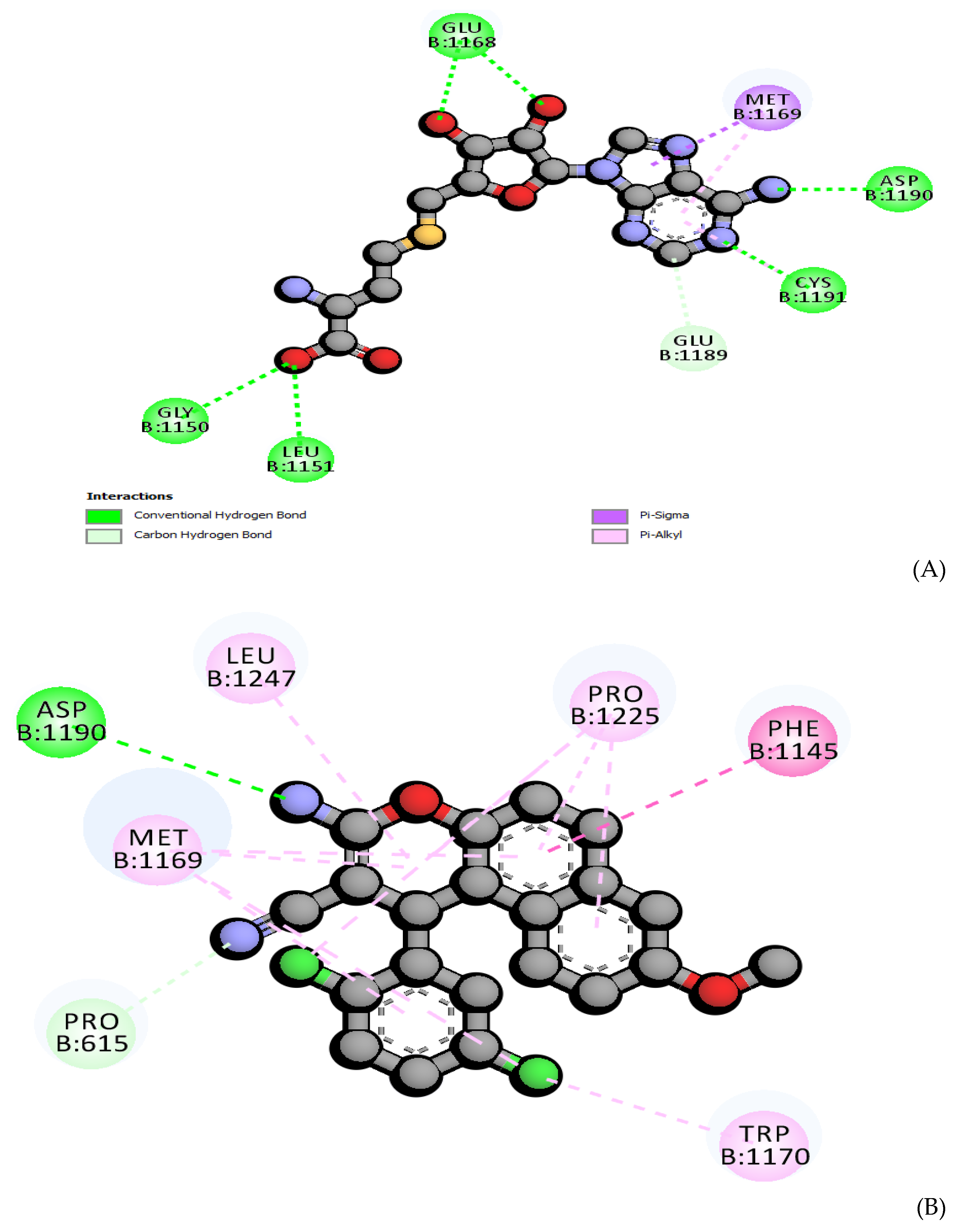
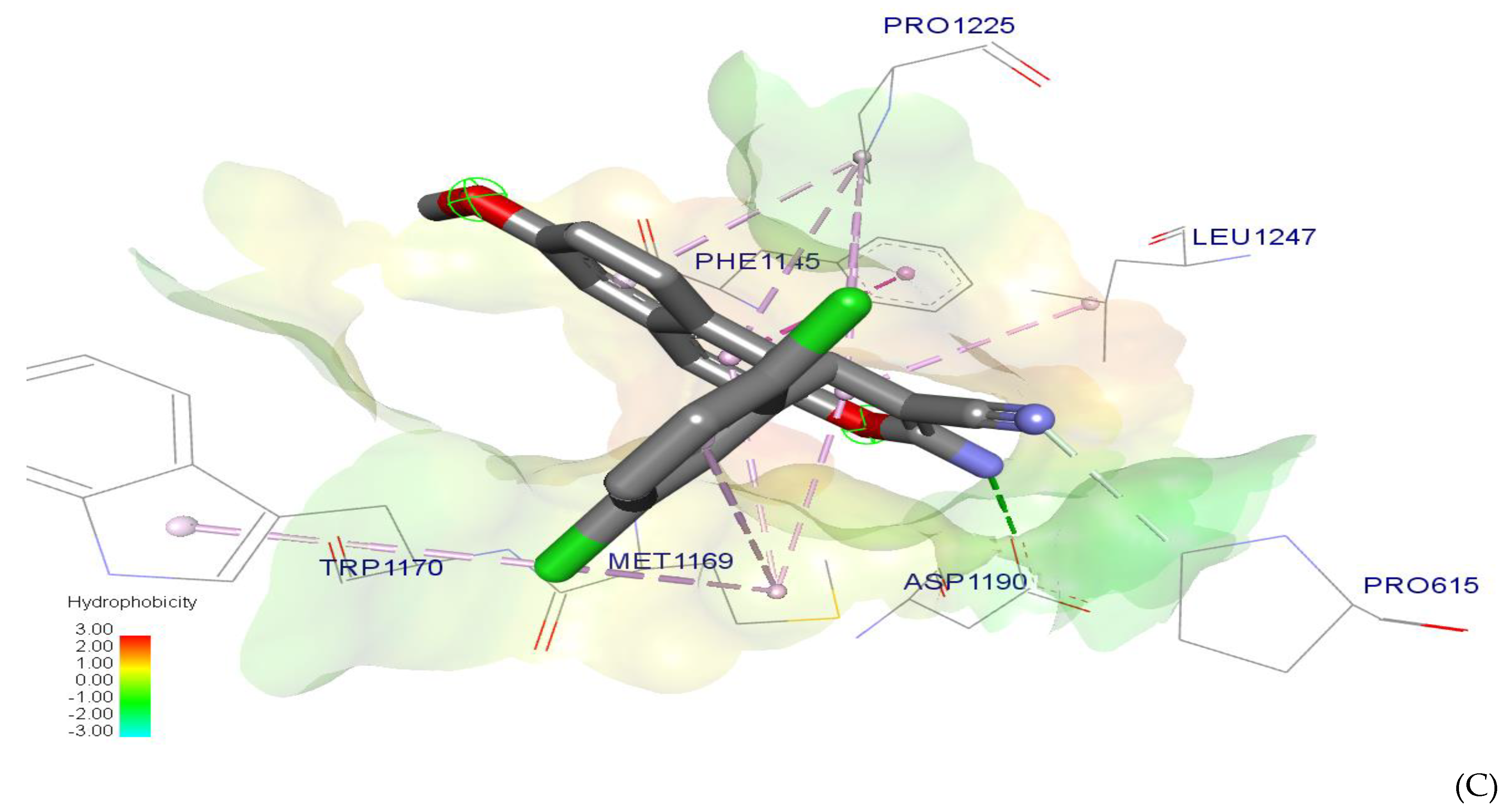
2.7. Molecular Docking

DNA methyltransferases (DNMTs) catalyze the transfer of the CH3 group between S-adenosyl methionine (SAM) and cytosine in CpG dinucleotides. Three active forms of DNMTs are encoded as DNMT1, DNMT2, DNMT3A, DNMT3B, and DNMT3L [75]. DNMT1 is fundamental for normal mammalian growth, and it has been recommended as the major attractive target in anticancer therapies [76]. The genetic changes in breast cancer take place due to the high expression of DNMT1. The inhibition of DNA-methylation through DNMT1 inhibition is an important target for epigenetic-therapy [77]. Human DNMT1 includes 1616 amino acids. It contains two domains: catalytic and regulatory at C- and N-terminals, respectively [78,79]. The catalytic C-terminal domain includes 10 amino acids. The IX- motif includes DNA recognition. The I / X-motifs, and VI/ VIII-motifs belong to the cofactor, substrate, and binding pocket, respectively [80]. The DNMT1 crystal structure has been solved using a modeling-homology of this protein. The primary-sequence of amino acids for DNMTs was retrieved from the NCBI (GenBank: AAD51619.1). The structural alignments by pBLAST [81] have been applied to get insights into the quality of the protein-folding. Its alignments allowed us to compare between several structures for the crystallized templates, suggested the similarity of the structures, and then assumed a satisfactory protein-folding form. The (5WVO_C) [82] and (5YDR_B) [83] models have been suggested for alignment by pBLAST as catalytic domain templates. Then, 3D structures of the alignment models were generated using HOMOLOGY module as implemented in Maestro [84] based on multiple-alignments, then minimization occurred in 200 illustration steps using an OPLS-2005 force field [85], followed by molecular-mechanics minimization. Finally, the model has been qualified using PROCHECK [86] for docking experiments. The pocket of the binding site was defined by CASTp server [87]. The 12 Å × 10 Å × 15 Å grid box size with coordinates X:142 Y:16 Z:16 was configured in molecular docking simulations. To validate the grid box, we found a protein model with a similar fold as the query and which had a known crystal structure. The (PDB code: 4WXX) [88] was applied to redocking with the same reference ligand and tested compounds. The 400 poses were generated using the Schrödinger model [84]. The pose selected has the lowest docking score and the lowest RMSD and possesses a shorter distance than (3 Å) with vital amino acid residues of the active site, as shown in Figure 8. The obtained poses were energetically minimized with a default force field.
Compound 4 exhibited a binding score (ΔG = −5.658 Kcal/mol) that was nearly equal with the original inhibitor S-adenosyl-L-homocysteine (ΔG = −6.412 Kcal/mol), as shown in Table 6. Compound 4 exhibited a strong interaction with the same part of DNMT1 as S-adenosyl-L-homocysteine, as established with a tolerable overlay and is shown in Figure 8. The dichlorobenzene ring formed a H-bond interaction with ASP119 through Cl group which acts as an acceptor, while benzo[f]chromene moiety interacts with the vital amino acids backbone such as through PRO615, LEU1247, PHE1145, and MET1169 by a favorable hydrophobic contact with PHE1145 (see Figure 8). These data represent a circular interaction into the DNMT1 pocket that promises a suitable filling for this enzyme part. All these of interaction-kinds contributed to the promising docking affinity and ΔG for compound 4. Compound 4 similarly as the reference drug appeared to block the same region formed by PRO615, LEU1247, PHE1145, MET1169, and ASP119 that fulfils the gateway of the protein pocket. This exposes the categories of DNMT1 inhibitors filtered as anticancer agents with promising IC50, as shown in Table 1.

Efficiency of Ligand based on Binding Energy

The variation between binding energy and heavy atoms is termed as a Efficiency ligand (LE) [89]. LE is considered to be a simple measurement to assess if the ligand is effective for an ideal fit against a targeted protein. The compound with a strong affinity to a small number of heavy atoms possesses high efficacy. The compound 4 showed similar values to S-adenosyl-L-homocysteine against DNMT1, as shown in Table 6.

3. Materials and Methods

3.1. General Information

All chemicals were purchased from Sigma-Aldrich Chemical Co. (St. Louis, MO, USA). The melting point was measured with a Stuart Scientific Co. Ltd apparatus and is uncorrected. The IR spectrum was recorded on a KBr disc on a Jasco FT/IR 460 plus spectrophotometer. The 1H NMR (500 MHz) and 13C NMR (125 MHz) spectra were measured on BRUKER AV 500 MHz spectrometer in DMSO-d6 as a solvent, using tetramethylsilane (TMS) as an internal standard, and chemical shifts were expressed as δ (ppm). The microwave apparatus used is Milestone Sr1, Microsynth. The mass spectrum was determined on a Shimadzu GC/MS-QP5050A spectrometer. Elemental analysis was carried out at the Regional Centre for Mycology and Biotechnology (RCMP), Al-Azhar University, Cairo, Egypt, and the results were within ± 0.25 %. Reaction courses and product mixtures were routinely monitored by thin layer chromatography (TLC) on silica gel precoated F254 Merck plates.

3.2. Synthesis of 3-Amino-1-(2,5-dichlorophenyl)-8-methoxy-1H-benzo[f]chromene-2-carbonitrile (4)

A reaction mixture of 6-methoxynaphthalen-2-ol (1) (0.01 mol), 2,5-dichlorobenzaldehyde (2) (0.01 mol), malononitrile (3) (0.01 mol), and piperidine (0.5 mL) in ethanol (25 mL) was heated under microwave irradiation conditions for 2 min at 140 °C. After completion of the reaction, the reaction mixture was cooled to room temperature and the precipitated solid was filtered off, washed with methanol, and was recrystallized from ethanol to give compound 4 as colorless crystals; yield 82%; m.p. 270–271 οC; IR (KBr) υ (cm−1): 3450, 3337, 3213 (NH2), 2193 (CN); 1H NMR δ: 7.88-6.96 (m, 8H, aromatic), 7.07 (bs, 2H, NH2), 5.74 (s, 1H, H-1), 3.83 (s, 3H, OCH3); 13C NMR δ: 160.08 (C-3), 156.53 (C-8), 145.23 (C-4a), 131.98 (C-6a), 128.79 (C-10a), 128.64 (C-6), 123.95 (C-10), 119.77 (C-10b), 119.50 (C-9), 117.09 (C-7), 114.44 (CN), 107.65 (C-5), 55.59 (C-2), 55.21 (CH3), 36.23 (C-1), 145.63, 132.16, 129.15, 129.01, 128.93, 124.92 (aromatic); MS m/z (%): 400 (M++4, 45.79), 398 (M++2, 15.27), 396 (M+, 53.43) with a base peak at 180 (100); Anal. Calcd for C21H14Cl2N2O2 (397.24): C, 63.49; H, 3.55; N, 7.05. Found: C, 63.45; H, 3.51; N, 7.01%.

3.3. Cytotoxic Activity

3.3.1. Cell Culture

The cancer cell lines, the triple negative breast cancer (TNBC) MDA-MB-231, the non-small cell lung cancer (NSCLC) A549, and the pancreatic adenocarcinoma MIA PaCa-2 were obtained from the American Type Culture Collection (ATCC, Rockville, MD, USA) and cultured as recommended by the suppliers.

3.3.2. Cytotoxicity Evaluation Using Cell Viability Assay

The cytotoxic activity was appraised using sodium 3′-[1-(phenylaminocarbonyl)-3,4-tetrazolium]-bis (4-methoxy-6-nitro) benzene sulfonic acid hydrate (XTT), as described in the literature [35,36].

3.4. X-ray Crystallography Analysis

A suitable single crystal for the compound 4 was obtained by a slow evaporation technique for the ethanolic solution at room temperature. The Bruker APEX-II D8 Venture diffractometer was employed for data collection, and was equipped with graphite monochromatic Mo Kα radiation, λ = 0.71073 Å at 293 (2) K. Data reduction and cell refinement were carried out by Bruker SAINT. SHELXT was used to develop the structure [90,91]. CCDC 2054709 containing all crystallographic data of the compound 4 can be obtained free of charge from the Cambridge Crystallographic Data Centre via www.ccdc.cam.ac.uk/data_request/cif (accessed on 18 February 2021). The full-matrix least-squares techniques with anisotropic thermal data for non-hydrogen atoms on F2 were employed for the final refinement of the structure.

3.5. Computational Model

All the quantum chemical computations were performed, using the DFT theory in Gaussian 09W [92] program package with the Becke3-Lee-Yang-parr (B3LYP) level using 6-311++G(d,p) basis. The single point calculations with wfn were used to calculate the following types of topological analysis for the molecular density: (i) the quantum theory of atoms in molecules (QTAIM); (ii) the non-covalent interactions index (NCI); (iii) an ELF (electron localization function). QTAIM, NCI, and ELF calculations were obtained from Multiwfn and the NCI plot program.

3.6. Protein Preparation

A 3D Crystal-structure for DNMT1 models was generated by a protein-preparation tool as implemented in Schrödinger-Suite-Maestro 20. Charges with bond orders were given and then H-atoms were added, respectively. The H2O molecules were omitted. OPLS_2005 force field was applied for minimization with RMSD to 0.30 Å. Potential binding pockets were identified using a CASTp server [80], which applies the modern algorithmic and geometrical analysis for computational chemistry through the analytical-validation pockets and cavities.

3.7. Molecular Docking

The Molecular docking was performed by Glide-tools [93]. A grid was generated for protein using the default factors, and then charged by assignment with a force field OPLS [94]. A cubic-box was applied on the centroid-binding pocket to generate the receptor. The size of the box was assigned to 12 Å × 12 Å × 12 Å for docking. Then, other molecular docking procedures were applied according to an original precision (SP) scoring function of the glide scoring function generated for measuring the binding affinity and the charges were assigned with a force field OPLS [94]. The structure with the lowest RMSD score was used to generate different poses of ligands.

3.8. ADMET Predictions

We used the ADMET profile in an attempt to identify of ADMET profile in silico (absorption, distribution, metabolism, excretion, and toxicity). The MOE and admetSAR tools were used for the prediction of pharmacokinetic parameters. The potential bioactivity was calculated, as well as the activity scores for distribution, excretion, and absorption parameters. The calculated drug-likeness scores of the tested compound were compared with each compound’s specific activity and reference drugs.

4. Conclusions

In conclusion, 3-amino-1-(2,5-dichlorophenyl)-8-methoxy-1H-benzo[f]-chromene-2- carbonitrile (4) was synthesized and characterized by 1H NMR, 13C NMR, elemental analysis, and X-ray single diffraction. The X-ray structure of the titled compound shows that bond lengths and angles were in normal ranges, while the dichlorophenyl moiety makes a plane which is perpendicular to the plane of 1H-benzo[f]chromene moiety, with a dihedral angle equal to 89.33°. The antitumor activity was evaluated against the human cancer cell lines MDA-MB-231, A549, and MIA PaCa-2 in comparison to the positive controls etoposide and camptothecin, the obtained results revealed promising cytotoxic activities against the three cancer cell lines. The experimental data were co-related with DFT theoretical calculations using B3LYP/6-311++G(d,p) basis set. Furthermore, DFT has been used to gain a clear view of global with local molecular reactivity, and HOMO, LUMO and MEP were plotted to examine intermolecular interaction through the charge transfer within the molecule. Also, the molecular QTAIM and NCI to non-covalent interactions supported the importance of the C-HO interactions in the molecular self-packing of co-crystal. The ADMET profile in silico showed that its compound has a good oral bioavailability and high ability BBB transport, with no marked carcinogenic and health effects observed for the rodent toxicity profile. Molecular docking demonstrated high interaction-affinity with high stabilty in DNMT1. The theoretical and experimental investigations suggested that the compound has a promising anticancer activity with good oral bioavailability without any observed carcinogenesis effect.

Supplementary Materials

The following are available online at https://0-www-mdpi-com.brum.beds.ac.uk/2073-4352/11/2/184/s1, Figure S1: 1H NMR of compound 4 in DMSO-d6; Figure S2: 1H NMR 8-6 ppm of compound 4 in DMSO-d6 and Figure S3: 13C NMR of compound 4 in DMSO-d6; Figure S4: The full and decomposed fingerprint plots of the intermolecular contacts observed in the crystal structure of the compound 4; Figure S5: Calculated optimization structure of compound 4 using DFT with a B3LYP/6-311++G(d,p); Figure S6: density gradient (RDG) versus sign(λ_2)×ρ(r) of BZims monomer. Table S1; Summary of various contacts and their contributions to the Hirshfeld Surface; Table S2: Optimization geometry parameters for compound 4; Table S3: ADMET predicted profile; Table S4: Calculated local reactivity (in eV) for the interacting atoms of compound 4; Table S5: Calculated NMR for 4 at B3LYP/6-311+G(d,p).

Author Contributions

H.M.M., A.M.E.-A., A.E.-G.E.A., and A.A.A. designed the proposed methods and analyzed the spectral data; A.M.E.-A. performed the experiments and implemented the biological study; A.A.E. performed DFT theoretical calculations; M.E.G. performed biological experiments. M.E.G. and T.S. analyzed the biological data, and reviewed and edited the draft. H.A.G. carried out and wrote the X-ray processes. All authors have read and agreed to the published version of the manuscript.

Funding

Funding was obtained via the Vice Deanship of Scientific Research Chairs.

Acknowledgments

The authors are grateful to the Deanship of Scientific Research, King Saud University for funding through the Vice Deanship of Scientific Research Chairs.

Conflicts of Interest

The authors declare no conflict of interest.

References

  1. Wang, Y.; Alenazy, R.; Gu, X.; Polyak, S.W.; Zhang, P.; Sykes, M.J.; Zhang, N.; Venter, H.; Ma, S. Design and structural optimization of novel 2H-benzo[h]chromene derivatives that target AcrB and reverse bacterial multidrug resistance. Eur. J. Med. Chem. 2020, 113049. [Google Scholar] [CrossRef]
  2. Radwan, H.A.M.; El-Mawgoud, H.K.A.; El-Mariah, F.; El-Agrody, A.M.; Amr, A.E.; Al-Omar, M.A.; Ghabbour, H.A. Single-Crystal Structure and Antimicrobial Activity of Ethyl 3-Amino-1-(4-chlorophenyl)-9-hydroxy-1H-benzo[f] chromene-2- carboxylate Combined with Ethyl α-Cyano-4-chlorocinnamate. Russ. J. Gen. Chem. 2020, 90, 299–304. [Google Scholar] [CrossRef]
  3. Fouda, A.M.; Hassan, A.H.; Eliwa, E.M.; Ahmed, H.E.A.; Al-Dies, A.A.M.; Omar, A.M.; Nassar, H.S.; Halawa, A.H.; Aljuhani, N.; El-Agrody, A.M. Targeted potent antimicrobial benzochromene-based analogues: Synthesis, computational studies, and inhibitory effect against 14α-Demethylase and DNA Gyrase. Bioorg. Chem. 2020, 105, 104387. [Google Scholar] [CrossRef]
  4. Abd El-Mawgoud, H.K.; Radwan, H.A.M.; El-Mariah, F.; El-Agrody, A.M. Synthesis, Characterization, Biological Activity of Novel 1H-benzo[f]chromene and 12H-benzo[f]chromeno[2,3-d]pyrimidine Derivatives. Lett. Drug Des. Discov. 2018, 15, 857–865. [Google Scholar] [CrossRef]
  5. Abdella, A.M.; Moatasim, Y.; Ali, M.A.; Elwahy, A.H.M.; Abdelhamid, I.A. Synthesis and Anti-influenza Virus Activity of Novel bis(4H-chromene-3-carbonitrile) Derivatives. J. Heterocycl. Chem. 2017, 54, 1854–1862. [Google Scholar] [CrossRef]
  6. Jayaprakash Rao, Y.; Yadaiah Goud, E.; Hemasri, Y.; Jain, N.; Gabriella, S. Synthesis and antiproliferative activity of 6,7-aryl/hetaryl coumarins. Russ. J. Gen. Chem. 2016, 86, 184–189. [Google Scholar] [CrossRef]
  7. Parthiban, A.; Kumaravel, M.; Muthukumaran, J.; Rukkumani, R.; Krishna, R.; Rao, H.S.P. Synthesis, in vitro and in silico anti-proliferative activity of 4-aryl-4H-chromene derivatives. Med. Chem. Res. 2016, 25, 1308–1315. [Google Scholar] [CrossRef]
  8. Ahagh, M.H.; Dehghan, G.; Mehdipour, M.; Teimuri-Mofrad, R.; Payami, E.; Sheibani, N.; Ghaffari, M.; Asadid, M. Synthesis, characterization, anti-proliferative properties and DNA binding of benzochromene derivatives: Increased Bax/Bcl-2 ratio and caspasedependent apoptosis in colorectal cancer cell. Bioorg. Chem. 2019, 93, 103329. [Google Scholar] [CrossRef]
  9. Fu, Z.-Y.; Jin, Q.-H.; Qu, Y.-L.; Guan, L.-P. Chalcone derivatives bearing chromen or benzo[f]chromen moieties: Design, synthesis, and evaluations of anti-inflammatory, analgesic, selective COX-2 inhibitory activities. Bioorg. Med. Chem. Lett. 2019, 29, 1909–1912. [Google Scholar] [CrossRef]
  10. Dehkordi, M.F.; Dehghan, G.; Mahdavi, M.; Feizi, M.A.H. Multispectral studies of DNA binding, antioxidant and cytotoxic activities of a new pyranochromene derivative. Spectrochim. Acta Part A 2015, 145, 353–359. [Google Scholar] [CrossRef]
  11. Schmitt, F.; Gold, M.; Rothemund, M.; Andronache, I.; Biersack, B.; Schobert, R.; Mueller, T. New naphthopyran analogues of LY290181 as potential tumor vascular-disrupting agents. Eur. J. Med. Chem. 2019. [Google Scholar] [CrossRef] [PubMed]
  12. Alblewi, F.F.; Okasha, R.M.; Eskandrani, A.A.; Afifi, T.H.; Mohamed, H.M.; Halawa, A.H.; Fouda, A.M.; Al-Dies, A.A.M.; Mora, A.; El-Agrody, A.M. Design and Synthesis of Novel Heterocyclic-Based 4H-benzo[h]chromene Moieties: Targeting antitumor caspase 3/7 activities and cell cycle analysis. Molecules 2019, 24, 1060. [Google Scholar] [CrossRef] [PubMed] [Green Version]
  13. Haiba, M.E.; Al-Abdullah, E.S.; Ghabbour, H.A.; Riyadh, S.M.; Abdel-Kader, R.M. Inhibitory activity of benzo[h]quinoline and benzo[h]chromene in human glioblastoma cells. Trop. J. Pharm. Res. 2016, 15, 2337–2343. [Google Scholar] [CrossRef] [Green Version]
  14. Haiba, M.E.; Al-Abdullah, E.S.; Ahmed, N.S.; Ghabbour, H.A.; Awad, H.M. Efficient and easy synthesis of new Benzo[h]chromene and Benzo[h]quinoline derivatives as a new class of cytotoxic agents. J. Mol. Struct. 2019, 1195, 702–711. [Google Scholar] [CrossRef]
  15. Kheirollahi, A.; Pordeli, M.; Safavi, M.; Mashkouri, S.; Naimi-Jamal, M.R.; Ardestani, S.K. Cytotoxic and apoptotic effects of synthetic benzochromene derivatives on human cancer cell lines. Naunyn-Schmiedeberg’s Arch. Pharmacol. 2014, 387, 1199–1208. [Google Scholar] [CrossRef]
  16. El-Agrody, A.M.; Abd El-Mawgoud, H.K.; Fouda, A.M.; Khattab, E.S.A.E.H. Synthesis, in-vitro cytotoxicity of 4H-benzo[h]chromene derivatives and structure-activity relationships of 4-aryl group and 3-, 7-positions. Chem. Pap. 2016, 70, 1279–1292. [Google Scholar] [CrossRef]
  17. El-Agrody, A.M.; Fouda, A.M.; Khattab, E.S.A.E.H. Synthesis, antitumor activity of 2-amino-4H-benzo[h]chromene derivatives, and structure-activity relationships of the 3- and 4-positions. Med. Chem. Res. 2013, 22, 6105–6120. [Google Scholar] [CrossRef]
  18. El-Agrody, A.M.; Fouda, A.M.; Khattab, E.S.A.E.H. Halogenated 2-amino-4H-benzo[h]chromene derivatives as antitumor agents and the relationship between lipophilicity and antitumor activity. Med. Chem. Res. 2017, 26, 691–700. [Google Scholar] [CrossRef]
  19. Okasha, R.M.; Alblewi, F.F.; Afifi, T.H.; Naqvi, A.; Fouda, A.M.; Al-Dies, A.A.M.; El-Agrody, A.M.; Belmont, P.; Bunce, R.A. Design of new Benzo[h]chromene derivatives: Antitumor activities and structure-activity relationships of the 2,3-Positions and fused rings at the 2,3-positions. Molecules 2017, 22, 479. [Google Scholar] [CrossRef]
  20. Halawa, A.H.; Fouda, A.M.; Al-Dies, A.-A.M.; El-Agrody, A.M. Synthesis, Biological Evaluation and Molecular Docking Studies of 4H-benzo[h]chromenes, 7H-benzo[h]chromeno[2,3-d]pyrimidines as Antitumor Agents. Lett. Drug Des. Discov. 2015, 13, 77–88. [Google Scholar] [CrossRef]
  21. El-Agrody, A.M.; Fouda, A.M.; Al-Dies, A.A.M. Studies on the synthesis, in vitro antitumor activity of 4H-benzo[h]chromene, 7H-benzo[h]chromene[2,3-d]pyrimidine derivatives and structure-Activity relationships of the 2-,3- and 2,3-positions. Med. Chem. Res. 2014, 23. [Google Scholar] [CrossRef]
  22. Ahmed, H.E.A.; El-Nassag, M.A.A.; Hassan, A.H.; Okasha, R.M.; Ihmaid, S.; Fouda, A.M.; Afifi, T.H.; Aljuhani, A.; El-Agrody, A.M. Introducing novel potent anticancer agents of 1H-benzo[f]chromene scaffolds, targeting c-Src kinase enzyme with MDA-MB-231 cell line anti-invasion effect. J. Enzyme Inhib. Med. Chem. 2018, 33. [Google Scholar] [CrossRef] [PubMed] [Green Version]
  23. Piazzi, L.; Cavalli, A.; Belluti, F.; Bisi, A.; Gobbi, S.; Rizzo, S.; Bartolini, M.; Andrisano, V.; Recanatini, M.; Rampa, A. Extensive SAR and computational studies of 3-{4-[(benzylmethylamino)methyl] phenyl}-6,7-dimethoxy-2H-2-chromenone (AP2238) derivatives. J. Med. Chem. 2007, 50. [Google Scholar] [CrossRef] [PubMed]
  24. Gorle, S.; Maddila, S.; Maddila, S.; Naicker, K.; Singh, M.; Singh, P.; Jonnalagadda, S. Synthesis, Molecular Docking Study and in vitro Anticancer Activity of Tetrazole Linked Benzochromene Derivatives. Anticancer Agents Med. Chem. 2017, 17. [Google Scholar] [CrossRef]
  25. Afifi, T.H.; Okasha, R.M.; Ahmed, H.E.A.; Ilaš, J.; Saleh, T.; Abd-El-aziz, A.S. Structure-activity relationships and molecular docking studies of chromene and chromene based azo chromophores: A novel series of potent antimicrobial and anticancer agents. EXCLI J. 2017, 16, 868. [Google Scholar] [CrossRef]
  26. Fouda, A.M.; Assiri, M.A.; Mora, A.; Ali, T.E.; Afifi, T.H.; El-Agrody, A.M. Microwave synthesis of novel halogenated β-enaminonitriles linked 9-bromo-1H-benzo[f]chromene moieties: Induces cell cycle arrest and apoptosis in human cancer cells via dual inhibition of topoisomerase I and II. Bioorg. Chem. 2019, 93. [Google Scholar] [CrossRef]
  27. Fouda, A.M.; Okasha, R.M.; Alblewi, F.F.; Mora, A.; Afifi, T.H.; El-Agrody, A.M. A proficient microwave synthesis with structure elucidation and the exploitation of the biological behavior of the newly halogenated 3-amino-1H-benzo[f]chromene molecules, targeting dual inhibition of topoisomerase II and microtubules. Bioorg. Chem. 2020, 95. [Google Scholar] [CrossRef]
  28. Okasha, R.M.; Amr, A.E.; El-Agrody, A.M.; Al-Omar, M.A.; Ghabbour, H.A. Crystal structure of 3-amino-8-methoxy-1-phenyl-1H-benzo[f]chromene-2-carbonitrile, C21H16N2O2. Z. Kristallogr. NCS 2017, 232. [Google Scholar] [CrossRef] [Green Version]
  29. Mohamed, H.M.; Amr, A.E.; El-Agrody, A.M.; Al-Omar, M.A.; Ghabbour, H.A. Crystal structure of 3-amino-1-(4-bromophenyl)-9-methoxy-1H-benzo[f]chromene-2-carbonitrile, C21H15BrN2O2. Z. Kristallogr. NCS 2017, 232. [Google Scholar] [CrossRef] [Green Version]
  30. Fouda, A.M.; Amr, A.E.; El-Agrody, A.M.; Al-Omar, M.A.; Ghabbour, H.A. Crystal structure of 3-amino-8-methoxy-1-(4-methoxy phenyl)-1H-benzo[f]chromene-2-carbonitrile, C22H18N2O3. Z. Kristallogr. NCS 2017, 232. [Google Scholar] [CrossRef] [Green Version]
  31. Okasha, R.M.; Alblewi, F.F.; Assiri, M.A.; Amr, A.E.; Ghabbour, H.A.; Afifi, T.H.; El-Agrody, A.M. Crystal Structure and Spectral Studies of 3-Amino-9-Methoxy-1-(4-methoxyphenyl)-1H-Benzo[f]Chromene-2-Carbonitrile. J. Comput. Theor. Nanosci. 2018, 15. [Google Scholar] [CrossRef]
  32. Okasha, R.M.; El-Agrody, A.M.; Amr, A.E.; Al-Omar, M.A.; Ghabbour, H.A. Synthesis and X-ray Single Crystals Characterizations of 2-Amino-4-(2-chlorophenyl)-6-Chloro-4H-Benzo[h]Chromene-3-Carbonitrile. J. Comput. Theor. Nanosci. 2017, 14, 5286–5291. [Google Scholar] [CrossRef]
  33. Amr, A.E.; Abd El-Mawgoud, H.K.; El-Agrody, A.M.; Al-Omar, A.E.; Alsultan, M.S. X-ray, Microwave Assisted Synthesis and Spectral Data of 3-Amino-1-(3,5-dibromo-2-methoxyphenyl)-8-methoxy-1H-benzo[f]-chromene-2-carbonitrile. J. Comput. Theor. Nanosci. 2017, 14, 3930–3935. [Google Scholar] [CrossRef]
  34. Afifi, T.H.; El-Agrody, A.M.; Amr, A.E.; Al-Omar, A.E.; Ghabbour, H.A. X-ray Characterization and Antimicrobial Activity of Synthesized New 3-Amino-8-Bromo-1-(3,4-dimethoxyphenyl)-1H-Benzo[f]-Chromene-2-Carbonitrile. J. Comput. Theor. Nanosci. 2017, 14, 3924–3929. [Google Scholar] [CrossRef]
  35. Paull, K.D.; Shoemaker, R.H.; Boyd, M.R.; Parsons, J.L.; Risbood, P.A.; Barbera, W.A.; Sharma, M.N.; Baker, D.C.; Hand, E.; Scudiero, D.A.; et al. The synthesis of XTT: A new tetrazolium reagent that is bioreducible to a water-soluble formazan. J. Heterocycl. Chem. 1988, 25, 911–914. [Google Scholar] [CrossRef]
  36. Scudiere, D.A.; Shoemaker, R.H.; Paull, K.D.; Monks, A.; Tierney, S.; Nofziger, T.H.; Currens, M.J.; Seniff, D.; Boyd, M.R. Evaluation of a Soluble Tetrazolium/Formazan Assay for Cell Growth and Drug Sensitivity in Culture Using Human and Other Tumor Cell Lines. Cancer Res. 1988, 48, 4827–4833. [Google Scholar]
  37. Allen, F.H.; Kennard, O.; Watson, D.G.; Brammer, L.; Orpen, A.G.; Taylor, R. Tables of bond lengths determined by x-ray and neutron diffraction. Part 1. Bond lengths in organic compounds. J. Chem. Soc. Perkin Trans. 2 1987, S1–S19. [Google Scholar] [CrossRef]
  38. McKinnon, J.J.; Fabbiani, F.P.A.; Spackman, M.A. Comparison of polymorphic molecular crystal structures through hirshfeld surface analysis. Cryst. Growth Des. 2007, 7. [Google Scholar] [CrossRef]
  39. Dalal, J.; Sinha, N.; Yadav, H.; Kumar, B. Structural, electrical, ferroelectric and mechanical properties with Hirshfeld surface analysis of novel NLO semiorganic sodium p-nitrophenolate dihydrate piezoelectric single crystal. RSC Adv. 2015, 5. [Google Scholar] [CrossRef]
  40. Mackenzie, C.F.; Spackman, P.R.; Jayatilaka, D.; Spackman, M.A. CrystalExplorer model energies and energy frameworks: Extension to metal coordination compounds, organic salts, solvates and open-shell systems. IUCrJ 2017, 4. [Google Scholar] [CrossRef] [PubMed] [Green Version]
  41. Fukui, K. Role of frontier orbitals in chemical reactions. Science 1982, 218, 747–754. [Google Scholar] [CrossRef] [Green Version]
  42. Hofmann, P. Arvi Rauk:“Orbital Interaction Theory of Organic Chemistry” Wiley & Sons: New York, NY, USA, 1994. ISBN 0-471-59389-3. 307 Seiten, mit HMO-Programmdiskette, Preis: $45.50. Ber. Bunsenges. Phys. Chem. 1995, 99, 997–999. [Google Scholar]
  43. Wildman, S.A.; Crippen, G.M. Prediction of physicochemical parameters by atomic contributions. J. Chem. Inf. Comput. Sci. 1999, 39, 868–873. [Google Scholar] [CrossRef]
  44. Ayers, P.W.; Parr, R.G. Variational principles for describing chemical reactions: The Fukui function and chemical hardness revisited. J. Am. Chem. Soc. 2000, 122, 2010–2018. [Google Scholar] [CrossRef]
  45. Lamaka, S.V.; Zheludkevich, M.L.; Yasakau, K.A.; Serra, R.; Poznyak, S.K.; Ferreira, M.G.S. Nanoporous titania interlayer as reservoir of corrosion inhibitors for coatings with self-healing ability. Prog. Org. Coat. 2007, 58, 127–135. [Google Scholar] [CrossRef]
  46. Komorowski, L.; Lipinski, J.; Szarek, P.; Ordon, P. Polarization justified Fukui functions: The theory and applications for molecules. J. Chem. Phys. 2011, 135, 14109. [Google Scholar] [CrossRef] [PubMed]
  47. Mendoza-Huizar, L.H.; Rios-Reyes, C.H.; Álvarez-Romero, G.A.; Palomar-Pardavé, M.E.; Ramírez-Silva, M.T. Electrophilic and nucleophilic chemical reactivity of neutral and anionic forms of 4-cpa, 24d-cpa, 34-cpa and 245t-cpa through conceptual dft reactivity descriptors. J. Chil. Chem. Soc. 2017, 62, 3411–3416. [Google Scholar] [CrossRef]
  48. Elhenawy, A.A.; Al-Harbi, L.M.; El-Gazzar, M.A.; Khowdiary, M.M.; Moustfa, A. Synthesis, molecular properties and comparative docking and QSAR of new 2-(7-hydroxy-2-oxo-2H-chromen-4-yl)acetic acid derivatives as possible anticancer agents. Spectrochim. Acta Part A Mol. Biomol. Spectrosc. 2019, 218, 248–262. [Google Scholar] [CrossRef]
  49. Luque, F.J.; López, J.M.; Orozco, M. Perspective on Electrostatic interactions of a solute with a continuum. A direct utilization of ab initio molecular potentials for the prevision of solvent effects. In Theoretical Chemistry Accounts; Springer: Berlin, Germany, 2000; pp. 343–345. [Google Scholar]
  50. Mendoza-Huizar, L.H.; Rios-Reyes, C.H. Chemical reactivity of atrazine employing the Fukui function. J. Mex. Chem. Soc. 2011, 55, 142–147. [Google Scholar]
  51. Sureshkumar, B.; Mary, Y.S.; Panicker, C.Y.; Suma, S.; Armaković, S.; Armaković, S.J.; Van Alsenoy, C.; Narayana, B. Quinoline derivatives as possible lead compounds for anti-malarial drugs: Spectroscopic, DFT and MD study. Arab. J. Chem. 2017, 13, 632–648. [Google Scholar] [CrossRef]
  52. War, J.A.; Jalaja, K.; Mary, Y.S.; Panicker, C.Y.; Armaković, S.; Armaković, S.J.; Srivastava, S.K.; Van Alsenoy, C. Spectroscopic characterization of 1-[3-(1H-imidazol-1-yl) propyl]-3-phenylthiourea and assessment of reactive and optoelectronic properties employing DFT calculations and molecular dynamics simulations. J. Mol. Struct. 2017, 1129, 72–85. [Google Scholar] [CrossRef]
  53. Elhenawy, A.A.; Al-Harbi, L.M.; Moustafa, G.O.; El-Gazzar, M.A.; Abdel-Rahman, R.F.; Salim, A.E. Synthesis, comparative docking, and pharmacological activity of naproxen amino acid derivatives as possible anti-inflammatory and analgesic agents. Drug Des. Dev. Ther. 2019, 13. [Google Scholar] [CrossRef] [PubMed] [Green Version]
  54. Ali, I.O.; Nassar, H.S.; El-Nasser, K.S.; Bougarech, A.; Abid, M.; Elhenawy, A.A. Synthesis and characterization of MnII and CoII complexes with poly (vinyl alcohol-nicotinic acid) for photocatalytic degradation of Indigo carmine dye. Inorg. Chem. Commun. 2021, 124, 108360. [Google Scholar] [CrossRef]
  55. Theophilou, A.K. A novel density functional theory for atoms, molecules, and solids. J. Chem. Phys. 2018. [Google Scholar] [CrossRef] [PubMed]
  56. Bader, R.F.W. Atoms in Molecules: A Quantum Theory, International Series of Monographs on Chemistry 22. Oxford University Press. Oxford Henkelman G, Arnaldsson A, Jónsson H A fast robust algorithm Bader decomposition of charge density. Comput. Mater. Sci. 1990, 88, 899. [Google Scholar]
  57. Lane, J.R.; Contreras-García, J.; Piquemal, J.P.; Miller, B.J.; Kjaergaard, H.G. Are bond critical points really critical for hydrogen bonding? J. Chem. Theory Comput. 2013, 9, 3263–3266. [Google Scholar] [CrossRef]
  58. Johnson, E.R.; Keinan, S.; Mori-Sánchez, P.; Contreras-García, J.; Cohen, A.J.; Yang, W. Revealing noncovalent interactions. J. Am. Chem. Soc. 2010, 132, 6498–6506. [Google Scholar] [CrossRef] [PubMed] [Green Version]
  59. Contreras-García, J.; Yang, W.; Johnson, E.R. Analysis of hydrogen-bond interaction potentials from the electron density: Integration of noncovalent interaction regions. J. Phys. Chem. A 2011, 115, 12983–12990. [Google Scholar] [CrossRef] [Green Version]
  60. Lu, T.; Chen, F. Multiwfn: A multifunctional wavefunction analyzer. J. Comput. Chem. 2012. [Google Scholar] [CrossRef]
  61. Wolinski, K.; Hinton, J.F.; Pulay, P. Efficient Implementation of the Gauge-Independent Atomic Orbital Method for NMR Chemical Shift Calculations. J. Am. Chem. Soc. 1990, 397–409. [Google Scholar] [CrossRef]
  62. Sheikh, J.; Hatzade, K.; Bader, A.; Shaheen, U.; Sander, T.; Ben Hadda, T. Computational evaluation and experimental verification of antibacterial and antioxidant activity of 7-hydroxy-3-pyrazolyl-4H-chromen-4-ones and their o-glucosides: Identification of pharmacophore sites. Med. Chem. Res. 2014, 23, 243–251. [Google Scholar] [CrossRef]
  63. Ahsan, M.J.; Govindasamy, J.; Khalilullah, H.; Mohan, G.; Stables, J.P.; Pannecouque, C.; Clercq, E. De POMA analyses as new efficient bioinformatics’ platform to predict and optimise bioactivity of synthesized 3a,4-dihydro-3H-indeno[1,2-c]pyrazole-2-carboxamide/carbothioamide analogues. Bioorg. Med. Chem. Lett. 2012, 22, 7029–7035. [Google Scholar] [CrossRef] [PubMed]
  64. Mabkhot, Y.N.; Arfan, M.; Zgou, H.; Genc, Z.K.; Genc, M.; Rauf, A.; Bawazeer, S.; Ben Hadda, T. How to improve antifungal bioactivity: POM and DFT study of some chiral amides derivatives of diacetyl-L-tartaric acid and amines. Res. Chem. Intermed. 2016, 42, 8055–8068. [Google Scholar] [CrossRef]
  65. Rachedi, K.O.; Ouk, T.-S.; Bahadi, R.; Bouzina, A.; Djouad, S.-E.; Bechlem, K.; Zerrouki, R.; Ben Hadda, T.; Almalki, F.; Berredjem, M. Synthesis, DFT and POM analyses of cytotoxicity activity of α-amidophosphonates derivatives: Identification of potential antiviral O,O-pharmacophore site. J. Mol. Struct. 2019, 1197, 196–203. [Google Scholar] [CrossRef]
  66. Lipinski, C.A. Lead-and drug-like compounds: The rule-of-five revolution. Drug Discov. Today Technol. 2004, 1, 337–341. [Google Scholar] [CrossRef] [PubMed]
  67. Sliwoski, G.; Kothiwale, S.; Meiler, J.; Lowe, E.W. Computational methods in drug discovery. Pharmacol. Rev. 2014, 66, 334–395. [Google Scholar] [CrossRef] [PubMed] [Green Version]
  68. Zhao, Y.H.; Abraham, M.H.; Le, J.; Hersey, A.; Luscombe, C.N.; Beck, G.; Sherborne, B.; Cooper, I. Rate-limited steps of human oral absorption and QSAR studies. Pharm. Res. 2002, 19, 1446–1457. [Google Scholar] [CrossRef] [PubMed]
  69. Zheng, M.; Liu, X.; Xu, Y.; Li, H.; Luo, C.; Jiang, H. Computational methods for drug design and discovery: Focus on China. Trends Pharmacol. Sci. 2013, 34, 549–559. [Google Scholar] [CrossRef]
  70. Husain, A.; Ahmad, A.; Khan, S.A.; Asif, M.; Bhutani, R.; Al-Abbasi, F.A. Synthesis, molecular properties, toxicity and biological evaluation of some new substituted imidazolidine derivatives in search of potent anti-inflammatory agents. J Saudi Pharm. J. 2016, 24, 104–114. [Google Scholar] [CrossRef] [Green Version]
  71. Amin, M.L. P-glycoprotein inhibition for optimal drug delivery. Drug Target Insights 2013, 7, 27–34. [Google Scholar] [CrossRef]
  72. Schinkel, A.H. P-Glycoprotein, a gatekeeper in the blood-brain barrier. Adv. Drug Deliv. Rev. 1999, 36, 179–194. [Google Scholar] [CrossRef]
  73. Mortelmans, K.; Zeiger, E. The Ames Salmonella/microsome mutagenicity assay. J Mutat. Res. Mol. Mech. Mutagen. 2000, 455, 29–60. [Google Scholar] [CrossRef]
  74. Shen, J.; Cheng, F.; Xu, Y.; Li, W.; Tang, Y. Estimation of ADME properties with substructure pattern recognition. J. Chem. Inf. Model. 2010, 50, 1034–1041. [Google Scholar] [CrossRef] [PubMed]
  75. Lyko, F. The DNA methyltransferase family: A versatile toolkit for epigenetic regulation. Nat. Rev. Genet. 2018, 19, 81–92. [Google Scholar] [CrossRef]
  76. Dueñas-González, A.; Jesús Naveja, J.; Medina-Franco, J.L. Introduction of Epigenetic Targets in Drug Discovery and Current Status of Epi-Drugs and Epi-Probes. In Epi-Informatics: Discovery and Development of Small Molecule Epigenetic Drugs and Probes; Elsevier: Amsterdam, The Netherlands, 2016; ISBN 9780128028094. [Google Scholar]
  77. Fahy, J.; Jeltsch, A.; Arimondo, P.B. DNA methyltransferase inhibitors in cancer: A chemical and therapeutic patent overview and selected clinical studies. Expert Opin. Ther. Pat. 2012, 22, 1427–1442. [Google Scholar] [CrossRef] [PubMed]
  78. Jurkowska, R.Z.; Jurkowski, T.P.; Jeltsch, A. Structure and Function of Mammalian DNA Methyltransferases. ChemBioChem 2011, 12, 206–222. [Google Scholar] [CrossRef] [PubMed]
  79. Edwards, J.R.; Yarychkivska, O.; Boulard, M.; Bestor, T.H. DNA methylation and DNA methyltransferases. Epigenetics Chromatin 2017, 10, 23. [Google Scholar] [CrossRef] [Green Version]
  80. Lan, J.; Hua, S.; He, X.; Zhang, Y. DNA methyltransferases and methyl-binding proteins of mammals. Acta Biochim. Biophys. Sin. 2010, 42, 243–252. [Google Scholar] [CrossRef] [PubMed] [Green Version]
  81. Madden, T. The BLAST Sequence Analysis Tool; National Center for Biotechnology Information (US): Bethesda, MA, USA, 2013.
  82. Ishiyama, S.; Nishiyama, A.; Saeki, Y.; Moritsugu, K.; Morimoto, D.; Yamaguchi, L.; Arai, N.; Matsumura, R.; Kawakami, T.; Mishima, Y.; et al. Structure of the Dnmt1 Reader Module Complexed with a Unique Two-Mono-Ubiquitin Mark on Histone H3 Reveals the Basis for DNA Methylation Maintenance. Mol. Cell 2017, 68, 350–360.e7. [Google Scholar] [CrossRef] [Green Version]
  83. Li, T.; Wang, L.; Du, Y.; Xie, S.; Yang, X.; Lian, F.; Zhou, Z.; Qian, C. Structural and mechanistic insights into UHRF1-mediated DNMT1 activation in the maintenance DNA methylation. Nucleic Acids Res. 2018, 46, 3218–3231. [Google Scholar] [CrossRef] [Green Version]
  84. Schrödinger Maestro|Schrödinger. 2018. Available online: https://www.Schrödinger.com/materials-science (accessed on 27 January 2021).
  85. Siu, S.W.I.; Pluhackova, K.; Böckmann, R.A. Optimization of the OPLS-AA force field for long hydrocarbons. J. Chem. Theory Comput. 2012, 39–40. [Google Scholar] [CrossRef] [PubMed]
  86. Laskowski, R.A.; MacArthur, M.W.; Thornton, J.M. Procheck: Validation of Protein-Structure Coordinates; Wiley: Hoboken, NJ, USA, 2012. [Google Scholar]
  87. Stewart, J.J.P. Optimization of parameters for semiempirical methods VI: More modifications to the NDDO approximations and re-optimization of parameters. J. Mol. Model. 2013, 19, 1–32. [Google Scholar] [CrossRef] [PubMed] [Green Version]
  88. Assumpção, J.H.M.; Takeda, A.A.S.; Sforcin, J.M.; Rainho, C.A. Abstract A12: Brazilian Propolis as a Source of Novel DNA Methyltransferase Inhibitors: A Computer-Aided Discovery and in Vitro Approaches. Available online: https://clincancerres.aacrjournals.org/content/24/1_Supplement/A12 (accessed on 27 January 2021).
  89. Tarcsay, Á.; Nyíri, K.; Keserú, G.M. Impact of lipophilic efficiency on compound quality. J. Med. Chem. 2012, 55, 1252–1260. [Google Scholar] [CrossRef] [PubMed]
  90. Saha, B.; Thapa, R.; Chattopadhyay, K.K. Bandgap widening in highly conducting CdO thin film by Ti incorporation through radio frequency magnetron sputtering technique. Solid State Commun. 2008, 145, 33–37. [Google Scholar] [CrossRef]
  91. Siemens Analytical X-Ray Instruments, Inc. Anal. Chem. 1990. [CrossRef]
  92. Frisch, M.J.; Schlegel, H.B.; Scuseria, G.E.; Robb, M.A.; Cheeseman, J.R.; Scalmani, G.; Barone, V.; Mennucci, B.; Petersson, G.A.; Nakatsuji, H.; et al. Gaussian 16, Revision B.01; Gaussian, Inc.: Wallingford, CT, USA, 2013. [Google Scholar]
  93. Friesner, R.A.; Banks, J.L.; Murphy, R.B.; Halgren, T.A.; Klicic, J.J.; Mainz, D.T.; Repasky, M.P.; Knoll, E.H.; Shelley, M.; Perry, J.K.; et al. Glide: A New Approach for Rapid, Accurate Docking and Scoring. 1. Method and Assessment of Docking Accuracy. J. Med. Chem. 2004, 47, 1739–1749. [Google Scholar] [CrossRef]
  94. Tapp, H.S.; Kemsley, E.K. Notes on the practical utility of OPLS. TrAC Trends Anal. Chem. 2009, 28, 1322–1327. [Google Scholar] [CrossRef]
Scheme 1. Synthesis of 3-amino-1-(2,5-dichlorophenyl)-8-methoxy-1H-benzo[f]chromene-2-carbonitrile (4).
Crystals 11 00184 sch001
Figure 1. IC50 values expressed in (µM) of compound 4 against MDA-MB-231, A549, and MIA PaCa-2 tumor cells.
Crystals 11 00184 g001
Figure 2. ORTEP diagram of the titled compound. Displacement ellipsoids are plotted at the 40% probability level for non-H atoms.
Crystals 11 00184 g002
Figure 3. Molecular packing of compound 4 viewed hydrogen bonds, which are drawn as dashed lines along the c axis.
Crystals 11 00184 g003
Figure 4. Molecular hirshfeld fingerprints dnorm, di, de, shape index, and curvedness and possible percentage contacts in compound 4.
Crystals 11 00184 g004
Figure 5. Plotting the HOMO, LUMO molecular orbitals, and MEP for the most stable optimization geometry of compound 4 based on B3LYP/6-311++G(d,p).
Crystals 11 00184 g005
Figure 6. (A) bond paths and Bond Critical Points (BCP) in the studied system. Dashed bonds show weak intramolecular interactions of the C–H⋯O type. (B) NCI isosurface and plot of the reduced density gradient (RDG) versus sign(λ_2)×ρ(r) of monomer, (C) ELF with a valence basin population.
Crystals 11 00184 g006
Figure 7. (A) The proposed potential antitumor pharmacophore sites according to POM analysis theory. (B,C) Microspecies distribution (%) of neutral, protonated, and deprotonated forms of compound 4 as represented in Scheme 2, leading to regeneration of antitumor/antibacterial pharmacophore sites.
Crystals 11 00184 g007
Scheme 2. Microspecies distribution (%) of neutral (A), protonated (B), and deprotonated (C) forms for compound 4.
Crystals 11 00184 sch002
Figure 8. (A,B) 2D, and (C) 3D Binding patterns of compound 4 and S-ADENOSYL-L-HOMOCYSTEINE with an active site of DNMT1.
Crystals 11 00184 g008aCrystals 11 00184 g008b
Table 1. Cytotoxic activity of the target compound 4 against MDA-MB-231, A549, and MIA PaCa-2 cell lines.
Crystals 11 00184 i001
CompoundAr Cell Lines
IC50(µM) a 24h
MDA-MB-231A549MIA PaCa-2
42,5-Cl2C6H310.7 ± 1.57.7 ± 0.77.3 ± 0.7
Etoposide->30>30>30
Camptothecin-22.4 ± 20.620.0 ± 10.20.9 ± 0.02
a IC50 values expressed in µM as the mean values of triplicate wells from at least three experiments and are reported as the mean ± standard error.
Table 2. Crystal data and structure refinement parameters for compound 4.
Crystal Data
Chemical formulaC21H14Cl2N2O2
Formula weight397.24
Crystal system, space groupOrthorhombic, Pbca
Temperature (K)293
a, b, c (Å)9.3186 (5), 19.0894 (10), 20.7892 (12)
V (Å3)3698.1 (3)
Z8
Radiation typeMo Kα
µ (mm−1)0.37
Crystal size (mm)0.32 × 0.27 × 0.25
Data collection
DiffractometerBruker APEX-II D8 venture diffractometer
Absorption correctionMulti-scan, SADABS Bruker 2018
Tmin, Tmax0.932, 0.944
No. of measured, independent and observed [I > 2σ(I)] reflections62944, 4248, 3181
Rint0.049
Refinement
R[F2 > 2σ( F2)], wR( F2), S0.059, 0.170, 1.05
No. of reflections4248
No. of parameters253
No. of restraints0
H-atom treatmentH atoms treated by a mixture of independent and constrained refinement
Δρmax, Δρmin (e Å−3)0.91, −0.73
Table 3. Selected bond lengths (Å), angles (◦), and theoretical calculations for the title compound.
Bond
Lengths
X-ray
Crystal
DFT*
6-311++G(d,p)
Bond
Lengths
X-ray
Crystal
DFT*
6-311++G(d,p)
Cl1—C161.740 (3)1.832O2—C81.373 (3)1.411
Cl2—C191.734 (3)1.845O2—C211.411 (4)1.470
O1—C11.357 (3)1.384N1—C11.336 (4)1.361
O1—C131.395 (3)1.419N2—C201.140 (4)1.173
Bond
Angles
X-ray
Crystal
DFT*
6-311++G(d,p)
Bond
Angles
X-ray
Crystal
DFT*
6-311++G(d,p)
C1—O1—C13118.33 (19)119.38O1—C13—C4123.1 (2)122.36
C8—O2—C21117.7 (2)114.80O1—C13—C12113.8 (2)11.6.97
O1—C1—N1110.7 (2)110.8Cl1—C16—C15118.7 (2)1180.66
O1—C1—C2122.4 (2)122.23Cl1—C16—C17120.0 (2)119.10
N1—C1—C2126.9 (3)127.21Cl2—C19—C14120.8 (3)120.77
O2—C8—C7114.1 (2)119.91Cl2—C19—C18118.2 (2)11631
O2—C8—C9125.3 (2)119.67N2—C20—C2179.3 (3)178.66
* Calculated bond lengths (Å), angles (°) at DFT using 6-311++G(d,p) basis set.
Table 4. Hydrogen-bond geometry (Å, °).
D—H···AD—HH···AD···AD—H···A
N1—H2N1···O2 i0.84(3)2.52(3)3.247(4)146(3)
N1—H1N1···N2 ii0.85(4)2.31(4)3.145(4)165(3)
C7—H7A···N2 iii0.932.603.369(4)141.0
Symmetry codes: (i) −x + 1/2, y + 1/2, z; (ii) −x, −y + 1, −z + 1; (iii) x + 1/2, −y + 1/2, −z + 1.
Table 5. Calculated energetic global reactivity parameters for compound 4 at DFT with B3LYP/6-311++G(d,p) basics sets.
HOMOLUMOΔGIAηSχωiµ+µΔNmaxΔEmaxΔEmin
−5.947−2.389−3.5585.9472.3891.7790.562−4.1684.882−3.278−5.057−1.171−4.685 −2.342
Table 6. Docking energy scores (kcal/mol) for compound 4 and the reference ligand.
Cpd.4Reference Inhibitor
ΔG−5.458−6.508
RMSD0.8021.637
Int.−46.7218−30.9447
H.B.−4.30374−4.55283
E ele−87.417−100.927
Evdw−104.168−72.201
L.E.−0.246 ± 0.08 *−0.217 ± 0.11 *
ΔG: Free binding energy of the ligand from a given conformer, Int.: Affinity binding energy of the hydrogen bond interaction with a receptor, H.B.: Hydrogen bonding energy between the protein and the ligand, E ele: Electrostatic interaction with the receptor, Evdw: Van der Waals energies between the ligand and the receptor, L.E.: Ligand efficiency. Excel was used to apply the Mean± SD test for the analyses of the ligand efficiency * = statistically significant at the 5% level.
Publisher’s Note: MDPI stays neutral with regard to jurisdictional claims in published maps and institutional affiliations.

