Article

Characterization and Antibacterial Response of Silver Nanoparticles Biosynthesized Using an Ethanolic Extract of Coccinia indica Leaves

by
Suresh V. Chinni
1,*,
Subash C. B. Gopinath
2,
Periasamy Anbu
3,
Neeraj Kumar Fuloria
4,
Shivkanya Fuloria
4,
Praveena Mariappan
1,
Kishonthani Krusnamurthy
1,
Lebaka Veeranjaneya Reddy
5,
Gobinath Ramachawolran
1,
Subramaniam Sreeramanan
6,7,8 and
Sumitha Samuggam
1
1
Department of Biotechnology, Faculty of Applied Sciences, AIMST University, Bedong 08100, Kedah, Malaysia
2
Institute of Nano Electronic Engineering and Faculty of Chemical Engineering Technology, Universiti Malaysia Perlis, Kangar 01000, Perlis, Malaysia
3
Department of Biological Engineering, Inha University, Incheon 402-751, Korea
4
Department of Pharmaceutical Chemistry, Faculty of Pharmacy, AIMST University, Bedong 08100, Kedah, Malaysia
5
Department of Microbiology, Yogi Vemana University, Kadapa-516005, India
6
School of Biological Sciences, Universiti Sains Malaysia (USM), Minden Heights, Gelugor 11800, Penang, Malaysia
7
School of Bioprocess Engineering, Universiti Malaysia Perlis (UNIMAP), 02600 Arau, Perlis, Malaysia
8
National Poison Centre, Universiti Sains Malaysia (USM), Gelugor 11800, Penang, Malaysia
*
Author to whom correspondence should be addressed.
Submission received: 27 December 2020 / Revised: 20 January 2021 / Accepted: 21 January 2021 / Published: 22 January 2021

Abstract

:
The present study was planned to characterize and analyze the antimicrobial activity of silver nanoparticles (AgNP) biosynthesized using a Coccinia indica leaf (CIL) ethanolic extract. The present study included the preparation of CIL ethanolic extract using the maceration process, which was further used for AgNP biosynthesis by silver nitrate reduction. Biosynthetic AgNPs were characterized using UV–Visible spectrometry, zeta potential analysis, transmission electron microscopy (TEM), scanning electron microscopy (SEM), atomic force microscopy (AFM), X-ray diffraction (XRD), and energy-dispersive X-ray (EDX) spectrometry. The biogenic AgNP and CIL extracts were further investigated against different bacterial strains for their antimicrobial activity. The surface plasmon resonance (SPR) signal at 425 nm confirmed AgNP formation. The SEM and TEM data revealed the spherical shape of biogenic AgNPs and size in the range of 8 to 48 nm. The EDX results verified the presence of Ag. The AgNPs displayed a zeta potential of −55.46 mV, suggesting mild AgNP stability. Compared to Gram-positive bacteria, the biogenic AgNPs demonstrated high antibacterial potential against Gram-negative bacteria. Based on the results, the current study concluded that AgNPs based on CIL extract have strong antibacterial potential, and it established that AgNP biosynthesis using CIL ethanol extract is an effective process.

1. Introduction

Evidence shows that humans possess a 1:1 ratio of bacteria and human cells; minor disturbances of this ratio can result in multiple illnesses and diseases [1]. Widespread use of antibiotics results in multiple drug resistance (MDR) against infections and presents a high mortality risk [2]. To resolve the barriers associated with traditional antibiotic preparation, an extensive body of research on metallic nanoparticles has been documented over the past decade. Widespread application of silver nanoparticles (AgNPs) to improve antimicrobials and other biological medicines, among the suggested metallic nanocomposites, has drawn much research attention [3].
AgNPs of 1–100 nm in size are more extensively used in nanoscience and technology. AgNPs offer high catalytic reactivity and antimicrobial activity [4,5]. AgNPs can be produced using various processes, such as chemical reduction, microwave irradiation, heat evaporation, and electrochemical reduction. However, the synthesis of AgNPs through these methods demands surface passivators (such as thiourea) to inhibit agglomeration, which may pollute the environment [6]. Another solution is to use a chemical process for AgNP synthesis. However, this may contribute to the adsorption of toxic elements over the particle surface and result in negative impacts. Hence, techniques that result in high yield while at the same time are environmentally-friendly, safe, and non-toxic are seriously being sought [7]. The merits associated with green synthesis, such as simplicity, stability, environmental friendliness, reproducibility, and cost-effectiveness, rationalize the importance of green AgNP synthesis [8]. Nanoparticle synthesis that involves plant material is considered an excellent green strategy [9]. Although AgNP synthesis using plant extracts as reductants does not produce harmful by-products, the organic compounds in leaf extracts impact the stability and reduction mechanisms of AgNPs [10,11,12].
Research suggests that, across the globe, antimicrobial resistance may cost around $1 trillion annually by the year 2050 [13]. The rising cost of medicines or antibiotics globally has stimulated investigators to conduct research and develop antibiotics using plant sources. Antimicrobials from plant sources offer high therapeutic efficacy, as they effectively treat various infectious diseases and mitigate common side effects associated with conventional antimicrobials [14]. The medicinal effects of plant products generally derive from combinations of various secondary metabolites found in the plants, such as tannins, alkaloids, steroids, flavonoids, phenolic compounds, fatty acids, steroids, resins, and gums [15]. Coccinia indica (C. indica), the "perennial climber", has long drawn research attention due to its high antimicrobial potential. C. indica bearing tendrils is also known as ivy gourd. C. indica belongs to the Cucurbitaceae family and has been reported to be found in China, tropical Asia, Australia, Philippines, Indonesia, Thailand, Malaysia, and Myanmar. Various parts of the C. indica plant have been reported as having potential in the treatment of ringworm, smallpox, ulcers, itchy skin eruption, and psoriasis [16]. The phytochemicals present in C. indica, such as like saponins, cardenolides, flavonoids, and polyphenols possess high antibacterial potential [17]. In Ayurvedic medicine, C. indica is known for its hypoglycemic and antidiabetic properties. Evidence suggests that C. indica leaves (CIL) possess high antimicrobial, antioxidant, antidiabetic, hepato-protective, anti-inflammatory, chemo-protective, and antihyperlipidemic activities [18]. The concerns over the rising costs of antibiotics, as well as the associated benefits and antimicrobial potential of green AgNP and C. indica together were the motivation for the present investigation, which is to perform an antimicrobial evaluation of AgNPs biosynthesized from CIL extract. Hence, the present study was designed with a view to biosynthesizing cost-effective AgNPs with high antibacterial potential.

2. Materials and Methods

2.1. Plant Sample Collection

The leaves of C. indica were obtained from Semeling, Sungai Petani (Sungai Petani, Kedah, Malaysia) during the month of November, 2016. The leaves were washed, dried at room temperature in the shade, and reduced into small pieces. The chemicals, including silver nitrate (AgNO3), Muller–Hinton agar, and dimethyl sulfoxide (DMSO) were procured from Fisher chemicals (Hampton, NH, USA), Hi-Media (Mumbai, Maharashtra, India), SD Fine (Mumbai, Maharashtra, India), and Sigma–Aldrich, (St. Louis, MI, USA). The glassware was cleaned and rinsed with deionized water and kept at 160 °C for 2 h. Plasticware was autoclaved before initiation of the antimicrobial experiment.

2.2. Extract Preparation

The CIL extract was prepared as per the standard procedure given in the systematic research literature with slight modification [19]. Briefly, 50 g of plant sample was transferred into 250 mL of 99.98% ethanol (1:5 ratio) in a volumetric flask. The conical flask was covered with aluminum foil and placed in an incubator shaker at 180 rpm at 37 °C for 1 week. Next, Whatman No. 1 filter paper was used to filter the plant extract. The extract was then condensed at low temperature (32–40 °C) by evaporation using a rotary evaporator. The concentrated extract was poured into a glass petri plate and left inside a fume chamber overnight for removal of the excess solvent. The petri plate was then sealed with parafilm, protected with foil, and placed in a refrigerator at 4 °C until further use.

2.3. Green Synthesis of AgNP

Then, 1 mM of silver nitrate solution was prepared by correctly dissolving 0.0085 g of silver nitrate in 45 mL of autoclaved distilled H2O. A magnetic stirrer was used to stir the mixture for 10 min. To the stirred AgNO3 solution, 5 mL of CIL extract was accurately applied drop by drop until the color changed from colorless to brownish green. The obtained mixture was incubated overnight at room temperature in completely dark conditions. Once the AgNO3 solution was reduced (color changed to brown), it was then centrifuged at 10,000 rpm for 15 min to separate the AgNP. A few drops of distilled water were mixed with the resulting AgNP pellet. The pellet was scraped out, poured onto watch glass, and kept in air for complete drying. After complete drying, the dried particles were scraped out using a sterile scalpel blade and stored at room temperature [2].

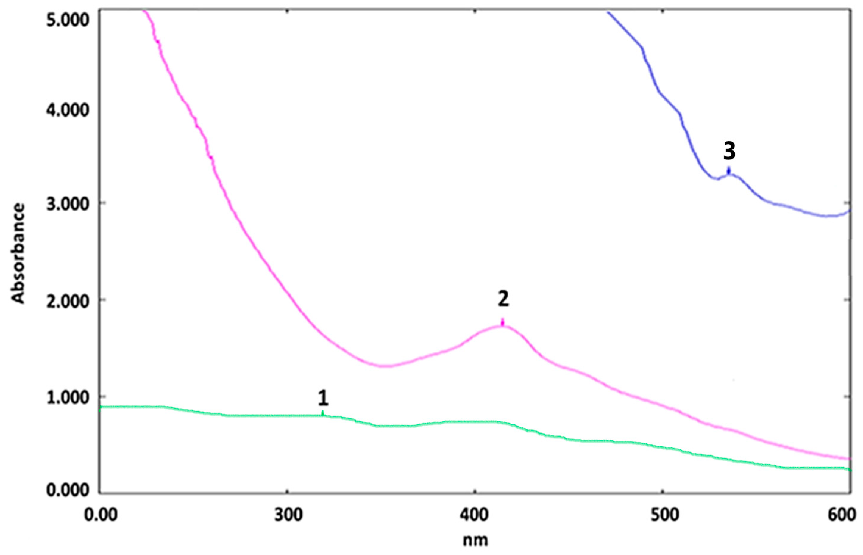
2.4. UV–Visible Analysis of AgNP

The success of AgNP biosynthesis was verified using UV–Visible spectrometry. A small aliquot of AgNP was diluted in deionized water. The surface plasmon resonance (SPR) signal was detected by testing the reaction mixture with the UV–Visible spectrometer (Shimadzu UV–VIS-U2800 (Shimadzu, Kyoto, Kyoto, Japan)) at room temperature with a scanning speed of 300 nm/min. The measurements were made between 400 and 800 nm. The decrease in Ag+ ions was determined by the UV–visible absorption spectrum of AgNP. At 430 nm, the AgNP solution exhibited an SPR peak.

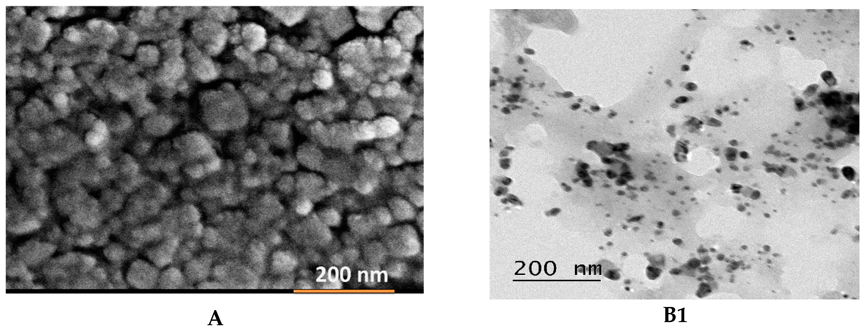
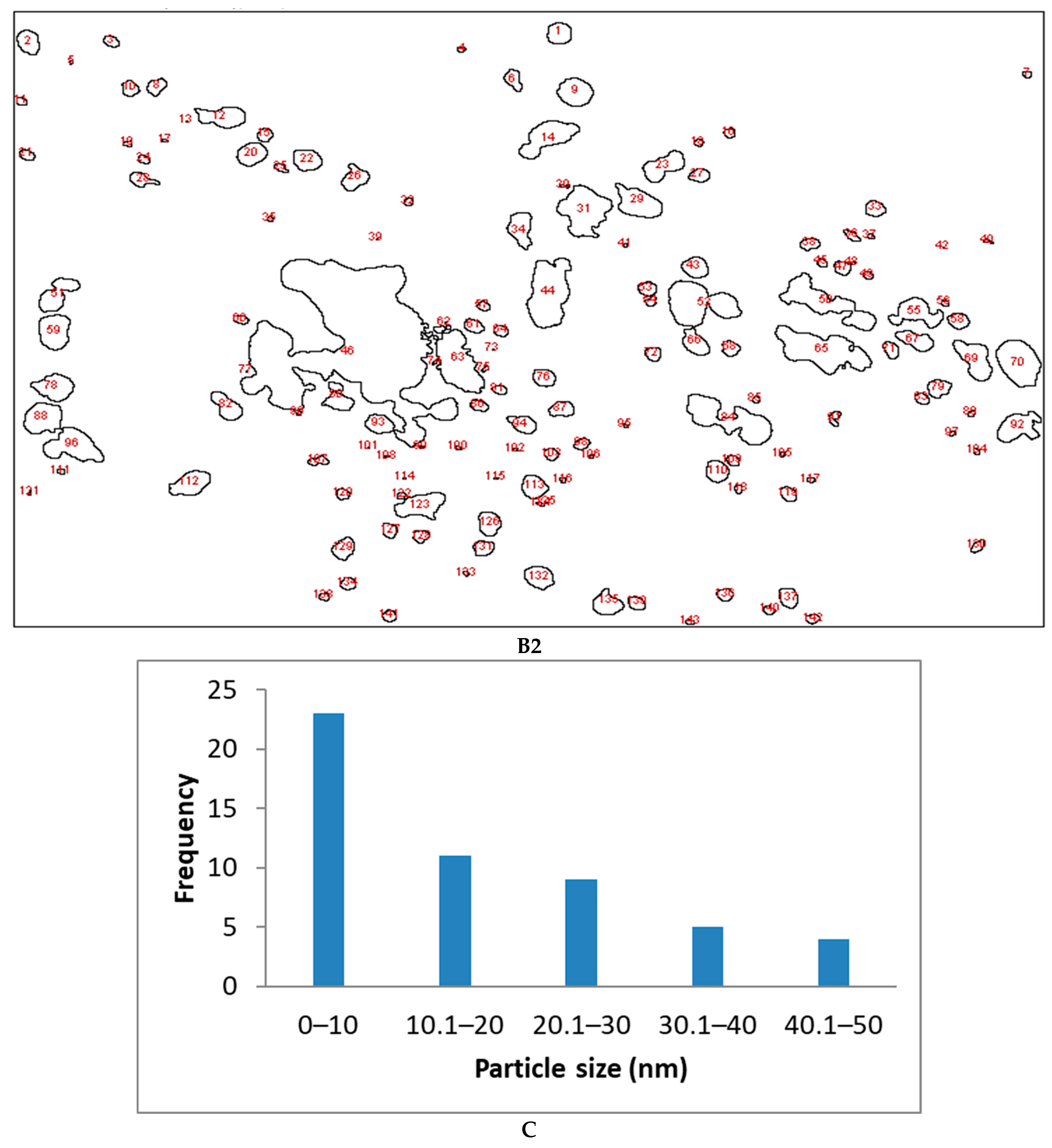
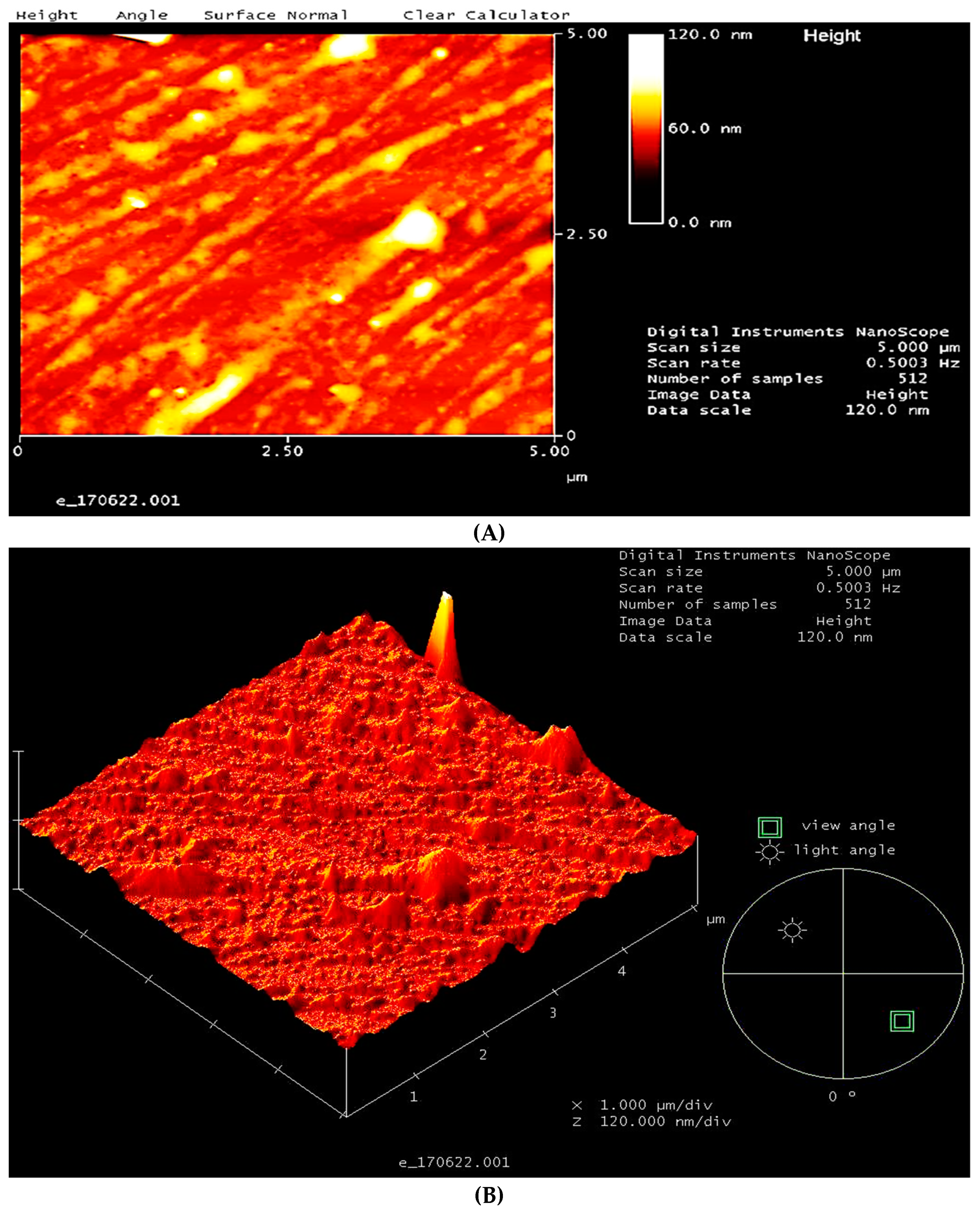
2.5. Characterization of AgNP

As described in other research studies, pure AgNPs were used for characterization studies [20,21]. AgNP were routinely washed and centrifuged using deionized water prior to characterization with AgNP characterization data in order to prevent interaction of unbound residual biochemical entities of CIL extract. Different analytical techniques including zetasizer analysis, atomic force microscopy (AFM), energy-dispersive X-ray (EDX), X-ray diffraction (XRD), scanning electron microscopy (SEM), and transmission electron microscopy (TEM) were used to characterize AgNPs. The hydrodynamic diameter of AgNPs was analyzed by a Zeta-PAL zeta potential analyzer (Brookhaven Instruments, Holtsville, NY, USA). To determine the morphology of the AgNPs, TEM, and SEM measurements were carried out. The crystal nature of AgNPs was determined based on the analysis of their XRD spectrum recorded on a PANalytical X’Pert PRO MRD PW 3040/60 X-ray diffractometer (Malvern panalytical, Malvern, UK) using CuKα radiation (λ = 1.5406) at 40 kV to 40 mA in 2θ/θ scanning mode. The EDX spectrum was recorded using an FEI Nova NanoSEM 450 EDX unit (FEI, Hillsboro, OR, USA). AFM assisted in the determination of the presence and size distribution of biosynthesized AgNPs. For AFM analysis, dilute samples (0.05 mg/mL in water) of AgNP were spread over zinc substrate. Evaluation of sample topography (with 1 × 1 µm2 scanned area) was performed at a set point of 10 nm with a scanning rate of 1 µm/s. The images were analyzed using a Bruker Dimension 3100 with Nanoscope 5 software (Bruker, Dynamostraße 19, Mannheim, Germany).

2.6. Antimicrobial Activity of AgNP

2.6.1. Disk Preparation

The plant extract (20 mg) and AgNPs (20 mg) were diluted separately in 1 mL of sterile 10% DMSO and mixed well. Next, the 50 µL of prepared plant extract and AgNP solution were separately added drop by drop on the top of 6 layered autoclaved Whatman® 70 mm Microfiber Filter Paper (Sigma-Aldrich, St. Louis, MI, USA), Grade CF/C disc, which was 6 mm in diameter. The disk was left to dry in a laminar hood at room temperature for 2 h. A ciprofloxacin disc and 10% DMSO were used as positive and negative controls, respectively.

2.6.2. Inoculum Preparation

To determine the antimicrobial potential of AgNP, 14 bacteria were used, namely Escherichia coli (E. coli), Salmonella typhi (S. typhi), Proteus mirabilis (P. mirabilis), Enterobacter cloacae (E. cloacae), Vibrio cholera (V. cholera), Acinetobacter baumannii (A. baumannii), Staphylococcus aureus (S. aureus), Staphylococcus haemolyticus (S. haemolyticus), Staphylococcus epidermidis (S. epidermidis), Streptococcus pyogenes (S. pyogenes), Pseudomonas aeruginosa (P. aeruginosa), Klebsiella pneumoniae (K. pneumoniae), Lactobacillus, and Bacillus subtilis (B. subtilis). These bacteria were streaked on nutrient agar from glycerol stock to obtain a single colony, and the plates were incubated overnight. A single colony from each bacterium was inoculated in nutrient broth (7 mL) and incubated for 5 h in an incubator shaker until it reached 0.6–0.7 OD at 600 nm absorbance level using a UV–Vis spectrometer. Next, each bacterial culture was diluted to achieve a viable cell count of 106 CFU/mL, standardized by 0.5 MacFarland.

2.6.3. Antibacterial Study

Antimicrobial evaluation of AgNP was based on disk diffusion methodology [22]. The strains of bacteria were swabbed over Mueller–Hinton agar (MHA) using sterile cotton swabs. Disks with ethanolic plant extract, AgNPs, 10% DMSO (negative control), and ciprofloxacin antibiotics (positive control) were applied over MHA agar. After 16 h of incubation, the zone of inhibition (ZOI) diameters (in mm) of each plates were measured.

3. Results and Discussion

3.1. Characterization of Synthesized Nanoparticles

The success of synthesis of AgNPs was based on results of visual inspection and UV–Visible spectrometric analysis. For color shift control, mixtures of AgNO3 solution and CIL extract were held apart for 60 min at 60 °C. The change in color from yellow to brown after 60 min indicated the formation of AgNPs. The formation of AgNP in brown color solution was further confirmed by UV–Visible analysis, which generated an absorption spectrum comprising curves 1, 2, and 3 (Figure 1). Curve 1 represented the AgNO3, curve 2 represented AgNP, and curve 3 represented CIL extract solution. The presence of an SPR peak at 425 in curve 2 of the UV–Visible spectrum confirmed formation of AgNPs. In the spectrum, curve 3 of the CIL extract did not exhibit a signal near 425 nm.
The AgNP formation was attributed to exposure of AgNO3 to CIL extract, which reduced Ag+ to Ag0. The transition in color from yellow to brown as well as the UV–Visible signal at 425 nm were attributed to the property of surface plasmon resonance and stimulated by the possibility of plasmon vibrations [23].
The SEM and TEM derived size distribution histogram data assisted in the determination of the size and shape of AgNPs. The SEM data given in Figure 2A indicated that synthesized AgNP are spherical in shape and poly-dispersed. The average size distribution histogram based on TEM data (given in Figure 2(B1,B2,C), and Table 1) revealed AgNPs to exist in sizes ranging between 1 and 50 nm.
Scanning of AgNPs in tapping mode generated two-dimensional (2D) (Figure 3A and three-dimensional (3D) (Figure 3B images. The images confirmed the uniform distribution of AgNPs, as most of the particles sizes were consistent with the SEM and TEM measurements.
The EDX analysis by FESEM generated a spectrum, as seen in Figure 4. The spectrum revealed the elemental composition of AgNPs. EDX spectra determined silver (64.05%) as a major constituent element in comparison to chlorine (20.75%) and calcium (15.20%).
Prominent peak at 3 keV confirmed the presence of AgNP elemental silver. The presence of strong signals for silver and other elemental peaks may be attributed to biomolecules bounded to the surface of silver nanoparticles, indicating the reduction in silver ions to elemental silver.
For zeta potential analysis, the AgNP samples (50 µg/mL) were suspended in deionized water and measured in triplicate at 25 °C [24]. Zeta potential assists in determining the stability of AgNP. It is important to note that particles with zeta potential values more positive than +30 mV or more negative than −30 mV are considered to be stable [25]. In the present study the zeta potential of AgNP with −55.46 mV indicated and supported the stability of AgNP biosynthesized using CIL (Figure 5).
The XRD data specified in Figure 6 exhibited the crystalline nature of AgNP. The spectrum exhibited various diffraction signals at the angle of 2θ ranging from 10 to 90°. The four distinctive diffraction Braggs signals at 2θ values of 32°, 46°, 68°, and 78° could be indexed to the 113, 210, 220, and 311 reflection planes, respectively, of face-centered cubic structures of silver. Such a pattern confirmed the crystallinity of AgNPs. In addition to the Bragg peaks representative of AgNPs, some additional peaks were also observed. These peaks were attributed to biomolecules of CIL extract and were responsible for the reduction and stabilization of silver ions into the resultant silver nanoparticles [26,27].

3.2. Antibacterial Potential

The antimicrobial potential of CIL extract and AgNPs was evaluated against six Gram-positive bacteria (S. haemolyticus, S. epidermidis, B. subtilis, Lactobacillus, S. aureus, and S. pyogenes) and eight Gram-negative bacteria (E. coli, P. mirabilis, S. typhi, E. cloacae, V. cholerae, P. aeruginosa, A. baumannii, and K. pneumoniae). Table 2 and Table 3 present the zones of inhibition (ZOI) exhibited by AgNP and CIL extracts against each bacterial strain.
The ZOI was calculated by measuring the zone in millimeters (mm). A clear ZOI indicates the inability of bacteria to grow or multiply around the sample-loaded disk (Table 2 and Table 3). The CIL extract inhibited growth of all test microorganisms except Lactobacillus, P. mirabilis, and A. baumannii. Lactobacillus is a non-pathogenic microorganism in which it produces lactic acid as a by-product of glucose metabolism. The bacterial species of the Lactobacillus genus (component of normal flora) are found in the gastrointestinal and genital tract of humans and animals [28]. Therefore, it is beneficial for humans that Coccinia indica plant extract did not exhibit any antimicrobial potential against Lactobacillus. The antimicrobial study of CIL extract against Gram-positive bacteria (GPB) revealed that CIL extract exhibited the maximum ZOI against S. epidermidis and the lowest ZOI against S. pyogenes, whereas the antimicrobial study of CIL extract against Gram-negative bacteria (GNB) revealed that CIL extract exhibited maximum ZOI against V. cholerae and a minimum ZOI against S. typhi and K. pneumoniae.
The antimicrobial activity of CIL extract was compared with the positive control (ciprofloxacin) and negative control (10% DMSO). The ZOIs of ciprofloxacin were larger and more clearly seen in comparison to those created by the CIL extract. A. baumannii did not show any ZOI in the ciprofloxacin disk. The resistance mechanism of A. baumannii is attributed to a single mutation that occurs from serine 83 to leucine in the quinolone resistance determining region (QRDRs) in gyrase subunit A (gyrA) [29].
The C. indica capped AgNPs exhibited substantial antimicrobial activity and ZOI against GPB and GNB. The AgNPs, when tested against GPB, exhibited the largest ZOI against B. subtilis, whereas when tested against GNB, they showed the largest ZOI against V. cholerae. The antimicrobial results revealed that the ZOI formed by AgNPs was larger than that formed by CIL extract. Large surface area per volume and easy penetrating characteristics of AgNPs best explain the antimicrobial activity of AgNPs. Therefore, AgNPs can easily diffuse through the cell walls of bacteria and disrupt microbial cell functions. The CIL extract comprises numerous components and has lesser penetration in comparison to AgNPs, which makes CIL extract less effective in damages bacterial cell walls [5]. Overall, in the present investigation, CIL extract and AgNPs displayed significant antimicrobial potential against GNB and GPB. The findings of present study are supported by previous studies that also reported the antimicrobial potential of C. grandis against K. pneumonia, S. aureus, and B. cereus [17].

4. Conclusions

The current research investigation shows that green synthesis of AgNP using an ethanolic extract of C. indica leaves is an eco-friendly, fast, and cost-effective method. In the present study, the antimicrobial activity of a 20 mg/mL concentration of CIL extract was shown to be sufficient to inhibit the growth of pathogenic bacteria. This study proves that compared to CIL extract, the ZOI of AgNP is larger against both GPB and GNB. Hence, the present study establishes the broad-spectrum antimicrobial potential of CIL extract derived AgNPs against Gram-negative and Gram-positive bacteria and recommends CIL ethanolic extract as an efficient biomaterial for green synthesis of silver nanoparticles.

Author Contributions

Conceptualization, S.V.C., S.S. (Sumitha Samuggam), S.C.B.G., G.R., P.A., L.V.R., N.K.F., and S.F.; validation, S.V.C., S.C.B.G., G.R., P.A., L.V.R., P.M., N.K.F., and S.F.; formal analysis, S.V.C., S.C.B.G., S.S. (Sumitha Samuggam), G.R., P.A., N.K.F., and S.F.; methodology, S.V.C., S.C.B.G., S.S. (Sumitha Samuggam), P.A., P.M., and K.K.; investigation, S.V.C., S.C.B.G., P.A., G.R., and L.V.R.; resources, S.V.C., S.C.B.G., P.A., L.V.R., N.K.F., S.F., G.R., and S.S. (Subramaniam Sreeramanan); data curation, S.V.C., S.C.B.G., P.A., L.V.R., N.K.F., S.F., G.R., and S.S. (Subramaniam Sreeramanan); writing—original draft preparation, S.V.C., S.C.B.G., P.A., L.V.R., N.K.F., S.F., G.R., and S.S. (Sumitha Samuggam); writing—review and editing, S.V.C., S.C.B.G., G.R., P.A., L.V.R., N.K.F., and S.F.; visualization, S.V.C., S.C.B.G., G.R., and P.A.; supervision, S.V.C.; project administration, S.V.C., S.C.B.G., and P.A.; funding acquisition, S.V.C. All authors have read and agreed to the published version of the manuscript.

Funding

This research was funded by the Ministry of Higher Learning Malaysia under the Fundamental Research Grant Scheme, FRGS/1/2018/STG03/AIMST/02/1. S.C.B.G. was supported by a special grant (9001-00596) from Universiti Malaysia Perlis.

Institutional Review Board Statement

Not applicable.

Informed Consent Statement

Not applicable.

Data Availability Statement

Not applicable.

Acknowledgments

The authors are thankful to Asian Institute of Medicine, Science and Technology AIMST University (Malaysia), Universiti Sains Malaysia (Malaysia), University Malaysia Perlis (Malaysia), Inha University (Korea), and Yogi Vemana University (India) for their support to successfully complete this study.

Conflicts of Interest

The authors have no conflict of interest to declare.

References

  1. Sender, R.; Fuchs, S.; Milo, R. Revised estimates for the number of human and bacteria cells in the body. PLoS Biol. 2016, 14, e1002533. [Google Scholar] [CrossRef] [PubMed] [Green Version]
  2. Hang, C.Z.; Fuloria, N.K.; Hong, O.J.; Kim, C.B.; Ting, B.Y.S.; Ru, C.S.; Ko, M.Y.; Fuloria, S. Biosynthesis of DLLAE blended silver nanoparticles and their response against periodontitis triggering bacteria. Int. J. Res. Pharm. Sci. 2020, 11, 1849–1856. [Google Scholar] [CrossRef] [Green Version]
  3. Muthukumar, H.; Matheswaran, M. Amaranthus spinosus leaf extract mediated FeO nanoparticles: Physicochemical traits, photocatalytic and antioxidant activity. ACS Sustain. Chem. Eng. 2015, 3, 3149–3156. [Google Scholar] [CrossRef]
  4. Song, J.Y.; Kim, B.S. Rapid biological synthesis of silver nanoparticles using plant leaf extracts. Bioproc. Biosyst. Eng. 2009, 32, 79. [Google Scholar] [CrossRef] [PubMed]
  5. Ramanathan, S.; Gopinath, S.C.; Anbu, P.; Lakshmipriya, T.; Kasim, F.H.; Lee, C.-G. Eco-friendly synthesis of Solanum trilobatum extract-capped silver nanoparticles is compatible with good antimicrobial activities. J. Mol. Struct. 2018, 1160, 80–91. [Google Scholar] [CrossRef]
  6. Fuloria, N.K.; Fuloria, S.; Chia, K.Y.; Karupiah, S.; Sathasivam, K. Response of green synthesized drug blended silver nanoparticles against periodontal disease triggering pathogenic microbiota. J. Appl. Biol. Biotchnol. 2019, 7, 46–56. [Google Scholar]
  7. Gomathi, M.; Rajkumar, P.V.; Prakasam, A.; Ravichandran, K. Green synthesis of silver nanoparticles using Datura stramonium leaf extract and assessment of their antibacterial activity. Res. Eff. Technol. 2017, 3, 280–284. [Google Scholar] [CrossRef]
  8. Mittal, J.; Jain, R.; Sharma, M.M. Phytofabrication of silver nanoparticles using aqueous leaf extract of Xanthium strumerium L. and their bactericidal efficacy. Adv. Nat. Sci. Nanosci. 2017, 8, 025011. [Google Scholar] [CrossRef] [Green Version]
  9. Lade, B.D.; Shanware, A.S. Phytonanofabrication: Methodology and Factors Affecting Biosynthesis of Nanoparticles. In Nanosystems; Limited 5 Princes Gate Court; IntechOpen: London, UK, 2020. [Google Scholar] [CrossRef] [Green Version]
  10. Makarov, V.V.; Makarova, S.S.; Love, A.J.; Sinitsyna, O.V.; Dudnik, A.O.; Yaminsky, I.V.; Taliansky, M.E.; Kalinina, N.O. Biosynthesis of stable iron oxide nanoparticles in aqueous extracts of Hordeum vulgare and Rumex acetosa plants. Langmuir 2014, 30, 5982–5988. [Google Scholar] [CrossRef]
  11. El-Kassas, H.Y.; Aly-Eldeen, M.A.; Gharib, S.M. Green synthesis of iron oxide (Fe3O4) nanoparticles using two selected brown seaweeds: Characterization and application for lead bioremediation. Acta Oceanol. Sin. 2016, 35, 89–98. [Google Scholar] [CrossRef]
  12. Silveira, C.; Shimabuku, Q.L.; Silva, M.F.; Bergamasco, R. Iron-oxide nanoparticles by the green synthesis method using Moringa oleifera leaf extract for fluoride removal. Environ. Technol. 2018, 39, 2926–2936. [Google Scholar] [CrossRef] [PubMed]
  13. Dadgostar, P. Antimicrobial resistance: Implications and costs. Infect. Drug Resist. 2019, 12, 3903. [Google Scholar] [CrossRef] [PubMed] [Green Version]
  14. Cheesman, M.J.; Ilanko, A.; Blonk, B.; Cock, I.E. Developing new antimicrobial therapies: Are synergistic combinations of plant extracts/compounds with conventional antibiotics the solution? Pharmacogn. Rev. 2017, 11, 57–72. [Google Scholar] [PubMed] [Green Version]
  15. Hussein, R.A.; El-Anssary, A.A. Plants secondary metabolites: The key drivers of the pharmacological actions of medicinal plants. Herb. Med. 2018. [Google Scholar] [CrossRef] [Green Version]
  16. Shaheen, S.Z.; Bolla, K.; Vasu, K.; Charya, M.S. Antimicrobial activity of the fruit extracts of Coccinia indica. Afr. J. Biotechnol. 2009, 8, 7073–7076. [Google Scholar]
  17. Sivaraj, A.; Jenifa, B.P.; Kavitha, M.; Inbasekar, P.; Senthilkumar, B.; Panneerselvam, A. Antibacterial activity of Coccinia grandis leaf extract on selective bacterial strains. J. Appl. Pharm. Sci. 2011, 1, 120–123. [Google Scholar]
  18. Nitharwal, R.K.; Patel, H.; Karchuli, M.S.; Ugale, R.R. Chemoprotective Potential of Coccinia Indica against Cyclophos-phamide-Induced Toxicity. Indian J. Pharmacol. 2013, 45, 502. [Google Scholar]
  19. Sumitha, S.; Vasanthi, S.; Shalini, S.; Chinni, S.V.; Gopinath, S.C.B.; Kathiresan, S.; Anbu, P.; Ravichandran, V. Durio zibethinus rind extract mediated green synthesis of silver nanoparticles: Characterization and biomedical applications. Pharmacogn. Mag. 2019, 15, 52. [Google Scholar]
  20. Sumitha, S.; Vasanthi, S.; Shalini, S.; Chinni, S.V.; Gopinath, S.C.; Anbu, P.; Bahari, M.B.; Harish, R.; Kathiresan, S.; Ravichandran, V. Phyto-mediated photo catalysed green synthesis of silver nanoparticles using Durio zibethinus seed extract: Antimicrobial and cytotoxic activity and photocatalytic applications. Molecules 2018, 23, 3311. [Google Scholar] [CrossRef] [Green Version]
  21. Anbu, P.; Gopinath, S.C.; Yun, H.S.; Lee, C.-G. Temperature-dependent green biosynthesis and characterization of silver nanoparticles using balloon flower plants and their antibacterial potential. J. Mol. Struct. 2019, 1177, 302–309. [Google Scholar] [CrossRef]
  22. Buszewski, B.; Railean-Plugaru, V.; Pomastowski, P.; Rafińska, K.; Szultka-Mlynska, M.; Golinska, P.; Wypij, M.; Laskowski, D.; Dahm, H. Antimicrobial activity of biosilver nanoparticles produced by a novel Streptacidiphilus durhamensis strain. J. Microbiol. Immunol. Infect. 2018, 51, 45–54. [Google Scholar] [CrossRef] [PubMed] [Green Version]
  23. Velusamy, P.; Su, C.-H.; Venkat Kumar, G.; Adhikary, S.; Pandian, K.; Gopinath, S.C.; Chen, Y.; Anbu, P. Biopolymers regulate silver nanoparticle under microwave irradiation for effective antibacterial and antibiofilm activities. PLoS ONE 2016, 11, e0157612. [Google Scholar] [CrossRef] [PubMed]
  24. Aljabali, A.A.; Akkam, Y.; Al Zoubi, M.S.; Al-Batayneh, K.M.; Al-Trad, B.; Abo Alrob, O.; Alkilany, A.M.; Benamara, M.; Evans, D.J. Synthesis of Gold Nanoparticles Using Leaf Extract of Ziziphus Zizyphus and Their Antimicrobial Activity. Nanomaterials 2018, 8, 174. [Google Scholar] [CrossRef] [PubMed] [Green Version]
  25. Saeb, A.; Alshammari, A.S.; Al-Brahim, H.; Al-Rubeaan, K.A. Production of Silver Nanoparticles with Strong and Stable Antimicrobial Activity against Highly Pathogenic and Multidrug Resistant Bacteria. Sci. World J. 2014, 2014, 704708. [Google Scholar] [CrossRef] [PubMed]
  26. Suvith, V.S.; Philip, D. Catalytic degradation of methylene blue using biosynthesized gold and silver nanoparticles. Spectrochim. Acta A Mol. Biomol. Spectrosc. 2014, 118, 526–532. [Google Scholar] [CrossRef] [PubMed]
  27. Fuloria, S.; Hong, O.; Kim, C.; Ting, B.; Karupiah, S.; Paliwal, N.; Kumari, U.; Sathasivam, K.; Vetriselvan, S.; Fuloria, N. Synthesis of Silver Nanoparticles of Corn Silk Agrowaste and Their Bioactivities. Asian J. Chem. 2020, 32, 1497–1504. [Google Scholar] [CrossRef]
  28. Hynönen, U.; Palva, A. Lactobacillus surface layer proteins: Structure, function and applications. Appl. Microbiol. Biotechnol. 2013, 97, 5225–5243. [Google Scholar] [CrossRef] [Green Version]
  29. Maleki, M.-H.; Jalilian, F.A.; Khayat, H.; Mohammadi, M.; Pourahmad, F.; Asadollahi, K.; Pakzad, I.; Sadeghifard, N.; Soroush, S.; Emaneini, M. Detection of highly ciprofloxacin resistance Acinetobacter baumannii isolated from patients with burn wound infections in presence and absence of efflux pump inhibitor. Maedica 2014, 9, 162. [Google Scholar]
Figure 1. UV–Visible spectrum of silver nitrate solution (curve 1), AgNP (curve 2), and Coccinia indica leaf (CIL) extract (curve 3).
Crystals 11 00097 g001
Figure 2. Morphological features of synthesized AgNPs. (A) SEM image at the 200 nm scale; (B1,B2) TEM images; (C) average size distribution histogram representing AgNP size range.
Crystals 11 00097 g002aCrystals 11 00097 g002b
Figure 3. AFM images of AgNPs. (A) 2D image; (B) 3D image.
Crystals 11 00097 g003
Figure 4. EDX spectrum of synthesized AgNPs.
Crystals 11 00097 g004
Figure 5. Zeta potential of synthesized AgNP from leaf extract of C. indica.
Crystals 11 00097 g005
Figure 6. XRD spectrum of synthesized AgNPs.
Crystals 11 00097 g006
Table 1. Silver nanoparticle size distribution based on TEM data (Figure 2B2).
AgNP
S. No.
Area
(nm)
Radius
(nm)
Diameter
(nm)
AgNP
S. No.
Area
(nm)
Radius
(nm)
Diameter
(nm)
AgNP
S. No.
Area
(nm)
Radius
(nm)
Diameter
(nm)
1306.779.88170119.76344942.9233.6963267.3926529716.4122.285634.571261
2304.2459.84094919.6819501352.05920.7454641.490929893.425.45312210.90624
393.425.45312210.9062451528.95712.9758425.95168996.3121.4174532.834906
423.9862.7631485.526296521512.38821.9410243.8820410012.6242.0045824.009163
56.3121.4174532.83490653146.4426.82744313.654891011.2620.6338041.267608
6178.0027.52727315.054555453.0224.1082168.2164331027.5751.5528043.105607
732.8233.232326.46464155646.36314.3437828.6875510393.425.45312210.90624
8165.3787.25544614.510895626.5112.9049485.80989510421.4612.6136675.227334
9554.20513.2819126.563825764.3844.5270399.05407810515.1492.1959244.391847
10142.6546.73856213.4771258186.8397.71185815.4237210610.0991.7929353.585869
1146.713.8559397.71187859643.83814.3157328.6314610790.8955.37892210.75784
12541.58113.1297726.259536070.6964.743769.4875191085.051.2678592.535718
131.2620.6338041.26760861145.1796.79793713.5958710975.7464.9102679.820534
14866.02516.6031533.2063622.5250.8965121.793023110278.9969.4237618.84752
15113.6186.01379812.0276631426.54321.3092342.6184511122.7242.6894765.378952
1659.3344.3458738.6917476489.6325.34142110.68284112511.28312.7572225.51443
178.8371.6771723.354345651804.00823.9631847.92636113328.23110.2215120.44302
1835.3483.3543456.70868966338.3310.3775720.755131142.5250.8965121.793023
1926.5112.9049485.80989567345.90510.493120.986191153.7871.0979262.195851
20378.72810.9796621.9593268147.7046.85679813.713611617.6742.371884.74376
2189.6325.34142110.6828469665.29914.5523729.1047411721.4612.6136675.227334
22349.69210.5503821.10076701090.73718.6331137.2662111836.613.4136986.827396
23540.31913.1144626.2289271140.1296.67865913.35732119130.036.43349612.86699
2456.8094.2523978.50479472126.2436.33911912.6782412079.5335.03151710.06303
2553.0224.1082168.216433731.2620.6338041.2676081217.5751.5528043.105607
26323.18110.1425720.285157410.0991.7929353.58586912232.8233.232326.464641
27154.0167.00177514.003557516.4122.285634.571261123641.31314.2876328.57526
28175.4777.47369514.9473976224.7128.45742916.9148612415.1492.1959244.391847
29735.99515.3060330.61207771.2620.6338041.2676081251.2620.6338041.267608
3015.1492.1959244.39184778563.04213.3873826.77476126300.4589.77951119.55902
311285.15120.2256440.4512879231.0248.57538817.15078127117.4066.11322512.22645
3239.1353.5294577.05891480345.90510.493120.98619128107.3065.84436411.68873
33175.4777.47369514.947398173.2214.8277319.655462129255.019.00956618.01913
34443.11211.8763223.7526582377.46610.9613521.9227113077.0084.9510039.902006
3516.4122.285634.5712618388.375.30368410.60737131178.0027.52727315.05455
3671.9584.7859139.571826841455.57821.5249943.04998132367.36610.8137121.62742
3718.9362.4551014.9102028526.5112.9049485.80989513313.8872.1024684.204937
38130.036.43349612.8669986112.3565.98030611.96061134103.5195.74030911.48062
391.2620.6338041.26760887215.8758.28946316.57893135371.15410.8693221.73864
4016.4122.285634.57126188679.18614.7034629.40692136111.0945.94662511.89325
417.5751.5528043.1056078923.9862.7631485.526296137213.358.24084216.48168
421.2620.6338041.2676089010.0991.7929353.58586913845.4473.8034517.606903
43304.2459.84094919.68199178.274.9914069.982812139127.5056.37072512.74145
441542.68622.1597144.3194192636.26314.2312728.4625314051.764.0590318.118062
4545.4473.8034517.60690393300.4589.77951119.5590214188.375.30368410.60737
461952.66124.9309449.8618894257.5359.0540618.1081214270.6964.743769.487519
47128.7686.402212.8044958.8371.6771723.35434514323.9862.7631485.526296
486.3121.4174532.83490696919.04717.1038634.20772
Table 2. Antibacterial activity of CIL ethanolic extract and synthesized AgNPs against Gram-positive bacteria.
Gram-Positive BacteriaZOI (mm)
CIL Extract (Ethanolic)AgNPControls
PositiveNegative
S. haemolyticus910.67 ± 0.3325NA
S. epidermidis10.67 ± 0.6711.67 ± 0.3327 ± 1.00NA
B. subtilis9.33 ± 0.6712.67 ± 1.2025NA
S. aureus9.33 ± 0.3311.67 ± 0.6725.67 ± 0.67NA
S. pyogenes8.67 ± 0.339.67 ± 0.3323.33 ± 0.33NA
LactobacillusNA11.67 ± 0.6724.67 ± 0.67NA
Note: The data are presented in the form of mean (±standard error), p < 0.05.
Table 3. Antibacterial activity of CIL ethanolic extract and synthesized AgNPs against Gram-negative bacteria.
Gram-Negative BacteriaZOI (mm)
CIL Extract (Ethanolic)AgNPControls
PositiveNegative
P. mirabilisNA9.67 ± 0.3325.33 ± 0.33NA
S. typhi9.33 ± 0.3311.67 ± 0.3330 ± 1.53NA
V. cholerae10.33 ± 0.3312.67 ± 0.3329.33 ± 0.67NA
E. cloacae10 ± 0.581229.33 ± 0.67NA
K. pneumoniae9.33 ± 0.6712 ± 0.5826 ± 1.00NA
E. coli9.67 ± 0.3310.33 ± 0.3327 ± 1.00NA
P. aeruginosa1011.33 ± 0.3329.67 ± 0.33NA
A. baumanniiNA11.67 ± 0.88NANA
Note: The data are presented in the form of mean (±standard error), p < 0.05.
Publisher’s Note: MDPI stays neutral with regard to jurisdictional claims in published maps and institutional affiliations.

