
Tin Disulfide-Coated Microfiber for Humidity Sensing with Fast Response and High Sensitivity

 , ,
, ,
Abstract
:1. Introduction
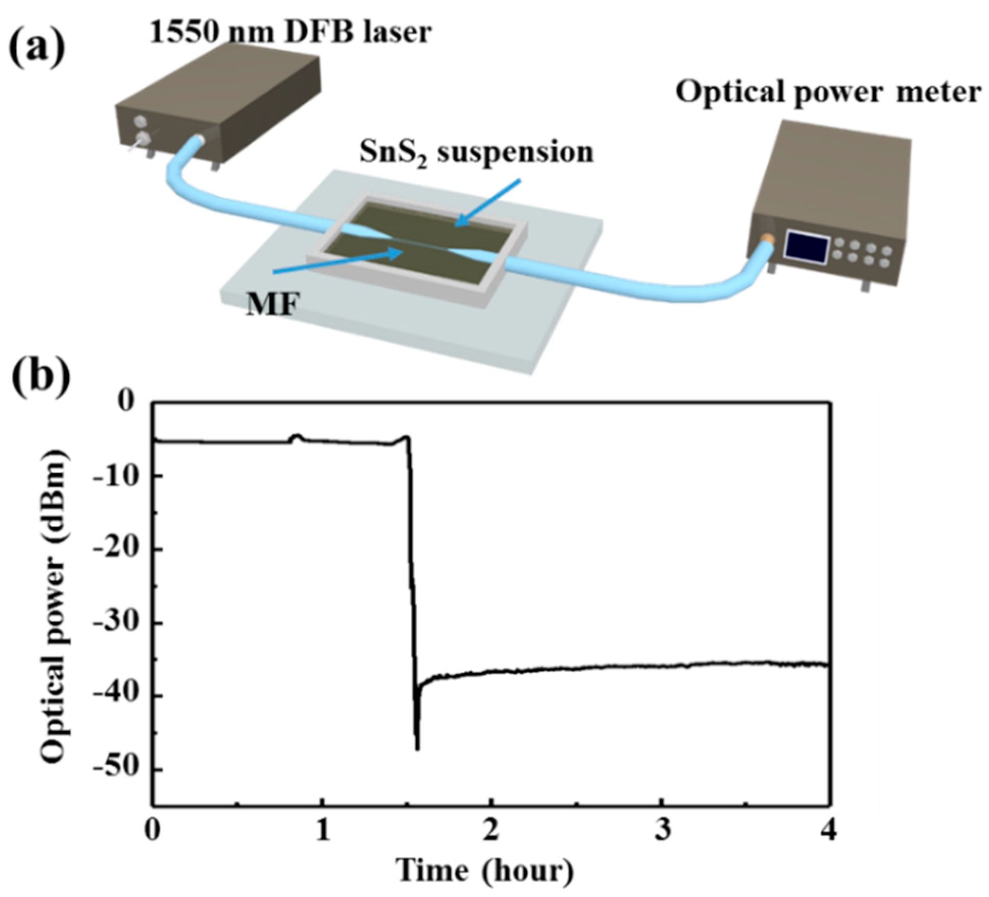
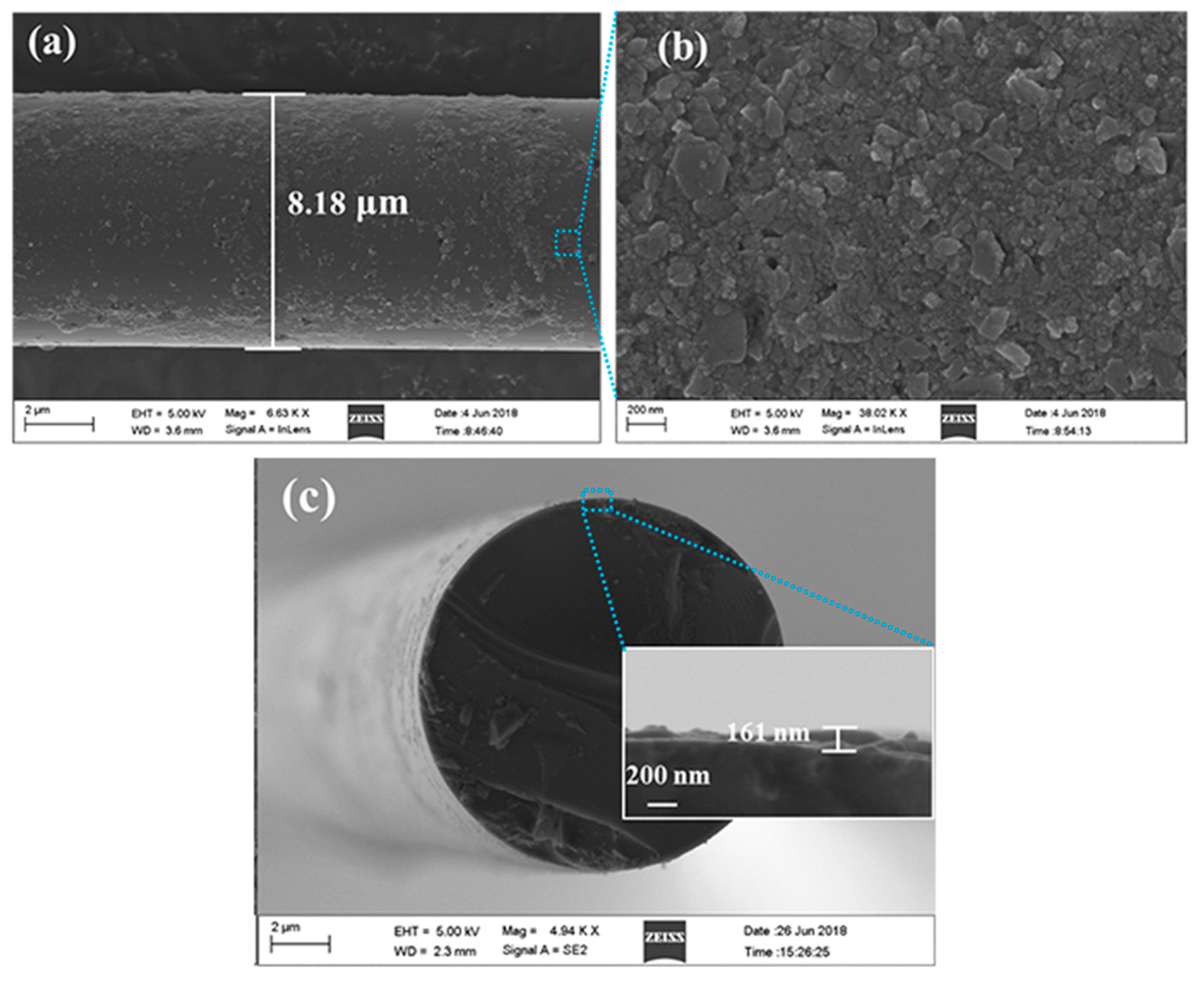
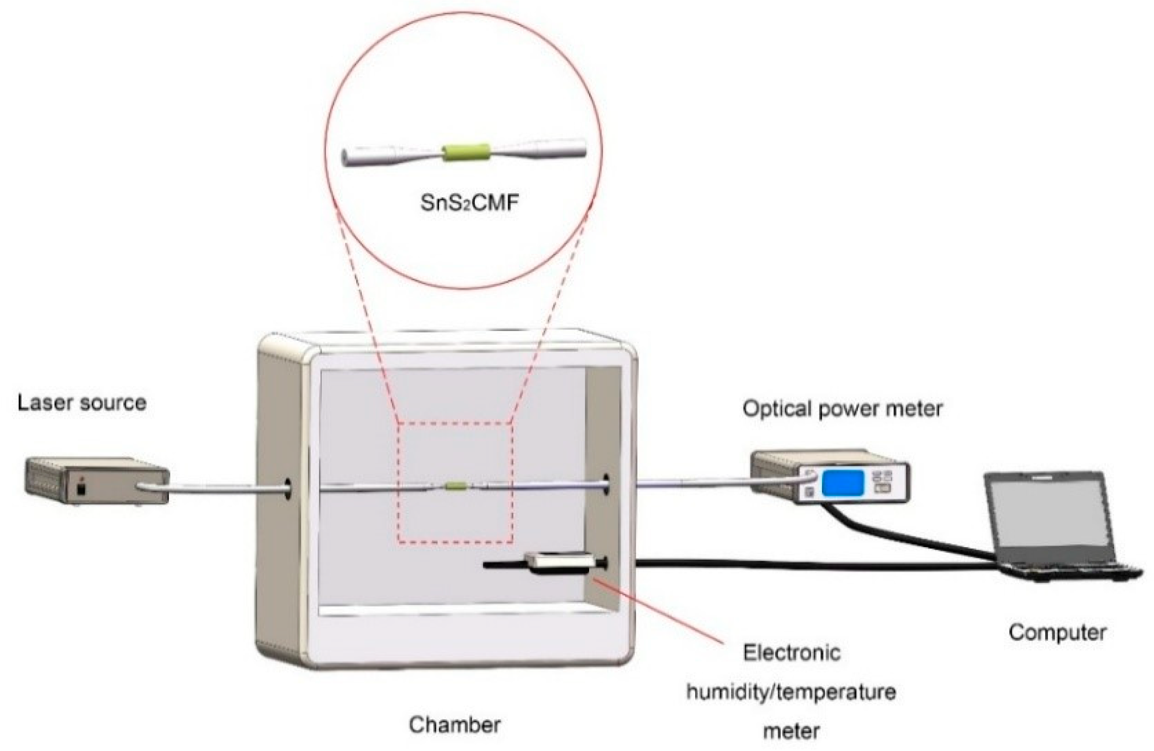
2. Materials and Methods
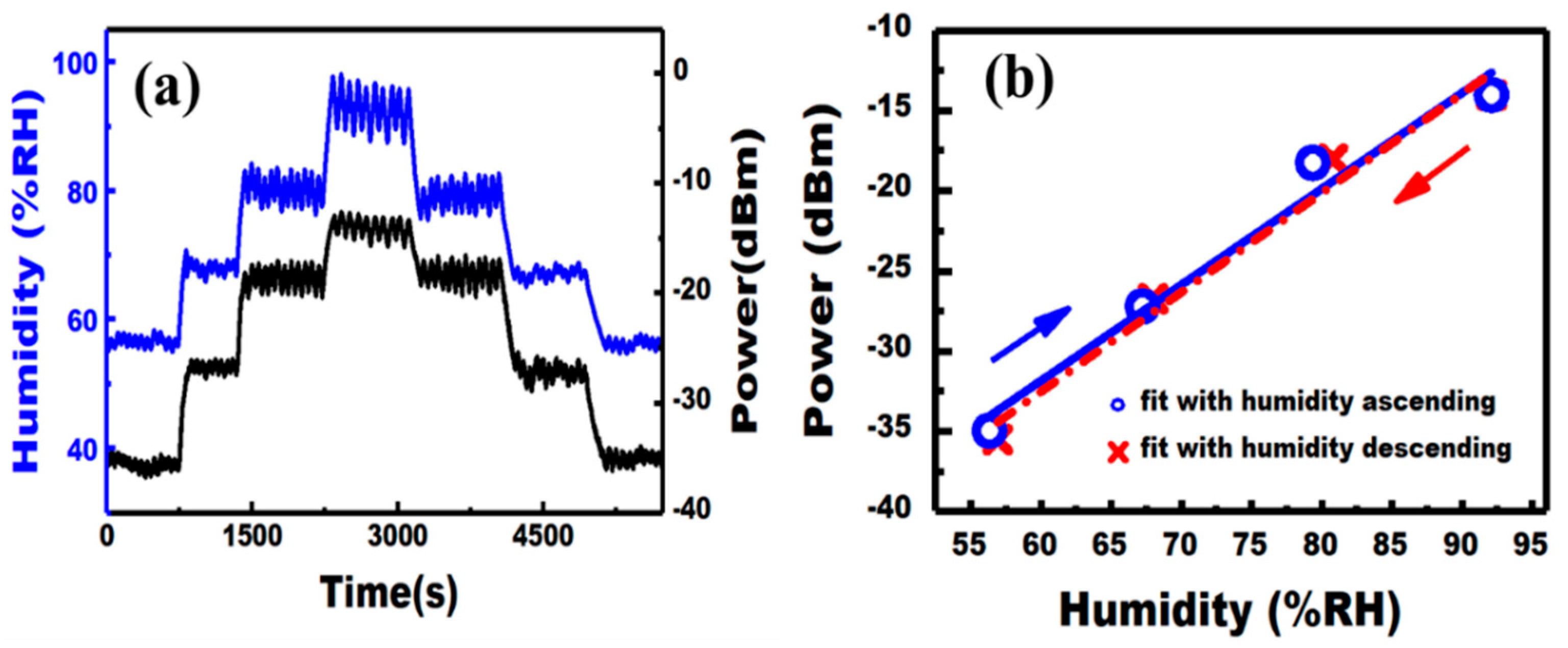
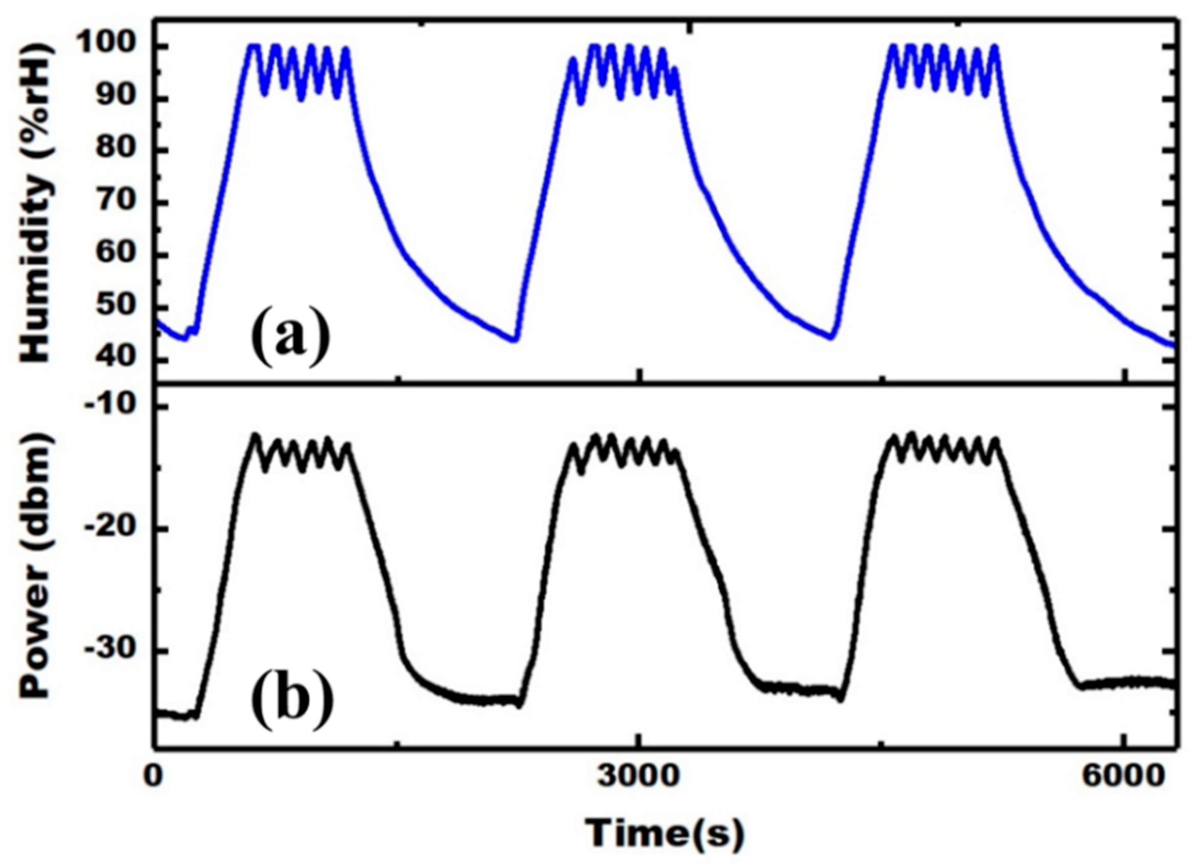
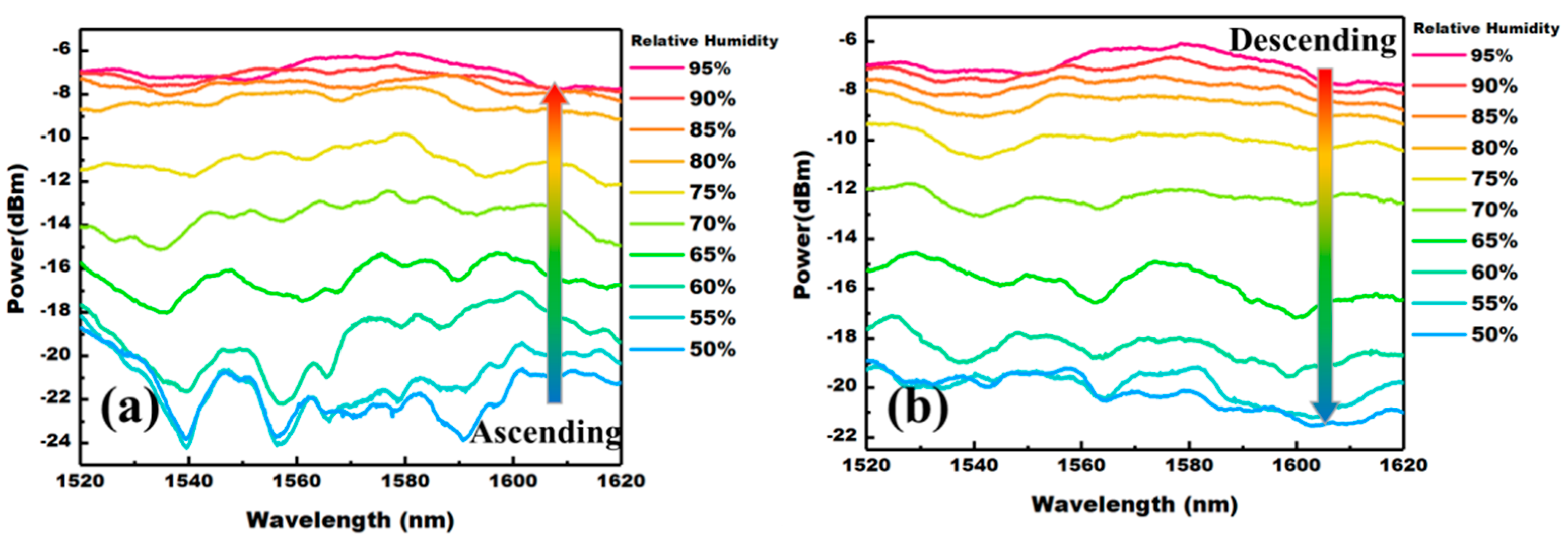
3. Results
4. Conclusions
Author Contributions
Funding
Institutional Review Board Statement
Informed Consent Statement
Data Availability Statement
Acknowledgments
Conflicts of Interest
References
- Ascorbe, J.; Corres, J.M.; Arregui, F.J.; Matias, I.R. Recent developments in fiber optics humidity sensors. Sensors 2017, 17, 893. [Google Scholar] [CrossRef] [Green Version]
- Gomez, D.; Morgan, S.P.; Hayes-Gill, B.R.; Correia, R.G.; Korposh, S. Polymeric optical fibre sensor coated by SiO2 nanoparticles for humidity sensing in the skin microenvironment. Sens. Actuators B Chem. 2018, 254, 887–895. [Google Scholar] [CrossRef]
- Yang, M.; Xie, W.; Dai, Y.; Lee, D.; Dai, J.; Zhang, Y.; Zhuang, Z. Dielectric multilayer-based fiber optic sensor enabling simultaneous measurement of humidity and temperature. Opt. Express 2014, 22, 11892. [Google Scholar] [CrossRef]
- Lou, J.; Wang, Y.; Tong, L. Microfiber optical sensors: A review. Sensors 2014, 14, 5823–5844. [Google Scholar] [CrossRef] [Green Version]
- Chen, J.; Xiong, Y.; Xu, F.; Lu, Y. Silica optical fiber integrated with two-dimensional materials: Towards opto-electro-mechanical technology. Light Sci. Appl. 2021, 10. [Google Scholar] [CrossRef]
- Guan, H.; Xia, K.; Chen, C.; Luo, Y.; Tang, J.; Lu, H.; Yu, J.; Zhang, J.; Zhong, Y.; Chen, Z. Tungsten disulfide wrapped on micro fiber for enhanced humidity sensing. Opt. Mater. Express 2017, 7, 1686. [Google Scholar] [CrossRef]
- Ascorbe, J.; Corres, J.M.; Matias, I.R.; Arregui, F.J. High sensitivity humidity sensor based on cladding-etched optical fiber and lossy mode resonances. Sens. Actuators B Chem. 2016, 233, 7–16. [Google Scholar] [CrossRef] [Green Version]
- Bariáin, C.; Matías, I.R.; Arregui, F.J.; López-Amo, M. Optical fiber humidity sensor based on a tapered fiber coated with agarose gel. Sens. Actuators B Chem. 2000, 69, 127–131. [Google Scholar] [CrossRef]
- Li, D.; Lu, H.; Qiu, W.; Dong, J.; Guan, H.; Zhu, W.; Yu, J.; Luo, Y.; Zhang, J.; Chen, Z. Molybdenum disulfide nanosheets deposited on polished optical fiber for humidity sensing and human breath monitoring. Opt. Express 2017, 25, 28407. [Google Scholar] [CrossRef]
- Wang, Y.; Shen, C.; Lou, W.; Shentu, F.; Zhong, C.; Dong, X.; Tong, L. Fiber optic relative humidity sensor based on the tilted fiber Bragg grating coated with graphene oxide. Appl. Phys. Lett. 2016, 109, 1–6. [Google Scholar] [CrossRef]
- Luo, Y.; Chen, C.; Xia, K.; Peng, S.; Guan, H.; Tang, J.; Lu, H.; Yu, J.; Zhang, J.; Xiao, Y.; et al. Tungsten disulfide (WS2) based all-fiber-optic humidity sensor. Opt. Express 2016, 24, 8956. [Google Scholar] [CrossRef]
- Huang, Y.; Zhu, W.; Li, Z.; Chen, G.; Chen, L.; Zhou, J.; Lin, H.; Guan, J.; Fang, W.; Liu, X.; et al. High-performance fibre-optic humidity sensor based on a side-polished fibre wavelength selectively coupled with graphene oxide film. Sens. Actuators B Chem. 2018, 255, 57–69. [Google Scholar] [CrossRef]
- Du, B.; Yang, D.; She, X.; Yuan, Y.; Mao, D.; Jiang, Y.; Lu, F. MoS2-based all-fiber humidity sensor for monitoring human breath with fast response and recovery. Sens. Actuators B Chem. 2017, 251, 180–184. [Google Scholar] [CrossRef]
- Yasuma, F.; Hayano, J.I. Respiratory Sinus Arrhythmia: Why Does the Heartbeat Synchronize with Respiratory Rhythm? Chest 2004, 125, 683–690. [Google Scholar] [CrossRef]
- Bates, J.H.T.; Schmalisch, G.; Filbrun, D.; Stocks, J. Tidal breath analysis for infant pulmonary function testing. Eur. Respir. J. 2000, 16, 1180–1192. [Google Scholar] [CrossRef] [PubMed] [Green Version]
- Schaibley, J.R.; Yu, H.; Clark, G.; Rivera, P.; Ross, J.S.; Seyler, K.L.; Yao, W.; Xu, X. Valleytronics in 2D materials. Nat. Rev. Mater. 2016, 1. [Google Scholar] [CrossRef]
- Mahmood, F.; Alpichshev, Z.; Lee, Y.H.; Kong, J.; Gedik, N. Observation of Exciton-Exciton Interaction Mediated Valley Depolarization in Monolayer MoSe2. Nano Lett. 2018, 18, 223–228. [Google Scholar] [CrossRef] [PubMed] [Green Version]
- Ye, G.; Gong, Y.; Lei, S.; He, Y.; Li, B.; Zhang, X.; Jin, Z.; Dong, L.; Lou, J.; Vajtai, R.; et al. Synthesis of large-scale atomic-layer SnS2 through chemical vapor deposition. Nano Res. 2017, 10, 2386–2394. [Google Scholar] [CrossRef]
- Liu, J.; Xia, C.; Li, H.; Pan, A. High on/off ratio photosensitive field effect transistors based on few layer SnS2. Nanotechnology 2016, 27, 1–6. [Google Scholar] [CrossRef]
- Su, G.; Hadjiev, V.G.; Loya, P.E.; Zhang, J.; Lei, S.; Maharjan, S.; Dong, P.M.; Ajayan, P.; Lou, J.; Peng, H. Chemical vapor deposition of thin crystals of layered semiconductor SnS2 for fast photodetection application. Nano Lett. 2015, 15, 506–513. [Google Scholar] [CrossRef]
- Zhou, T.; Pang, W.K.; Zhang, C.; Yang, J.; Chen, Z.; Liu, H.K.; Guo, Z. Enhanced sodium-ion battery performance by structural phase transition from two-dimensional hexagonal-SnS2 to orthorhombic-SnS. ACS Nano 2014, 8, 8323–8333. [Google Scholar] [CrossRef] [PubMed]
- Qu, B.; Ma, C.; Ji, G.; Xu, C.; Xu, J.; Meng, Y.S.; Wang, T.; Lee, J.Y. Layered SnS2-reduced graphene oxide composite—A high-capacity, high-rate, and long-cycle life sodium-ion battery anode material. Adv. Mater. 2014, 26, 3854–3859. [Google Scholar] [CrossRef] [Green Version]
- Zhang, Y.; Zhang, F.; Yang, Z.; Xue, H.; Dionysiou, D.D. Development of a new efficient visible-light-driven photocatalyst from SnS2 and polyvinyl chloride. J. Catal. 2016, 344, 692–700. [Google Scholar] [CrossRef]
- Xu, K.; Li, N.; Zeng, D.; Tian, S.; Zhang, S.; Hu, D.; Xie, C. Interface bonds determined gas-sensing of SnO2-SnS2 hybrids to ammonia at room temperature. ACS Appl. Mater. Interfaces 2015, 7, 11359–11368. [Google Scholar] [CrossRef]
- Huang, J.; Yu, K.; Gu, C.; Zhai, M.; Wu, Y.; Yang, M.; Liu, J. Preparation of porous flower-shaped SnO2 nanostructures and their gas-sensing property. Sens. Actuators B Chem. 2010, 147, 467–474. [Google Scholar] [CrossRef]
- Ou, J.Z.; Ge, W.; Carey, B.; Daeneke, T.; Rotbart, A.; Shan, W.; Wang, Y.; Fu, Z.; Chrimes, A.F.; Wlodarski, W.; et al. Physisorption-Based Charge Transfer in Two-Dimensional SnS2 for Selective and Reversible NO2 Gas Sensing. ACS Nano 2015, 9, 10313–10323. [Google Scholar] [CrossRef] [PubMed]
- Bharatula, L.D.; Erande, M.B.; Mulla, I.S.; Rout, C.S.; Late, D.J. SnS2 nanoflakes for efficient humidity and alcohol sensing at room temperature. RSC Adv. 2016, 6, 105421–105427. [Google Scholar] [CrossRef]
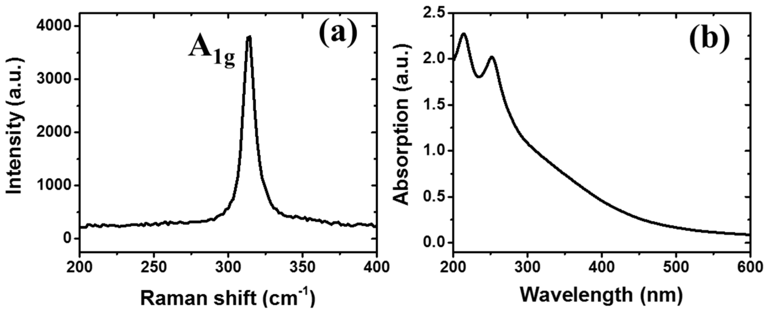
- Fu, X.; Ilanchezhiyan, P.; Mohan Kumar, G.; Cho, H.D.; Zhang, L.; Chan, A.S.; Lee, D.J.; Panin, G.N.; Kang, T.W. Tunable UV-visible absorption of SnS2 layered quantum dots produced by liquid phase exfoliation. Nanoscale 2017, 9, 1820–1826. [Google Scholar] [CrossRef]
- Wang, C.; Tang, K.; Aa, Q.Y.; Qian, Y. Raman scattering, far infrared spectrum and photoluminescence of SnS2 nanocrystallites. Chem. Phys. Lett. 2002, 357, 371–375. [Google Scholar] [CrossRef]
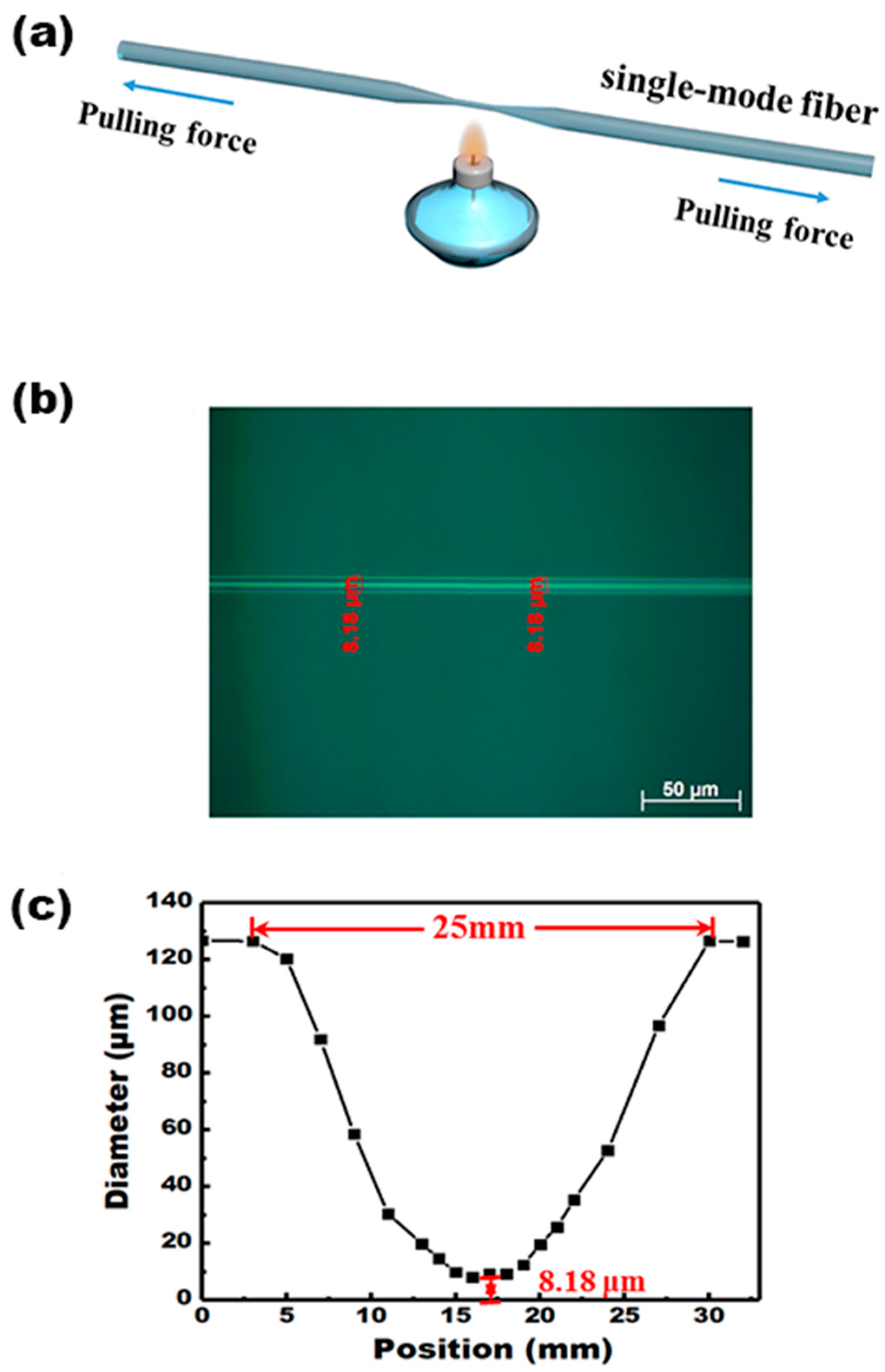
- Wu, X.; Tong, L. Optical microfibers and nanofibers. Nanophotonics 2013, 2, 407–428. [Google Scholar] [CrossRef]
- Zhong, Y.; Wang, Y.; Wang, Z.; Xing, Z.; Xiao, Y.; Yu, J.; Guan, H.; Luo, Y.; Lu, H.; Zhu, W.; et al. Ultrafast freestanding microfiber humidity sensor based on three-dimensional graphene network cladding. Opt. Express 2020, 28, 4362. [Google Scholar] [CrossRef] [PubMed]
- Hirunpinyopas, W.; Prestat, E.; Worrall, S.D.; Haigh, S.J.; Dryfe, R.A.W.; Bissett, M.A. Desalination and Nanofiltration through Functionalized Laminar MoS2 Membranes. ACS Nano 2017, 11, 11082–11090. [Google Scholar] [CrossRef]
- Amroun, M.N.; Khadraoui, M.; Miloua, R.; Kebbab, Z.; Sahraoui, K. Investigation on the structural, optical and electrical properties of mixed SnS2—CdS thin films. Optik 2017, 131, 152–164. [Google Scholar] [CrossRef]
- Lang, Y.; Ouyang, T.; Lin, L.; Xia, K.; Jiang, M.; Guan, H.; Yu, J.; Li, D.; Chen, G.; Zhu, W.; et al. Side polished fiber coated with molybdenum diselenide (MoSe2) for humidity sensing. Sensors 2017, 25, SeW1E.2. [Google Scholar] [CrossRef]


| Device Structure | Response Time (s) | Recovery Time (s) | Total Time(s) | Dynamic Range of Response |
|---|---|---|---|---|
| MoS2 nanosheets based SPF [9] | 0.85 | 0.85 | 1.70 | 0.33 dB/%RH (40 %RH–85 %RH) |
| MoS2-coated etched single-mode fiber [13] | 0.066 | 2.395 | 2.461 | 0.008 dB/%RH (20 %RH–80 %RH) |
| MoSe2-coated fiber-optic sensor [34] | 1 | 4 | 5 | 0.26 dB/%RH (32 %RH–73 %RH) |
| Tungsten disulphide (WS2)-coated | 1 | 4 | 5 | 0.17 dB/%RH (37 %RH–90 %RH) |
| Graphene oxide (GO)-coated fiber-optic sensor [11] | 2.73 | 7.27 | 10.0 | 0.427 dB/%RH (59 %RH–93 %RH) |
| Agarose gel with tapered fiber [7] | 5 | 55 | 60 | 0.13 dB/%RH (30 %RH–80 %RH) |
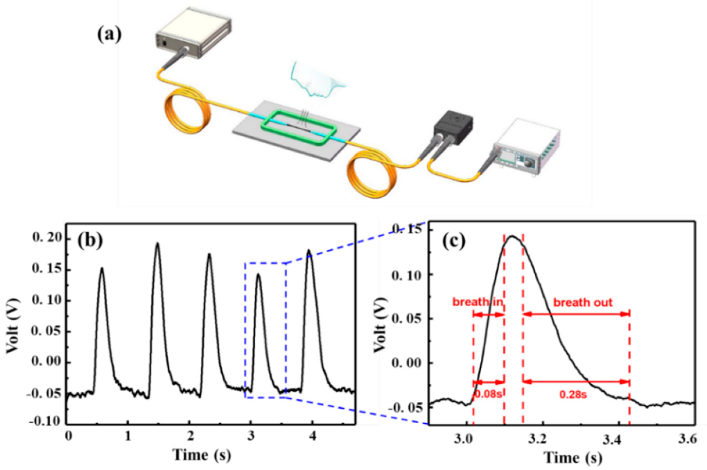
| SnS2-coated MF (this paper) | 0.08 | 0.28 | 0.36 | 0.57 dB/%RH (55 %RH–95 %RH) |
Publisher’s Note: MDPI stays neutral with regard to jurisdictional claims in published maps and institutional affiliations. |
© 2021 by the authors. Licensee MDPI, Basel, Switzerland. This article is an open access article distributed under the terms and conditions of the Creative Commons Attribution (CC BY) license (https://creativecommons.org/licenses/by/4.0/).
Share and Cite
Liang, A.; Ming, J.; Zhu, W.; Guan, H.; Han, X.; Zhang, S.; Lin, Y.; Dong, J.; Huang, Y.; Qiu, W.; et al. Tin Disulfide-Coated Microfiber for Humidity Sensing with Fast Response and High Sensitivity. Crystals 2021, 11, 648. https://0-doi-org.brum.beds.ac.uk/10.3390/cryst11060648
Liang A, Ming J, Zhu W, Guan H, Han X, Zhang S, Lin Y, Dong J, Huang Y, Qiu W, et al. Tin Disulfide-Coated Microfiber for Humidity Sensing with Fast Response and High Sensitivity. Crystals. 2021; 11(6):648. https://0-doi-org.brum.beds.ac.uk/10.3390/cryst11060648
Chicago/Turabian StyleLiang, Aijie, Jingyuan Ming, Wenguo Zhu, Heyuan Guan, Xinyang Han, Shuo Zhang, Yuxin Lin, Jiangli Dong, Yaoming Huang, Wentao Qiu, and et al. 2021. "Tin Disulfide-Coated Microfiber for Humidity Sensing with Fast Response and High Sensitivity" Crystals 11, no. 6: 648. https://0-doi-org.brum.beds.ac.uk/10.3390/cryst11060648