Article

Complexes of 1,3-Diisobutyl Thiourea with Copper(I), Zinc(II) and Mercury(II): Their Antioxidant and Antibacterial Evaluation

1
Department of Chemistry, University of Malakand, Chakdara 18800, Pakistan
2
Institute of Chemical Sciences, University of Swat, Swat 18800, Pakistan
3
Department of Chemistry, College of Science, University of Bahrain, Sakhir 32038, Bahrain
4
Anorganische Chemie II, Universität Bayreuth, 95440 Bayreuth, Germany
5
Department of Biochemistry, University of Malakand, Chakdara 18800, Pakistan
6
Department of Pharmacognosy, College of Pharmacy, King Saud University, P.O. Box 2457, Riyadh 11451, Saudi Arabia
7
Department of Pharmaceutical Chemistry, College of Pharmacy, King Saud University, P.O. Box 2457, Riyadh 11451, Saudi Arabia
*
Author to whom correspondence should be addressed.
Submission received: 19 July 2021 / Revised: 12 August 2021 / Accepted: 18 August 2021 / Published: 20 August 2021

Abstract

:
The reaction of 1,3-Diisobutyl thiourea (Tu) with metal salts, {[CuX (X = Cl, I)], [ZnCl2] and [HgI2] in an appropriate stoichiometric ratio afforded the corresponding metal complexes [Tu2CuCl] (1), [Tu3CuI] (2), [Tu2ZnCl2] (3) and [Tu2HgI2] (4) in good yields. The FT-IR data show typically broad signals (3278–3288 cm−1) attributed to the involvement of NH bonds in extensive hydrogen bonding. The structures of complexes were proposed based on a spectroscopic data set. Compounds 1 and 2 were additionally characterized by single-crystal X-ray analysis. Complexes 1–4 were tested for their free radical scavenging efficiency using 2,2-diphenyl-1-picrylhydrazyl free radical (hereafter abbreviated as DPPH). The free radical scavenging activity was a function of decrease in the resultant absorption of DPPH solution after the mixing of an appropriate concentration of the respective complex. The activity of complexes was determined to be dose dependent and increased concentration of the complex resulted in improved antioxidant activity. Compound 1 was found to be the most efficient, with 79.9% free radical scavenging activity. Complexes were also tested for their efficiency against selected strains of bacteria (E. coli, S. flexneri, S. typhi, and P. aeruginosa) and the activities were compared to commercially available standard drug cephradine. Compound 1 was more active against P. aeruginosa (ZI 13.25), while compound 4 was found to be more active against E. coli (ZI 11.0), S. flexneri (ZI 11.2), and S. typhi (ZI 10.5).

1. Introduction

Thiourea derivatives make a versatile group of compounds, with applications in organic, coordination, and material chemistry [1,2]. These compounds act as organocatalysts in a variety of reactions which lead to outstanding products [3,4,5,6]. There are two potential functional groups in thiourea derivatives, the C=S and NH groups. These sites provide interesting coordination modes and make thiourea derivatives very attractive candidates for several applications. The NH function is mainly responsible for the establishment of H-bonding and is able to make these compounds usable as sensors [7,8]. The C=S function has also been effectively used for sensor applications of metal ions (cations) [9,10] and has a very rich coordination chemistry with late transition metals [11,12]. Since the discovery of cisplatin, coordination compounds have been in use against various ailments. Thus, the design and synthesis of coordination compounds has continued to remain a hot topic. The two main segments in the designing of a coordination compound for biological function are the ligand and the metal ion. The selection of the ligand is made on its coordination ability to afford stable synthons, the least toxicity and easy accessibility. Metal ions should be biologically acceptable, and their complex is supposed to tolerate physiological conditions for better efficiency. Since thiourea is the active part of bioorganic chemistry for a handsome number of applications, its selection as a ligand raises no question [13,14,15,16]. Complexes with late transition metals, such as Zn and Hg, possess photoluminescence properties and applications in nonlinear optics [17,18,19]. Complexes with Cu and Zn have been synthesized and tested in the field of bioinorganic chemistry [20,21,22,23]. The wide range of biological activities of thiourea complexes such as antimicrobial, pesticidal, herbicidal, rodenticidal, and anticancer exponentially increases the importance of these compounds. The modification of properties in a desired direction such as hydrophobicity, biocompatibility, and the potency of compounds can be varied by changing the nature and size of certain substituents.
Recently, we reported Cu complexes stabilized by monodentate non-thiourea ligands bearing coordination numbers (CN) four [24,25] and six [26,27], where steric bulk and/or M-X fragment (X = halogen, AcO) played a vital role in coordination around the metal ion. These complexes with thiourea ligands [28,29] showed better biological activities. Thus, we extended our studies and treated the thiourea ligand with Cu(I) metal salts, where halogen function was found responsible for defining the CN of the metal ion. Under identical conditions, Zn and Hg afforded the proposed complexes. Besides structural studies, these complexes were evaluated for their antioxidant and antibacterial efficiency.

2. Experimental Section

2.1. General and Spectroscopy

The handling of chemicals and other reagents was carried out under aerobic conditions. Solvents were distilled prior to use. The ligand was prepared by a literature procedure [30] and analytical-grade metal salts CuI, CuCl, ZnCl2 and HgCl2 were purchased from TCI, Japan, or Sigma Aldrich and were used as received. The 1H- and 13C-NMR spectra (for ca. 5–10% solutions) were recorded by VARIAN INOVA 300 MHz in deuterated chloroform at room temperature. Chemical shifts are given relative to TMS (δ 1H(CHCl3) = 7.26 and δ13C(CHCl3) = 77.16 ppm). FT-IR spectra of complexes were recorded by the SHIMADZU model 8400s as KBr pellets, in the range 4000–400 cm−1. The EIMS analyses were carried out with the help of a FOCUS DSQ (thermo) mass spectrometer and the m/z refer to isotopes 1H and 12C, 14N, and 63Cu. The crystal structure of complex 1 and 2 was determined by STOE IPDSII, fitted with a low-temperature unit. In both experiments/diffractometers, Mo-Ka source with λ = 0.71073 Å was used. Crystal structure refinements and solutions were accomplished by software SIR97, SHELXL97, WinGX, and PLATON [31,32,33,34,35].

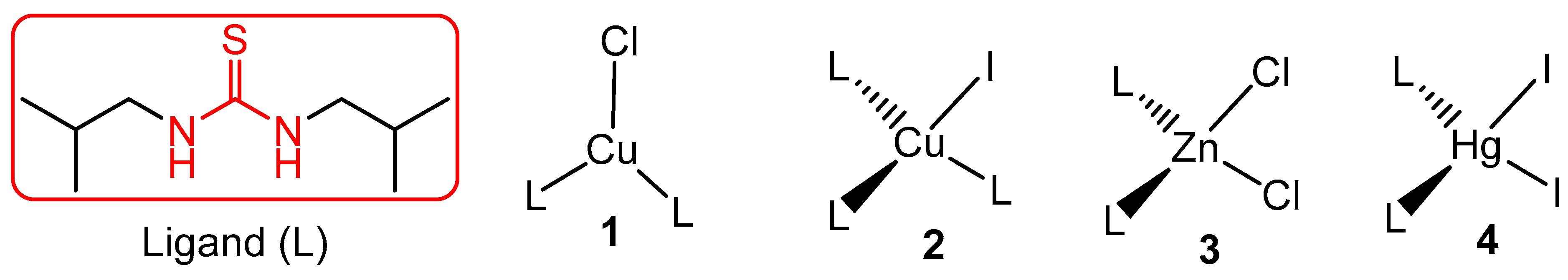
2.2. Synthesis of Compounds 1–4

For the synthesis of compound 1, a solution of CuCl (0.0501 g, 0.51 mmol) was prepared in acetonitrile and was kept stirring for 2 h, followed by the dropwise addition of 1,3-diisobutyl thiourea (0.2802 g, 1.49 mmol, solution in acetonitrile) over a time of ca. 10 min. The color of the resultant solution turned from yellow to colorless which indicated the reaction was complete. Stirring of the reaction mixture was continued for 12 h, in order to ensure maximum product formation. The reaction mixture was filtered for the separation of unreacted salt and other insoluble material and the filtrate was kept for slow evaporation at room temperature. Colorless crystals of the desired compound 1 appeared after a few days and were separated from the mother liquor, then studied with the help of EI-MS, UV/Vis spectroscopy, FT-IR, and X-ray diffraction. Following the same procedure, compound 2 was prepared by treating CuI (0.0500 g, 0.26 mmol) and the ligand (0.148 g, 0.79 mmol) together; colorless block crystals were grown after 6 days. Compound 3 was obtained by treating ZnCl2 (0.0602 g, 0.44 mmol) with the ligand (0.167 g, 0.89 mmol), using MeOH as solvent. Colorless crystals appeared after 2 days, were separated and then studied. Compound 4 was prepared in the same way, with HgI2 (0.2407 g, 0.529 mmol) and the ligand (0.2003 g, 1.06 mmol) in MeOH.
Complex 1, bis(1,3-Diisobutylthiourea)copper(I) chloride: Yield: 60%; C18H40N4ClS2Cu; Found (Cal): C 45.54 (45.45), H 8.36 (8.48), N 12.06 (11.78); EI-MS: m/z (%) 188 (99) Tu, 255 (85) Cu-Tu, 96 (25) Cu-Cl; FT-IR (KBr) ν(cm−1) = 3280br, 2965s, 2878s, 1734s.
Complex 2, tris(1,3-diisobutyl thiourea)copper(I) iodide: Yield: 65%; C27H60N6IS3Cu; Found (Cal): C 38.47 (40.93), H 7.18 (8.01), N 10.65(11.12); MS (+ESI): m/z 188 (99) Tu, 254 (45) Cu-Tu, 380 (20) I-Cu-Tu; FT-IR (KBr) ν(cm−1) = 3288br, 2971s, 2882s, 1751s.
Complex 3, bis(1,3-diisobutylthiourea)zinc(II) chloride: Yield: 76 %; C18H40N4Cl2S2Zn; Found (Cal): C 40.61 (42.15), H 7.39 (7.86), N 10.43 (10.92); 1H-NMR (300 MHz, CDCl3) δ (ppm) = 0.95 (br, Me), 1.96 (br, CH), 3.36 (br, CH2), 6.06 (br, NH): 13C-NMR (75.8 MHz, CDCl3); δ (ppm) = 20.2 (Me), 28.3 (CH), 52.4 (N-CH2), 174.8 (C=S); MS(EI): m/z (%): 188 (99) Tu, 253 (25) Zn-Tu; FT-IR (KBr) ν(cm−1) = 3278br, 2956s, 2868s, 1748s.
Complex 4, bis(1,3-diisobutylthiourea)mercury(II) iodide: Yield: 79 %; C18H40N4l2S2Hg; Found (Cal): C 26.24 (26.01), H 5.6 (4.85), N 7.57 (6.74); 1H-NMR (300 MHz, CDCl3) δ (ppm) = 0.97 (d, 24H, Me, J(1H,1H) = 6.63 Hz), 2.01 (Sep, 4H, CH, J(1H,1H) = 6.67), 3.21 (br, 8H, CH2), 5.80 (br, 4H, NH); 13C-NMR (75.8 MHz, CDCl3); δ (ppm) = 20.5 (Me), 27.9 (CH), 51.9 (N-CH2), 176.9 (C=S); MS(EI): m/z (%) 188 (99) Tu, 328 (7) HgI, 456 (35) HgI2; FT-IR (KBr) ν(cm−1) = 3281br, 2961s, 2872s, 1741s.

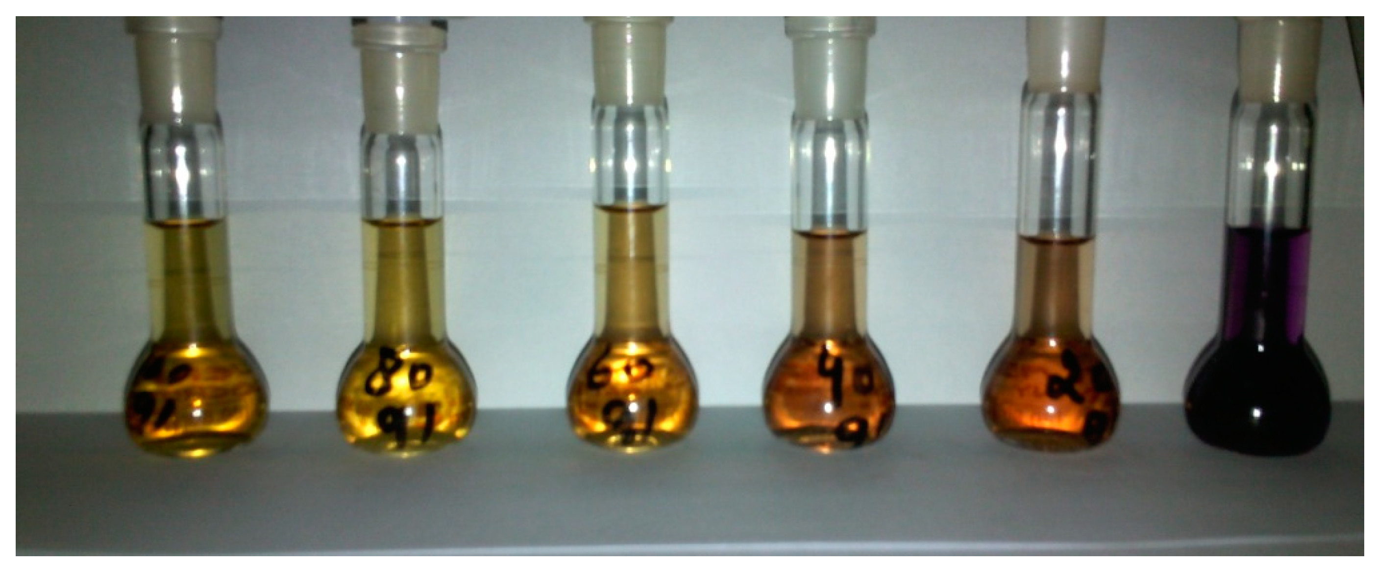
2.3. Determination of Antioxidant Potentials

DPPH is a stable free radical and is capable of utilizing its unpaired electron in a chemical interaction with any other species. The involvement of this unpaired radical with foreign species is a function of free radical scavenging of that species/compound. Compounds 1–4 were tested for their antioxidant activities against DPPH. A solution containing 0.039 g/100 mL of DPPH was prepared and used as a standard/control. Absorbance of this solution was measured at 517 nm under normal conditions of temperature. After mixing the solution of the respective compound with DPPH solution, a change in the resultant absorbance was observed. The decrease in maximum absorbance of the solution was taken as a function of the compound. Prior to the determination of absorbance, all solutions were incubated in the dark for 30 min at room temperature (23 ± 1°C). The percent inhibition capacity of each complex (I)%, was calculated as below [36].
Perecent   Inhibition   I % = A D P P H A s a m p l e A D P P H × 100

2.4. Antibacterial Screenings of Selected Compounds

The agar well diffusion assay was used to evaluate antimicrobial potentials of complexes 1–4 [37,38]. Sterile, nutrient agar was prepared and poured in Petri dishes. Bacterial cultures were evenly applied on the surface of the agar Petri dishes by sterile swab sticks. Wells of 6 mm diameter were bored (five per plate) with a sterile borer. An amount of 6.25 mg/mL of each compound was applied to each well. The commercially available antibiotic cephradine was used for comparing the efficiency of each complex. For the sake of accuracy, the same concentration of each complex and the standard was applied. The agar plates were covered with lids and were incubated at 37 ± 1°C for 24 h in an oven. Growth inhibition was observed in each bore and the respective zones were measured manually. The diameter of the zones of inhibition is a measure of antimicrobial activity of the corresponding complex. The data presented for antibacterial activity are the average diameter of the zones of inhibition in mm.

3. Results and Discussion

3.1. General Chemistry and Spectroscopy

The proposed structures of compounds and the corresponding ligand (inserted) are shown in Scheme 1. The treatment of ligand (1,3-diisobutylthiourea) with copper(I) chloride in molar ratio 3:1, respectively, afforded Complex 1. The reaction of CuCl with a two-fold excess of the ligand was carried out under identical conditions and the same products were obtained. By changing the reaction medium from acetonitrile to MeOH, all attempts were unsuccessful in obtaining exclusively tetrahedral complex of geometry [CuL3Cl], [CuL2Cl2], or [CuL4]Cl, where L = thiourea ligand [39,40,41]. The ligand reacted with CuI in a molar ratio 3:1 and the proposed compound 2 was thus obtained. These observations reveal that the presence of halogen function can affect the coordination sphere around the metal ion. The reaction of the ligand with Zn and Hg metal ions was straightforward, as was reported for structurally analogous species [42].
Complexes 1–4 were obtained as crystalline material from their respective reaction mixtures and structures of complexes were deduced from a consistent set of spectroscopic data. The FT-IR spectra of all compounds contain typically broad signals (3278–3288 cm−1) that correspond to the involvement of NH bonds in extensive hydrogen bonding. NMR spectroscopy is a reliable technique in the structural elucidation of compounds and is of particular importance in coordination chemistry, where the coordination behavior of thiourea ligands can easily be determined to be an N or S donor. The 1H-NMR of complex 3 and 4 show distinct typically broad signals at 4.01 and 3.47 ppm, respectively, which can be assigned unambiguously to NH group. These signals show considerable shift with respect to the free ligand (5.89 ppm, given in supporting information). The 13C-NMR process is quite useful in this regard: the C=S signal appeared at 181.7 ppm in the free thiourea ligand and was found to be considerably shifted in complex 3 and 4 to 174.8 and 176.9 ppm, respectively. These data indicate the bond formation through the S atom, thus affording the proposed coordination behavior of the TU ligand [43]. The mass fragmentation pattern of compounds 1–3 was also studied using the EI-MS technique. The information retrieved from these data were not too informative because of the high-energy ionization technique. All the complexes gave a base peak at m/z 188, which corresponds to the ion of the thiourea ligand. A molecular ion peak for these complexes was not observed.

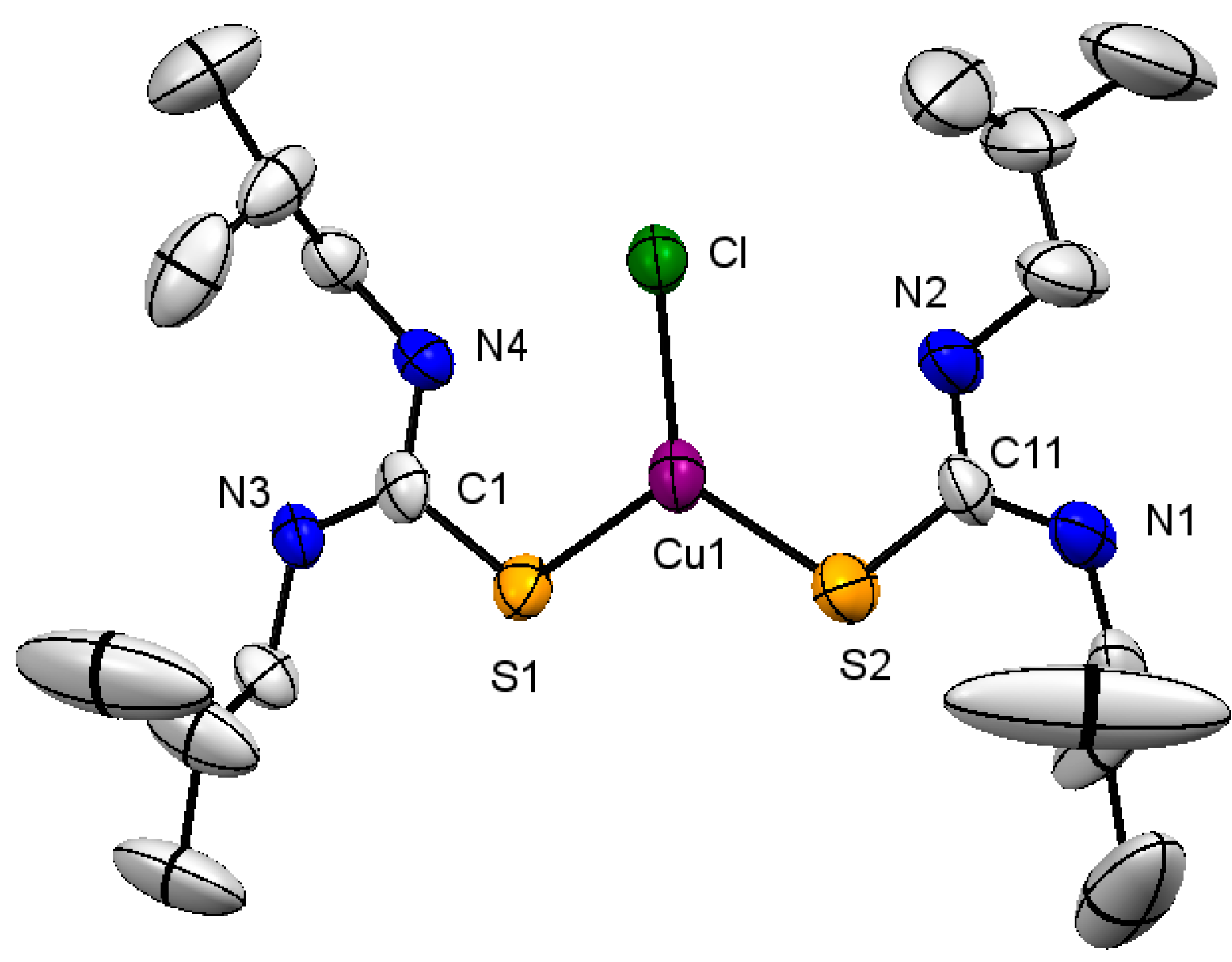
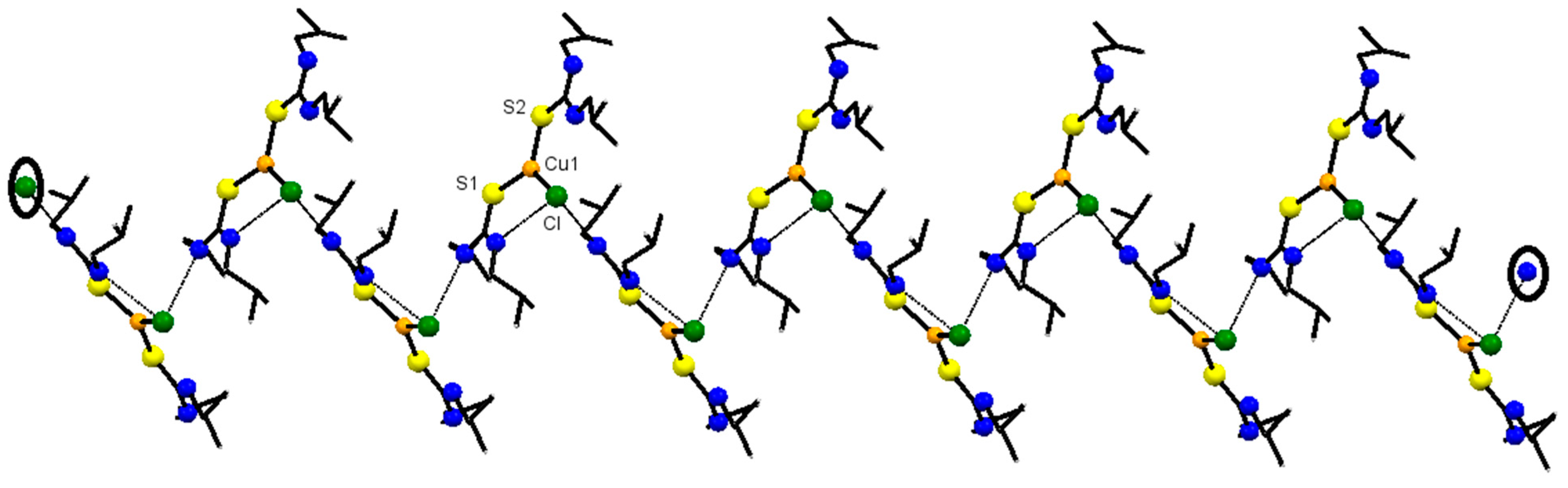
3.2. Structural Description of Complex 1

Ethyl fragments in the structure of compound 1 (shown in Figure 1) suffer from some sort of disorder which makes it very difficult to precisely solve and refine the structure. The diffraction data provide enough information which is sufficient for the establishment of connectivity. The reaction of the ligand with CuCl in molar ratio 3:1 exclusively afforded compound 1. The molecule is monoclinic, bearing space group Cc (further details pertaining to refinements and crystal structure solution are summarized in Table 1). The geometry around the metal ion is trigonal planar. Two thiourea ligands are attached to the metal ion through the S atom, which is normal behavior of thiourea derivatives [28]. Bond angles around the metal ion are 115.76(13)°, (S1-Cu1-S2), 120.94(11)°, and 123.30(12)° (S1-Cu1-C1 and S2-Cu1-Cl, respectively). The sum of all three angles around metal ion is 360°, which clearly supports the trigonal environment. The Cu-S bond lengths with negligible difference, 2.213(4) and 2.216(3) Å, are shorter than the Cu-Cl bond (2.272(3) Å) and are within the expected limit [44] (Table 2). Coordination with the metal ion through S reduces the bond order between C and S; therefore, it results in C=S bond elongation as compared to the uncoordinated thiourea derivative and are comparable to structurally analogous compounds [28,30,45,46]. A close view of molecules of complex 1 in solid state reveals that chloro function is actively involved in intra- as well as intermolecular secondary interactions. The intramolecular separation distance between N4Cl is 3.268 Å, and the (intermolecular) ClN3 distance is 3.268 Å (Figure 2). The difference in bond lengths, C1-N3 1.306(14) and C1-N4 1.349(14) Å, can possibly be explained on the basis of intramolecular interactions wherein N atoms of one coordinated thiourea ligand are involved (Figure 2). Each molecule offers an N3 of a coordinated ligand to participate in secondary interactions, which is the probable reason for slightly different C-N bond lengths.

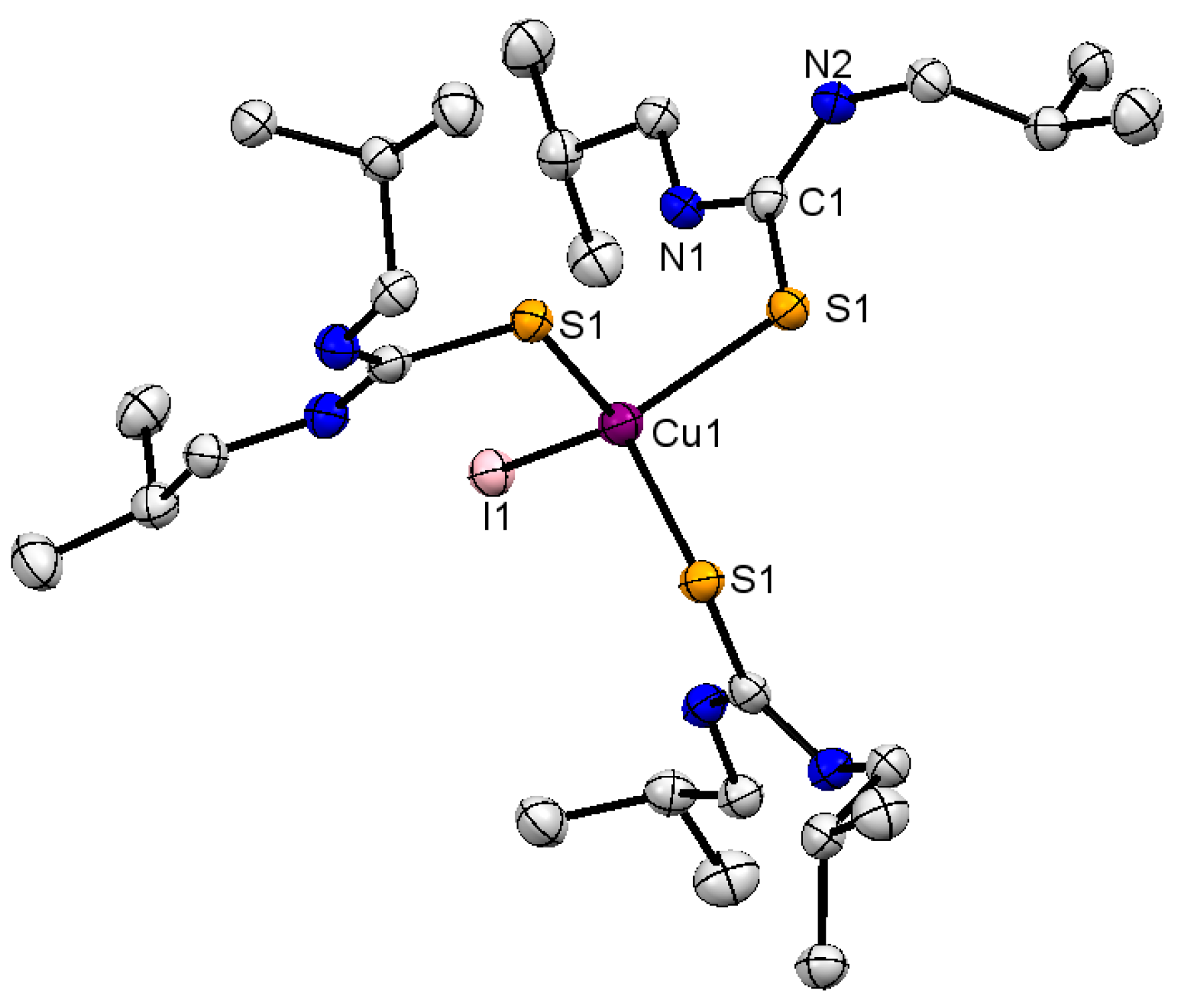
3.3. Structural Description of Complex 2

The structure of complex 2 is depicted in Figure 3, together with selected bond lengths and angles. The structure solution and refinements were carried out as per details given in Table 1. The crystal structure is trigonal with space group P-3. The Cu+ ion is surrounded by three thiourea ligands and a halogen (iodide), making the tetrahedral geometry with expected deviation owing to hetero-ligands [47,48]. The ligand is attached through the S atom, as normally expected for this class of compounds. The angles S1-Cu1-S1 and S1-Cu1-I1 are 100.32(2) and 117.55(16), respectively, with the sum of all six angles amounting to 653.6°, which supports distorted tetrahedral geometry around the metal ion [49]. These features are within the expected limit and are very close to structurally analogous complexes [50,51]. The M-S bond distance of 2.336(6) Å for complex 2 is within the expected limit for analogous thiourea complexes [41]. The C=S bond slightly elongates (1.713(2) Å) with respect to the uncoordinated ligand (1.698 Å) because of electronic flow towards the metal ion. The abovementioned behavior of the ligand causes an electron delocalization over the S-C-N fragment [1,52]. There are a number of secondary short-range interactions within the molecules of the complex, which can be useful for further studies and in predicting certain applications of the complex. The hydrogen of C2H and N2H of each ligand are involved in interactions with S atoms of neighboring molecules. Each S atom of the ligand is pseudo-four-coordinated, and the supramolecular structure of complex 2 extends in a 3D fashion.

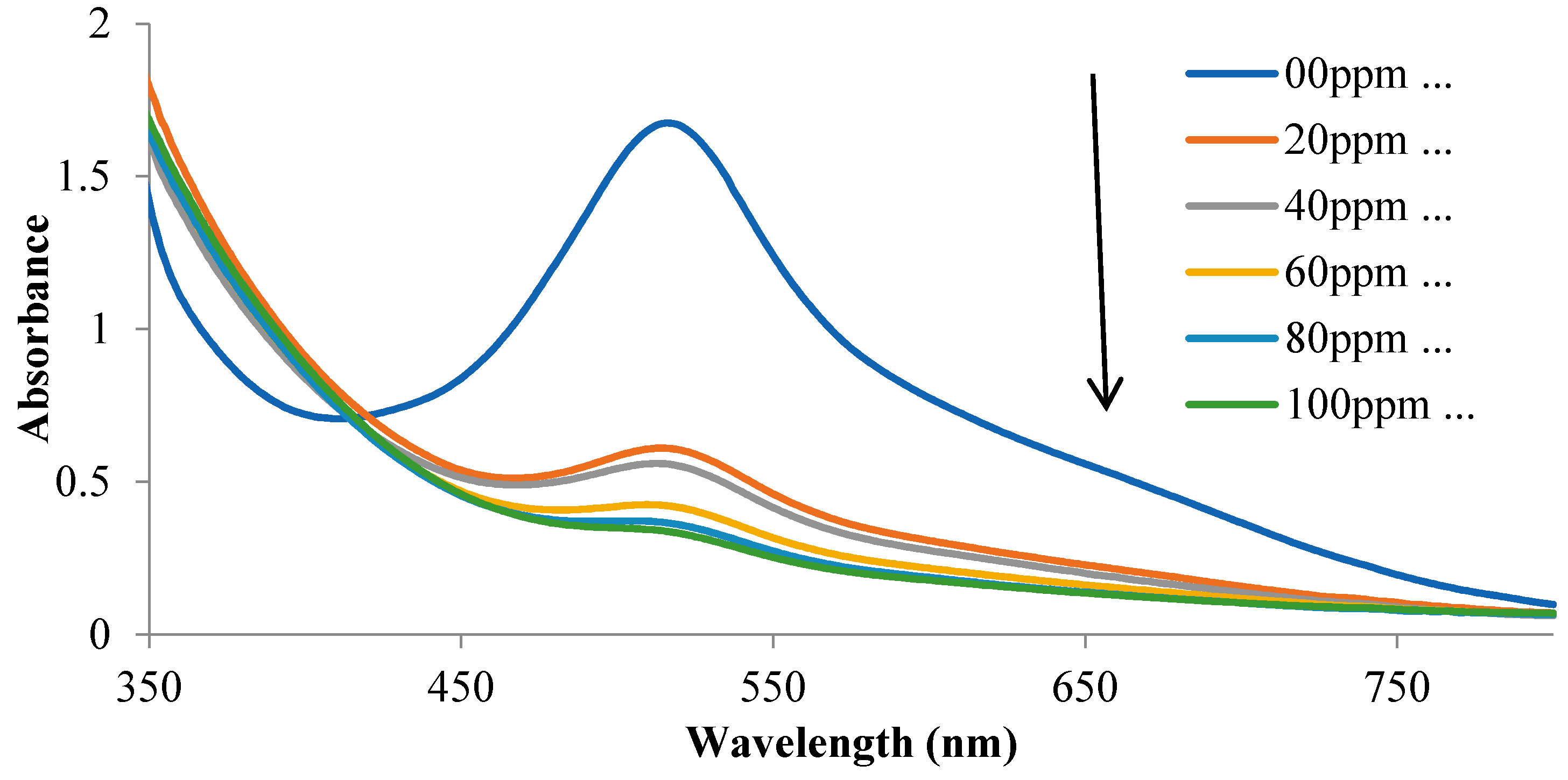
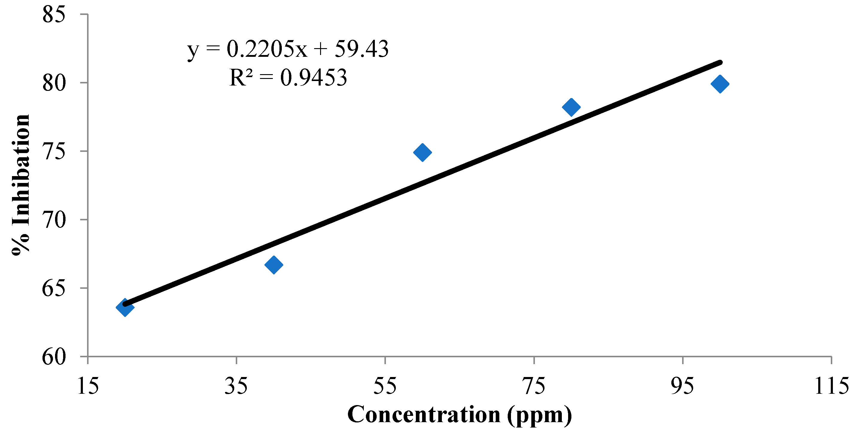
3.4. Free Radical Scavenging Activities of Complexes 1–4

The inhibitory effects of thiourea complexes 1–4 were studied using DPPH as the free radical. For these complexes, which include S-coordinated 1,3-diisobutyl thiourea molecules, high antioxidant activity was discovered. The absorption intensity of DPPH was reduced by the addition of complexes 1–4, in a dose-dependent manner ranging from 00–100 ppm. The decrease in absorbance of the resultant solution was quite regular, as expected for an antioxidant reagent. The percent RSA (radical scavenging activity) of compounds was at its lowest at a lower concentration and was at its maximum at a 100 ppm concentration of each complex, in a dose-dependent manner. A comparison of the activity of 1–4 at 100 ppm (maximum concentration) was found to be 79.9%, 19.5%, 29.3%, and 19.2%, respectively, which indicates that compound 1 is more potent as compared to 24. Data pertaining to the free radical scavenging efficiency of complexes are summarized in Table 3 and the same are graphically described in Figure 4. The complex reveals a reasonable R2 value of 0.9453, shown in Figure 5 (see Figures S1–S6 for complexes 2, 3 and 4) (see Supplementary Materials). Among these four complexes, 1 is a far better antioxidant and the activity can be observed by the naked eye due to the change in the color of the solution after mixing the appropriate concentration with DPPH (Figure 6). The activity of compounds 24 were the least, and color change was difficult to be observed with the naked eye. The activity of compound 1 was better than the Cu complex stabilized by phosphine ligands, as has been reported recently [25].

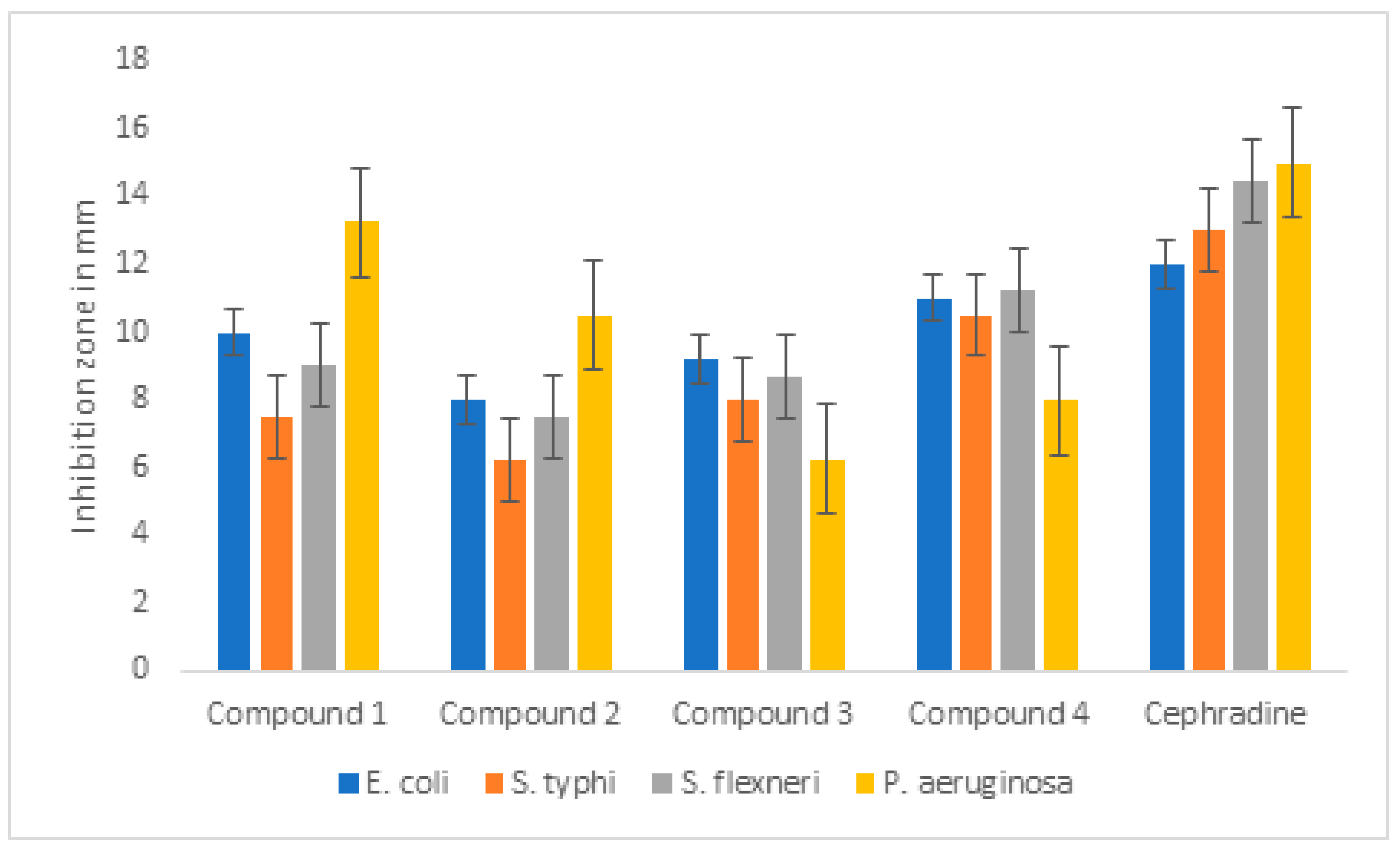
3.5. Antibacterial Activity

The agar well diffusion method was used to assess the antimicrobial activity of complexes 1–4. Escherichia coli, Shigella flexneri, Pseudomonas aeruginosa, and Salmonella typhi were selected for this study and complexes 1–4 were studied against these strains. The antibacterial evaluation of these complexes reveal that they exhibit moderate activity in comparison to cephradine. The overall inhibition range of 6.2–13.25 mm (Table 4) was observed for these complexes. Compound 1 shows comparatively better activity against E. coli and P. aeruginosa, comparable with standard drugs, and compound 4 is most active against three strains: E. coli, S. typhi and S. flexneri. Compounds 2 and 3 were found to be poor antibacterial agents among these four complexes. The activity of all complexes and the standard is graphically represented in Figure 7.

4. Conclusions

Four new heteroleptic transition metal complexes (14) were successfully obtained by reacting 1,3-diisobutyl thiourea with CuCl, CuI, ZnCl2, and HgI2, respectively. Despite the same reaction conditions, reaction with CuCl and CuI afforded bis-ligated and tris-ligated copper halide complexes, respectively. Compounds 1 and 2 were also characterized by single-crystal X-ray analysis. The Zn and Hg metals were included in the study with the intent to explore the coordination behavior of the ligand, and the proposed geometries were obtained. All complexes were tested for their free radical scavenging ability and antimicrobial potentials; the data reveal that compound 1 is an excellent antioxidant and the activity could be observed with the naked eye. All compounds showed antioxidant potentials in a dose-dependent manner. In comparison to the standard, compound 1 was active against E. coli and P. aeruginosa, while compound 4 was a potent antibacterial agent against E. coli, S. typhi and S. flexneri. Future work regarding functionalization can explore potentials of thiourea complexes as antioxidant and antibacterial reagents.

Supplementary Materials

CCDC No 1,994,970 (1) and 1,979,130 (2) contain the supplementary crystallographic data for complex 1 and 2. These data can be obtained free of charge via http://www.ccdc.cam.ac.uk/conts/retrieving.html accessed on 17 August 2021, or from the Cambridge Crystallographic Data Centre, 12 Union Road, Cambridge CB2 1EZ, UK; fax: (+44) 1223-336-033; or e-mail: [email protected]. Figures S1–S6 are available online at https://0-www-mdpi-com.brum.beds.ac.uk/article/10.3390/cryst11080989/s1, Figure S1. Absorption spectra of DPPH in the absence of compound 2 (Top spectra) and presence of increasing concentration of the compound (20, 40, 60, 80 & 100 ppm). Arrow shows the change in absorption as a function of activity with respect to increasing concentration of compound 2. Figure S2. Plot of % Inhibition versus concentration of compound 2 for radical scavenging activity. Figure S3. Absorption spectra of pure DPPH, the absorbance decreased when compounds 3 was added, in a dose dependent manner. Figure S4. Percent inhibition versus concentration of compound 3 for radical scavenging activity. Figure S5. Absorption spectra of free radical (DPPH) in the absence (Top spectra) and presence of increased concentration of the compound 4 (20, 40, 60, 80 & 100 ppm). Arrow show the change in spectra on increasing concentration of compound. Figure S6. Plot of % Inhibition versus concentration of compound 4 for radical scavenging activity.

Author Contributions

A.S. Data curation, writing of paper original draft, E.K. Conceptualization, Data curation, Project Administration, Formal analysis; Funding acquisition; Methodology; Supervision, writing of paper original and revised draft. M.S., G.S.K., M.G.S. and M.Z. equally contributed in formal analysis; Software; Writing—review and editing, A.N. collected Crystal data, visualization; R.U. Characterization of samples, help in write up, revisions and funding acquisition, A.B. Characterization of samples, help in write up, revisions and funding acquisition. All authors have read and agreed to the published version of the manuscript.

Funding

This research was funded by King Saud University Research Supporting Project Number: RSP-2021/346.

Institutional Review Board Statement

Not applicable.

Informed Consent Statement

Not applicable.

Data Availability Statement

Data which support this work is included in the manuscript.

Acknowledgments

Authors wish to thanks Research Supporting Project Number: RSP-2021/346 King Saud University Riyadh Saudi Arabia for their financial support.

Conflicts of Interest

The authors declare no conflict of interest.

References

  1. Rahman, F.U.; Bibi, M.; Khan, E.; Shah, A.B.; Muhammad, M.; Tahir, M.N.; Shahzad, A.; Ullah, F.; Zahoor, M.; Alamery, S.; et al. Thiourea Derivatives, Simple in Structure but Efficient Enzyme Inhibitors and Mercury Sensors. Molecules 2021, 26, 4506. [Google Scholar] [CrossRef] [PubMed]
  2. Khan, E.; Khan, S.; Gul, Z.; Muhammad, M. Medicinal Importance, Coordination Chemistry with Selected Metals (Cu, Ag, Au) and Chemosensing of Thiourea Derivatives. A Review. Crit. Rev. Anal. Chem. 2021, 1–23. [Google Scholar] [CrossRef]
  3. Zhang, Z.; Schreiner, P.R. (Thio)urea organocatalysis—What can be learnt from anion recognition? Chem. Soc. Rev. 2009, 38, 1187–1198. [Google Scholar] [CrossRef] [PubMed]
  4. Sun, Y.; Wei, Y.; Shi, M. Applications of Chiral Thiourea-Amine/Phosphine Organocatalysts in Catalytic Asymmetric Reactions. ChemCatChem 2017, 9, 718–727. [Google Scholar] [CrossRef]
  5. Esteban, F.; Cieślik, W.; Arpa, E.M.; Guerrero-Corella, A.; Díaz-Tendero, S.; Perles, J.; Fernandez-Salas, J.A.; Fraile, A.; Alemán, J. Intramolecular Hydrogen Bond Activation: Thiourea-Organocatalyzed Enantioselective 1,3-Dipolar Cycloaddition of Salicylaldehyde-Derived Azomethine Ylides with Nitroalkenes. ACS Catal. 2018, 8, 1884–1890. [Google Scholar] [CrossRef] [PubMed] [Green Version]
  6. Steppeler, F.; Iwan, D.; Wojaczyńska, E.; Wojaczyński, J. Chiral Thioureas—Preparation and Significance in Asymmetric Synthesis and Medicinal Chemistry. Molecules 2020, 25, 401. [Google Scholar] [CrossRef] [Green Version]
  7. Li, A.-F.; Wang, J.-H.; Wang, F.; Jiang, Y.-B. Anion complexation and sensing using modified urea and thiourea-based receptors. Chem. Soc. Rev. 2010, 39, 3729–3745. [Google Scholar] [CrossRef] [Green Version]
  8. Bregović, V.B.; Basaric, N.; Mlinarić-Majerski, K. Anion binding with urea and thiourea derivatives. Coord. Chem. Rev. 2015, 295, 80–124. [Google Scholar] [CrossRef]
  9. Udhayakumari, D.; Velmathi, S.; Venkatesan, P.; Wu, S.-P. Anthracene coupled thiourea as a colorimetric sensor for F/Cu2+ and fluorescent sensor for Hg2+/picric acid. J. Lumin. 2015, 161, 411–416. [Google Scholar] [CrossRef]
  10. Zhang, Z.; Lu, S.; Sha, C.; Xu, D. A single thiourea-appended 1,8-naphthalimide chemosensor for three heavy metal ions: Fe3+, Pb2+, and Hg2+. Sens. Actuators B Chem. 2015, 208, 258–266. [Google Scholar] [CrossRef]
  11. Koch, K.R. New chemistry with old ligands: N-alkyl- and N,N-dialkyl-N′-acyl(aroyl)thioureas in co-ordination, analytical and process chemistry of the platinum group metals. Coord. Chem. Rev. 2001, 216-217, 473–488. [Google Scholar] [CrossRef]
  12. Saeed, A.; Qamar, R.; Fattah, T.A.; Flörke, U.; Erben, M.F. Recent developments in chemistry, coordination, structure and biological aspects of 1-(acyl/aroyl)-3-(substituted) thioureas. Res. Chem. Intermed. 2016, 43, 3053–3093. [Google Scholar] [CrossRef]
  13. Reddy, V.L.; Avula, V.K.R.; Zyryanov, G.V.; Vallela, S.; Anireddy, J.S.; Pasupuleti, V.R.; Chamarthi, N.R. Hunig’s base catalyzed synthesis of new 1-(2,3-dihydro-1H-inden-1-yl)-3-aryl urea/thiourea derivatives as potent antioxidants and 2HCK enzyme growth inhibitors. Bioorganic Chem. 2020, 95, 103558. [Google Scholar] [CrossRef] [PubMed]
  14. Thomas, S.J.; Balónová, B.; Cinatl, J.; Wass, M.; Serpell, C.J.; Blight, B.A.; Michaelis, M. Thiourea and Guanidine Compounds and Their Iridium Complexes in Drug-Resistant Cancer Cell Lines: Structure-Activity Relationships and Direct Luminescent Imaging. ChemMedChem 2019, 15, 349–353. [Google Scholar] [CrossRef]
  15. Hu, H.; Lin, C.; Ao, M.; Ji, Y.; Tang, B.; Zhou, X.; Fang, M.; Zeng, J.-Z.; Wu, Z. Synthesis and biological evaluation of 1-(2-(adamantane-1-yl)-1H-indol-5-yl)-3-substituted urea/thiourea derivatives as anticancer agents. RSC Adv. 2017, 7, 51640–51651. [Google Scholar] [CrossRef] [Green Version]
  16. Ghorab, M.M.; El-Gaby, M.; Alsaid, M.S.; Elshaier, Y.; Soliman, A.M.; Elsenduny, F.; Badria, F.A.; Sherif, A.Y. Novel Thiourea Derivatives Bearing Sulfonamide Moiety as Anticancer Agents Through COX-2 Inhibition. Anti-Cancer Agents Med. Chem. 2017, 17, 1411–1425. [Google Scholar] [CrossRef] [PubMed]
  17. Estévez-Hernández, O.; Duque, J.; Rodríguez-Hernández, J.; Reguera, E. Dinuclear and polymeric Hg(II) complexes with 1-(2-furoyl)thiourea derivatives: Their crystal structure and related properties. Polyhedron 2015, 97, 148–156. [Google Scholar] [CrossRef]
  18. Cole, J.M.; Hickstein, D.D. Molecular origins of nonlinear optical activity in zinc tris (thiourea) sulfate revealed by high-resolution x-ray diffraction data and ab initio calculations. Phys. Rev. B 2013, 88, 184105. [Google Scholar] [CrossRef]
  19. Shkir, M.; Ganesh, V.; AlFaify, S.; Maurya, K.K.; Vijayan, N. Effect of phenol red dye on monocrystal growth, crystalline perfection, and optical and dielectric properties of zinc (tris) thiourea sulfate. J. Appl. Crystallogr. 2017, 50, 1716–1724. [Google Scholar] [CrossRef]
  20. Chetana, P.; Srinatha, B.; Somashekar, M.; Policegoudra, R. Synthesis, spectroscopic characterisation, thermal analysis, DNA interaction and antibacterial activity of copper(I) complexes with N, N′- disubstituted thiourea. J. Mol. Struct. 2016, 1106, 352–365. [Google Scholar] [CrossRef] [Green Version]
  21. Mahendiran, D.; Amuthakala, S.; Bhuvanesh, N.S.P.; Kumar, R.S.; Rahiman, A.K. Copper complexes as prospective anticancer agents: In vitro and in vivo evaluation, selective targeting of cancer cells by DNA damage and S phase arrest. RSC Adv. 2018, 8, 16973–16990. [Google Scholar] [CrossRef] [Green Version]
  22. Jose, E.S.; Philip, J.E.; Shanty, A.; Kurup, M.; Mohanan, P. Novel class of mononuclear 2-methoxy-4-chromanones ligated Cu (II), Zn (II), Ni (II) complexes: Synthesis, characterisation and biological studies. Inorganica Chim. Acta 2018, 478, 155–165. [Google Scholar] [CrossRef]
  23. Iliş, M.; Cîrcu, V. Discotic Liquid Crystals Based on Cu (I) Complexes with Benzoylthiourea Derivatives Containing a Perfluoroalkyl Chain. J. Chem. 2018, 2018, 1–10. [Google Scholar] [CrossRef]
  24. Khan, S.A.; Noor, A.; Kempe, R.; Subhan, H.; Shah, A.; Khan, E. Syntheses, molecular structure, and electrochemical investigations of cobalt (II), copper (II), palladium (II), and zinc (II) complexes with 3-methylpyrazole. J. Coord. Chem. 2014, 67, 2425–2434. [Google Scholar] [CrossRef]
  25. Khan, E.; Shahzad, A.; Tahir, M.N.; Noor, A. Antioxidant potential and secondary reactivity of bis{diphenyl(2-pyridyl)phosphino} copper (II) complex. Turk. J. Chem. 2018, 42, 1299–1309. [Google Scholar] [CrossRef] [Green Version]
  26. Khan, E.; Gul, Z.; Shahzad, A.; Jan, M.S.; Ullah, F.; Tahir, M.N.; Noor, A. Coordination compounds of 4,5,6,7-tetrahydro-1H-indazole with Cu(II), Co(II) and Ag(I): Structural, antimicrobial, antioxidant and enzyme inhibition studies. J. Coord. Chem. 2017, 70, 4054–4069. [Google Scholar] [CrossRef]
  27. Gul, Z.; Din, N.U.; Khan, E.; Ullah, F.; Tahir, M.N. Synthesis, molecular structure, anti-microbial, anti-oxidant and enzyme inhibition activities of 2-amino-6-methylbenzothiazole and its Cu(II) and Ag(I) complexes. J. Mol. Struct. 2020, 1199, 126956. [Google Scholar] [CrossRef]
  28. Rahman, F.U.; Bibi, M.; Altaf, A.A.; Tahir, M.N.; Ullah, F.; Khan, E. Zn, Cd and Hg complexes with unsymmetric thiourea derivatives; syntheses, free radical scavenging and enzyme inhibition essay. J. Mol. Struct. 2020, 1211, 128096. [Google Scholar] [CrossRef]
  29. Khan, U.A.; Badshah, A.; Tahir, M.N.; Khan, E. Gold (I), silver (I) and copper (I) complexes of 2, 4, 6-trimethylphenyl-3-benzoylthiourea; synthesis and biological applications. Polyhedron 2020, 181, 114485. [Google Scholar] [CrossRef]
  30. Altaf, A.A.; Shahzad, A.; Gul, Z.; Khan, S.A.; Badshah, A.; Tahir, M.N.; Zafar, Z.I.; Khan, E. Synthesis, Crystal Structure, and DFT Calculations of 1,3-Diisobutyl Thiourea. J. Chem. 2015, 2015, 1–5. [Google Scholar] [CrossRef] [Green Version]
  31. Altomare, A. A new tool for crystal structure determination and refinement. J. Appl. Cryst. 1994, 27, 435. [Google Scholar] [CrossRef]
  32. Sheldrick, G.M. Crystal structure refinement with SHELXL. Acta Crystallogr. Sect. C Struct. Chem. 2015, 71, 3–8. [Google Scholar] [CrossRef] [PubMed]
  33. Farrugia, L.J. WinGX suite for small-molecule single-crystal crystallography. J. Appl. Crystallogr. 1999, 32, 837–838. [Google Scholar] [CrossRef]
  34. Farrugia, L.J. ORTEP-3 for Windows—A version ofORTEP-III with a Graphical User Interface (GUI). J. Appl. Crystallogr. 1997, 30, 565. [Google Scholar] [CrossRef]
  35. Spek, A.L. Structure validation in chemical crystallography. Acta Crystallogr. Sect. D Biol. Crystallogr. 2009, 65, 148–155. [Google Scholar] [CrossRef] [PubMed]
  36. Khan, E.; Khan, A.; Gul, Z.; Ullah, F.; Tahir, M.N.; Khalid, M.; Asif, H.M.; Asim, S.; Braga, A.A.C. Molecular salts of terephthalic acids with 2-aminopyridine and 2-aminothiazole derivatives as potential antioxidant agents; Base-Acid-Base type architectures. J. Mol. Struct. 2020, 1200, 127126. [Google Scholar] [CrossRef]
  37. Irshad, S.; Mahmood, M.; Perveen, F. In vitro antibacterial activities of three medicinal plants using agar well diffusion method. Res. J. Biol. 2012, 2, 1–8. [Google Scholar]
  38. Athanassiadis, B.; Abbott, P.V.; George, N.; Walsh, L.J. An in vitro study of the antimicrobial activity of some endodontic medicaments and their bases using an agar well diffusion assay. Aust. Dent. J. 2009, 54, 141–146. [Google Scholar] [CrossRef]
  39. Mufakkar, M.; Isab, A.A.; Rüffer, T.; Lang, H.; Ahmad, S.; Arshad, N.; Waheed, A. Synthesis, characterization, and antibacterial activities of copper (I) bromide complexes of thioureas: X-ray structure of [Cu (Metu) 4] Br. Transit. Met. Chem. 2011, 36, 505–512. [Google Scholar] [CrossRef]
  40. Bombicz, P.; Mutikainen, I.; Krunks, M.; Leskelä, T.; Madarász, J.; Niinistö, L. Synthesis, vibrational spectra and X-ray structures of copper(I) thiourea complexes. Inorganica Chim. Acta 2004, 357, 513–525. [Google Scholar] [CrossRef]
  41. Bowmaker, G.A.; Hanna, J.V.; Pakawatchai, C.; Skelton, B.; Thanyasirikul, Y.; White, A.H. Crystal Structures and Vibrational Spectroscopy of Copper (I) Thiourea Complexes. Inorg. Chem. 2009, 48, 350–368. [Google Scholar] [CrossRef] [PubMed]
  42. Malik, M.R.; Vasylyeva, V.; Merz, K.; Metzler-Nolte, N.; Saleem, M.; Ali, S.; Isab, A.A.; Munawar, K.S.; Ahmad, S. Synthesis, crystal structures, antimicrobial properties and enzyme inhibition studies of zinc (II) complexes of thiones. Inorganica Chim. Acta 2011, 376, 207–211. [Google Scholar] [CrossRef]
  43. Ajibade, P.A.; Zulu, N.H. Metal Complexes of Diisopropylthiourea: Synthesis, Characterization and Antibacterial Studies. Int. J. Mol. Sci. 2011, 12, 7186–7198. [Google Scholar] [CrossRef] [Green Version]
  44. Saxena, A.; Dugan, E.C.; Liaw, J.; Dembo, M.D.; Pike, R.D. Copper (I) complexes of heterocyclic thiourea ligands. Polyhedron 2009, 28, 4017–4031. [Google Scholar] [CrossRef] [Green Version]
  45. Zhao, X.-Y.; Zhu, C.-B.; Li, H.-P.; Yang, Y.; Roesky, H.W. Synthesis and Characterization of Copper (I) Halide Complexes withN-(2, 6-Diisopropylphenyl)-N′-benzoylthiourea: Monomeric, Dimeric, and Cage Structures. Z. Für Anorg. Und Allg. Chem. 2014, 640, 1614–1621. [Google Scholar] [CrossRef]
  46. Jia, D.; Zhu, A.M.; Ji, M.; Zhang, Y. Copper (I) halide complexes with a sterically hindered thiourea: Synthesis and crystal structures of [Cu (dchtu) 2Cl] and [Cu (dchtu) 2Br](dchtu = N,N′-dicyclohexylthiourea). J. Coord. Chem. 2008, 61, 2307–2314. [Google Scholar] [CrossRef]
  47. Piro, O.E.; Piatti, R.C.V.; Bolzan, A.E.; Salvarezza, R.C.; Arvia, A.J. X-ray diffraction study of copper (I) thiourea complexes formed in sulfate-containing acid solutions. Acta Crystallogr. Sect. B Struct. Sci. 2000, 56, 993–997. [Google Scholar] [CrossRef]
  48. Pakawatchai, C.; Thanyasirikul, Y.; Saepae, T.; Pansook, S.; Fun, H.-K.; Chinnakali, K. Hexakis (μ-N-ethylthiourea-S) tetrakis [iodocopper (I)] Monohydrate. Acta Crystallogr. Sect. C Cryst. Struct. Commun. 1998, 54, 1750–1752. [Google Scholar] [CrossRef]
  49. McNelis, E.; Blandino, M. 657, a method for estimating tetrahedral bond angles. New J. Chem. 2001, 25, 772–774. [Google Scholar] [CrossRef]
  50. Ahmad, S.; Altaf, M.; Stoeckli-Evans, H.; Rüffer, T.; Lang, H.; Mufakkar, M.; Waheed, A. Crystal Structures of Trinuclear Chlorido (N,N′-diethylthiourea) copper (I) and a Second Polymorph of Iodidotris (N,N′-diethylthiourea) copper (I). J. Chem. Crystallogr. 2010, 40, 639–645. [Google Scholar] [CrossRef]
  51. Fun, H.K.; Razak, I.A.; Pakawatchai, C.; Khaokong, C.H.U.A.N.P.I.T.; Chantrapromma, S.; Saithong, S.A.P.W.A.N.I.T. Tris (N,N’-diethylthiourea-S) iodocopper (I) and Tris (N,N’-diethylthiourea-S) iodosilver (I). Acta Crystallogr. Sect. C Cryst. Struct. Commun. 1998, 54, 453–456. [Google Scholar] [CrossRef]
  52. Binzet, G.; Kavak, G.; Külcü, N.; Özbey, S.; Flörke, U.; Arslan, H. Synthesis and Characterization of Novel Thiourea Derivatives and Their Nickel and Copper Complexes. J. Chem. 2013, 2013, 1–9. [Google Scholar] [CrossRef] [Green Version]
Scheme 1. Structures of ligand (left inserted) and its corresponding complexes with Cu, Zn and Hg ions.
Crystals 11 00989 sch001
Figure 1. Molecular structure of Complex 1, ball and stick representation with partial numbering scheme. All hydrogen atoms are omitted for clarity, summarized bond lengths (Å) and angles (°) are shown in Table 2. Two of the ethyl groups show disorder, which is the reason for relatively large displacement parameters and unusual bond distances (C21A-C51 1.08(3), C21A-C32 1.68(4) and C14-C35 1.29(3) Å).
Crystals 11 00989 g001
Figure 2. 1D representation of Complex 1, the infinite pseudo-polymeric zigzag chain stabilized by intermolecular NHCl interactions. Hydrogen atoms are omitted for clarity and hanging contacts at both ends of the chain are encircled to make them prominent. Intramolecular NHCl interactions are also indicated.
Crystals 11 00989 g002
Figure 3. Molecular structure of compound 2 with partial numbering scheme. Thermal ellipsoid drawn at 50% probability level, hydrogens are omitted for simplicity, summarized bond lengths (Å) and angles (°) are given in Table 2.
Crystals 11 00989 g003
Figure 4. Absorption spectra of DPPH in the absence (top spectra) and presence of the compound 1 (20, 40, 60, 80, and 100 ppm). There is abrupt drop in the absorbance of the solution by addition of the antioxidant agent (compound 1).
Crystals 11 00989 g004
Figure 5. Graphical representation of percent inhibition versus concentration of compound 1 for radical scavenging activity.
Crystals 11 00989 g005
Figure 6. Naked eye detection of DPPH radical scavenging activity of compound 1. Sample flask to the right-hand side contains free DPPH, and concentration of compound 1 increases in the preceding flasks from right to left.
Crystals 11 00989 g006
Figure 7. Graphical representation of antimicrobial activities of compounds 14.
Crystals 11 00989 g007
Table 1. Crystal structure solution and refinement parameters of complexes 1 and 2.
Compound 12
Empirical formulaC36H80Cl2Cu2N8S4C27H60N6CuIS3
Formula weight951.30755.43
Temperature (K)296133
Crystal systemMonoclinic Trigonal
Space groupCcP-3
a, Å 19.675(5)13.592(7)
b, Å12.226(5)13.592(7)
c, Å10.772(8)11.254(5)
Β,deg95.04(5)90
Volume Å32581.2(13)1800.5(2)
Z/Z’2/0.52/0.5
μ (mm−1)1.121.66
F (000)1016788
Wavelength (Å)0.710690.71069
DiffractometerSTOE-IPDSIISTOE-IPDSII
θminmax, deg1.963–26.2401.730–26.174
Range of indices−24 ≤ h ≤ 24−16 ≤ h ≤ 16
−15 ≤ k ≤ 15−16 ≤ k ≤ 16
−13 ≤ l ≤ 13−13 ≤ l ≤ 13
Total number of reflections 1778025350
Rint 0.1230.048
Completeness of data to θmax, %99.499.7
Tmax/TminNone0.813/0.915
Number of observed reflections (I > 2σ(I))24502238
Number of refined parameters 243127
Goodness of Fit 0.8441.088
Crystal size 0.26 × 0.19 × 0.140.28 × 0.27 × 0.13
R ((I > 2σ(I))R1 = 0.0586
wR2 = 0.1176
R1 = 0.0279
wR2 = 0.0683
R for all reflections R1 = 0.1411
wR2 = 0.1418
R1 = 0.0301
wR2 = 0.0713
Residual electron density (max/min), (e/Å3)0.46/−0.390.80/−0.59
Table 2. Selected bond lengths (Å) and angles (°) of structures 1 and 2.
Compound Atoms Bond LengthAtoms Angles
1Cu1-S22.213(4)S2-Cu1-S1115.76(13)
Cu1-S1 2.216(3)S2-Cu1-Cl123.30(12)
Cu1-Cl2.272(3)S1-Cu1-Cl120.94(11)
S1-C11.736(12)N3-C1-N4121.4(10)
N3-C11.306(14)N3-C1-S1120.5(9)
N4-C11.349(14)N4-C1-S1118.0(8)
N2-C111.342(15)N1-C11-N2117.6(10)
N1-C111.337(14)N1-C11-S2122.1(10)
2I1-Cu12.610(5)S1-Cu1-S1100.32(2)
Cu1-S12.336(6)S1-Cu1-I1117.55(16)
S1-C11.713(2)C1-S1-Cu1112.72(7)
N1-C11.329(3)N1-C1-N2118.08(19)
N1-C21.456(3)N1-C1-S1120.88(19)
N2-C11.335(3)N2-C1-S1120.03(16)
N2-C61.456(3)
Table 3. Percent free radical scavenging activities of compound 14.
Concentration (ppm)1 234
2063.65.29.46.5
4066.713.716.812.5
6074.918.218.215.6
8078.219.227.916.0
100 79.9 19.5 29.3 19.2
Table 4. Antimicrobial activities of compounds 1–4, ZI were measured in mm.
Bacterial Strain1234Cephradine
E. coli10.08.09.211.012.01
S. typhi7.56.28.010.513.02
S. flexenari9.07.58.711.214.50
P. aeruginosa13.2510.56.258.015
Publisher’s Note: MDPI stays neutral with regard to jurisdictional claims in published maps and institutional affiliations.

