
Investigation of the Biological Applications of Biosynthesized Nickel Oxide Nanoparticles Mediated by Buxus wallichiana Extract

 ,
,  ,
,  , ,
, ,
Abstract
:1. Introduction
2. Materials and Methods
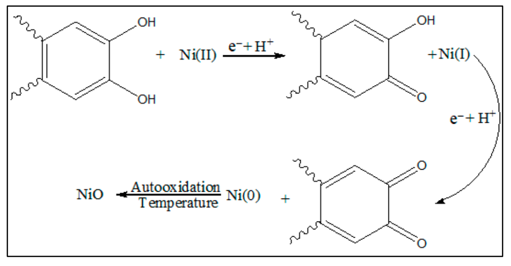
2.1. Synthesis of NiO NPs
2.2. Characterization
2.3. Antioxidant Assay
2.4. Antibacterial Assay
3. Results
3.1. Characterization of NiO NPs
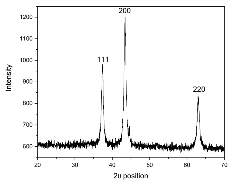
3.1.1. XRD Analysis
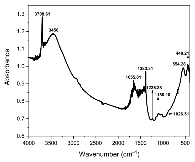
3.1.2. FTIR Analysis
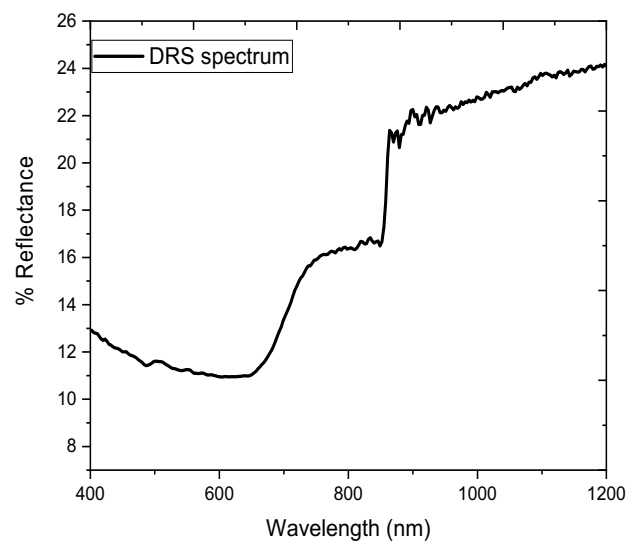
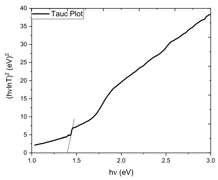
3.1.3. DRS Analysis
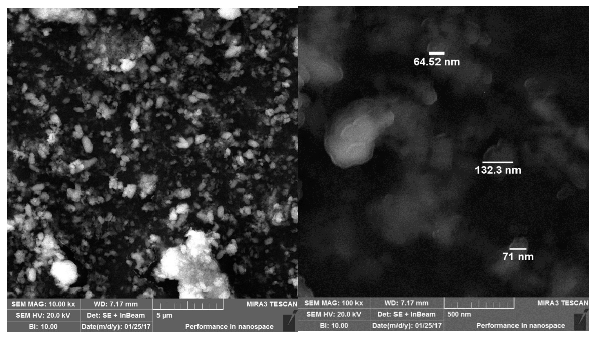
3.1.4. SEM Analysis
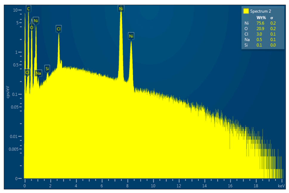
3.1.5. EDX Analysis
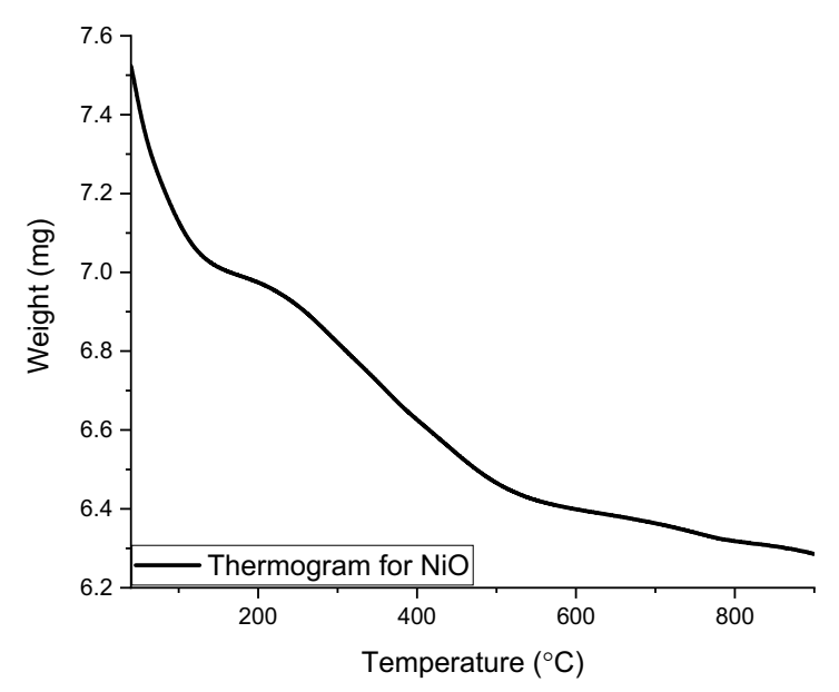
3.1.6. TGA Analysis
3.2. Biological Activities
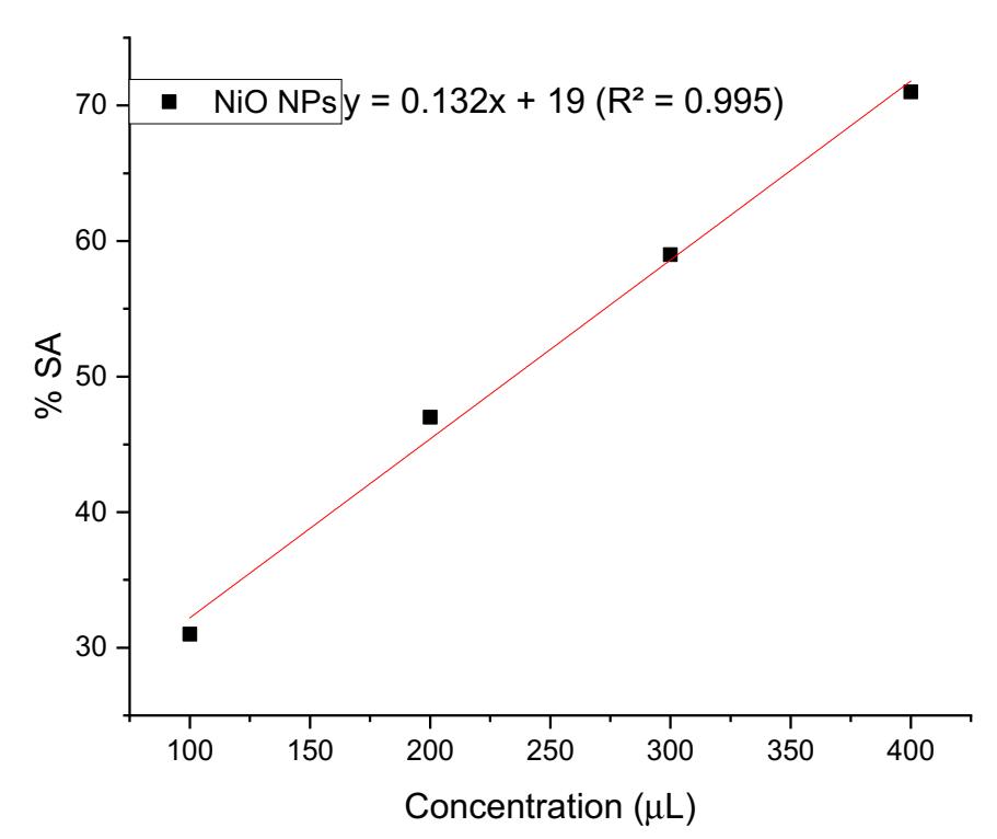
3.2.1. Antioxidant Activity
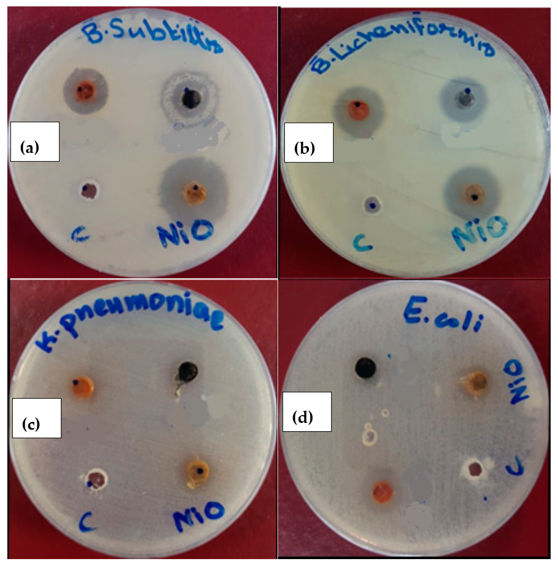
3.2.2. Bactericidal Activity
4. Conclusions
Author Contributions
Funding
Data Availability Statement
Acknowledgments
Conflicts of Interest
References
- Din, M.I.; Nabi, A.G.; Rani, A.; Aihetasham, A.; Mukhtar, M. Single step green synthesis of stable nickel and nickel oxide nanoparticles from Calotropis gigantea: Catalytic and antimicrobial potentials. Environ. Nanotechnol. Monit. Manag. 2018, 9, 29–36. [Google Scholar] [CrossRef]
- Chen, Z.; Wang, Q.; Zhang, Z.; Lei, H. Preparation and properties of antibacterial fluorinated acrylic emulsion. React. Funct. Polym. 2021, 163, 104901. [Google Scholar] [CrossRef]
- Hafeez, M.; Shamim, W.; Ehsan, R.; ul Abdin, Z.; Din, S.U.; Haq, S.; Khan, A.; Shahida, S.; Hameed, U. Structural and biological investigation of biogenically synthesized titanium dioxide nanoparticles: Calcination and characterization. Microsc. Res. Tech. 2021, 84, 2372–2380. [Google Scholar] [CrossRef] [PubMed]
- Wang, X.D.; Wei, W.; Wang, P.F.; Tang, Y.T.; Deng, R.C.; Li, B.; Zhou, S.S.; Zhang, J.W.; Zhang, L.; Xiao, Z.P.; et al. Novel 3-arylfuran-2(5H)-one-fluoroquinolone hybrid: Design, synthesis and evaluation as antibacterial agent. Bioorg. Med. Chem. 2014, 22, 3620–3628. [Google Scholar] [CrossRef]
- Haq, S.; Rehman, W.; Waseem, M.; Meynen, V.; Awan, S.U.; Saeed, S.; Iqbal, N. Fabrication of pure and moxifloxacin functionalized silver oxide nanoparticles for photocatalytic and antimicrobial activity. J. Photochem. Photobiol. B Biol. 2018, 186, 116–124. [Google Scholar] [CrossRef]
- Stankic, S.; Suman, S.; Haque, F.; Vidic, J. Pure and multi metal oxide nanoparticles: Synthesis, antibacterial and cytotoxic properties. J. Nanobiotechnol. 2016, 14, 73. [Google Scholar] [CrossRef] [Green Version]
- Shah, A.; Haq, S.; Rehman, W.; Muhammad, W.; Shoukat, S.; Rehman, M. ur Photocatalytic and antibacterial activities of Paeonia emodi mediated silver oxide nanoparticles. Mater. Res. Express 2019, 6, 045045. [Google Scholar] [CrossRef]
- Xiao, Z.P.; Wei, W.; Wang, P.F.; Shi, W.K.; Zhu, N.; Xie, M.Q.; Sun, Y.W.; Li, L.X.; Xie, Y.X.; Zhu, L.S.; et al. Synthesis and evaluation of new tyrosyl-tRNA synthetase inhibitors as antibacterial agents based on a N2-(arylacetyl)glycinanilide scaffold. Eur. J. Med. Chem. 2015, 102, 631–638. [Google Scholar] [CrossRef]
- Zorkipli, N.N.M.; Kaus, N.H.M.; Mohamad, A.A. Synthesis of NiO Nanoparticles through Sol-gel Method. Procedia Chem. 2016, 19, 626–631. [Google Scholar] [CrossRef] [Green Version]
- Rajith Kumar, C.R.; Betageri, V.S.; Nagaraju, G.; Pujar, G.H.; Suma, B.P.; Latha, M.S. Photocatalytic, nitrite sensing and antibacterial studies of facile bio-synthesized nickel oxide nanoparticles. J. Sci. Adv. Mater. Devices 2020, 5, 48–55. [Google Scholar] [CrossRef]
- Wei, W.; Shi, W.K.; Wang, P.F.; Zeng, X.T.; Li, P.; Zhang, J.R.; Li, Q.; Tang, Z.P.; Peng, J.; Wu, L.Z.; et al. Adenosine analogs as inhibitors of tyrosyl-tRNA synthetase: Design, synthesis and antibacterial evaluation. Bioorg. Med. Chem. 2015, 23, 6602–6611. [Google Scholar] [CrossRef]
- Haq, S.; Dildar, S.; Ali, M.B.; Mezni, A.; Hedfi, A.; Shahzad, M.I.; Shahzad, N.; Shah, A. Antimicrobial and antioxidant properties of biosynthesized of NiO nanoparticles using Raphanus sativus (R. sativus) extract. Mater. Res. Express 2021, 8, 055006. [Google Scholar] [CrossRef]
- Pant, S. Buxus wallichiana L., a multipurpose Himalayan tree in peril. Int. J. Biodivers. Conserv. 2015, 3, 175–177. [Google Scholar]
- Perachiselvi, M.; Jenson Samraj, J.; Bagavathy, S.; Feiona, T.A.; Krishnaveni, P.; Laksmi, E.P.; Swetha, V.; Leema, M.M.; Britto, S.J.; Annadurai, G. Fabrication of Nickel Oxide Nanoparticles for Ntibacterial and Photocatalytic Activity. Res. J. Pharm. Biol. Chem. Sci. 2018, 4, 749–760. [Google Scholar] [CrossRef]
- Haq, S.; Abbasi, F.; Ben Ali, M.; Hedfi, A.; Mezni, A.; Rehman, W.; Waseem, M.; Khan, A.R.; Shaheen, H. Green synthesis of cobalt oxide nanoparticles and the effect of annealing temperature on their physiochemical and biological properties. Mater. Res. Express 2021, 8, 075009. [Google Scholar] [CrossRef]
- Elhaddad, E.; Rehman, W.; Waseem, M.; Nawaz, M.; Haq, S.; Guo, C.Y. Fabrication of Highly Efficient Bi2Sn2O7/C3N4 Composite with Enhanced Photocatalytic Activity for Degradation of Organic Pollutants. J. Inorg. Organomet. Polym. Mater. 2020, 31, 172–179. [Google Scholar] [CrossRef]
- Lingaraju, K.; Raja Naika, H.; Nagabhushana, H.; Jayanna, K.; Devaraja, S.; Nagaraju, G. Biosynthesis of Nickel oxide Nanoparticles from Euphorbia heterophylla (L.) and their biological application. Arab. J. Chem. 2020, 13, 4712–4719. [Google Scholar] [CrossRef]
- Anand, G.T.; Nithiyavathi, R.; Ramesh, R.; John Sundaram, S.; Kaviyarasu, K. Structural and optical properties of nickel oxide nanoparticles: Investigation of antimicrobial applications. Surf. Interfaces 2020, 18, 100460. [Google Scholar] [CrossRef]
- Abdallah, A.M.; Basma, H.; Awad, R. Preparation, Characterization, and Application of Nickel Oxide Nanoparticles in Glucose and Lactose Biosensors. Mod. Appl. Sci. 2019, 13, 99. [Google Scholar] [CrossRef] [Green Version]
- Ezhilarasi, A.A.; Vijaya, J.J.; Kaviyarasu, K.; Zhang, X.; Kennedy, L.J. Green synthesis of nickel oxide nanoparticles using Solanum trilobatum extract for cytotoxicity, antibacterial and photocatalytic studies. Surf. Interfaces 2020, 20, 100553. [Google Scholar] [CrossRef]
- Shoukat, S.; Haq, S.; Rehman, W.; Waseem, M.; Shahzad, M.I.; Shahzad, N.; Hafeez, M.; Din, S.U.; ul-Abdin, Z.; Shah, A.; et al. Fabrication and Characterization of Zinc Titanate Heterojunction for Adsorption and Photocatalytic Applications. J. Inorg. Organomet. Polym. Mater. 2020, 30, 4944–4953. [Google Scholar] [CrossRef]
- Yuvakkumar, R.; Suresh, J.; Nathanael, A.J.; Sundrarajan, M.; Hong, S.I. Rambutan (Nephelium lappaceum L.) peel extract assisted biomimetic synthesis of nickel oxide nanocrystals. Mater. Lett. 2014, 128, 170–174. [Google Scholar] [CrossRef]
- Sabouri, Z.; Akbari, A.; Hosseini, H.A.; Hashemzadeh, A.; Darroudi, M. Eco-Friendly Biosynthesis of Nickel Oxide Nanoparticles Mediated by Okra Plant Extract and Investigation of Their Photocatalytic, Magnetic, Cytotoxicity, and Antibacterial Properties. J. Clust. Sci. 2019, 30, 1425–1434. [Google Scholar] [CrossRef]
- Abbasi, B.A.; Iqbal, J.; Mahmood, T.; Ahmad, R.; Kanwal, S.; Afridi, S. Plant-mediated synthesis of nickel oxide nanoparticles (NiO) via Geranium wallichianum: Characterization and different biological applications. Mater. Res. Express 2019, 6. [Google Scholar] [CrossRef]
- Hamid, A.; Haq, S.; Ur Rehman, S.; Akhter, K.; Rehman, W.; Waseem, M.; Ud Din, S.; ul-Abdin, Z.; Hafeez, M.; Khan, A.; et al. Calcination temperature-driven antibacterial and antioxidant activities of fumaria indica mediated copper oxide nanoparticles: Characterization. Chem. Pap. 2021, 75, 4189–4198. [Google Scholar] [CrossRef]
- Haq, S.; Ahmad, P.; Khandaker, M.U.; Faruque, M.R.I.; Rehman, W.; Waseem, M.; Din, S.U. Antibacterial, antioxidant and physicochemical investigations of tin dioxide nanoparticles synthesized via microemulsion method. Mater. Res. Express 2021, 8. [Google Scholar] [CrossRef]
- Rehman, F.U.; Mahmood, R.; Ali, M.B.; Hedfi, A.; Mezni, A.; Haq, S.; Din, S.U.; Ehsan, R. Physicochemical, Photocatalytic, Antibacterial, and Antioxidant Screening of Bergenia Ciliata Mediated Nickel Oxide Nanoparticles. Crystal 2021, 11, 1137. [Google Scholar] [CrossRef]
- Srihasam, S.; Thyagarajan, K.; Korivi, M.; Lebaka, V.R.; Mallem, S.P.R. Phytogenic generation of NiO nanoparticles using stevia leaf extract and evaluation of their in-vitro antioxidant and antimicrobial properties. Biomolecules 2020, 10, 89. [Google Scholar] [CrossRef] [Green Version]
- Ramalingam, R.; Hussain, M.; Turabe, U.; Kumar, N.; Deivi, K. Green synthesis, characterization and antibacterial evaluation of electrospun nickel oxide nanofibers. Mater. Lett. 2019, 256, 126616. [Google Scholar] [CrossRef]


| Miller Indices (hkl) | Peak Position Theta Value in Degrees | Interplanar Space (d) 2dsinθ = λ | Lattice Constant a = dhkl (h2 + k2 + l2)1/2 |
|---|---|---|---|
| 111 | 37.31 | 0.24 | 0.41 |
| 200 | 43.43 | 0.22 | 0.44 |
| 220 | 63.01 | 0.29 | 0.81 |
| Planck’s Constant (h) | Velocity of Light (c) | Band Gap Reflectance Edge (λ) | Optical Band Gap Energy (Eg) |
|---|---|---|---|
| 6.626 × 10⁻³⁴ Js | 3 × 10⁸ ms−1 | 645 nm | 1.99 eV |
| Samples Code | Concentration (µL) | % Scavenging Activity | IC50 |
|---|---|---|---|
| 100 | 31 | ||
| NiO NPs | 200 | 47 | 234.84 |
| 300 | 59 | ||
| 400 | 71 |
| Microorganism | Volume of NiO Suspension | Correlation between Utilized Dose and Obtained Data (p = 0.05) | C = Solvent DMSO | ||
|---|---|---|---|---|---|
| 25 μL | 50 μL | 75 μL | |||
| B. subtilis | 10.72 | 17.38 | 23.83 | 0.046 | 00 |
| B. licheniformis | 8.01 | 12.93 | 18.57 | 0.042 | 00 |
| K. pneumoniae | 00 | 01 | 2.08 | 0.036 | 00 |
| E. coli | 2.10 | 4.95 | 8.23 | 0.035 | 00 |
| S. No | Methods | Particle Size (nm) | Activity against Gram-Negative Bacteria | Activity against Gram-Positive Bacteria | References |
| 01 | Green synthesis | 23.21 | 9 mm (E. hermannii) | 21 mm (S. aureus) | [20] |
| 02 | Green synthesis | 20 to 50 | 16 mm (E. coli) | 12 mm (B. subtilis) | [28] |
| 03 | Green synthesis | 20 to 50 | 16 mm (E. coli) | 12 mm (B. subtilis) | [29] |
| 04 | Green synthesis | 89.28 | 8.23 (E. coli) | 23.83 (B. subtilis) | Present study |
Publisher’s Note: MDPI stays neutral with regard to jurisdictional claims in published maps and institutional affiliations. |
© 2022 by the authors. Licensee MDPI, Basel, Switzerland. This article is an open access article distributed under the terms and conditions of the Creative Commons Attribution (CC BY) license (https://creativecommons.org/licenses/by/4.0/).
Share and Cite
Din, S.U.; Iqbal, H.; Haq, S.; Ahmad, P.; Khandaker, M.U.; Elansary, H.O.; Al-Harbi, F.F.; Abdelmohsen, S.A.M.; El-Abedin, T.K.Z. Investigation of the Biological Applications of Biosynthesized Nickel Oxide Nanoparticles Mediated by Buxus wallichiana Extract. Crystals 2022, 12, 146. https://0-doi-org.brum.beds.ac.uk/10.3390/cryst12020146
Din SU, Iqbal H, Haq S, Ahmad P, Khandaker MU, Elansary HO, Al-Harbi FF, Abdelmohsen SAM, El-Abedin TKZ. Investigation of the Biological Applications of Biosynthesized Nickel Oxide Nanoparticles Mediated by Buxus wallichiana Extract. Crystals. 2022; 12(2):146. https://0-doi-org.brum.beds.ac.uk/10.3390/cryst12020146
Chicago/Turabian StyleDin, Salah Ud, Hina Iqbal, Sirajul Haq, Pervaiz Ahmad, Mayeen Uddin Khandaker, Hosam O. Elansary, Fatemah F. Al-Harbi, Shaimaa A. M. Abdelmohsen, and Tarek K. Zin El-Abedin. 2022. "Investigation of the Biological Applications of Biosynthesized Nickel Oxide Nanoparticles Mediated by Buxus wallichiana Extract" Crystals 12, no. 2: 146. https://0-doi-org.brum.beds.ac.uk/10.3390/cryst12020146