Soft X-ray Absorption Spectroscopy Study of Spin Crossover Fe-Compounds: Persistent High Spin Configurations under Soft X-ray Irradiation

 ,
, 
Abstract
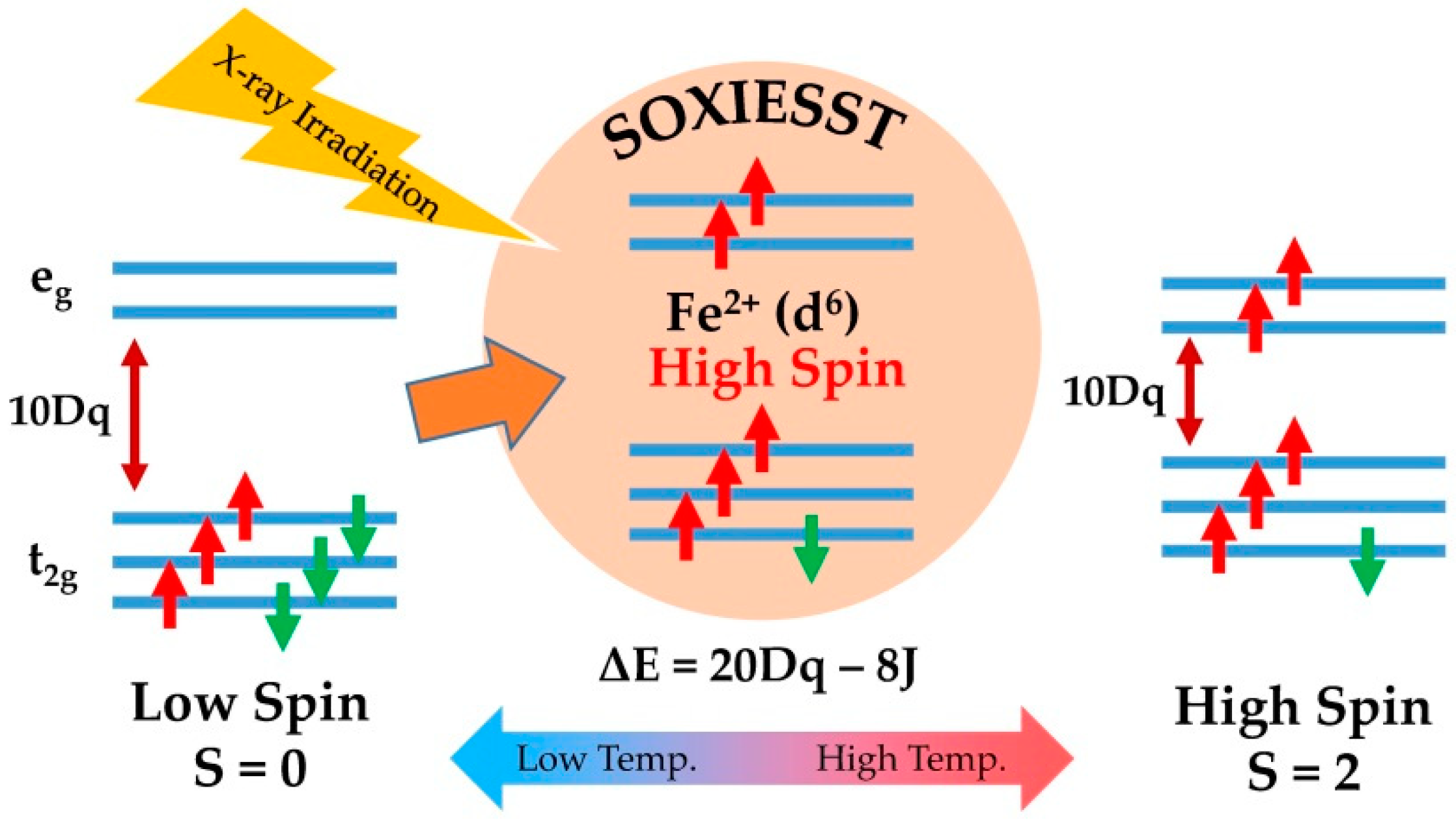
:1. Introduction
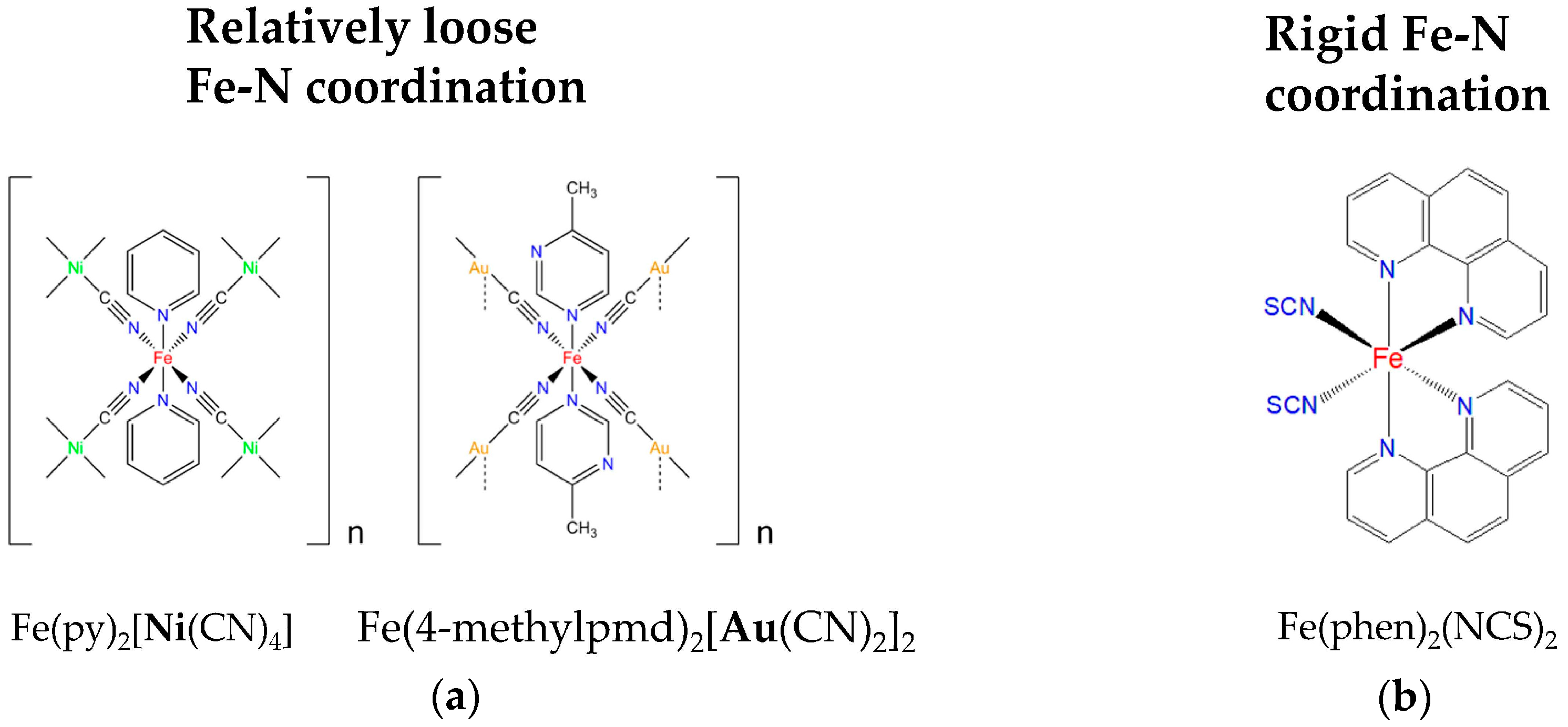
2. Materials and Methods
3. Results and Discussion
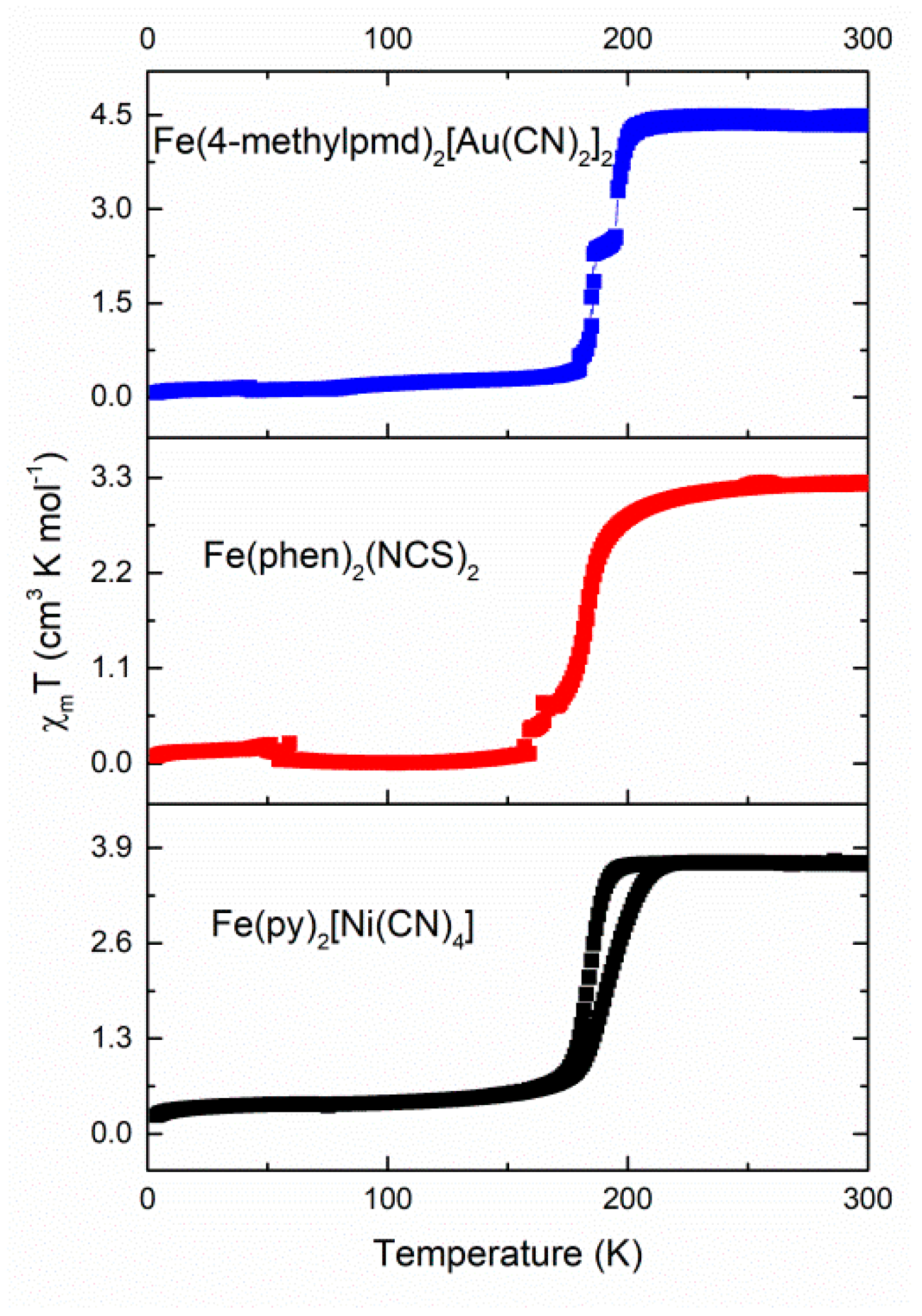
3.1. Magnetic Susceptibility
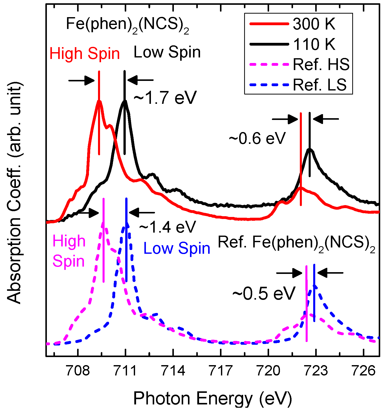
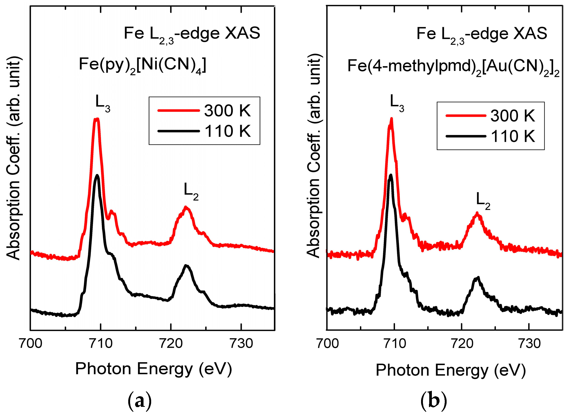
3.2. Soft X-ray Absorption Spectroscopy
4. Conclusions
Supplementary Materials
Author Contributions
Funding
Acknowledgments
Conflicts of Interest
References
- Cambi, L.; SzegzI, L. Über die magnetische Suszeptibilität der komplexen Verbindungen. Berichte Dtsch. Chem. Gesellachaft B 1931, 64, 2591–2598. [Google Scholar] [CrossRef]
- Gütlich, P.; Goodwin, H.A. Spin Crossover in Transition Metal I; Gütlich, P., Goodwin, H.A., Eds.; Springer: Berlin/Heidelberg, Germany, 2004. [Google Scholar] [CrossRef]
- Gütlich, P.; Garcia, Y.; Goodwin, H.A. Spin crossover phenomena in Fe(ii) complexes. Chem. Soc. Rev. 2000, 29, 419–427. [Google Scholar] [CrossRef]
- Real, J.A.; Gaspar, A.B.; Munoz, M.C. Thermal, pressure and light switchable spin-crossover materials. Dalton. Trans. 2005, 2062–2079. [Google Scholar] [CrossRef] [PubMed]
- Murray, K.S. Spin-Crossover Materials; Halcrow, M.A., Ed.; John Wiley & Sons, Ltd.: London, UK, 2013. [Google Scholar]
- Létard, J.-F.; Guionneau, P.; Goux-Capes, L. Towards Spin Crossover Applications. Top. Curr. Chem. 2004, 235, 221–249. [Google Scholar] [CrossRef]
- Marchivie, M.; Guionneau, P.; Howard, J.A.K.; Chastanet, G.; Létard, J.-F.; Goeta, A.E.; Chasseau, D. Structural Characterization of a Photoinduced Molecular Switch. J. Am. Chem. Soc. 2002, 124, 194–195. [Google Scholar] [CrossRef] [PubMed]
- Kitazawa, T.; Gomi, Y.; Takahashi, M.; Takeda, M.; Enomoto, M.; Miyazakib, A.; Enoki, T. Spin-crossover behaviour of the coordination polymer FeII(C5H5N)2NiII(CN)4. J. Mater. Chem. 1996, 6, 119–121. [Google Scholar] [CrossRef]
- Kosone, T.; Tomori, I.; Kanadani, C.; Saito, T.; Mochida, T.; Kitazawa, T. Unprecedented three-step spin-crossover transition in new 2-dimensional coordination polymer {FeII(4-methylpyridine)2[AuI(CN)2]2}. Dalton Trans. 2010, 39, 1719–1721. [Google Scholar] [CrossRef] [PubMed]
- Kosone, T.; Kawasaki, T.; Tomori, I.; Okabayashi, J.; Kitazawa, T. Modification of Cooperativity and Critical Temperatures on a Hofmann-Like Template Structure by Modular Substituent. Inorganics 2017, 5, 55. [Google Scholar] [CrossRef]
- Wu, Z.; Justo, J.F.; da Silva, C.R.S.; de Gironcoli, S.; Wentzcovitch, R.M. Anomalous thermodynamic properties in ferropericlase throughout its spin crossover. Phys. Rev. B 2009, 80. [Google Scholar] [CrossRef]
- Wentzcovitch, R.M.; Justo, J.F.; Wu, Z.; da Silva, C.R.; Yuen, D.A.; Kohlstedt, D. Anomalous compressibility of ferropericlase throughout the iron spin cross-over. Proc. Natl. Acad. Sci. USA 2009, 106, 8447–8452. [Google Scholar] [CrossRef] [PubMed] [Green Version]
- Ksenofontov, V.; Gaspar, A.B.; Gütlich, P. Pressure Effect Studies on Spin Crossover and Valence Tautomeric Systems. In Spin Crossover in Transition Metal III; Gütlich, P., Goodwin, H.A., Eds.; Springer: Berlin/Heidelberg, Germany, 2004. [Google Scholar] [CrossRef]
- Boillot, M.-L.; Zarembowitch, J.; Sour, A. Ligand-Driven Light-Induced Spin Change (LD-LISC): A Promising Photomagnetic Effect. In Spin Crossover in Transition Metal II; Gütlich, P., Goodwin, H.A., Eds.; Springer: Berlin/Heidelberg, Germany, 2004. [Google Scholar] [CrossRef]
- Naggert, H.; Bannwarth, A.; Chemnitz, S.; von Hofe, T.; Quandt, E.; Tuczek, F. First observation of light-induced spin change in vacuum deposited thin films of iron spin crossover complexes. Dalton Trans. 2011, 40, 6364–6366. [Google Scholar] [CrossRef] [PubMed]
- Cannizzo, A.; Milne, C.J.; Consani, C.; Gawelda, W.; Bressler, C.; van Mourik, F.; Chergui, M. Light-induced spin crossover in Fe(II)-based complexes: The full photocycle unraveled by ultrafast optical and X-ray spectroscopies. Coord. Chem. Rev. 2010, 254, 2677–2686. [Google Scholar] [CrossRef]
- Herber, R.; Casson, L.M. Light-Induced Excited-Spin-State Trapping: Evidence from VTFTIR Measurements. Inorg. Chem. 1986, 25, 847–852. [Google Scholar] [CrossRef]
- Hauser, A. Light-Induced Spin Crossover and the High-Spin → Low-Spin Relaxation. In Spin Crossover in Transition Metal II; Gütlich, P., Goodwin, H.A., Eds.; Springer: Berlin/Heidelberg, Germany, 2004. [Google Scholar] [CrossRef]
- Decurtins, S.; Gutlich, P.; Hasselbach, K.M.; Hauser, A.; Spieringt, H. Light-Induced Excited-Spin-State Trapping in Iron(II) Spin-Crossover Systems. Optical Spectroscopic and Magnetic Susceptibility Study. Inorg. Chem. 1985, 24, 2174–2178. [Google Scholar] [CrossRef]
- Hauser, A. Reversibility of light-induced excited spin state trapping in the Fe(ptz)6(BF4)2 and the Zn1-xFex(ptz)6(BF4)2 spin-crossover systems. Chem. Phys. Lett. 1986, 124, 543–548. [Google Scholar] [CrossRef]
- Davesne, V.; Gruber, M.; Miyamachi, T.; Da Costa, V.; Boukari, S.; Scheurer, F.; Joly, L.; Ohresser, P.; Otero, E.; Choueikani, F.; et al. First glimpse of the soft X-ray induced excited spin-state trapping effect dynamics on spin cross-over molecules. J. Chem. Phys. 2013, 139, 074708. [Google Scholar] [CrossRef] [PubMed]
- Collison, D.; Garner, C.D.; McGrath, C.M.; Mosselmans, J.F.W.; Roper, M.D.; Seddon, J.M.W.; Sinn, E.; Young, N.A. Soft X-ray induced excited spin state trapping and soft X-ray photochemistry at the iron L2,3 edge in [Fe(phen)2(NCSe)2] and [Fe(phen)2(NCS)2] (phen = 1,10-phenanthroline). J. Chem. Soc. Dalton Trans. 1997, 22, 4371–4376. [Google Scholar] [CrossRef]
- Bousseksou, A.; Varret, F.; Goiran, M.; Boukheddaden, K.; Tuchagues, J.P. The Spin Crossover Phenomenon Under High Magnetic Field. In Spin Crossover in Transition Metal III; Gütlich, P., Goodwin, H.A., Eds.; Springer: Berlin/Heidelberg, Germany, 2004. [Google Scholar] [CrossRef]
- Miyamachi, T.; Gruber, M.; Davesne, V.; Bowen, M.; Boukari, S.; Joly, L.; Scheurer, F.; Rogez, G.; Yamada, T.K.; Ohresser, P.; et al. Robust spin crossover and memristance across a single molecule. Nat. Commun. 2012, 3, 938. [Google Scholar] [CrossRef] [PubMed] [Green Version]
- Cotton, F.; Wilkinson, G.; Gaus, P. Basic Inorganic Chemistry, 3rd ed.; Cotton, F., Wilkinson, G., Gaus, P., Eds.; Wiley: New York, NY, USA, 1995. [Google Scholar] [CrossRef]
- Wäckerlin, C.; Donati, F.; Singha, A.; Baltic, R.; Decurtins, S.; Liu, S.-X.; Rusponi, S.; Dreiser, J. Excited Spin-State Trapping in Spin Crossover Complexes on Ferroelectric Substrates. J. Phys. Chem. C 2018, 122, 8202–8208. [Google Scholar] [CrossRef]
- Davesne, V. Organic Spintronics: An Investigation on Spin-Crossover Complexes from Isolated Molecules to the Device. Ph.D. Thesis, Physique et Chimie Physique Université de strasbourg, Strasbourg, France, 2013. [Google Scholar]
- Kitazawa, T.; Kawasaki, T.; Shiina, H.; Takahashi, M. Mössbauer Spectroscopic Study on Hofmann-like Coordination Polymer Fe(4-Clpy)2[Ni(CN)4]. Croat. Chem. Acta 2016, 89, 111–115. [Google Scholar] [CrossRef]
- Kitazawa, T.; Kishida, T.; Kawasaki, T.; Takahashi, M. Spin crossover behaviour in Hofmann-like coordination polymer Fe(py)2[Pd(CN)4] with 57Fe Mössbauer spectra. Hyperfine Interact. 2017, 238. [Google Scholar] [CrossRef]
- Gütlich, P.; Goodwin, H.A. Spin Crossover—An Overall Perspective. Top. Curr. Chem. 2004, 233, 1–47. [Google Scholar] [CrossRef]
- Ueki, Y.; Okabayashi, J.; Kitazawa, T. Guest Molecule Inserted Spin Crossover Complexes: Fe[4-(3-Pentyl)pyridine]2[Au(CN)2]2·Guest. Chem. Lett. 2017, 46, 747–749. [Google Scholar] [CrossRef]
- Moulin, C.C.D.; Rudolf, P.; Flank, A.M.; Chen, C.T. Spin Transition Evidenced by Soft X-ray Absorption Spectroscopy. J. Phys. Chem. 1992, 96, 6196–6198. [Google Scholar] [CrossRef]
- Moulin, C.C.D.; Flank, A.M.; Rudolf, P.; Chen, C.T. Electronic Structure from Iron L-edge Spectroscopy: An Example of Spin Transition Evidenced by Soft X-ray Absorption Spectroscopy. Jpn. J. Appl. Phys. 1993, 32 (Suppl. 2), 308–310. [Google Scholar] [CrossRef]
- Briois, V.; Moulin, C.C.D.; Sainctavit, P.; Brouder, C.; Flank, A.-M. Full Multiple Scattering and Crystal Field Multiplet Calculations Performed on the Spin Transition FeII(phen)2(NCS)2 Complex at the Iron K and L2,3 X-ray Absorption Edges. J. Am. Chem. Soc. 1995, 117, 1019–1026. [Google Scholar] [CrossRef]
- Kipgen, L.; Bernien, M.; Ossinger, S.; Nickel, F.; Britton, A.J.; Arruda, L.M.; Naggert, H.; Luo, C.; Lotze, C.; Ryll, H.; et al. Evolution of cooperativity in the spin transition of an iron(II) complex on a graphite surface. Nat. Commun. 2018, 9, 2984. [Google Scholar] [CrossRef] [PubMed]
- Davesne, V.; Gruber, M.; Studniarek, M.; Doh, W.H.; Zafeiratos, S.; Joly, L.; Sirotti, F.; Silly, M.G.; Gaspar, A.B.; Real, J.A.; et al. Hysteresis and change of transition temperature in thin films of Fe{[Me2Pyrz]3BH}2, a new sublimable spin-crossover molecule. J. Chem. Phys. 2015, 142, 194702. [Google Scholar] [CrossRef] [PubMed]
- Madeja, K. Darstellung und magnetisches Verhalten von [Fe(phen)2X2]-Komplexen. Chemické Zvesti 1965, 19, 186–191. [Google Scholar]
- Baker, W.A., Jr.; Bobonich, H.M. Magnetic Properties of Some High-Spin Complexes of Iron(II). Inorg. Chem. 1964, 3, 1184–1188. [Google Scholar] [CrossRef]
- Blundell, S. Magnetism in Condensed Matter; Oxford University Press Inc.: New York, NY, USA, 2001. [Google Scholar]
- Lee, J.-J.; Sheu, H.; Lee, C.-R.; Chen, J.-M.; Lee, J.-F.; Wang, C.-C.; Huang, C.-H.; Wang, Y. X-ray Absorption Spectroscopic Studies on Light-Induced Excited Spin State Trapping of an Fe(II) Complex. J. Am. Chem. Soc. 2000, 122, 5742–5747. [Google Scholar] [CrossRef]


© 2018 by the authors. Licensee MDPI, Basel, Switzerland. This article is an open access article distributed under the terms and conditions of the Creative Commons Attribution (CC BY) license (http://creativecommons.org/licenses/by/4.0/).
Share and Cite
Mohamed, A.Y.; Lee, M.; Kitase, K.; Kitazawa, T.; Kim, J.-Y.; Cho, D.-Y. Soft X-ray Absorption Spectroscopy Study of Spin Crossover Fe-Compounds: Persistent High Spin Configurations under Soft X-ray Irradiation. Crystals 2018, 8, 433. https://0-doi-org.brum.beds.ac.uk/10.3390/cryst8110433
Mohamed AY, Lee M, Kitase K, Kitazawa T, Kim J-Y, Cho D-Y. Soft X-ray Absorption Spectroscopy Study of Spin Crossover Fe-Compounds: Persistent High Spin Configurations under Soft X-ray Irradiation. Crystals. 2018; 8(11):433. https://0-doi-org.brum.beds.ac.uk/10.3390/cryst8110433
Chicago/Turabian StyleMohamed, Ahmed Yousef, Minji Lee, Kosuke Kitase, Takafumi Kitazawa, Jae-Young Kim, and Deok-Yong Cho. 2018. "Soft X-ray Absorption Spectroscopy Study of Spin Crossover Fe-Compounds: Persistent High Spin Configurations under Soft X-ray Irradiation" Crystals 8, no. 11: 433. https://0-doi-org.brum.beds.ac.uk/10.3390/cryst8110433