Choosing the Method of Crystallization to Obtain Optimal Results


Affiliation Computational and Systems Medicine, Department of Surgery and Cancer, Faculty of Medicine, Imperial College London, London SW7 2AZ, UK
*
Author to whom correspondence should be addressed.
Crystals 2019, 9(2), 106; https://0-doi-org.brum.beds.ac.uk/10.3390/cryst9020106
Submission received: 21 January 2019
/
Revised: 11 February 2019
/
Accepted: 16 February 2019
/
Published: 19 February 2019
(This article belongs to the Special Issue Advanced Technologies for Analysis, Directed Optimization and Delivery of Protein Crystallization)
Abstract
:Anyone who has ever attempted to crystallise a protein or other biological macromolecule has encountered at least one, if not all of the following scenarios: No crystals at all, tiny low quality crystals; phase separation; amorphous precipitate and the most frustrating; large, beautiful crystals that do not diffract at all. In this paper we review a number of simple ways to overcome such problems, which have worked well in our hands and in other laboratories. It brings together information that has been dispersed in various publications and lectures over the years and includes further information that has not been previously published.
1. Introduction
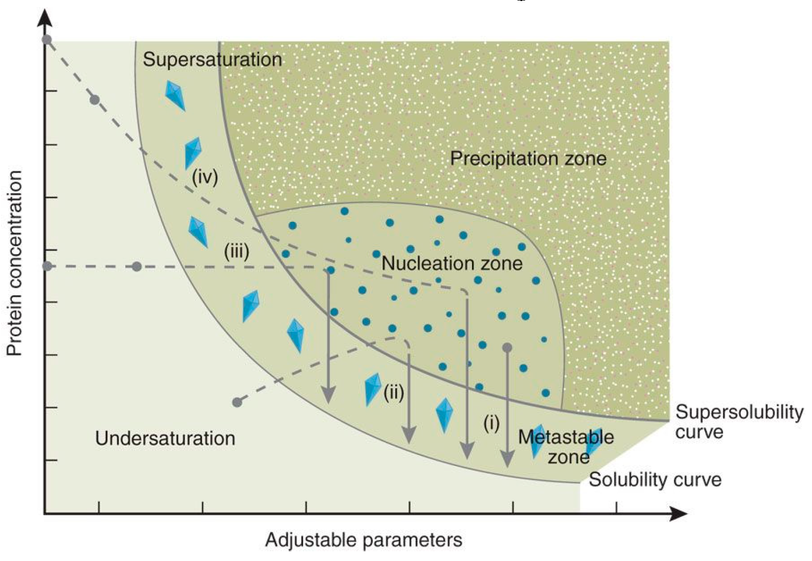
The crystallization phase diagram forms the basis for crystal growth and it is central to every crystallization experiment. Obtaining a phase diagram is commonly done after finding a hit during the screening experiments by dispensing (manually or by a robot) 10–24 trials varying the protein and precipitant concentrations in steps. Alternatively, the pH, temperature, or another parameter to which solubility is sensitive can be varied. These experiments provide the super-solubility curve, which separates the nucleation conditions from the metastable conditions which are ideal for growth [1].
Crystallization methods follow different routes on the phase diagram. In the case of the batch method, the drops reach super-saturation instantaneously upon mixing the protein and crystallizing solutions. On the other hand, in vapour diffusion, both free interface diffusion and dialysis trials, the protein solution is under-saturated to start with, and attains super-saturation on equilibrating with the crystallizing solutions over the course of the trial (Figure 1).
There are various outcomes when setting up crystallization trials:
- Obtaining no crystals at all.
- Obtaining showers of small crystals which are not suitable for diffraction.
- Obtaining large crystals or twinned crystals that do not diffract.
- Obtaining reasonable crystals that require improvement.
- Obtaining good crystals that are unreproducible or difficult to repeat
It is well known that sample preparation, and its quality, have a crucial impact on the crystallization results. Hence, prior to setting up crystallization trials, it is essential for the protein sample to be as pure as possible (e.g., [3]), to be soluble, and to be stable ([4,5,6] and references within). A useful approach for establishing the optimal buffer for protein stability is to test a variety of buffer conditions using a thermo-stability assay [7].
Choosing which crystallization method to use depends on the problem, and in this paper, we review a number of simple ways to overcome such problems.
2. Obtaining No Crystals or Crystals that Do Not Diffract in the Screening Stage of Crystallization
2.1. Adopting A Variety of Crystallization Methods
The different crystallization methods lead to the formation of crystals via different routes, as described in Section 1 above. Vapour diffusion and micro-batch are the most common methods. In the case of a micro-batch, supersaturation is achieved upon mixing, while vapour diffusion is a dynamic system in which a reservoir acts to gradually concentrate the crystallization drop, thus the change in concentration may lead to crystallization. Baldock et al. reported a comparison of screening, using vapour diffusion and variations of the microbatch method thereby concluding that both methods should be applied to ensure all possibilities are covered [8]. Since that study, a variety of new methods for screening have been established, the most common being microfluidics and chips, which in some laboratories are preferred to microbatch, and even vapour diffusion. The first microfluidic device was developed by Hansen et al. for high-throughput screening of protein crystallization. It applied a miniaturized free interface diffusion technique [9]. Following that, more advanced microfluidic systems have been developed [10,11,12].
2.2. Concentration of Drops that Would Remain Clear Indefinitely
Drops, set up for screening experiments, that remain clear after a two-week incubation period, are mostly ignored and considered a ‘dead end’. This is because the protein solution is either under-saturated or in a metastable condition. These clear drops can be utilized in a hanging drop vapour diffusion set up using Easy Xtal crystallization plates. The screw caps, which seal the well of an Easy Xtal tray, can be unsealed by twisting them for a desired period and resealing them again. An under-saturated/metastable trial can thus be driven to supersaturation, resulting in nucleation and subsequent crystal growth Khurshid et al reported 11 new hits on 3 proteins tested [13].
This technique can also be used for optimisation as reported by Govada and Chayen, who obtained better diffracting crystals of the cardiac Myosin Binding Protein-C with a resolution limit of up to 1.5Å compared to 3.2 Å using conventional techniques, which led to the determination of its structure [14].
3. Obtaining Showers of Small Crystals
It is often the case that a large number of very small useless crystals appear in the trial. This can occur because the crystallization process is taking place too rapidly [15] or the crystallization solution contains dust and other random particles that serve as unintended nucleation sites.
There is a variety of ways to improve the quality of such crystals.
3.1. Rigorous Filtration
It is standard practice to filter the sample solution though a 0.2 µm mesh size filter before setting up the crystallization trials, but often this is not sufficient and filtration needs to be done more rigorously. Filtration is relevant to all methods of crystallization at the optimization stage. Filtration of a protein sample with or without its crystallizing agents, through filters of different sizes (e.g., from 0.1 µm to 100KDa filters), can determine the number, the size and quality of the resulting crystals. The filtration can remove unwanted material and particles such as dust, protein aggregates and fungi thereby reducing the numbers of crystals from many useless ones to a few single diffracting ones and increasing the reproducibility of the experiments. Filtration, done prior to setting up the experiments, does not necessitate any change to the crystallization method, the vessel or the conditions. It can, therefore, be used as a one-step optimization strategy when numerous small worthless crystals, twinned crystals, or even precipitates are formed and no improvement is obtained by conventional means. For example, fine-tuning the concentrations of the protein and crystallizing agents, inserting additives, changing pH or temperature. Rigorous filtration is also very useful after the storage of proteins and is especially valuable in improving the results of seeding and the application of nucleants [16].
3.2. Slowing Down the Crystallization Process
A means to improving the size and quality of crystals, which appear as showers of small crystals, twinned crystals or even a crystalline precipitate, is done by inserting an oil barrier over the reservoir of vapour diffusion hanging drops or sitting drops trials. The barrier affects the rate of equilibration. The speed of equilibration depends on the ratio of paraffin to silicon oils and on the thickness of the oil layer. Using Linbro or the Qiagen Easy X-tal plates a layer of 250 µL of a mixture of 1:1 paraffin and silicon oils works well in most cases. The trials are set up in the same conditions in the control and in trials containing the oil barrier, the only difference is the presence of the layer of oil above the reservoirs. The limitation of this technique is that it cannot be used with PEG or MPD concentrations above 13%, but it is very effective at concentrations below 13%, and at all concentrations of all salts [17]. In trials containing an oil barrier, crystals require longer periods (e.g., 8–10 days compared to 12–24 h) to grow to full size, but their quality is more improved. One example are the crystals of lobster apocrustacyanin C1 that are grown in trials with an oil barrier, which diffract to higher resolution, compared to the controls (1.3Å compared to 2.1Å)
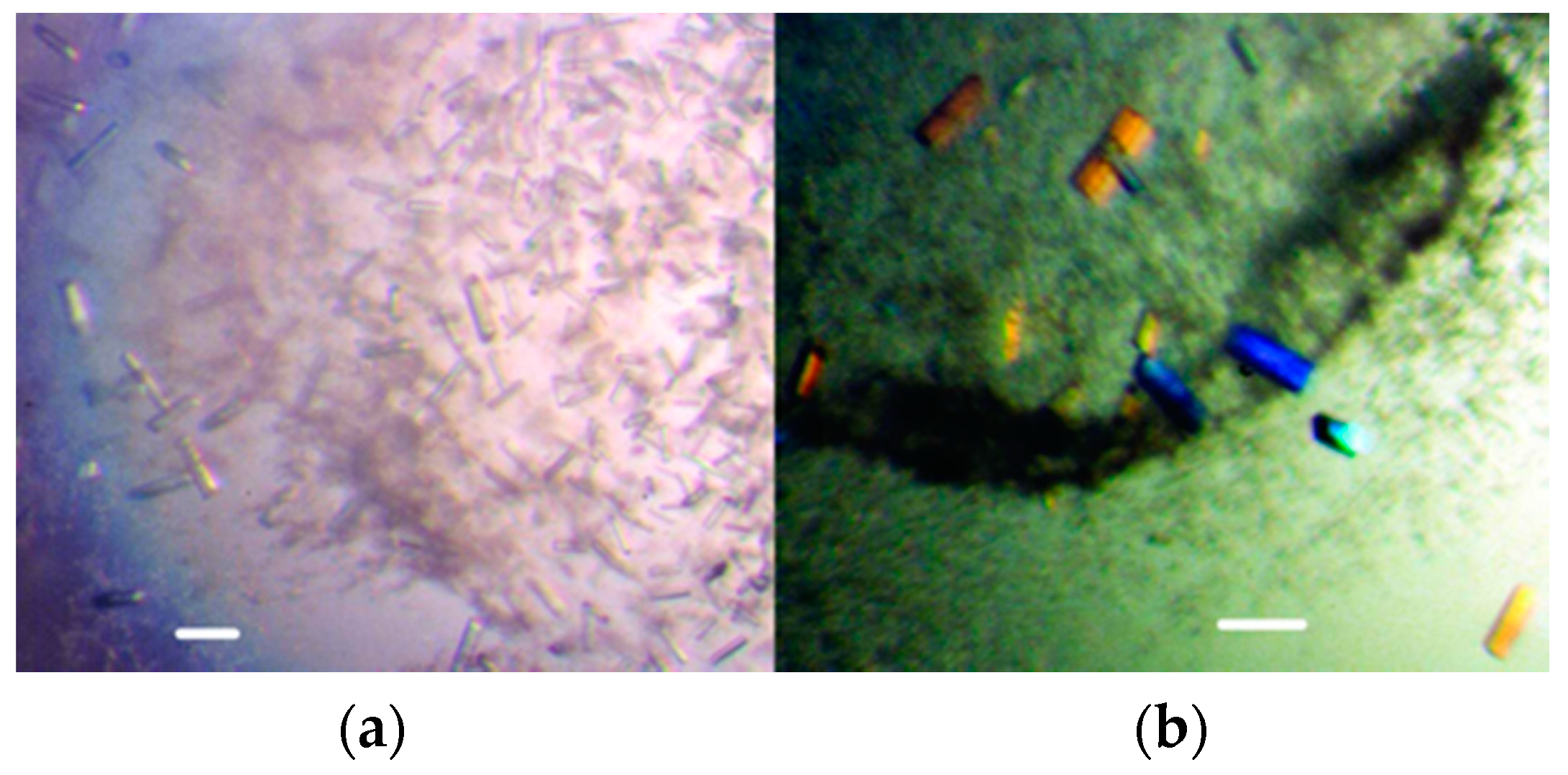
Further examples where placing an oil barrier over the reservoir dramatically reduced the nucleation, were the rat liver ribosomal P2 protein thereby resulting in higher diffracting crystals, with a resolution limit of 2.4 Å (Figure 2) [18] and GH42 intracellular beta-galactosidase diffracting to 2.45Å [19].
At the moment, such trials can only be done manually, but an oil barrier technique, which is miniaturized and automated, will soon be published.
3.3. Separation of the Nucleation and Growth Stages of Crystallization
Volume Adjustments during Crystallization
It is well documented that nucleation requires different condition to those of growth, therefore methods that separate the phases of nucleation and growth can lead to remarkable results.
One way of achieving such separation, is by dilution of trials, in order to transfer the system from nucleation to metastable conditions before nucleation becomes excessive. This is an alternative means of achieving a similar outcome to seeding, but without the need to handle crystals or nucleants. When the clusters of non-diffracting crystals, which cannot be improved by conventional fine-tuning of the crystallisation parameters are produced, a working phase diagram that provides the super-solubility curve can be generated (either manually or using a robot) (Figure 1).
The experiments related to microbatch were reported by Saridakis et al back in 1994. The Authors have established a working phase diagram for the enzyme carboxypeptidase G2, with pH and temperature being kept constant, while varying the concentrations of the protein and precipitant. The working phase diagram produced the super-solubility curve and established the conditions of nucleation (conditions that would produce crystals spontaneously). Crystallization trials were set up at these conditions, using a crystallization robot and at various time intervals after the set-up of the experiments, the robot was programmed to add a buffer or protein solution resulting in the dilution of the trials. Single diffracting crystals were routinely attained, equivalent to the best, very rarely without using the dilution procedure [20,21].
In the case of vapour diffusion, the crystallisation drops were not physically diluted, but coverslips holding hanging drops were incubated over reservoir solutions that would give many small crystals. At time intervals (before crystals are visible), the coverslips are transferred over reservoirs, containing lower precipitant concentrations that would normally yield clear drops [22]. This technique has produced significant improvement in crystal order of a number of proteins e.g., *MtCM (Figure 3) [23] and most remarkably, crystals of C-phycocyanin, which reproducibly diffracted to 1.45 Å, compared to 2 Å, using standard optimisation techniques [24]. This resolution was the highest ever obtained for this protein.
4. The Application of Light-Scattering Techniques
The short-comings of the volume adjustment and transfer techniques, using any crystallization method, is that one does not know when nucleation takes place. It can only be estimated by reference to the time that it took to see the first crystals. The most effective moment to intervene with a crystallisation experiment is soon after the formation of the first critical size nuclei, which will eventually form the crystal. By the time crystals are observed under the microscope, it is too late to act, as nuclei and crystals have already been formed [25].
A means to pinpoint the appropriate time for transfer/dilution or arrest of nucleation, is by using dynamic light scattering (DLS) and static light scattering. DLS offers a size resolution of particles in optically transparent aqueous samples, some three orders of magnitude below an optical microscope. It is therefore a useful tool for an early, non-invasive, in situ observation of a crystallisation event, before it becomes visible with a light microscope [21,26,27,28].
Light scattering was applied to separate nucleation and growth in the crystallization of lysozyme by Rosenberger et al. who successfully limited the number of crystals that were grown [29]. The procedure involved scintillation, and required 50–100 µL of a sample, which was acceptable at the time, but the procedure needed to be improved and miniaturised for application to other proteins. Almost 10 years later, Saridakis et al. used DLS to monitor the crystallisation of proteins that were mixed with their crystallising agents in microbatch, with the aim of getting an indication as to when to dilute the trial in order to lead it out of the nucleation phase and into the growth phase. This was achieved using a DLS-apparatus (Made by RiNA GmbH, Berlin, Germany), which is able to measure a crystallisation trial as it takes place in standard cuvettes containing 10–20 µL. When the DLS spectrum showed a change in the size-distribution profile of species in the solution, the trial was diluted with buffer to metastable conditions, leading to the growth of fewer and larger crystals, compared to crystals grown in control solutions that were left undisturbed [Patent of Chayen, Dieckmann and Dierks [30].
In the case of vapour diffusion, Collingthworth et al. applied static light scattering to detect a change in the profile of crystallising solutions. The drops were evaporated using nitrogen gas flow and the evaporation was arrested when the light-scattering sensor detected aggregation [31]. Varying the evaporation rate of the crystallisation solutions resulted in the formation of fewer, larger crystals than those obtained in controls.
Both dynamic and static light-scattering techniques have more recently been adapted for automated routine use in a micro-batch, vapour diffusion, and other crystallisation set-ups. Transfer from nucleation to metastable conditions can now be achieved in nanoscale and picolitre volumes, by changing parameters such as pH, temperature, humidity, and other parameters, as well as the protein and precipitant concentrations [32]. Recent advances in DLS technology are also able to provide new and detailed insights of two-step crystal nucleation mechanisms [33].
5. Summary
This article highlights the progress that has gradually been made in the design of methods, by describing a selection of the original methods some, which have led to practices that are much improved technically, but are still based on the same principles and ideas. The techniques have been miniaturised and automated to nanoscale by using robotics, special crystallisation trays, microfluidics, and micro-chips, thereby also increasing the reproducibility of the results. Moreover, combinations of techniques, such as vapour batch, gels and oils [34] as well as conbining counter-diffusion and microseeding [35,36] are increasingly being used.
Funding
The UK Engineering and Physical Sciences Research Council.
Acknowledgments
We are grateful to the UK Engineering and Physical Sciences Research Council (EPSRC) for financial support.
Conflicts of Interest
The authors declare no conflict of interest.
References
- Saridakis, E.; Chayen, N.E. Systematic improvement of protein crystals by determining the supersolubility curves of phase diagrams. Biophys. J. 2003, 84, 1218–1222. [Google Scholar] [CrossRef]
- Chayen, N.E.; Saridakis, E. Protein crystallization: From purified protein to diffraction-quality crystal. Nat. Methods 2008, 5, 147–153. [Google Scholar] [CrossRef] [PubMed]
- Giegé, R.; Dock, A.C.; Kern, D.; Lorber, B.; Thierry, J.C.; Moras, D. The role of purification in the crystallization of proteins and nucleic acids. J. Cryst. Growth 1986, 76, 554–561. [Google Scholar] [CrossRef]
- Bergfors, T.M. Protein Crystallization. Techniques, Strategies, and Tips; Bergfors, T.M., Ed.; International University Line: San Diego, CA, USA, 1999. [Google Scholar]
- McPherson, A. Crystallization of Biological Macromolecules; Cold Spring Harbor Lab Press: Cold Spring Harbor, NY, USA, 1999. [Google Scholar]
- Ducruix, A.; Giegé, R. Crystallization of Nucleic Acids and Proteins: A Practical Approach, 2nd ed.; Oxford University Press: Oxford, UK, 1999. [Google Scholar]
- Ericsson, U.B.; Hallberg, B.M.; DeTitta, G.T.; Dekker, N.; Nordlund, P. Thermofluor-based high-throughput stability optimization of proteins for structural studies. Anal. Biochem. 2006, 357, 289–298. [Google Scholar] [CrossRef] [PubMed]
- Baldock, P.; Mills, V.; Stewart, P.S. A comparison of microbatch and vapour diffusion for initial screening of crystallization conditions. J. Cryst. Growth 1996, 168, 170–174. [Google Scholar] [CrossRef]
- Hansen, C.L.; Skordalakes, E.; Berger, J.M.; Quake, S.R. A robust and scalable microfluidic metering method that allows protein crystal growth by free interface diffusion. Proc. Natl. Acad. Sci. USA 2002, 99, 16531. [Google Scholar] [CrossRef] [PubMed]
- Sauter, C.; Dhouib, K.; Lorber, B. From macrofluidics to microfluidics for the crystallization of biological macromolecules. Cryst. Growth Des. 2007, 7, 2247–2250. [Google Scholar] [CrossRef]
- Abdallah, B.G.; Roy-Chowdhury, S.; Fromme, R.; Fromme, P.; Ros, A. Protein Crystallization in an Actuated Microfluidic Nanowell Device. Cryst. Growth Des. 2016, 16, 2074–2082. [Google Scholar] [CrossRef] [Green Version]
- Otálora, F.; Gavira, J.A.; Ng, J.D.; García-Ruiz, J.M. Counterdiffusion methods applied to protein crystallization. Prog. Biophys. Mol. Biol. 2009, 101, 26–37. [Google Scholar] [CrossRef]
- Khurshid, S.; Govada, L.; Chayen, N.E. Dynamic Screening Experiments to Maximize Hits for Crystallization. Cryst. Growth Des. 2007, 7, 2171–2175. [Google Scholar] [CrossRef]
- Govada, L.; Chayen, N.E. Crystallization by Controlled Evaporation Leading to High Resolution Crystals of the C1 Domain of Cardiac Myosin Binding Protein-C (cMyBP-C). Cryst. Growth Des. 2009, 9, 1729–1732. [Google Scholar] [CrossRef]
- Chayen, N.E. The role of oil in macromolecular crystallization. Structure 1997, 5, 1269–1274. [Google Scholar] [CrossRef] [Green Version]
- Chayen, N.E. Rigorous filtration for protein crystallization. J. Appl. Crystallogr. 2009, 42, 743–744. [Google Scholar] [CrossRef]
- Chayen, N.E. A novel technique to control the rate of vapour diffusion, giving larger protein crystals. J. Appl. Crystallogr. 1997, 30, 198–202. [Google Scholar] [CrossRef] [Green Version]
- Mandelman, D.; Gonzalo, P.; Lavergne, J.P.; Corbier, C.; Reboud, J.P.; Haser, R. Crystallization and preliminary X-ray study of an N-terminal fragment of rat liver ribosomal P2 protein. Acta Crystallogr. D 2002, 58, 668–671. [Google Scholar] [CrossRef] [PubMed] [Green Version]
- Solomon, H.V.; Tabachnikov, O.; Feinberg, H.; Govada, L.; Chayen, N.E.; Shoham, Y.; Shoham, G. Crystallization and preliminary crystallographic analysis of GanB, a GH42 intracellular beta-galactosidase from Geobacillus stearothermophilus. Acta Crystallogr. F 2013, 69, 1114–1119. [Google Scholar] [CrossRef]
- Saridakis, E.E.G.; Stewart, P.D.S.; Lloyd, L.F.; Blow, D.M. Phase-Diagram and Dilution Experiments in the Crystallization of Carboxypeptidase-G(2). Acta Crystallogr. D 1994, 50, 293–297. [Google Scholar] [CrossRef] [PubMed]
- Chayen, N.E. Methods for separating nucleation and growth in protein crystallisation. Prog. Biophys. Mol. Biol. 2005, 88, 329–337. [Google Scholar] [CrossRef]
- Saridakis, E.; Chayen, N.E. Improving protein crystal quality by decoupling nucleation and growth in vapor diffusion. Protein Sci. 2000, 9, 755–757. [Google Scholar] [CrossRef]
- Krengel, U.; Dey, R.; Sasso, S.; Okvist, M.; Ramakrishnan, C.; Kast, P. Preliminary X-ray crystallographic analysis of the secreted chorismate mutase from Mycobacterium tuberculosis: A tricky crystallization problem solved. Acta Crystallogr. Sect. F Struct. Biol. Cryst. Commun. 2006, 62, 441–445. [Google Scholar] [CrossRef]
- Nield, J.; Rizkallah, P.; Barber, J.; Chayen, N.E. The 1.45 Angstrom three-dimensional structure of C-phycocyanin from the thermophilic cyanobacterium Synechococcus elongatus. J. Struct. Biol. 2003, 141, 149–155. [Google Scholar] [CrossRef]
- Saridakis, E.; Dierks, K.; Moreno, A.; Dieckmann, M.W.M.; Chayen, N.E. Separating nucleation and growth in protein crystallization using dynamic light scattering. Acta Crystallogr. D 2002, 58, 1597–1600. [Google Scholar] [CrossRef] [PubMed] [Green Version]
- Shmitz, S. An introduction to Dynamic Light Scattering by Macromoelcules. Phys. Today 1990, 44, 66. [Google Scholar] [CrossRef]
- Dierks, K.; Meyer, A.; Einspahr, H.; Betzel, C. Dynamic Light Scattering in Protein Crystallization Droplets: Adaptations for Analysis and Optimization of Crystallization Processes. Cryst. Growth Des. 2008, 8, 1628–1634. [Google Scholar] [CrossRef]
- Proteau, A.; Shi, R.; Cygler, M. Application of Dynamic Light Scattering in Protein Crystallization. Curr. Protoc. Protein Sci. 2010, 61, 10–17. [Google Scholar]
- Rosenberger, F.; Howard, S.B.; Sowers, J.W.; Nyce, T.A. Temperature dependence of protein solubility—Determination and application to crystallization in X-ray capillaries. J. Cryst. Growth 1993, 129, 1–12. [Google Scholar] [CrossRef]
- Chayen, N.E.; Dieckmann, M.; Dierks, K. Use of Dynamic Light Scattering in a Method for Producing Macromolecular Crystals. WO 03080901 A1, 23 May 2002. [Google Scholar]
- Collingsworth, P.; Bray, T.; K Christopher, G. Crystal Growth via Computer Controlled Vapor Diffusion. J. Cryst. Growth 2000, 219, 283–289. [Google Scholar] [CrossRef]
- Meyer, A.; Dierks, K.; Hilterhaus, D.; Klupsch, T.; Muhlig, P.; Kleesiek, J.; Schopflin, R.; Einspahr, H.; Hilgenfeld, R.; Betzel, C. Single-drop optimization of protein crystallization. Acta Crystallogr. Sect. F 2012, 68, 994–998. [Google Scholar] [CrossRef]
- Schubert, R.; Meyer, A.; Baitan, D.; Dierks, K.; Perbandt, M.; Betzel, C. Real-Time Observation of Protein Dense Liquid Cluster Evolution during Nucleation in Protein Crystallization. Cryst. Growth Des. 2017, 17, 954–958. [Google Scholar] [CrossRef]
- Moreno, A.; Saridakis, E.; Chayen, N.E. Combination of oils and gels for enhancing the growth of protein crystals. J. Appl. Crystallogr. 2002, 35, 140–142. [Google Scholar] [CrossRef] [Green Version]
- Gavira, J.A.; Hernandez-Hernandez, M.A.; Gonzalez-Ramirez, L.A.; Briggs, R.A.; Kolek, S.A.; Shaw Stewart, P.D. Combining Counter-Diffusion and Microseeding to Increase the Success Rate in Protein Crystallization. Cryst. Growth Des. 2011, 11, 2122–2126. [Google Scholar] [CrossRef]
- Bolanos-Garcia, V.M.; Chayen, N.E. New directions in conventional methods of protein crystallization. Prog. Biophys. Mol. Biol. 2009, 101, 3–12. [Google Scholar] [CrossRef] [PubMed]
- Moreno, A. Protein Crystallization under the Presence of an Electric Field; MDPI Books: Basel, Switzerland, 2018. [Google Scholar]
- Johansson, L.C.; Stauch, B.; Ishchenko, A.; Cherezov, V. A Bright Future for Serial Femtosecond Crystallography with XFELs. Trends Biochem. Sci. 2017, 42, 749–762. [Google Scholar] [CrossRef]
- McSweeney, S.; Fromme, P. Crystallography: Sources of inspiration. Nature 2014, 505, 620–621. [Google Scholar] [CrossRef] [PubMed]
Figure 1.
Phase Diagram illustrating the different routes of attaining supersaturation. (i) Micro-batch trials where instant supersaturation is reached. (ii) Vapour diffusion (iii) Dialysis (iv) free interface diffusion trials. The super-solubility curve separates the conditions under which spontaneous nucleation occurs and the metastable zone, ideal for crystals growth [2]. Reprinted by permission from Springer Nature [Nature Methods] (Protein crystallization: from purified protein to diffraction-quality crystal, Naomi E Chayen, Emmanuel Saridakis) (2008).

Figure 2.
Crystals of an N-terminal fragment of rat liver ribosomal P2 protein with and without an oil barrier. (a) Control (b) with oil barrier [18].
Figure 2.
Crystals of an N-terminal fragment of rat liver ribosomal P2 protein with and without an oil barrier. (a) Control (b) with oil barrier [18].

Figure 3.
*MtCM crystals obtained by transfer from higher to lower precipitant concentration. (a) Control (b) after transfer [23].
Figure 3.
*MtCM crystals obtained by transfer from higher to lower precipitant concentration. (a) Control (b) after transfer [23].

© 2019 by the authors. Licensee MDPI, Basel, Switzerland. This article is an open access article distributed under the terms and conditions of the Creative Commons Attribution (CC BY) license (http://creativecommons.org/licenses/by/4.0/).
Share and Cite
MDPI and ACS Style
Govada, L.; Chayen, N.E. Choosing the Method of Crystallization to Obtain Optimal Results. Crystals 2019, 9, 106. https://0-doi-org.brum.beds.ac.uk/10.3390/cryst9020106
AMA Style
Govada L, Chayen NE. Choosing the Method of Crystallization to Obtain Optimal Results. Crystals. 2019; 9(2):106. https://0-doi-org.brum.beds.ac.uk/10.3390/cryst9020106
Chicago/Turabian StyleGovada, Lata, and Naomi E. Chayen. 2019. "Choosing the Method of Crystallization to Obtain Optimal Results" Crystals 9, no. 2: 106. https://0-doi-org.brum.beds.ac.uk/10.3390/cryst9020106
Note that from the first issue of 2016, this journal uses article numbers instead of page numbers. See further details here.