Review

Promising Anti-Mitochondrial Agents for Overcoming Acquired Drug Resistance in Multiple Myeloma

by
Vanessa Innao
1,
Vincenzo Rizzo
2,
Andrea Gaetano Allegra
1,
Caterina Musolino
1 and
Alessandro Allegra
1,*
1
Division of Hematology, Department of Human Pathology in Adulthood and Childhood “Gaetano Barresi”, University of Messina, 98125 Messina, Italy
2
Department of Clinical and Experimental Medicine, University of Messina, 98125 Messina, Italy
*
Author to whom correspondence should be addressed.
Submission received: 16 December 2020 / Revised: 12 February 2021 / Accepted: 18 February 2021 / Published: 19 February 2021
(This article belongs to the Collection Trends and Advances in Tumor Immunology)

Abstract

:
Multiple myeloma (MM) remains an incurable tumor due to the high rate of relapse that still occurs. Acquired drug resistance represents the most challenging obstacle to the extension of survival and several studies have been conducted to understand the mechanisms of this phenomenon. Mitochondrial pathways have been extensively investigated, demonstrating that cancer cells become resistant to drugs by reprogramming their metabolic assessment. MM cells acquire resistance to proteasome inhibitors (PIs), activating protection programs, such as a reduction in oxidative stress, down-regulating pro-apoptotic, and up-regulating anti-apoptotic signals. Knowledge of the mechanisms through which tumor cells escape control of the immune system and acquire resistance to drugs has led to the creation of new compounds that can restore the response by leading to cell death. In this scenario, based on all literature data available, our review represents the first collection of anti-mitochondrial compounds able to overcome drug resistance in MM. Caspase-independent mechanisms, mainly based on increased oxidative stress, result from 2-methoxyestradiol, Artesunate, ascorbic acid, Dihydroartemisinin, Evodiamine, b-AP15, VLX1570, Erw-ASNase, and TAK-242. Other agents restore PIs’ efficacy through caspase-dependent tools, such as CDDO-Im, NOXA-inhibitors, FTY720, GCS-100, LBH589, a derivative of ellipticine, AT-101, KD5170, SMAC-mimetics, glutaminase-1 (GLS1)-inhibitors, and thenoyltrifluoroacetone. Each of these substances improved the efficacy rates when employed in combination with the most frequently used antimyeloma drugs.

1. Introduction

Multiple myeloma (MM) is a malignant disease caused by plasma cell clonal proliferation in bone marrow, which can cause damage to the kidneys and skeletal fractures, but also involve any organ [1]. The advent of novel drugs has improved the overall survival of myeloma patients. Many experimental studies on innovative treatment strategies, such as vaccines and antagomirs, are proliferating, but the relapse rates remain unacceptably high [2,3]. In real life, combinations of proteasome inhibitors (PIs), immunomodulators, monoclonal antibodies, and anti-B-cell maturation antigen (anti-BCMA) represent the best treatment, but several prognostic factors impact the response rates of these patients. Three-drug combinations, using lenalidomide as the back bone, with carfilzomib (CFZ), ixazomib (Ixa), elotuzumab (Elo), and daratumumab (Dara), are widely used across the world in clinical practice, especially in relapsed/refractory multiple myeloma (RRMM) patients post-first treatment lines with bortezomib (BTZ) [4]. By improving the toxicity profile and effectiveness rates of first-generation compounds, these therapeutic regimes currently represent the best strategies for MM patients, significantly prolonging progression-free survival and overall survival [5]. In addition, the increasing use of antibiotic, antimycotic, and anti-viral prophylaxis, and growth factors, with awareness, and the more serious control of cardiovascular and neurological drugs’ collateral effects have also allowed infective, hematological, cardiological, and ocular complications related to all classes of drugs or some of them to be reduced [6]. Furthermore, infusion/hypersensitivity-related reactions (I/HRRs) to monoclonal antibodies (moAbs) are well-managed by the combination of H1 and H2 antihistamines, with steroids and paracetamol, used as pre-medication in the recommended premeditation schemes.
However, despite improvements in response rates, patients continue to die, and MM is still an unhealthy disease, acquiring aggressivity over the years, with extra-medullar localizations, also in the central nervous system (CNS) or transforming into plasma cellular leukemia. Therefore, in trying to outline the best prognostic drivers of disease since the onset, scientists have identified the minimal residual disease (MRD) negativity as the best marker of a good prognosis [7]. In this scenario, many scientists have focused their studies on intracellular and molecular mechanisms underlying the inadequate response of aberrant plasma cells to the most commonly used drugs. The high proteosynthetic activity of clonal plasma cells was identified as a nodal point in the development of PIs. In fact, the proteasome not only represents an active intracellular complex in protein degradation, but is also responsible for maintaining normal cellular functions. The control system of protein degradation represents an important Achilles heel in many tumor cells. Proteolysis partially begins in cytosol, where the proteins to be degraded are marked by ubiquitins, which are driven within the proteasome. Here, they are cut into small polypeptides or amino acids. Inhibition of the proteasome leads to cellular death, due to the accumulation of ubiquitinated, destructed, or malfunctioning proteins, by determining the development of a toxic intracellular environment, causing antiproliferative and pro-apoptotic effects. In some cell lines, the stabilization of p53, resulting from reduced proteolytic activity, may induce apoptosis. This capability, combined with antiangiogenic and antiproliferative activities, makes PIs potential anticancer agents. However, the prolonged use of PIs leads to adaptation by tumoral cells, because they tend to offset the blockade of the ubiquitin-proteasome system (UPS) using other proteolytic routes.
The UPS has been widely studied, but there are few studies on the stress- or chemical-induced mitochondrial effects, such as cell damage caused by chemotherapy. In this regard, Shadel et al. identified a new function of mitochondria: In response to chemical stress, they activate molecular alarm signals followed by nuclear DNA (nDNA) damage. In previous studies, the researchers showed that cells responded to mitochondrial DNA (mtDNA) changes similarly to an external pathogen, such as a virus, due to the release of mediators capable of activating cell protection programs [8]. Then, they revealed that some anti-tumoral drugs, such as doxorubicin, cause mtDNA and nDNA damage. This causes the expression of Interferon Stimulated Genes (ISG), normally activated by viruses, with the effect of protecting nDNA, often responsible for chemo-resistance. The increased sensitivity of mtDNA, due to the lack of repair tools, makes it a very good DNA stress sensor [9].
Bortezomib (BTZ) and carfilzomib (CFZ) inhibitors of the 26S proteasome in cancer cells, but not in normal cells, represent a powerful back bone in the modern approach to MM patients, although their therapeutic efficacy is often affected by the increasing emergence of an acquired resistance to them. In this regard, with the aim of overcoming these issues, several studies have been conducted. Nelfinavir (NFV), for example, has shown a high capacity to overcome the PI resistance via a voltage-dependent anion channel (VDAC) interaction. These porins represent mitochondrial proteins controlling cross-talk between mitochondria and the rest of the cell, involving cell death, via mitochondria-mediated apoptosis. In this regard, we recently reported the antitumorigenic action of NFV in MM cells resistant to PIs, acute myeloid leukemia (AML), chronic lymphoid leukemia (CLL), and diffuse large B cell lymphoma (DLBCL) [10]. The discovery of new cellular targets and new compounds often saw smaller molecules at the beginning, such as nanobodies and AntagomiRs, which could ensure long-term efficacy and sensitivity [3,11].
Focusing on proteotoxic stress and oxidative damage in the tumoral cells, whose survival is based on the activation of antiapoptotic mechanisms, or the inhibition of pro-apoptotic tools, in this review, we want to describe the most interesting mechanisms for the re-sensitization of myeloma cells to the action of the most commonly used drugs, through the induction of mitochondrial stress and caspase-independent and -dependent apoptotic mechanisms (Figure 1 and Figure 2). During the last few decades, several studies on the centrality of a mitochondrion in the acquired drug resistance in multiple myeloma have emerged [12,13].
A recent report described massive metabolic changes in BTZ and carfilzomib-resistant MM cells, including the activation of antioxidant mechanisms, such as the regeneration of glutathione and nicotinamide adenine dinucleotide phosphate (NAD(P)H), a high oxidative phosphorylation and tricarboxylic acid cycle, a shift to lipogenesis and amino acid biosynthesis, and ab increase in sphingomyelin synthesis and protein folding [14]. Finally, the authors showed that Venetoclax, which is a B-cell lymphoma 2 (BCL-2) inhibitor, and compound C, which is an AMP-activated protein kinase (AMPK) inhibitor, were more effective in BTZ- and CFZ-resistant cells, due to their potent effects on mitochondria. Using tricyclodecan-9-yl-xanthogenate (D609), which is an inhibitor of sphingomyelin synthase with known antiviral and antitumor properties, they proved the importance of sphingomyelin synthesis in PI-resistance, demonstrating a consistent synergic effect of D609 in combination with BTZ and CFZ in MM cells [14].
Considering these pathways, based on our knowledge, our report represents the first collection of several compounds targeting the mitochondria that are able to overcome drug resistance in MM.

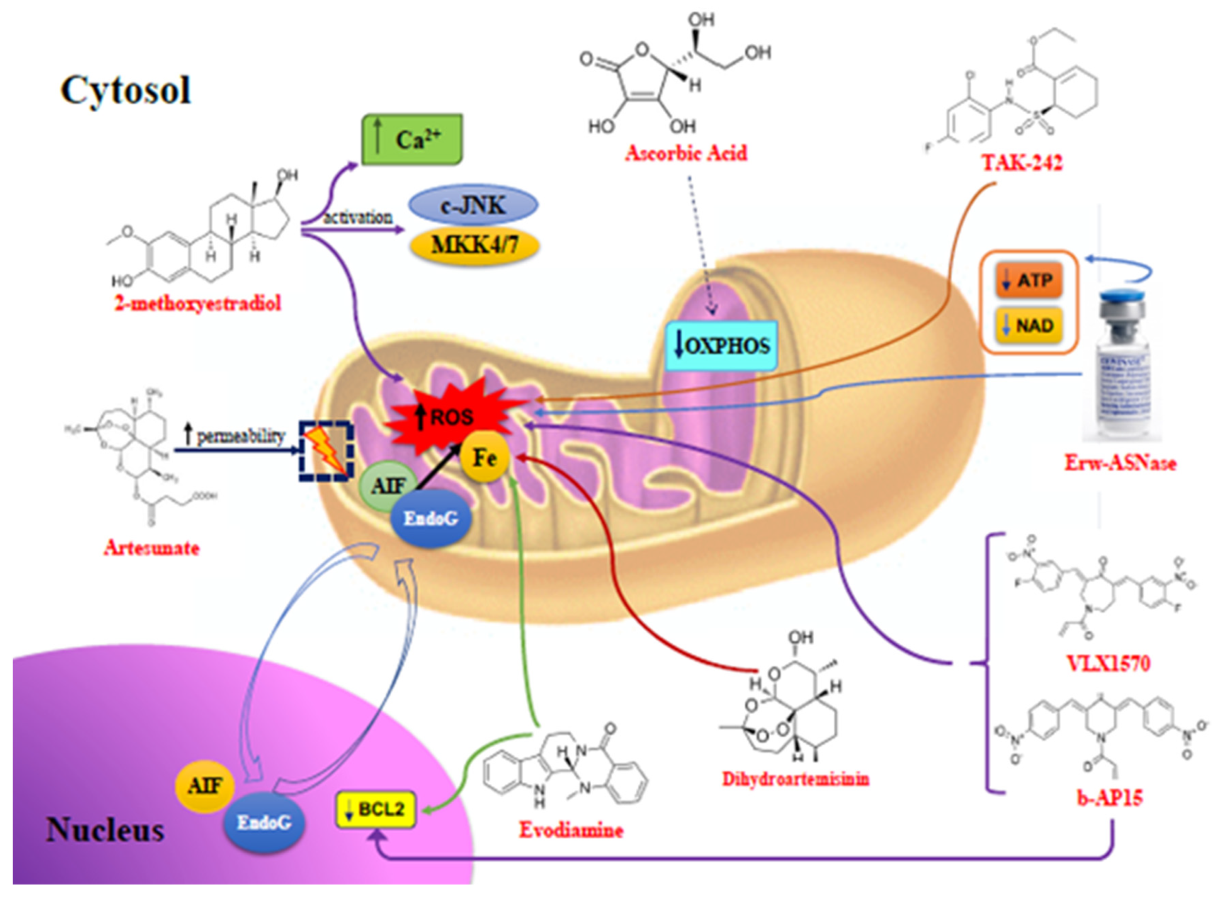
2. Re-Sensitization to Chemotherapy through Caspase-Independent Apoptosis via ROS

Despite therapeutic advances, and sometimes regardless of the prognostic risk, multiple myeloma patients still experience relapses after exposure to several agents and reduced survival [15]. The induction of cellular oxidative damage has opened the way to the study of new compounds that kill plasma cells with acceptable safety profiles, and we can consider some of them as promising adjuvants in the treatment of MM in clinical practice. Some of these molecules act as anticancer drugs by direct mitochondrial damage, while others determine wider cellular changes followed by mitochondrial changes.
The mitochondrial function plays a central role in the cellular reactive oxygen species (ROS) balance and, in this regard, to overcome bortezomib-resistance in MM cell lines, in 2013, Korean researchers identified a compound active via ROS overproduction [16]. Compared with BTZ alone, representing the first PI available for MM patients, they showed that 2-methoxyestradiol (2ME), which is a natural estrogen metabolite, induced a higher rate of MM cell death, due to increases in the mitochondrial ROS and Ca2+ levels in the cells, the activation of c-Jun N-terminal kinase (JNK), and mitogen-activated protein kinase kinase 4/7 (MKK4/7) [17]. The capability of 2ME to increase the death of cancer cells has also been proved to be powerful against other types of tumor, such as ovarian, lung, breast, and colorectal tumors [13,18,19,20,21].
In 2014, scientists from Little Rock explained the mechanism inducing apoptosis by Artesunate, which is a well-tolerated anti-malarial drug. Targeting mitochondria and increasing their membrane permeability in MM cell lines, due to an increased bivalent iron-dependent mitochondrial production of ROS levels, this compound turned out to be an effective cellular death stimulator, through non-caspase-mediated apoptosis, such as in lung adenocarcinoma cells [21,22]. The osmotic swelling of the mitochondrial matrix and compression of vesicles caused release into the cytosol and subsequent migration into the nucleus of pro-apoptotic proteins, such as cytochrome c, apoptosis inducing factor (AIF), and endonuclease G (EndoG). The role of EndoG in cell death is still unclear, whereas AIF, binding directly to DNA, causes chromatin condensation and DNA fragmentation. In addition, Artesunate has been shown to cause cell death independently of bortezomib-resistance, increasing its effectiveness [23].
The importance of cytoplasmic iron assessment in MM cells was underlined by Tricot el al. in 2017. Comparing the gene expression profiles (GEPs) of 18 mitochondrial genes in 22 normal plasma cells from 44 monoclonal gammopathy of undetermined significance (MGUS) patients and 351 newly diagnosed MM (NDMM) patients during a total therapy trial, they demonstrated that higher cytosolic iron levels promote cell proliferation and induce acquired drug resistance, due to its influence on the expression of many mitochondrial genes, including mitochondrial biogenesis signatures and oxidative phosphorylation. Due to inhibiting mitochondria oxidative phosphorylation and inducing MM cell death, ascorbic acid could become an old but revolutionary compound in synergic treatment in bortezomib-refractory MM patients [24].
Iron and ROS are also involved in the effectiveness of inducing the mitochondrial apoptotic pathway of Dihydroartemisinin (DHA), which is a derivative of artermisin (ART), employed for malaria treatment. Interacting with ferrous ions (Fe2+) and oxygen to produce ROS, DHA has been proven to be active in promoting myeloma cell death, overcoming the Dexamethasone resistance by reverting its action to upregulate the expression of the BCL2 protein. In addition, the high efficacy of DHA+Dexa in vitro and in vivo is due to mitochondrial cytochrome C translocation to the cytoplasm, resulting in caspase-mediated apoptosis, leading to high hopes regarding the therapeutic success of this substance [25]. In addition, DHA has exhibited a high anti-tumoral efficacy on MM cells by favoring autophagy and inhibiting angiogenesis, through reducing vascular endothelial growth factor (VEGF) levels [26,27].
An herbal substance from traditional Chinese medicine called Evodiamine (Evo) has displayed great anticancer activity in several tumor types [28,29,30,31]. Derived from the drying mature fruit of Evodia rutaecarpa, Evo induces apoptosis in MM cells, activating several pathways, including BCL2 inhibition and ROS accumulation. Similar to other compounds, Evo treatment increases mitochondrial cytochrome c release into the cytosol, upregulating ROS levels, due to mitochondrial superoxide production, and activating intrinsic mitochondrial apoptosis pathways [32]. Evo’s toxicity profile has still not been well-defined by giving cardiac arrhythmias and prolonging the bleeding times, although studies testing the acute toxicity seem to provide promising data on Evo’s development as a new anticancer remedy [33].
Additionally, deubiquitinase inhibitors (DUBs), such as b-AP15, have been shown to overcome the acquired resistance to BTZ in MM, Waldestrom macroglobulinemia (WM), and diffuse large B cell lymphoma (DLBCL), resulting in mitochondrial dysfunction [34,35,36]. Inducing oxidative stress in MM cells, b-AP15 revealed high antiproliferative activity, causing mitochondrial deformations, through the induction of the chaperones heat shock protein 70B’ (HSP70B’) and heat shock protein 40 (HSP40), resulting in nuclear factor erythroid 2-related factor 2 (Nrf-2) and its target heme-oxygenase 1 (HO-1) induction, but without lipid peroxidation [37]. The caspase-independent pro-apoptotic effects of b-AP15 were very high in tumor cells overexpressing BCL2 family proteins and defective in Tumor Protein p53 (TP53) [38], not just in MM [39,40,41,42,43,44]. However, these data have only been produced from cell line studies, and should be confirmed by in vivo tests.
Another recognized competitive DUB capable of inducing apoptosis in MM cells is VLX1570, which is a more potent and soluble analogue of b-AP15 able to strongly inhibit Ubiquitin carboxyl-terminal hydrolase 14 (UPS14) activity, inducing the accumulation of polyubiquitin chains, the expression of the chaperone HSP70B’, the oxidative stress marker heme oxygenase 1 (Hmox-1), and apoptosis in MM cells. A GEP study revealed that VLX1570 determines cell oxidative stress similar to b-AP15, in respect to which it has been shown to be more effective because of the best administration [45]. In vivo, a phase I/II clinical trial (NCT02372240) evaluated the efficacy and safety of this compound and Dexamethasone in relapsed/refractory MM (RRMM) patients. Although the efficacy was promising, its low safety profile, which resulted in death by respiratory failure of two of the 14 enrolled patients, caused the premature closure of the study, focusing on the identification of better tolerated substances [46].
Italian researchers recently reported mitochondrial damage and increased ROS generation by L-Asparaginase purified from Erwinia chrysanthemi (Erw-ASNase), which is a powerful enhancer of the carfilzomib response in resistant MM cells. Recognizing amino acid depletion as an instrument to better hit tumor cells, the authors analyzed the concept of amino acid starvation, induced by Erw-ASNase. In combination with Carfilzomib, Erw-ASNase caused cell death via increased mitochondrial oxidative stress, due to higher ROS generation, Nrf2 upregulation, and a reduced adenosine triphosphate (ATP) and nicotinamide adenine dinucleotide (NAD) intracellular content [47]. Since ERW-ASNase has been approved and is commonly used in clinical practice in the treatment of pediatric acute lymphoblastic leukemia, where the safety profile is fairly high, its tolerability in adult patients is still low, so in vivo studies of multiple myeloma patients are required to assess its applicability as an antimyeloma drug.
A recent report of Italian researchers sustained mitochondrial involvement in BTZ resistance in MM cells, due to the increased signal of Toll-like receptor 4 (TLR4). Combining BTZ with TAK-242 (Resatorvid), which is a selective TLR4 inhibitor, they overcame MM cell resistance, generating higher oxidative stress due to an ROS and reactive nitrogen species (RNS) increase, followed by depolarization of the mitochondrial membrane and cytochrome c release into the cytosol, finally resulting in the activation of caspase-9 [48].
Often, several cellular death mechanisms are compenetrated, without allowing clear distinctions. Frequently, in fact, the same substances may activate multiple mechanisms capable of killing tumor cells at the same time.

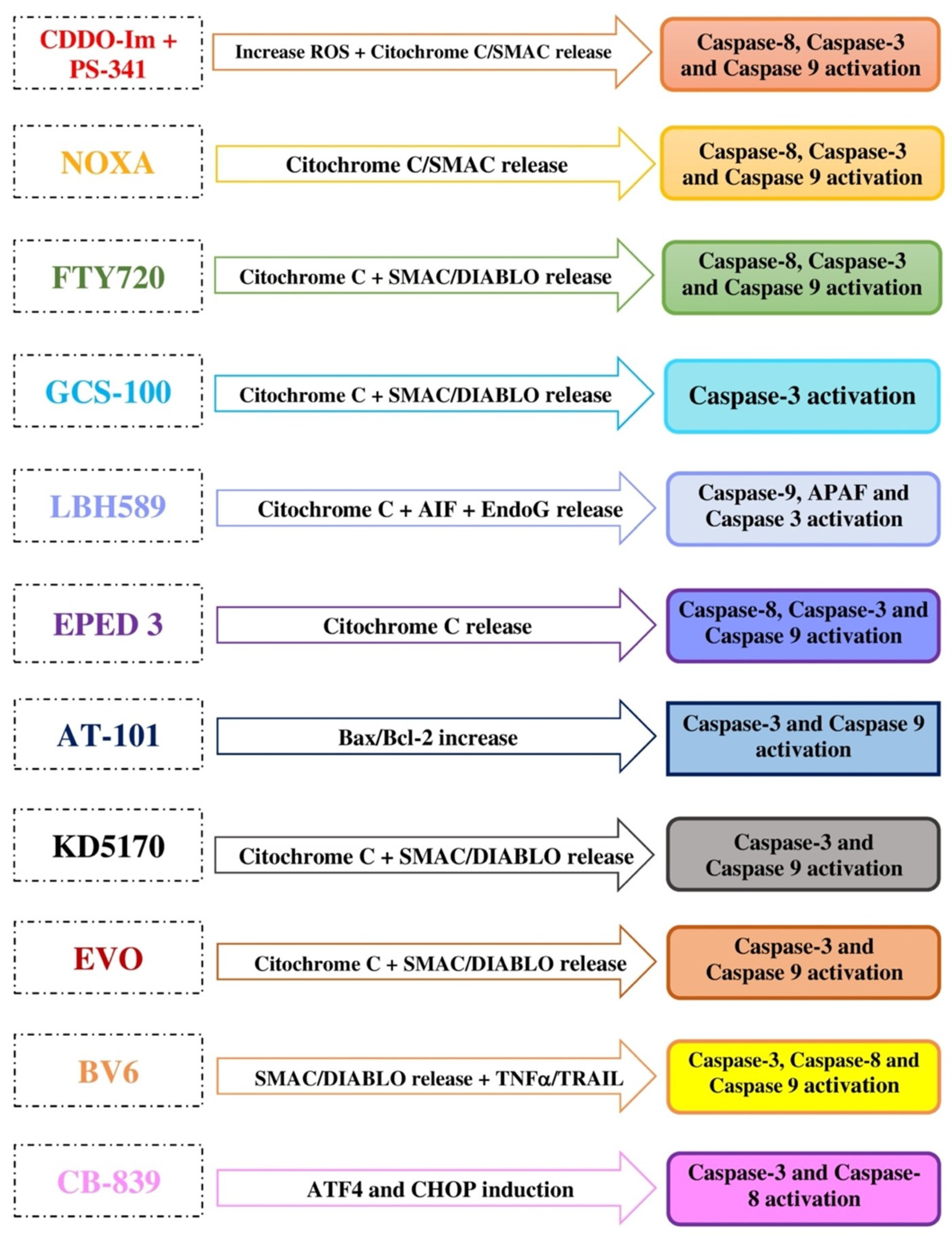
3. Re-Sensitization to Chemotherapy through Caspase-Dependent Apoptosis

Acquired resistance to proteasome inhibitors is not only based on the development of caspase-independent anti-apoptotic mechanisms; they also lose their effectiveness when tumor cells gain resistance to caspase-dependent mechanisms.
In this regard, an old report published in 2004 identified the role of low-dose CDDO-Im (triterpenoid 2-cyano-3, 12-dioxooleana-1, 9-dien-28-oic acid, Imidazoline) associated with PS-341, which is a bortezomib-proteasome inhibitor, in the induction of apoptosis in MM cells resistant to conventional therapy, including doxorubicin, melphalan, and dexamethasone. Studies on leukemia and lymphoma cells have demonstrated that CDDO-Im induces apoptosis in several ways [49,50,51], such as through the activation of caspase-8 and -3, and the release of mitochondrial cytochrome c. This study showed that PS-341, in combination with low-dose CDDO-Im, exhibited high anti-MM activity in BTZ-resistant MM cell lines and MM patients, without effects on normal lymphocytes, even due to mitochondria-dependent apoptotic signaling pathways. Their effectiveness in cell death included the rapid depletion of mitochondrial glutathione, generation of ROS, loss of mitochondrial membrane potential, release of mitochondrial cytochrome c and the second mitochondria-derived activator of caspases (SMAC) into the cytosol, and activation of caspase-9 and caspase-3. Moreover, mitochondria-independent apoptosis was also described, via the death receptor-mediated-caspase-8-caspase-3 pathway [52]. However, in vivo data refer to studies in murine models, so phase 1 studies are required to establish its applicability in the anticancer therapeutic strategy.
In 2005, the same targeted pathways were described by protein phorbol-12-myristate-13-acetate-induced protein 1 (NOXA), which is responsible for the mitochondrial-based apoptotic resistance to PIs, based on the release of cytochrome c, followed by a proteolytic cascade involving caspase-9, -3, and -8. The scientists were able to overcome the PI resistance via NOXA induction, which was p53-independent, because its block, using an antisense oligonucleotide, caused a 30% to 50% reduced apoptotic response in melanoma and myeloma cells, suggesting a therapeutic role of agents triggering NOXA induction in these patients [53]. This pathway is involved in the sensitivity to apoptosis in several types of cancer, such as mantle cell lymphoma and small cell lung cancer [54,55].
In 2005, Swiss researchers reported results on an immunomodulator agent, named Fingolimod (FTY720), which has been approved by the FDA as an oral compound to treat relapsed multiple sclerosis. This agent demonstrated high antitumoral activity in micromolar concentrations in drug-sensitive and -resistant MM cell lines and MM patients, due to the well-known activation of proapoptotic Bax and depolarization with an increased permeability of the mitochondrial membrane, followed by cytochrome c and SMAC/direct inhibitor of apoptosis-binding protein with low isoelectric point (DIABLO) translocation into the cytosol, finally resulting in the activation of caspase-8, -9, and -3. In combination with dexamethasone and anti-Fas antibodies, FYT720 improved the response rates, managing to overcome the acquired drug resistance [56]. Recently, Chinese scientists confirmed these results, proving the synergic action of Fingolimod with metformin that is able to induce apoptosis in MM cells [57]. Despite the study being conducted on MM cell lines, the safety profiles of both compounds are well-known, so there would appear to be no limits in testing them in vivo in MM patients.
The activation of the same mitochondrial targets was described in a report of 2005 on GCS-100, which is a derivative citrus pectin polysaccharide, developed for the treatment of several cancers. Alone or in combination with dexamethasone, this compound induced apoptosis in BTZ-resistant MM cells, activating mitochondrial apoptotic signals via the release of cytochrome c and SMAC/DIABLO, followed by caspase-3 induction [58]. However, data in the literature stops 10 years ago and this compound has not been developed, so it cannot be included among the potential drugs that may be expected to treat MM patients in the near future.
For in vitro and in vivo MM cells, caspase-dependent cell death has been observed for LBH589 Panobinostat (LBH589), a hydroxamic acid-derived Hystone Deacetylase (HDAC) inhibitor, which has been proven to display a potent antiproliferative action, showing promising results in myeloid leukemia [59]. In MM, LBH589 induces apoptosis via caspase-independent and -dependent mechanisms, increasing the action of the most frequently used antimyeloma drugs, including bortezomib, melphalan, and dexamethasone. Apoptosis occurs because LBH589 increases the mitochondrial outer membrane permeability, resulting in the release of cytochrome c into the cytosol, which is then responsible for the activation of the apoptosome, made up of caspase-9, APAF-1, and caspase-3, and the release of AIF and EndoG. These results supported the clinical use of LBH589 as a good partner for other antimyeloma drugs, so the FDA approved it in 2014 for the treatment of patients with RRMM after at least two previous therapy lines [60]. However, its low safety profile led clinics not to prefer this choice, so it is an orphan drug in Europe.
A few years later, in 2008, a study on the pro-apoptotic effects of a hydrophilic derivative of the plant alkaloid of Ellipticine (EPED3, 9-dimethyl amino-ethoxy ellipticine) in MM cell lines came to light. Although it is not yet clear whether the mitochondrion is the only or main cell target of EPED3 in the re-sensitization of myeloma cells to chemotherapy, its effectiveness is clear. In fact, intercalating between DNA base pairs and inducing G2-M-phase cycle arrest, EPED3 proved to be a powerful anti-myeloma agent in cells with CDC28 protein kinase regulatory subunit 1B (CKS1B) overexpression, which is a cell cycle regulator, even by the inhibition of topoisomerase II and P-glycoprotein-mediated multidrug resistance, without damage to stromal cells [61]. Its effectiveness also appeared to be high in BTZ- and dexamethasone-resistant cell lines, by destroying the mitochondrial membrane, followed by cytochrome c release into the cytosol, with the final activation of caspase-8, caspase-9, and caspase-3. Furthermore, using electron microscopy, the authors showed further ultrastructure changes due to apoptosis activation by EPED3 on MM cells, such as the destruction of mitochondrial inner cristae, nuclear pyknosis, and formation of multiple cytosolic vacuoles, hoping for its use in vivo as an antimyeloma agent in combination with conventional drugs [62]. However, in vitro studies are still ongoing regarding the identification of other effective water-soluble derivatives of Ellipticine, such as anticancer agents and immunomodulators for its low bioavailability [63].
In the same year, Rochester researchers provided similar results on AT-101, which is an R-(−)-gossypol small molecule active in MM resistant-cells in vivo and in vitro, mimicking the BH3 domain of cellular BCL-2 inhibitors. They proved that AT-101 decreased cell proliferation, increasing the Bax/BCL-2 ratio and activating apoptosis via mitochondrial pathways. Caspase-3 and caspase-9 activation resulted from mitochondrial membrane depolarization. Finally, AT-101 in combination with dexamethasone exhibited synergistic toxicity on MM cell lines extracted from MM patients [64]. In combination with Rituximab or Lenalidomide, AT-101 has shown a good efficacy in patients with chronic lymphocytic leukemia (CLL), but no study has yet been started in MM patients and its tolerability profile has not been well-defined (NCT00286780) [65]. In this regard, by enhancing the Noxa-mediated activation of Bak, followed by an increase in the mitochondrial apoptotic pathway, the BH3-mimetic GX15-070 demonstrated synergic activity with bortezomib in mantle cell lymphoma, while ABT-737 induced apoptosis in small cell lung cancer via Noxa/Mcl1 [54,55]. The ABT-analogous 737, 263, and 199 are other Bcl-2-specific BH3-mimetics active against the subgroup of t(11;14) MM patients presenting a Bcl2high/Mcl-1 low profile [66,67,68].
Caspase-3, caspase-8, and caspase-9 are also activated by KD5170, which is a mercaptoketone-based histone deacetylase inhibitor that has shown a high antiproliferative efficacy in MM cell lines. An article of 2008 reported the proapoptotic effect of this compound, caused by loss the mitochondrial membrane integrity, with cytochrome c and SMAC/DIABLO release into the cytosol, and thus apoptosis inducing factor activation. In association with BTZ, KD5170 enhanced its antimyeloma effectiveness [69]. In addition, KD5170 exhibited a broad spectrum of antitumoral activity in vitro and in vivo [70], and similar results emerged about another histone deacetylase inhibitor, named PXD101, capable of improving the antimyeloma effect of BTZ by the induction of oxidative stress and DNA damage [71]. Both compounds have not yet been tested in patients, so their real applicability as antimyeloma pharmacological agents cannot be established.
Similarly, in 2013, Chinese scientists supported the efficacy of gossypol in inducing apoptosis in myeloma cells in vitro and in vivo, by the inhibition of BCL-2/BCL-XL and caspase-3 and -9 activation [72]. Used as a male contraceptive, the safety data already obtained from its marketing would reduce the test times required to prove its effectiveness in MM patients.
Another natural derivative, called Evodiamine (Evo), is known for its anticancer activity in numerous tumor cells [28,29,30,31], including myeloma. Already mentioned as being responsible for cell damage by ROS, in MM, Evo also selectively blocks cell proliferation and increases apoptosis, vigorously activating caspase-3 and -9. In combination with BTZ, in a xenografted mice model, Evo enhances MM cell death, with a strong reduction in the cell viability, with respect to a BTZ single agent [32], but no data on MM patients are available and there are no negligible data on adverse events related to its use, such as cardiac arrhythmias and prolonged bleeding times, so further in vivo studies are necessary.
Caspase-dependent apoptosis is involved, along with independent mechanisms, in the abovementioned DHA, which is highly effective in association with dexamethasone in MM cells, due to mitochondrial cytochrome C translocation to the cytoplasm [24] and this action is valid in many types of cancer [73,74,75,76].
SMAC-mimetics are novel anticancer compounds developed to simulate the endogenous SMAC/DIABLO, which is an inhibitor of a class of proteins known as inhibitors of apoptosis proteins (IAPs). In this way, SMAC/DIABLO acts as an enhancer of the activation of caspases [77,78]. Drug resistance in MM is largely attributed to a reduction in SMAC release [79]. In recent years, fervent attention has been paid to these molecules because of their promising efficacy in several types of tumor [80,81,82,83,84]. In MM cells the novel bivalent SMAC mimetic BV6 induces the degradation of IAPs (cIAP1 and cIAP2), resulting in the apoptotic death of resistant cell lines via non-canonical NF-kB pathway activation. In the presence of cytochrome c, BV6 sensitizes MM cells to death ligands tumor necrosis factor-a (TNF-a) and TNF-related apoptosis-inducing ligands (TRAIL)-induced cell death, activating the caspase pathway. The sensitizing effect of BV6 on recombinant-TNF-a and killer TRAIL allows us to consider it a modern therapeutic tool in combination with conventional drugs in different MM cells [85], as well as in acute myeloid leukemia [86,87], chronic lymphocytic leukemia [88], and some solid tumors [89,90,91].
Using SMAC-mimetics, in recent years, several reports have underlined the role of controlling apoptosis by IAPs in multiple myeloma. In this regard, the administration of LCL161 has significantly reduced X-linked inhibitor of apoptosis protein (XIAP) activity and cellular inhibitor of apoptosis protein-1 (cIAP1) levels in both sensitive and resistant myeloma cells. In addition, LCL161 determines the up-regulation of the Janus kinase 2/Signaling transducer and activator of transcription (Jak2/Stat3) signaling pathway in resistant cells, showing synergic antimyeloma activity when used with specific Jak2 inhibitors, both in cell lines and patient cells. Finally, associated with death-inducing ligands, LCL161 re-sensitized MM cells to both Fas cell surface death receptor (FAS-L) and TNF-related apoptosis-inducing ligand (TRAIL) [92]. However, these are in vitro studies or preclinical models, and in vivo tests are required to understand the therapeutic impact of these compounds in the treatment of MM patients in the future.
Another SMAC-mimetic molecule with similar inducing apoptosis effects in drug-resistant MM cells is LBW242, which is a well-tolerated compound that acts via caspase-8, -9, and -3 activation. When combined with BTZ or melphalan, LBW242 improved the response rates synergistically [76].
The evolution of PIs with second-, third-, and fourth-generation Carfilzomib (CFZ), Ixazomib (Ixa), and Oprozomib (Opro) has improved response rates, but has not resolved the problem of acquired resistance in MM cells. A 2016 study showed that the mechanisms of MM cells based on a loss of susceptibility to CFZ are similar to those observed with BTZ. Specifically, using glutamine as the main energy source, resistant cells appeared to exhibit higher mitochondrial respiration. This assumption has allowed us to use glutaminase-1 (GLS1) inhibitors to overcome the PI resistance of MM cells. Therefore, CB-839 (Telaglenastat) in combination with BTZ, Ixa, Opro, or even better with CFZ, has been shown to be a powerful inducer of apoptosis, by intense induction of the ER stress markers Activating Transcription Factor 4 (ATF4) and C/EPB Homologous Protein (CHOP), and caspase-3 and caspase-8 activation [93,94]. Furthermore, GLS selective inhibitors have also displayed good anti-cancer activity that is able to proliferate studies in other tumor cells, as well as B cell lymphoma, breast, liver, ovarian, pancreatic, colorectal, liver, and non-small cell lung cancers [95,96,97,98,99].
According to what has been said so far, the results obtained from a very recent study on the action mechanisms of Venetoclax, which is a BCL-2 antagonist highly effective in t(11;14) MM cells, are not surprising. Its effectiveness is based on several cellular targets, mostly involving the mitochondrion, which showed a reduced respiration in sensitive cells. Using inhibitors of electron transport chain (ETC) Complex I and II, IACS-010759, and thenoyltrifluoroacetone (TTFA), respectively, the authors demonstrated that the same ETC activity could represent a predictor and a target for Venetoclax sensitivity in MM patients [68].
The mechanisms of action and pathways activated by the various compounds considered in our manuscript are described in Table 1.

4. Conclusions

This comprehensive collection of data present in the literature is born from the desire to refine our weapons to improve the response rates and overall survival of multiple myeloma patients, who too often experience relapses [100]. Improvement of diagnostic definitions and the adoption of the minimum residual disease as the best prognostic marker available are not sufficient for permanently healing this disease [7,101]. Therefore, many studies are being carried out to find compounds and mechanisms for the circumvention of this tumor intelligence. Indeed, over the last few years, MM has represented one of the cancers that has progressed the most in terms of survival, mainly thanks to the advent of modern monoclonal antibodies (moAbs), such as Daratumumab and Isatuximab, and anti-BCMA, such as Belantamab mafodotin; however, they often do not exceed cytogenetic high risk [102].
We have reported several substances, also of natural origin, such as Chinese herbs or those from common plants, effective in improving or restoring the response to the most commonly used drugs, as well as PIs, such as BTZ, CFZ, and ixazomib, but no data are available for the abovementioned novel anti-myeloma agents, whose resistance mechanisms remain largely unknown.
In this respect, some of the abovementioned molecules appear, more than others, to be promising adjuvant agents for the therapy of MM patients, such as 2ME, Artesunate, Ascorbic Acid, Fingolimod, Panobinostat, AT-101, Telaglenastat, and Venetoclax. For the others, which are Evodiamine, bAP15, VLX1570, ERW-ASNase, TAK-242, CDDO-Im, GCS-100, EPED3, KD5170, PXD101, BV6, and LBW242, additional preclinical and in vivo tests are required to prove their applicability in clinical practice.
Therefore, we believe that it is necessary to extend research on these compounds, perhaps by testing the same substances that have proved to be effective until now or by finding even better substances.

Author Contributions

Conceptualization: A.A. and C.M.; methodology: V.I.; software: V.I.; formal analysis: V.I., A.G.A., and V.R.; writing—original draft preparation: V.I.; writing—review and editing: V.I. and A.A. All authors have read and agreed to the published version of the manuscript.

Funding

This research received no external funding.

Conflicts of Interest

The authors declare no conflict of interest.

Abbreviations

2ME2-methoxyestradiol
ROSReactive Oxygen Species
c-JNKc-Jun N-terminal Kinase
MKK4/7Mitogen-activated protein Kinase Kinase 4/7
MMMultiple Myeloma
AIFApoptosis Inducing Factor
EndoGEndonuclease G
AMLAcute Myeloid Leukemia
CNSCentral Nervous System
OXPHOSOxidative Phosphorilation
Nrf-2Nuclear factor erythroid 2-Related Factor 2
VEGFVascular Endothelial Growh Factor
Bcl-2B-cell Lymphoma 2
EvoEvodiamine
Cdc2Cyclin-dependent protein kinase
IAPsInhibitors of Apoptosis
NF-kBNuclear Factor kappa light chain enhancer of activated B cells
HSP70B’Heat Shock Protein 70B’
HSP40Heat Shock Protein 40
HO-1Heme Oxygenase 1
WMWaldenstrom’s Macroglobulinemia
DLBLCDiffuse Large B Cell Lymphoma
UPS14Ubiquitin carboxyl-terminal hydrolase 14
MCLMantle cell Lymphoma
ATPAdenosine triphosphate
NADNicotinamide adenine dinucleotide
CMLChronic Myeloid Leukemia
RNSReactive Nitrogen Specie
TLR4Toll-like receptor 4
SMAC/DIABLOSecond Mitochondria-derived Activator of Caspases/Direct Inhibitor of Apoptosis-Binding protein with low isoelectric point
BAxBcl-2 Associated X
HCCHepato cellular carcinoma
CLLChronic Lymphocytic Leukemia
APAF-1Apoptotic Protease Activating Factor-1
CTCLCutaneous T Cell Lymphoma
HLHodgkin Lymphoma
APLAcute Promyelocytic Leukemia
NSCLCNon-Small Cell Lung Cancer
TRAILTNF-Related Apoptosis-Inducing Ligand

References

  1. Aragona, P.; Postorino, E.I.; Rania, L.; Innao, V.; Wylegala, E.; Pisani, A.; Puzzolo, D.; Micali, A.; Allegra, A.; Nowinska, A.; et al. Corneal Structural Changes in Nonneoplastic and Neoplastic Monoclonal Gammopathies. Investig. Opthalmol. Vis. Sci. 2016, 57, 2657. [Google Scholar] [CrossRef] [Green Version]
  2. Allegra, A.; Penna, G.; Innao, V.; Greve, B.; Maisano, V.; Russo, S.; Musolino, C. Vaccination of multiple myeloma: Current strategies and future prospects. Crit. Rev. Oncol. 2015, 96, 339–354. [Google Scholar] [CrossRef]
  3. Innao, V.; Allegra, A.; Pulvirenti, N.; Allegra, A.; Musolino, M. Therapeutic potential of antagomiRs in haematological and oncological neoplasms. Eur. J. Cancer Care 2020, 29, 2. [Google Scholar] [CrossRef]
  4. Conticello, C.; Romano, A.; Del Fabro, V.; Martino, E.; Calafiore, V.; Sapienza, G.; Leotta, V.; Parisi, M.; Markovic, U.; Garibaldi, B.; et al. Feasibility, Tolerability and Efficacy of Carfilzomib in Combination with Lenalidomide and Dexamethasone in Relapsed Refractory Myeloma Patients: A Retrospective Real-Life Survey of the Sicilian Myeloma Network. J. Clin. Med. 2019, 8, 877. [Google Scholar] [CrossRef] [Green Version]
  5. Holstein, S.A.; Suman, V.J.; McCarthy, P.L. Update on the role of lenaliomide in patients with multiple myeloma. Ther. Adv. Hematol. 2018, 9, 7. [Google Scholar] [CrossRef] [PubMed] [Green Version]
  6. Delforge, M.; Ludwig, H. How I manage the toxicities of myeloma drugs. Blood 2017, 129, 2359–2367. [Google Scholar] [CrossRef] [PubMed]
  7. Innao, V.; Allegra, A.G.; Russo, S.; Gerace, D.; Vaddinelli, D.; Alonci, A.; Musolino, C. Standardisation of minimal residual disease in multiple myeloma. Eur. J. Cancer Care 2017, 26, e12732. [Google Scholar] [CrossRef] [PubMed]
  8. West, A.P.; Shadel, G.S. Mitochondrial DNA in innate immune responses and inflammatory pathology. Nat. Rev. Immunol. 2017, 17, 363–375. [Google Scholar] [CrossRef] [PubMed]
  9. Wu, Z.; Oeck, S.; West, A.P.; Mangalhara, K.C.; Sainz, A.G.; Newman, L.E.; Zhang, X.-O.; Wu, L.; Yan, Q.; Bosenberg, M.; et al. Mitochondrial DNA stress signalling protects the nuclear genome. Nat. Metab. 2019, 1, 1209–1218. [Google Scholar] [CrossRef]
  10. Allegra, A.; Innao, V.; Allegra, A.G.; Pulvirenti, N.; Pugliese, M.; Musolino, C. Antitumorigenic action of nelfinavir: Effects on multiple myeloma and hematologic malignancies (Review). Oncol. Rep. 2020, 43, 6. [Google Scholar] [CrossRef] [PubMed]
  11. Allegra, A.; Innao, V.; Gerace, D.; Vaddinelli, D.; Allegra, A.G.; Musolino, C. Nanobodies and Cancer: Current Status and New Perspectives. Cancer Investig. 2018, 36, 221–237. [Google Scholar] [CrossRef] [PubMed]
  12. Pei, X.-Y.; Dai, Y.; Grant, S. Synergistic Induction of Oxidative Injury and Apoptosis in Human Multiple Myeloma Cells by the Proteasome Inhibitor Bortezomib and Histone Deacetylase Inhibitors. Clin. Cancer Res. 2004, 10, 3839–3852. [Google Scholar] [CrossRef] [PubMed] [Green Version]
  13. Chauhan, D.; Li, G.; Podar, K.; Hideshima, T.; Mitsiades, C.; Schlossman, R.; Munshi, N.; Richardson, P.; Cotter, F.; Anderson, K. Targeting mitochondria to overcome conventional and bortezomib/proteasome inhibitor PS-341 resistance in multiple my-eloma (MM). Cells 2004, 104, 8. [Google Scholar]
  14. Besse, L.; Besse, A.; Mendez-Lopez, M.; Vasickova, K.; Sedlackova, M.; Vanhara, P.; Kraus, M.; Bader, J.; Ferreira, R.; Castel-lano, R.; et al. A metabolic switch in proteasome inhibitor-resistant multiple myeloma ensures higher mito-chondrial metabolism, protein folding and sphingomyelin synthesis. Haematologica 2019, 104, 9. [Google Scholar] [CrossRef] [Green Version]
  15. Kumar, S.; Lee, J.H.; Lahuerta, J.J.; Morgan, G.; Richardson, P.G.; Crowley, J.; Haessler, J.; Feather, J.; Hoering, A.; Moreau, P.; et al. Risk of progression and survival in multiple myeloma relapsing after therapy with IMiDs and bortezomib: A multicenter international myeloma working group study. Leukemia 2011, 26, 149–157. [Google Scholar] [CrossRef] [Green Version]
  16. Song, I.; Kim, H.; Lee, S.; Jeong, S.; Kim, N.; Ko, K.; Rhee, B.; Han, J. Mitochondrial modulation decreases the bortezomib-resistance in multiple myeloma cells. Int. J. Cancer 2013, 133, 1357–1367. [Google Scholar] [CrossRef]
  17. Song, I.-S.; Jeong, Y.J.; Jeong, S.H.; Heo, H.J.; Kim, H.K.; Lee, S.R.; Ko, T.H.; Youm, J.B.; Kim, N.; Ko, K.S.; et al. Combination treatment with 2-methoxyestradiol overcomes bortezomib resistance of multiple myeloma cells. Exp. Mol. Med. 2013, 45, e50. [Google Scholar] [CrossRef] [Green Version]
  18. Pal, P.; Hales, K.; Hales, D.B. The pro-apoptotic actions of 2-methoxyestradiol against ovarian cancer involve catalytic activation of PKCδ signaling. Oncotarget 2020, 11, 3646–3659. [Google Scholar] [CrossRef] [PubMed]
  19. Romero, Y.; Castillejos-López, M.; Romero-García, S.; Aguayo, A.S.; Herrera, I.; Garcia-Martin, M.O.; Torres-Espíndola, L.M.; Negrete-García, M.C.; Olvera, A.C.; Huerta-Cruz, J.C.; et al. Antitumor Therapy under Hypoxic Microenvironment by the Combination of 2-Methoxyestradiol and Sodium Dichloroacetate on Human Non-Small-Cell Lung Cancer. Oxid. Med. Cell. Longev. 2020, 2020, 1–12. [Google Scholar] [CrossRef]
  20. Lebelo, M.T.; Joubert, A.M.; Visagie, M.H. Sulphamoylated Estradiol Analogue Induces Reactive Oxygen Species Generation to Exert Its Antiproliferative Activity in Breast Cancer Cell Lines. Molecules 2020, 25, 4337. [Google Scholar] [CrossRef] [PubMed]
  21. Oh, Y.; Jung, H.; Min, S.; Kang, J.; Jang, D.; Shin, S.; Kim, J.; Lee, S.; Sung, C.; Lee, W.; et al. Targeting antioxidant enzymes enhances the therapeutic efficacy of the BCL-X L inhibitor ABT-263 in KRAS-mutant colorectal cancers. Cancer Lett. 2021, 497, 123–136. [Google Scholar] [CrossRef] [PubMed]
  22. Zhou, C.; Pan, W.; Wang, X.P.; Chen, T.S. Artesunate induces apoptosis via a Bak-mediated caspase-independent intrinsic pathway in human lung adenocarcinoma cells. J. Cell. Physiol. 2012, 227, 3778–3786. [Google Scholar] [CrossRef]
  23. Papanikolaou, X.; Johnson, S.; Garg, T.; Tian, E.; Tytarenko, R.; Zhang, Q.; Stein, C.; Barlogie, B.; Epstein, J.; Heuck, C. Artesunate overcomes drug resistance in multiple myeloma by inducing mitochondrial stress and non-caspase apoptosis. Oncotarget 2014, 5, 4118–4128. [Google Scholar] [CrossRef]
  24. Zhan, X.; Yu, W.; Franqui-Machin, R.; Bates, M.; Nadiminti, K.; Cao, H.; Amendt, B.; Jethava, Y.; Frech, I.; Zhan, F.T.G. Research Paper Alteration of mitochondrial biogenesis promotes disease progression in multiple myeloma. Oncotarget 2017, 8, 67. [Google Scholar] [CrossRef] [PubMed]
  25. Chen, Y.; Li, R.; Zhu, Y.; Zhong, S.; Qian, J.; Yang, D.; Jurczyszyn, A.; Beksac, M.; Gu, C.; Yang, Y. Dihydroartemisinin Induces Growth Arrest and Overcomes Dexamethasone Resistance in Multiple Myeloma. Front. Oncol. 2020, 10, 10. [Google Scholar] [CrossRef] [PubMed]
  26. Wu, X.; Liu, Y.; Zhang, E.; Chen, J.; Huang, X.; Yan, H.; Cao, W.; Qu, J.; Gu, H.; Xu, R.; et al. Dihydroartemisinin Modulates Apoptosis and Autophagy in Multiple Myeloma through the P38/MAPK and Wnt/β−Catenin Signaling Pathways. Oxid. Med. Cell. Longev. 2020, 2020, 6096391. [Google Scholar] [CrossRef]
  27. Wu, X.H.; Zhou, H.J.; Lee, J. Dihydroartemisinin inhibits angiogenesis induced by multiple myeloma RPMI8226 cells under hypoxic conditions via downregulation of vascular endothelial growth factor expression and suppression of vascular endo-thelial growth factor secretion. Anticancer Drugs 2006, 17, 7. [Google Scholar] [CrossRef]
  28. Qiu, C.; Gao, L.; Yan, K.; Cui, Y.; Zhang, Y. A promising antitumor activity of evodiamine incorporated in hydroxypropyl-beta- cyclodextrin: Pro-apoptotic activity in human hepatoma HepG2 cells. Chem. Cent. J. 2016, 10, 1–11. [Google Scholar] [CrossRef] [Green Version]
  29. Zhang, Y.; Wu, L.-J.; Tashiro, S.-I.; Onodera, S.; Ikejima, T. Evodiamine induces tumor cell death through different pathways: Apoptosis and necrosis. Acta Pharmacol. Sin. 2004, 25, 1. [Google Scholar]
  30. Wang, R.; Deng, D.; Shao, N.; Xu, Y.; Xue, L.; Peng, Y.; Liu, Y.; Zhi, F. Evodiamine activates cellular apoptosis through sup-pressing PI3K/AKT and activating MAPK in glioma. Oncotargets Ther. 2018, 11, 1183. [Google Scholar] [CrossRef] [PubMed] [Green Version]
  31. Guo, Q.; Liu, Y.; Zhao, J.; Wang, J.; Li, Y.; Pang, Y.; Chen, J.; Wang, J. Evodiamine inactivates NF-kappaB and potentiates the antitumor effects of gemcitabine on tongue cancer both in vitro and in vivo. Oncotargets Ther. 2018, 12, 257. [Google Scholar] [CrossRef] [Green Version]
  32. Fang, Q.; Jiang, S.; Li, C. Evodiamine Selectively Inhibits Multiple Myeloma Cell Growth by Triggering Activation of Intrinsic Apoptosis Pathway. Oncotargets Ther. 2019, 12, 11383–11391. [Google Scholar] [CrossRef] [PubMed] [Green Version]
  33. Jiang, J.; Hu, C. Evodiamine: A novel anti-cancer alkaloid from Evodia retaecarpa. Molecules 2009, 14, 1852. [Google Scholar] [CrossRef] [PubMed]
  34. Tian, Z.; D’Arcy, P.; Wang, X.; Ray, A.; Tai, Y.-T.; Hu, Y.; Carrasco, R.D.; Richardson, P.; Linder, S.; Chauhan, D.; et al. A novel small molecule inhibitor of deubiquitylating enzyme USP14 and UCHL5 induces apoptosis in multiple myeloma and overcomes bortezomib resistance. Blood 2014, 123, 706–716. [Google Scholar] [CrossRef] [PubMed]
  35. Chitta, K.; Paulus, A.; Akhtar, S.; Blake, M.K.K.; Caulfield, T.R.; Novak, A.J.; Ansell, S.M.; Advani, P.; Ailawadhi, S.; Asher, C.-K.; et al. Targeted inhibition of the deubiquitinating enzymes, USP14 and UCHL5, induces proteotoxic stress and apoptosis in Waldenström macroglobulinaemia tumour cells. Br. J. Haematol. 2015, 169, 377–390. [Google Scholar] [CrossRef] [PubMed]
  36. Jiang, L.; Sun, Y.; Wang, J.; He, Q.; Chen, X.; Lan, X.; Chen, J.; Dou, Q.P.; Shi, X.; Liu, J. Proteasomal cysteine deubiquitinase inhibitor b-AP15 suppresses migration and induces apoptosis in diffuse large B cell lymphoma. J. Exp. Clin. Cancer Res. 2019, 38, 414–453. [Google Scholar] [CrossRef]
  37. Zhang, X.; Espinosa, B.; Saei, A.A.; D’Arcy, P.; Zubarev, R.A.; Linder, S. Oxidative Stress Induced by the Deubiquitinase Inhibitor b-AP15 Is Associated with Mitochondrial Impairment. Oxid. Med. Cell. Longev. 2019, 2019, 1–11. [Google Scholar] [CrossRef]
  38. Zhang, X.; Pellegrini, P.; Saei, A.; Hillert, E.; Mazurkiewicz, M.; Olofsson, M.; Zubarev, R.; D’Arcy, P.; Linder, S. The deubiq-uitinase inhibitor b-AP15 induces strong proteotoxic stress and mito- chondrial damage. Biochem. Pharmacol. 2018, 156, 291–301. [Google Scholar] [CrossRef] [Green Version]
  39. Didier, R.; Mallavialle, A.; Ben Jouira, R.; Domdom, M.; Tichet, M.; Auberger, P.; Luciano, F.; Ohanna, M.; Tartare-Deckert, S.; Deckert, M. Targeting the proteasome-associated deubiquitinating enzyme USP14 impairs melanoma cell survival and over-comes resistance to MAPK-targeting therapies. Mol. Cancer Ther. 2018, 17, 7. [Google Scholar] [CrossRef] [Green Version]
  40. Erdal, H.; Berndtsson, M.; Castro, J.; Brunk, U.; Shoshan, B.; Linder, S. Induction of lysosomal membrane perme- abilization by compounds that activate p53-independent apoptosis. Proc. Natl. Acad. Sci. USA 2005, 102, 1. [Google Scholar] [CrossRef] [PubMed] [Green Version]
  41. D’Arcy, P.; Linder, S. Proteasome deubiquitinases as novel targets for cancer therapy. Int J Biochem Cell Biol. 2012, 44, 1729–1738. [Google Scholar] [CrossRef]
  42. Kropp, K.N.; Maurer, S.; Rothfelder, K.; Schmied, B.J.; Clar, K.L.; Schmidt, M.; Strunz, B.; Kopp, H.-G.; Steinle, A.; Grünebach, F.; et al. The novel deubiquitinase inhibitor b-AP15 induces direct and NK cell-mediated antitumor effects in human mantle cell lymphoma. Cancer Immunol. Immunother. 2018, 67, 935–947. [Google Scholar] [CrossRef]
  43. Ding, Y.; Chen, X.; Wang, B.; Yu, B.; Ge, J. Deubiquitinase inhibitor b-AP15 activates endoplasmic reticulum (ER) stress and inhibits Wnt/Notch1 signaling pathway leading to the reduction of cell survival in hepatocellular carcinoma cells. Eur. J. Pharmacol. 2018, 825, 10–18. [Google Scholar] [CrossRef]
  44. Cai, J.; Xia, X.; Liao, Y.; Liu, N.; Guo, Z.; Chen, J.; Yang, L.; Long, H.; Yang, Q.; Zhang, X.; et al. A novel deubiquitinase inhibitor b-AP15 triggers apoptosis in both androgen receptor-dependent and -independent prostate cancers. Oncotarget 2017, 8, 63232–63246. [Google Scholar] [CrossRef] [Green Version]
  45. Wang, X.; Mazurkiewicz, M.; Hillert, E.; Olofsson, M.; Pierrou, S.; Hillertz, P.; Gullbo, J.; Selvaraju, K.; Paulus, A.; Akhtar, S.; et al. The proteasome deubiquitinase inhibitor VLX1570 shows selectivity for ubiqui-tin-specific protease-14 and induces apoptosis of multiple myeloma cells. Sci. Rep. 2016, 6, 1–15. [Google Scholar]
  46. Rowinsky, E.K.; Paner, A.; Berdeja, J.G.; Paba-Prada, C.; Venugopal, P.; Porkka, K.; Gullbo, J.; Linder, S.; Loskog, A.; Richardson, P.G.; et al. Phase 1 study of the protein deubiquitinase inhibitor VLX1570 in patients with relapsed and/or refractory multiple myeloma. Investig. New Drugs 2020, 38, 1448–1453. [Google Scholar] [CrossRef] [PubMed] [Green Version]
  47. Soncini, D.; Minetto, P.; Martinuzzi, C.; Becherini, P.; Fenu, V.; Guolo, F.; Todoerti, K.; Calice, G.; Contini, P.; Miglino, M.; et al. Amino acid depletion triggered by ʟ-asparaginase sensitizes MM cells to carfilzomib by inducing mitochondria ROS-mediated cell death. Blood Adv. 2020, 4, 4312–4326. [Google Scholar] [CrossRef]
  48. Giallongo, C.; Tibullo, D.; Puglisi, F.; Barbato, A.; Vicario, N.; Cambria, D.; Parrinello, N.L.; Romano, A.; Conticello, C.; Forte, S.; et al. Inhibition of TLR4 Signaling Affects Mito-chondrial Fitness and Overcomes Bortezomib Resistance in Myeloma Plasma Cells. Cancers 2020, 12, 1999. [Google Scholar] [CrossRef]
  49. Konopleva, M.; Tsao, T.; Ruvolo, P.; Stiouf, I.; Estrov, Z.; Leysath, C.E.; Zhao, S.; Harris, D.; Chang, S.; Jackson, C.E.; et al. Novel triterpenoid CDDO-Me is a potent inducer of apoptosis and differentiation in acute myelogenous leukemia. Blood 2002, 99, 326–335. [Google Scholar] [CrossRef] [PubMed] [Green Version]
  50. Pedersen, I.M.; Kitada, S.; Schimmer, A.; Kim, Y.; Zapata, J.M.; Charboneau, L.; Rassenti, L.; Andreeff, M.; Bennett, F.; Sporn, M.B.; et al. The triterpenoid CDDO induces apoptosis in refractory CLL B cells. Blood 2002, 100, 2965–2972. [Google Scholar] [CrossRef]
  51. Stadheim, T.A.; Suh, N.; Ganju, N.; Sporn, M.B.; Eastman, A. The Novel Triterpenoid 2-Cyano-3,12-dioxooleana-1,9-dien-28-oic acid (CDDO) Potently Enhances Apoptosis Induced by Tumor Necrosis Factor in Human Leukemia Cells. J. Biol. Chem. 2002, 277, 16448–16455. [Google Scholar] [CrossRef] [Green Version]
  52. Chauhan, D.; Li, G.; Podar, K.; Hideshima, T.; Shringarpure, R.; Catley, L.; Mitsiades, C.; Munshi, N.; Tai, Y.T.; Suh, N.; et al. The bortezomib/proteasome inhibitor PS-341 and triterpenoid CDDO-Im induce synergistic anti–multiple myeloma (MM) activity and overcome bortezomib resistance. Blood 2004, 103, 3158–3166. [Google Scholar] [CrossRef] [PubMed] [Green Version]
  53. Qin, J.-Z.; Ziffra, J.; Stennett, L.; Bodner, B.; Bonish, B.K.; Chaturvedi, V.; Bennett, F.; Pollock, P.M.; Trent, J.M.; Hendrix, M.J.C.; et al. Proteasome Inhibitors Trigger NOXA-Mediated Apoptosis in Melanoma and Myeloma Cells. Cancer Res. 2005, 65, 6282–6293. [Google Scholar] [CrossRef] [Green Version]
  54. Pérez-Galán, P.; Roué, G.; Villamor, N.; Campo, E.; Colomer, D. The BH3-mimetic GX15-070 synergizes with bortezomib in mantle cell lymphoma by enhancing Noxa-mediated activation of Bak. Blood 2007, 109, 4441–4449. [Google Scholar] [CrossRef] [Green Version]
  55. Hauck, P.; Chao, B.H.; Litz, J.; Krystal, G.W. Alterations in the Noxa/Mcl-1 axis determine sensitivity of small cell lung cancer to the BH3 mimetic ABT-737. Mol. Cancer Ther. 2009, 8, 883–892. [Google Scholar] [CrossRef] [PubMed] [Green Version]
  56. Yasui, H.; Hideshima, T.; Raje, N.; Roccaro, A.M.; Shiraishi, N.; Kumar, S.; Hamasaki, M.; Ishitsuka, K.; Tai, Y.-T.; Podar, K.; et al. FTY720 Induces Apoptosis in Multiple Myeloma Cells and Overcomes Drug Resistance. Cancer Res. 2005, 65, 7478–7484. [Google Scholar] [CrossRef] [PubMed] [Green Version]
  57. Zhao, Y.; Zhang, E.; Lv, N.; Ma, L.; Yao, S.; Yan, M.; Zi, F.; Deng, G.; Liu, X.; He, J.; et al. Metformin and FTY720 Sinergisticallu Induce Apoptosis in Multiple Myeloma Cells. Cell Physiol. Biochem. 2018, 48, 2. [Google Scholar] [CrossRef] [PubMed]
  58. Chauhan, D.; Li, G.; Podar, K.; Hideshima, T.; Neri, P.; He, D.; Mitsiades, N.; Richardson, P.; Chang, Y.; Schindler, J.; et al. A Novel Carbohydrate-Based Therapeutic GCS-100 Overcomes Bortezomib Resistance and Enhances Dexamethasone-Induced Apoptosis in Multiple Myeloma Cells. Cancer Res. 2005, 65, 18. [Google Scholar] [CrossRef] [Green Version]
  59. George, P.; Bali, P.; Annavarapu, S.; Scuto, A.; Fiskus, W.; Guo, F.; Sigua, C.; Sondarva, G.; Moscinski, L.; Atadja, P.; et al. Combination of the histone deacetylase inhibitor LBH589 and the hsp90 inhibitor 17-AAG is highly active against human CML-BC cells and AML cells with activating mutation of FLT-3. Blood 2005, 105, 1768–1776. [Google Scholar] [CrossRef] [Green Version]
  60. Maiso, P.; Carvajal-Vergara, X.; Ocio, E.; Lopez-Pérez, R.; Mateo, G.; Gutiérrez, N.; Atadja, P.; Pandiella, A.; San Miguel, J. The Histone Deacetylase Inhibitor LBH589 Is a Potent Antimyeloma Agent that Overcomes Drug Resistance. Cancer Res. 2006, 66, 1. [Google Scholar] [CrossRef] [Green Version]
  61. Bhuyan, B.K.; Fraser, T.J.; Li, L.H. Cell cycle phase specificity and biochemical effects of ellipticine on mammalian cells. Cancer Res. 1972, 32, 11. [Google Scholar]
  62. Tian, E.; Landowski, T.H.; Stephens, O.W.; Yaccoby, S.; Barlogie, B.; Shaughnessy, J.D. Ellipticine derivative NSC 338258 represents a potential new antineoplastic agent for the treatment of multiple myeloma. Mol. Cancer Ther. 2008, 7, 500–509. [Google Scholar] [CrossRef] [Green Version]
  63. De Oliveira, R.C.; Pontes, G.S.; Kostyuk, A.; Camargo, G.B.C.; Dhyani, A.; Shvydenko, T.; Shvydenko, K.; Grafov, A. Anticancer and Immunomodulatory Activities of a Novel Water-Soluble Derivative of Ellipticine. Molecules 2020, 25, 2130. [Google Scholar] [CrossRef]
  64. Kline, M.P.; Rajkumar, S.V.; Timm, M.M.; Kimlinger, T.K.; Haug, J.L.; Lust, J.A.; Greipp, P.R.; Kumar, S. R-(-)−gossypol (AT-101) activates programmed cell death in multiple myeloma cells. Exp. Hematol. 2008, 36, 568–576. [Google Scholar] [CrossRef] [Green Version]
  65. Masood, A.; Chitta, K.; Paulus, A.; Khan, A.N.H.; Sher, T.; Ersing, N.; Miller, K.C.; Manfredi, D.; Ailawadhi, S.; Borrelo, I.; et al. Downregulation of BCL2 by AT-101 enhances the antileukaemic effect of lenalidomide both by an immune dependant and independent manner. Br. J. Haematol. 2011, 157, 59–66. [Google Scholar] [CrossRef]
  66. Bodet, L.; Gomez-Bougie, P.; Touzeau, C.; Dousset, C.; Descamps, G.; Maïga, S.; Avet-Loiseau, H.; Bataille, R.; Moreau, P.; Le Gouill, S.; et al. ABT-737 is highly effective against molecular subgroups of multiple myeloma. Blood 2011, 118, 3901–3910. [Google Scholar] [CrossRef]
  67. Lavik, A.; Zhong, F.; Chang, M.J.; Greenberg, E.; Choudhary, Y.; Smith, M.R.; McColl, K.S.; Pink, J.; Reu, F.J.; Matsuyama, S.; et al. A syntetic peptide targeting the BH4 domain of Bcl-2 induces apoptosis in multiple myeloma and follicular lymphoma cells alone or in combination with agents targeting the BH3-binding pocket of Bcl-2. Oncotarget 2015, 6, 29. [Google Scholar] [CrossRef] [PubMed] [Green Version]
  68. Bajpai, R.; Sharma, A.; Achreja, A.; Edgar, C.L.; Wei, C.; Siddiqa, A.A.; Gupta, V.A.; Matulis, S.M.; McBrayer, S.K.; Mittal, A.; et al. Electron transport chain activity is a predictor and target for venetoclax sensitivity in multiple myeloma. Nat. Commun. 2020, 11, 1–16. [Google Scholar] [CrossRef] [PubMed]
  69. Feng, R.; Ma, H.; Hassig, C.A.; Payne, J.E.; Smith, N.D.; Mapara, M.Y.; Hager, J.H.; Lentzsch, S. KD5170, a novel mercaptoketone-based histone deacetylase inhibitor, exerts antimyeloma effects by DNA damage and mitochondrial signaling. Mol. Cancer Ther. 2008, 7, 1494–1505. [Google Scholar] [CrossRef] [Green Version]
  70. Hassig, C.A.; Symons, K.T.; Guo, X.; Nguyen, P.-M.; Annable, T.; Wash, P.L.; Payne, J.E.; Jenkins, D.A.; Bonnefous, C.; Trotter, C.; et al. KD5170, a novel mercaptoketone-based histone deacetylase inhibitor that exhibits broad spectrum antitumor activity in vitro and in vivo. Mol. Cancer Ther. 2008, 7, 1054–1065. [Google Scholar] [CrossRef] [PubMed] [Green Version]
  71. Feng, R.; Oton, A.; Mapara, M.Y.; Anderson, G.; Belani, C.; Lentzsch, S. The histone deacetylase inhibitor, PXD101, potentiates bortezomib-induced anti-multiple myeloma effect by induction of oxidative stress and DNA damage. Br. J. Haematol. 2007, 139, 385–397. [Google Scholar] [CrossRef] [PubMed]
  72. Lin, J.; Wu, Y.; Yang, D.; Zhao, Y. Induction of apoptosis and antitumor effects of a small molecule inhibitor of Bcl-2 and Bcl-xl, gossypol acetate, in multiple myeloma in vitro and in vivo. Oncol. Rep. 2013, 30, 731–738. [Google Scholar] [CrossRef] [Green Version]
  73. Lu, Y.; Chen, T.; Wang, X.; Li, L. Single-cell analysis of dihydroartemisinin-induced apoptosis through reactive oxygen spe-cies-mediated caspase-8 activation and mitochondrial pathway in ASTC-a-1 cells using fluorescence imaging techniques. J. Biomed. Opt. 2010, 15, 4. [Google Scholar] [CrossRef] [PubMed]
  74. Mao, H.; Gu, H.; Qu, X.; Sun, J.; Song, B.; Gao, W.; Liu, J.; Shao, Q. Involvement of the mitochondrial pathway and Bim/Bcl-2 balance in dihydroartemisinin-induced apoptosis in human breast cancer in vitro. Int. J. Mol. Med. 2012, 31, 213–218. [Google Scholar] [CrossRef]
  75. Lu, M.; Sun, L.; Zhou, J.; Yang, J. Dihydroartemisinin induces apoptosis in colorectal cancer cells through the mitochondria-dependent pathway. Tumor Biol. 2014, 35, 6. [Google Scholar] [CrossRef]
  76. Aghaei, M.; Poupel, F.; Movahedian, A.; Jafari, S.M.; Shahrestanaki, M.K. Dihydroartemisinin induces apoptosis in human bladder cancer cell lines through reactive oxygen species, mitochondrial membrane potential, and cytochrome C pathway. Int. J. Prev. Med. 2017, 8, 78. [Google Scholar] [CrossRef]
  77. Du, C.; Fang, M.; Li, Y.; Li, L.; Wang, X. Smac, a Mitochondrial Protein that Promotes Cytochrome c–Dependent Caspase Activation by Eliminating IAP Inhibition. Cell 2000, 102, 33–42. [Google Scholar] [CrossRef] [Green Version]
  78. Verhagen, A.M.; Ekert, P.G.; Pakusch, M.; Silke, J.; Connolly, L.M.; Reid, G.E.; Moritz, R.L.; Simpson, R.J.; Vaux, D.L. Identification of DIABLO, a Mammalian Protein that Promotes Apoptosis by Binding to and Antagonizing IAP Proteins. Cell 2000, 102, 43–53. [Google Scholar] [CrossRef] [Green Version]
  79. Chauhan, D.; Neri, P.; Velankar, M.; Podar, K.; Hideshima, T.; Fulciniti, M.; Tassone, P.; Raje, N.; Mitsiades, C.; Mitsiades, N.; et al. Targeting mitochondrial factor Smac/DIABLO as therapy for multiple myeloma (MM). Blood 2006, 109, 1220–1227. [Google Scholar] [CrossRef] [PubMed]
  80. Chen, D.J.; Huerta, S. Smac mimetics as new cancer therapeutics. Anticancer Drugs 2009, 20, 646–658. [Google Scholar] [CrossRef] [PubMed]
  81. Sun, H.; Nikolovska-Coleska, Z.; Lu, J.; Qiu, S.; Yang, C.-Y.; Gao, W.; Meagher, J.; Stuckey, J.; Wang, S. Design, Synthesis, and Evaluation of a Potent, Cell-Permeable, Conformationally Constrained Second Mitochondria Derived Activator of Caspase (Smac) Mimetic. J. Med. Chem. 2006, 49, 7916–7920. [Google Scholar] [CrossRef] [PubMed]
  82. Amaravadi, R.K.; Schilder, R.J.; Martin, L.P.; Levin, M.; Graham, M.A.; Weng, D.E.; Adjei, A.A. A Phase I Study of the SMAC-Mimetic Birinapant in Adults with Refractory Solid Tumors or Lymphoma. Mol. Cancer Ther. 2015, 14, 2569–2575. [Google Scholar] [CrossRef] [Green Version]
  83. Li, W.; Li, B.; Giacalone, N.; Torossian, A.; Sun, Y.; Niu, K.; Lin-Tsai, O.; Lu, B. BV6, an IAP antagonist, activates apoptosis and enhances radiosensi- tization of non-small cell lung carcinoma in vitro. J. Thorac. Oncol. 2011, 6, 11. [Google Scholar] [CrossRef] [PubMed] [Green Version]
  84. Hehlgans, S.; Oppermann, J.; Reichert, S.; Fulda, S.; Rödel, C.; Rödel, F. The SMAC mimetic BV6 sensitizes colorectal cancer cells to ionizing radiation by interfering with DNA repair processes and enhancing apoptosis. Radiat. Oncol. 2015, 10, 198. [Google Scholar] [CrossRef] [PubMed] [Green Version]
  85. El-Mesery, M.E.; Shaker, M.; Elgaml, A. The SMAC mimetic BV6 induces cell death and sensitizes different cell lines to TNF-α and TRAIL-induced apoptosis. Exp. Biol. Med. 2016, 241, 2015–2022. [Google Scholar] [CrossRef] [Green Version]
  86. Bake, V.; Roesler, S.; Eckhardt, I.; Belz, K.; Fulda, S. Synergistic interaction of Smac mimetic and IFNalpha to trigger apoptosis in acute myeloid leukemia cells. Cancer Lett. 2014, 355, 2. [Google Scholar] [CrossRef] [PubMed]
  87. Steinwascher, S.; Nugues, A.; Schoeneberger, H.; Fulda, S. Identificationof a novel synergistic induction of cell death by Smac mimetic and HDAC inhibitors in acute myeloid leukemia cells. Cancer Lett. 2015, 366, 1. [Google Scholar] [CrossRef] [PubMed]
  88. Opel, D.; Schnaiter, A.; Dodier, D.; Jovanovic, M.; Gerhardinger, A.; Idler, I.; Mertens, D.; Bullinger, L.; Stilgenbauer, S.; Fulda, S. Targeting inhibitor of apoptosis proteins by Smac mimetic elicits cell death in poor prognostic subgroups of chronic lym-phocytic leukemia. Int. J. Cancer 2015, 137, 12. [Google Scholar] [CrossRef] [PubMed]
  89. Cristofanon, S.; Abhari, B.; Krueger, M.; Tchoghandjian, A.; Momma, S.; Calaminus, C.; Vucic, D.; Pichler, B.; Fulda, S. Iden-tification of RIP1 as a critical mediator of Smac mimetic-mediated sensitization of glioblastoma cells for Drozitumab-induced apoptosis. Cell Death Dis. 2015, 6, 4. [Google Scholar] [CrossRef] [Green Version]
  90. Liese, J.; Abhari, B.; Fulda, S. Smac mimetic and oleanolic acid synergize to induce cell death in human hepatocellular carci-noma cells. Cancer Lett. 2015, 365, 1. [Google Scholar] [CrossRef]
  91. Marschall, V.; Fulda, S. Smac mimetic-induced upregulation of interferon-beta sensitizes glioblastoma to temozolomide-induced cell death. Cell Death Dis. 2015, 6, 9. [Google Scholar] [CrossRef] [Green Version]
  92. Thompson, R.M.; Dytfeld, D.; Reyes, L.; Robinson, R.M.; Smith, B.; Manevich, Y.; Jakubowiak, A.; Komarnicki, M.; Przybylowicz-Chalecka, A.; Szczepaniak, T.; et al. Glutaminase inhibitor CB-839 synergizes with carfilzomib in resistant multiple myeloma cells. Oncotarget 2017, 8, 35863–35876. [Google Scholar] [CrossRef] [Green Version]
  93. Vogl, D.T.; Younes, A.; Stewart, K.; Orford, K.W.; Bennett, M.; Siegel, D.; Berdeja, J.G. Phase 1 Study of CB-839, a First-in-Class, Glutaminase Inhibitor in Patients with Multiple Myeloma and Lymphoma. Blood 2015, 126, 3059. [Google Scholar] [CrossRef]
  94. Le, A.; Lane, A.N.; Hamaker, M.; Bose, S.; Gouw, A.; Barbi, J.; Tsukamoto, T.; Rojas, C.J.; Slusher, B.S.; Zhang, H.; et al. Glucose-Independent Glutamine Metabolism via TCA Cycling for Proliferation and Survival in B Cells. Cell Metab. 2012, 15, 110–121. [Google Scholar] [CrossRef] [Green Version]
  95. Wang, J.; Erickson, J.; Fuji, R.; Ramachandran, S.; Gao, P.; Dinavahi, R.; Wilson, K.; Ambrosio, A.; Dias, S.; Dang, C.; et al. Targeting mitochondrial glutaminase activity inhibits oncogenic transformation. Cancer Cell. 2010, 18, 3. [Google Scholar] [CrossRef]
  96. Vaidya, A.; Ayat, N.; Buford, M.N.; Wang, H.; Shankardass, A.; Zhao, Y.; Gilmore, H.; Wang, Z.; Lu, Z. Noninvasive assessment and therapeutic monitoring of drug-resistant colorectal cancer by MR molecular imaging of extradomain-B fibronectin. Theranostics 2020, 10, 24. [Google Scholar] [CrossRef] [PubMed]
  97. Jin, H.; Wang, S.A.; Zaal, E.; Wang, C.; Wu, H.; Bosma, A.; Jochems, F.; Isima, N.; Jin, G.; Lieftink, C.; et al. A powerful drug combination strategy targeting glutamine addiction for the treatment of human liver cancer. eLife 2020, 9, 9. [Google Scholar] [CrossRef] [PubMed]
  98. Riess, J.W.; Frankel, P.; Shackelford, D.; Dunphy, M.; Badawi, R.D.; Nardo, L.; Cherry, S.R.; Lanza, I.; Reid, J.; Gonsalves, W.I.; et al. Phase 1 Trial of MLN0128 (Sapanisertib) and CB-839 HCl (Telaglenastat) in Patients with Advanced NSCLC (NCI 10327): Rationale and Study Design. Clin. Lung Cancer 2021, 22, 67–70. [Google Scholar] [CrossRef] [PubMed]
  99. Sonneveld, P. Management of multiple myeloma in the relapsed/refractory patient. Hematology 2017, 2017, 508–517. [Google Scholar] [CrossRef] [PubMed] [Green Version]
  100. Rajkumar, S.V. Multiple myeloma: 2020 update on diagnosis, risk-stratification and management. Am. J. Hematol. 2020, 95, 548–567. [Google Scholar] [CrossRef] [Green Version]
  101. Jullien, M.; Touzeau, C.; Moreau, P. Monoclonal antibodies as an addition to current myeloma therapy strategies. Expert Rev. Anticancer Ther. 2020, 21, 1–11. [Google Scholar] [CrossRef] [PubMed]
  102. Demel, I.; Bago, J.R.; Hajek, R.; Jelinek, T. Focus on monoclonal antibodies targeting B-cell maturation antigen (BCMA) in multiple myeloma: Update 2020. Br. J. Haematol. 2020. [Google Scholar] [CrossRef] [PubMed]
Figure 1. Caspase-independent mechanism of action of anti-mitochondrial agents. Mitochondrial targets of compounds with pro-apoptotic actions on myeloma cells are shown: 2-methoxyestradiol increases mitochondrial ROS production and increases Ca2+ levels in the MM cells, also causing JNK and MKK4/7 activation; artesunate increases the mitochondrial membrane permeability and ROS concentration in MM cells, inducing mitochondrial protein transfer into the nucleus, which causes DNA damage, AIF, and EndoG activation; ascorbic acid reduces cell respiration by the direct inhibition of mitochondrial oxidative phosphorylation in MM cells; DHA induces a mitochondrial Fe2+-dependent ROS increase, also inhibiting BCL2 when associated with Dexa; Evo activates mitochondrial pathways and inhibits nuclear BCL2 with an ROS increase; b-AP15 overcomes BTZ-resistance, inducing oxidative stress via ROS, through the induction of chaperones HSP70B’ and HSP40, then resulting in Nrf-2 and its target HO-1 induction; VLX1570 determines cell oxidative stress, inducing the accumulation of polyubiquitin chains, causing MM cell death in combination with Dexa in vivo; Erw-ASNase in combination with CFZ causes MM cell death, increasing ROS generation and Nrf2 deregulation, and reducing the ATP and NAD intracellular content; TAK-242 associated with BTZ generates an ROS and RNS increase, causing MM cell death. Abbreviations: ROS, Reactive Oxygen Species; JNK, c-Jun N-terminal Kinase; MKK4/7, Mitogen-activated protein Kinase Kinase 4/7; MM, multiple myeloma; AIF, Apoptosis Inducing Factor; EndoG, Endonuclease G; OXPHOS, Oxidative Phosphorylation; DHA, Dihydroartemisinin; BCL2, B-Cell Lymphoma 2; Dexa, Dexamethasone; Evo, Evodiamine; b-AP15, deubiquitinase inhibitor b-AP15; BTZ, Bortezomib; HSP70B’, Heat Shock Protein 70B’; HSP40, Heat Shock Protein 40; Nrf-2, Nuclear factor erythroid 2-Related Factor 2; HO-1, Heme Oxygenase 1; VLX570, deubiquitinase inhibitor VLX570; Erw-ASNase, L-Asparaginase purified from Erwinia chrysanthemi; CFZ, carfilzomib; ATP, Adenosine TriPhosphate; NAD, Nicotinamide adenine dinucleotide; TAK-242, Toll-like receptor 4 Antagonist 242 or Resatorvid; and RNS, reactive nitrogen species.
Cells 10 00439 g001
Figure 2. Anti-mitochondrial agents causing caspase-dependent apoptosis. CDDO-Im associated with PS-341 induces apoptosis in multidrug-resistant MM cell lines targeting several pathways, such as caspase-8, capsase-3, and caspase-9 activation and mitochondrial cytochrome c and SMAC release into the cytosol, but also via ROS; NOXA causes MM cell death, inducing cytochrome c release into the cytosol, followed by a proteolytic cascade involving caspase-9, -3, and -8; FTY720 with Dexa and anti-Fas antibodies reverts MM cells’ drug resistance, causing an increase in the permeability of the mitochondrial membrane, followed by cytochrome c and SMAC/DIABLO release into the cytosol, and activates proapoptotic Bax, resulting in the activation of caspase-8, -9, and -3; GCS-100 induces caspase-3 activation in BTZ-resistant MM cells via cytochrome c and SMAC/DIABLO release into the cytosol; LBH589 causes apoptosis in MM drug-resistant cells via caspase-independent and -dependent mechanisms, principally due to an increase in the mitochondrial membrane permeability, release of cytochrome c into the cytosol, activation of apoptosome (made up of caspase-9, APAF-1, and caspase-3), and then the release of AIF and EndoG; EPED3 determines MM drug-resistant cell death, destroying the mitochondrial membrane, followed by cytochrome c release into the cytosol, with the final activation of caspase-8, caspase-9, and caspase-3; AT-101 increases the Bax/BCL-2 ratio, inducing apoptosis via caspase-3 and caspase-9 activation; KD5170 enhances MM cell death of BTZ, causing loss of the mitochondrial membrane integrity, followed by cytochrome c and SMAC/DIABLO release into the cytosol, with caspase activation; similarly, Evo in combination with BTZ blocks MM cell proliferation, due to caspase-3 and -9 activation; BV6 induces the degradation of IAPs, sensitizing MM cells to death ligands TNF-a and TRAIL-induced cell death, activating the caspase pathway; CB-839, in combination with BTZ, Ixa, Opro, or even better with CFZ, causes MM cell death due to induction of the ER stress markers ATF4 and CHOP, and caspase-3 and -8 activation. Abbreviations: CDDO-Im, triterpenoid 2-cyano-3, 12-dioxooleana-1, 9-dien-28-oic acid, Imidazoline; PS-341, Bortezomib; MM, multiple myeloma; SMAC, Second mitochondria-derived activator of caspases; ROS, Reactive oxygen species; NOXA, phorbol-12-myristate-13-acetate-induced protein 1; FTY720, Fingolimod; Dexa, Dexamethasone; Fas, Fas cell surface death receptor; SMAC/DIABLO, Second mitochondria-derived activator of caspases/direct inhibitor of apoptosis-binding protein with low isoelectric point; Bax, Bcl-2 Associated X; GCS-100, apoptosis inducer GCS-100; BTZ, Bortezomib; LBH589, Panobinostat; APAF-1, Apoptotic protease activating factor-1; AIF, Apoptosis inducing factor; EndoG, Endonuclease G; EPED3, 9-dimethyl amino-ethoxy ellipticine; AT-101, Gossypol; Bax/BCL-2, BCL-2-associated X protein/B-Cell Lymphoma 2; KD5170, mercaptoketone-based histone deacetylase inhibitor; Evo, Evodiamine; BV6, SMAC mimetic BV6; IAPs, Inhibitors of Apoptosis; TNF-a, Tumor Necrosis Factor-alpha; TRAIL, TNF-Related apoptosis-inducing ligand; CB-839, glutaminase 1, GSL1, inhibitor or Telaglenastat; Ixa, Ixazomib; Opro, oprozomib; CFZ, carfilzomib; ATF4, Activating Transcription Factor 4; and CHOP, C/EPB Homologous Protein.
Cells 10 00439 g002
Table 1. Mechanisms of action and pathways activated by anti-mitochondrial agents.
CompoundsMitochondrial ChangesPathways ActivatedTumors
2ME↑ Mitochondrial ROS and Ca2+c-JNK and MKK4/7MM, ovarian, lung, breast, and colorectal cancers
Artesunate↑ Mitochondrial ROS, loss of mitochondrial membrane integrity, release of cytochrome c, AIF, and EndoG into the cytosolChromatine condensation and DNA fragmentation by AIF and EndoGMM, AML, melanoma, osteosarcoma, pancreas, breast, prostate, ovarian, renal, CNS cancers
Ascorbic Acid↓ Iron levels, inhibition of mitochondrial OXPHOS, ↓ ATP↓ Nrf-2, p53 upregulation, cell cycle arrestMM, lung, pancreas, breast, cervix, urothelial cancers, and mesothelioma
DHA↑ Mitochondrial ROS, ↓ Iron levels, ↓ VEGF, loss of mitochondrial membrane integrity, release of cytochrome c into the cytosol↓ Bcl-2, ↑ caspases activityMM, neuroblastoma, cervix, liver, pancreas, prostate cancers
Evo↑ Mitochondrial ROS, loss of mitochondrial membrane integrity, release of cytochrome c into the cytosol↓ Bcl-2, ↑ caspases 3 and 9 activity, activation of Cdc2/Cyclin B, ↑ IAPs, ↓ NF-kB, ↓ Cyclin D1MM, T cell leukemia, melanoma, Cervix, colorectal, lung, breast, prostate cancers
b-AP15↑ Mitochondrial ROS, mitochondrial deformations, ↑ HSP70B’ and HSP40↓ Bcl-2, ↓ Nrf-2 and HO-1MM, WM, DLBCL, AML, pancreas, lung cancers
VLX1570↓ UPS14, ↑ HSP70B’↓ HO-1, ↓ NF-kB, ↑ caspases activityMM, WM, ALL, MCL, Ewings Sarcoma, ovarian cancer
Erw-ASNase↑ Mitochondrial ROS, ↓ mitochondrial ATP and NAD levels, ↓ amino acids↓ Nrf-2, ↓ genomic instability, ↓ DNA-repair toolsMM, ALL, AML, CML, NK/T cell lymphoma, colon and CNS cancers
TAK-242↑ Mitochondrial ROS and RNS, mitochondrial membrane depolarization, release of cytochrome c into the cytosol↓ TLR4, caspase 9 activationMM, breast, ovarian cancers
CDDO-Im↑ Mitochondrial ROS, ↓ mitochondrial glutathione, mitochondrial membrane depolarization, release of cytochrome c and SMAC/DIABLO into the cytosolInduction of caspases 8, 3, and 9MM, leukemia, lymphoma
FTY720↑ Mitochondrial ROS, mitochondrial membrane depolarization, release of cytochrome c and SMAC/DIABLO into the cytosol↑ proapoptotic Bax, activation of caspases 8, 9, and 3MM, leukemia, glioblastoma, mesothelioma, HCC, breast and bladder cancers
GCS-100Loss of mitochondrial membrane integrity, release of cytochrome c and SMAC/DIABLO into the cytosolInduction of caspases 9, 3, and 8, ↓ NF-kB, ↓VEGF, ↓ Bcl-2MM, CLL, DLBCL, colorectal, pancreatic, prostate, renal cancers
LBH589↑ Mitochondrial membrane permeability, release of cytochrome c, AIF and EndoG into the cytosolInduction of caspases 9 and 3, ↑ APAF-1MM, CTCL, DLBCL, AML, HL, breast, colon, prostate, pancreatic, ovarian, esophageal squamous cell cancers
EPED3Loss of mitochondrial membrane integrity, release of cytochrome c into the cytosolInhibition of topoisomerase II, induction of caspases 8, 9, and 3MM, APL, T cell leukemia, melanoma, CNS, thyroid, breast, lung, ovarian cancers
AT-101Mitochondrial membrane depolarization, release of cytochrome c into the cytosol↑ Bax/Bcl2 ratio, induction of caspases 3 and 9MM, MCL, lung cancer
KD5170Loss of mitochondrial membrane integrity, release of cytochrome c, SMAC/DIABLO and AIF into the cytosolInduction of caspases 3, 8, and 9, inhibition of Bcl2/Bcl-XLMM, CTCL, MCL, CLL, colorectal, NSCLC, prostate cancer
BV6Loss of mitochondrial membrane integrity, release of cytochrome c and SMAC/DIABLO into the cytosol, induced degradation of IAPsTRAIL-induced cell death, induction of caspasesMM, AML, CLL, glioblastoma, HCC
LBW242Loss of mitochondrial membrane integrity, release of cytochrome c and SMAC/DIABLO into the cytosolInduction of caspases 8, 9, and 3MM, neuroblastoma, glioma, breast, renal cancers
↑: increased concentrations ↓: reduced concentrations.
Publisher’s Note: MDPI stays neutral with regard to jurisdictional claims in published maps and institutional affiliations.

