Article

Comparison of the RNA Content of Extracellular Vesicles Derived from Paracoccidioides brasiliensis and Paracoccidioides lutzii

by
Roberta Peres da Silva
1,
Larissa G. V. Longo
1,
Julia P. C. da Cunha
2,
Tiago J. P. Sobreira
3,
Marcio L. Rodrigues
4,5,
Helisson Faoro
4,
Samuel Goldenberg
4,
Lysangela R. Alves
4,* and
Rosana Puccia
1,*
1
Departamento de Microbiologia, Imunologia e Parasitologia, Escola Paulista de Medicina-Universidade Federal de São Paulo (EPM-UNIFESP), São Paulo 04023-062, Brazil
2
Laboratório Especial de Ciclo Celular—Center of Toxins, Immune Response and Cell Signaling-Center (CeTICS), Butantan Institute, São Paulo 05503-900, Brazil
3
Bindley Bioscience Center, Purdue University, West Lafayette, IN 47907, USA
4
Instituto Carlos Chagas—FIOCRUZ PR, Curitiba 81350-010, Brazil
5
Instituto de Microbiologia da Universidade Federal do Rio de Janeiro, Rio de Janeiro 21941-590, Brazil
*
Authors to whom correspondence should be addressed.
Submission received: 30 May 2019 / Revised: 9 July 2019 / Accepted: 13 July 2019 / Published: 23 July 2019
(This article belongs to the Section Intracellular and Plasma Membranes)

Abstract

:
Paracoccidioides brasiliensis and P. lutzii cause human paracoccidioidomycosis. We have previously characterized the <200-nt RNA sub-populations contained in fungal extracellular vesicles (EVs) from P. brasiliensis Pb18 and other pathogenic fungi. We have presently used the RNA-seq strategy to compare the <200- and >200-nt RNA fractions contained in EVs isolated from culture supernatants of P. brasiliensis Pb18, Pb3, and P. lutzii Pb01. Shared mRNA sequences were related to protein modification, translation, and DNA metabolism/biogenesis, while those related to transport and oxidation-reduction were exclusive to Pb01. The presence of functional full-length mRNAs was validated by in vitro translation. Among small non-coding (nc)RNA, 15 were common to all samples; small nucleolar (sno)RNAs were enriched in P. brasiliensis EVs, whereas for P. lutzii there were similar proportions of snoRNA, rRNA, and tRNA. Putative exonic sRNAs were highly abundant in Pb18 EVs. We also found sRNA sequences bearing incomplete microRNA structures mapping to exons. RNA-seq data suggest that extracellular fractions containing Pb18 EVs can modulate the transcriptome of murine monocyte-derived dendritic cells in a transwell system. Considering that sRNA classes are involved in transcription/translation modulation, our general results may indicate that differences in virulence among fungal isolates can be related to their distinct EV-RNA content.

1. Introduction

Paracoccidioides brasiliensis and P. lutzii cause human paracoccidioidomycosis (PCM), which is an endemic systemic mycosis prevalent in Latin American countries, but predominantly reported in Brazil [1]. The successful establishment of this fungal infection depends on the thermo-dependent dimorphic transition from environmental conidia to the yeast phase in the lung alveoli. P. lutzii isolates are responsible for the majority of PCM cases in the Central and Northern Brazil and are represented by the highly studied Pb01 isolate [2]. P. brasiliensis is a complex of several phylogenetic groups originating from Brazilian regions and other countries [3,4]. The Pb18 and Pb3 isolates represent the phylogenetic main species S1 and a cryptic PS2 group, respectively [5]. PS2 isolates cause a milder form of murine PCM when compared to S1 isolates, apparently by stimulating a predominant Th1-type of cellular immune response [6], but the fungal features that contribute to their distinct virulence are not known. A broad analysis of genome diversity in Paracoccidioides showed that the phenotypic differences among phylogenetic groups and species are not due to differences in large protein families, but probably due to unique genes with no orthologs in other lineages [7].
Extracellular vesicles (EVs) constitute an important cellular mechanism of non-conventional secretion across all kingdoms. EV is a general term used to define spherical bi-layered-membrane structures that group, according with their origin, (a) exosomes of 30–100 nm deriving from multivesicular bodies; (b) microvesicles or ectosomes of 100–1000 nm originating from either budding or invagination of the plasma membrane; (c) apoptotic bodies that are larger than 1000 nm [8,9,10,11,12]. EVs can safely transport to the extracellular environment and distant sites a vast number of proteins, including active enzymes and toxins, lipoproteins, DNA, RNA, polysaccharides, and pigments [9]. EVs are involved in a broad array of intercellular communication and active biomolecule transfer that have implications in physiological cellular processes, infection, and disease [13,14]. EVs from microorganisms can directly interact with cells of the immune system and affect the infection process [15]. Distinct RNA specimens can be transported inside EV-like structures or associated with RNA-binding proteins and high-density lipoprotein complexes; therefore, the standardization of EV isolation and RNA purification is essential step to evaluate those populations [12,16]. After the detection of mRNA and small RNAs (sRNA) within EVs [17], the role of micro RNA (miRNA) from EVs in the genetic regulation of recipient cells has been widely addressed [18,19]. EVs also contain different classes of long non-coding RNA (lncRNAs) that are potentially able to regulate the transcription by recruiting epigenetic modifiers in the recipient cells [20].
We have previously characterized the sRNA fraction contained in EVs isolated from P. brasiliensis (isolate Pb18), Cryptococcus neoformans, Candida albicans, and also from Saccharomyces cerevisiae [21]. The sRNA fraction included sequences of various sizes shorter than 250 nt, specifically, 114 small non-coding (nc)RNA sequences of the small nucleolar (sno)RNA and transporter (t)RNA classes. We also identified 1246 conserved miRNA-like sequences, from which 20 were common to all samples and 47 had differentially represented levels. There was a small percentage of mRNA (10%) that co-fractionated in the <200 nt-enriched fraction and was also characterized. Interestingly, these sequences were unique to EVs from each species [21].
The present work aimed at comparing the RNA populations carried by EVs from two isolates (Pb18 and Pb3) representative of P. brasiliensis lineages S1 and PS2 and P. lutzii (Pb01), since differences in RNA content could be valuable to partially explain differences in virulence among isolates. We also tested the functionality of EVs mRNA and if extracellular fractions containing fungal EVs can evoke transcriptional changes in dendritic cells.

2. Materials and Methods

2.1. Fungal Growth Conditions

For EVs isolation from culture supernatant, P. brasiliensis isolates Pb18, Pb3, and P. lutzii Pb01 were cultivated for 2 days at 37 °C, under shaking, in defined Ham’s F12 medium supplemented with 1.5% glucose (500 mL), as previously described [21].

2.2. EVs Isolation

Paracoccidioides EV preparations were performed as described [21,22]. In brief, culture supernatants from two 500-mL cultures were centrifuged for 15 min at 4000× g, then for 30 min at 15,000× g, followed by filtration through a 45 μm membrane and concentration using Amicon ultrafiltration membranes (100-kDa cutoff). The cell-free, debris-free concentrated supernatant was ultracentrifuged at 100,000× g for 1 h at 4 °C and the pellets containing EVs were washed in phosphate-buffered saline (PBS). This protocol for EV isolation results in EVs with size peaks between 40 to 80 nm (suggestive of exosomes), as verified by nanoparticle-tracking analysis (NTA) for Pb18; small peaks over 150 nm are also visible. The final pellets were lyophilized for RNA extraction.

2.3. Murine Monocyte-Derived CD11c+ Cells (MoDC)

The cells extracted from the C57BL/6 mice bone marrow were cultured in 6-well plates (2.5 × 10⁵ cells/well) in 2 mL of complete DMEM high glucose (Gibco) supplemented with 10% FBS (fetal bovine serum, Gibco), glutamine (2 mM) (Gibco), 2-mercaptoethanol (0.05 mM) (Gibco), penicillin/streptomycin (100 U/mL; 100 μg/mL) (Gibco), and 20 ng/mL GM-CSF (Gibco). The plates were kept at 37 °C at 5% CO2 and, after four days of incubation, 1 mL of media was replaced by 1 mL of fresh DMEM supplemented with 40 ng/mL GM-CSF. By the seventh day, 500 µL of fresh medium without GM-CSF replaced the same volume of old medium. On day 9, non-adherent cells and loosely adherent cells were harvested, stained in ice-cold PBS containing 1% BSA using anti-CD11c monoclonal antibody-PerCP-Cyanine5.5 (eBioscience) and sorted by FACS.

2.4. Indirect Co-Culture of Murine Monocyte-Derived CD11c+ Cells (MoDC) and Pb18

Indirect co-culture of MoDC with Pb18 was performed using a transwell system (0.4-μm membrane porosity) in 1 mL DMEM supplemented with 10% FBS in each chamber. Pb18 yeast cells (1 × 10⁶) were seeded in the upper chamber and 1 × 10⁶ CD11c+ cells (dendritic cells, MoDC) were placed in the lower chamber of a six-well plate. The plates were incubated for 48 h at 37 °C, at 5% CO2, the MoDCs were harvested, washed and used for RNA extraction.

2.5. RNA Isolation and Sequencing

Total RNA extraction from Paracoccidioides EVs and fractionation were carried out as described [21]. Small and large RNA were fractionated using the miRNeasy mini kit (Qiagen, Germantown, MD, USA) and the RNeasy MinElute Cleanup Kit (Qiagen), according to the manufacturer’s protocol. We used an Agilent 2100 Bioanalyzer (Agilent Technologies, Santa Clara, CA, USA) to test the integrity of the RNA preparations before sequencing. Total RNA was extracted from 1 × 10⁶ MoDC cells using the RNeasy kit (Qiagen). The RNA was eluted in RNAse-free water and stored at −70 °C. The RNA-seq was carried out as previously described [21].

2.6. Data Analysis

The RNA sequences were analyzed by CLC Genomics Workbench© v 7.0 (Qiagen), using both the corresponding Paracoccidioides genomes from NCBI as reference (Pb18-ABKI02000000, Pb3-ABHV02000000, and Pb01-ABKH02000000) and the Saccharomyces cerevisiae non-coding database (https://www.yeastgenome.org/). All the sequencing analyses were performed in triplicate, except for the < 200nt enriched-fraction from Pb18 EV that had a duplicate evaluated. The parameters used for the alignments were: mismatch cost (2), insertion cost (3), deletion cost (3), length fraction (0.8), and similarity fraction (0.8). The abundance values are presented in reads per kilobase of exon model per million mapped reads (RPKM) [23]. For the analysis of secondary structure, the RNA-seq reads obtained for the < 200 nt fraction were directly evaluated in a PPFold plugin in the CLC Genomics Workbench v.10.1, using default parameters [24]. The sequences were up to 50-nt long.

2.7. Data Access

The RNA-seq data have been deposited at the Sequence Read Archive (SRA) database under the accession number (SRA: SRP022849).

2.8. In Vitro Translation and Protein Analysis

The in vitro translation assay was performed according to the manufacturer’s instructions. Briefly, 0.6 µg total RNA from Pb18 EVs were incubated for 90 min at 30 °C with 35 µL Rabbit Reticulocyte Lysate (Promega) containing leucine/methionine (0.5 pM) and 40 U RNAseOUT (Invitrogen). The reaction product was filtered through a Millipore® membrane (0.45 µm), diluted 5 times in PBS-NaCl 0.3 M and incubated at 4 °C with 200 µL Ni-NTA (Amersham) until the resin became red due to hemoglobin binding [25]. The suspension was centrifuged (5 min at 16,000× g at 4 °C) and the proteins were precipitated overnight (−20 °C) from the supernatant by addition of 3 volumes of cold acetone. The sample was spun down for 30 min at 16,000× g (4 °C) and washed with cold acetone. The precipitated proteins were denatured, reduced, trypsinized, and desalted as described [25]. The tryptic peptides were suspended in 0.1% formic acid (FA) and loaded to an LTQ-VelosOrbitrap (Thermo Fisher Scientific, Waltham, MA, USA) through a coupled nanoHPLC (Proxeon, Odense, Denmark). The samples were desalted and concentrated in a pre-column (10-μm C18 beads, Phenomenex, 100 μm × 2 cm, Torrance, CA, USA) and separated at 200 nL/min in a reverse-phase capillary column (5-μm beads, Phenomenex 10 cm × 75 mm). The peptides were eluted by a linear gradient from 5% to 40% of solvent A [solvent A: 5% acetonitrile (ACN)/0.1% FA; solvent B: 100% ACN/0.1% FA] over 90 min and for an additional 15 min with up to 95% of solvent B. The eluted peptides were directly injected in the mass spectrometer via a nanoelectrospray set at 2.2 kV. All analyses were performed in the positive ionization mode at the 50–2000 m/z range. The mass spectrometer was operated in the data-dependent acquisition mode to automatically switch between one orbitrap full-scan and ten ion trap tandem mass spectra. The *.raw data files were processed at MaxQuant 1.3.0.5 and the searches performed at Andromeda against a merged database of P. brasiliensis Pb18 (https://0-www-ncbi-nlm-nih-gov.brum.beds.ac.uk/genome/?term=txid502780[Organism:noexp]) and Oryctolagus cuniculus of TrEMBL (downloaded at 2015/02) using a 1% false discovery rate (FDR). The search parameters included: (i) carbamidomethylation of cysteine residues as a fixed modification; (ii) oxidation of methionine residues as a variable modification; and (iii) 6 ppm and 0.5 Da for MS1 and MS2 tolerance, respectively. Proteins identified as contaminants at a reverse database and “only identified by site” were excluded from further analysis.

3. Results

We characterized the ncRNA and mRNA sub-populations contained in EVs isolated from culture supernatant samples of the representative Paracoccidioides isolates Pb18, Pb3, and Pb01. Our results compile the analysis of three independent biological replicates for each fungal sample. In addition, we evaluated the effect of Pb18 extracellular fractions containing EVs on the transcription pattern of a murine monocyte-derived (CD11c+) cell line co-cultivated with Pb18.

3.1. Paracoccidioides EVs Carry Functional mRNA

In a previous study, we characterized mRNA sequences in fungal EVs that were co-purified in the < 200-nt RNA fraction [21]. We presently analyzed large RNA fractions to identify the most abundant mRNA sequences contained in Paracoccidioides EVs. The reads obtained from the mRNA libraries (>200 nt) were aligned with each isolate-specific genome at NCBI (Pb18-ABKI02000000, Pb3-ABHV02000000, and Pb01-ABKH02000000). For data validation, we only considered sequences with expression values of RPKM > 100 in all biological replicates. Those sequences were individually accessed and only mRNA transcripts with reads covering at least 50% of the CDS were validated (Table 1).
According to the aforementioned parameters, we validated a total of 30 mRNA sequences in EV samples from Pb18 (14), Pb3 (5), and Pb01 (18), as seen in Table 1 and Figure 1A. The sequences coding for polyubiquitin and histone H3 were found in EVs from all isolates, while those coding for calmodulin, elongation factor 1-alpha, and histone H2a were detected in two isolates (Table 1). The remaining sequences were exclusive to either Pb18 or Pb01. For Pb3, there was only one exclusive mRNA coding for a pre-mRNA splicing factor. In general, the mRNA sequences were mainly related to protein modification, translation, and DNA metabolism/biogenesis (Table 1). The latter function grouped most of the overlapping transcripts among the three samples. On the other hand, sequences related to transport and oxidation-reduction were only detected in Pb01. Twenty-seven out of the 30 validated sequences had over 69% of CDS coverage. Considering that we only validated mRNAs with over 50% coverage of each CDS, comparisons with our previous work were unfruitful because the validation parameters were distinct [21].
To confirm the presence of functional full-length mRNA in Paracoccidioides EVs, we carried out an in vitro translation assay. The Pb18 mRNA EV sample was translated using a rabbit reticulocyte lysate. Thereafter, the contaminating hemoglobin was removed by incubation with NI-NTA agarose and the remaining translated proteins were analyzed by LC-MS/MS. We found five proteins that were translated from EV mRNA (Table 2). Among them, only the transcript for a heat shock protein (PADG_07715) was included in Table 1 (RPKM > 100). Together, our results show that the Paracoccidiodes EVs contain functional full-length mRNAs and that they are differentially represented in Pb18, Pb3, and Pb01.

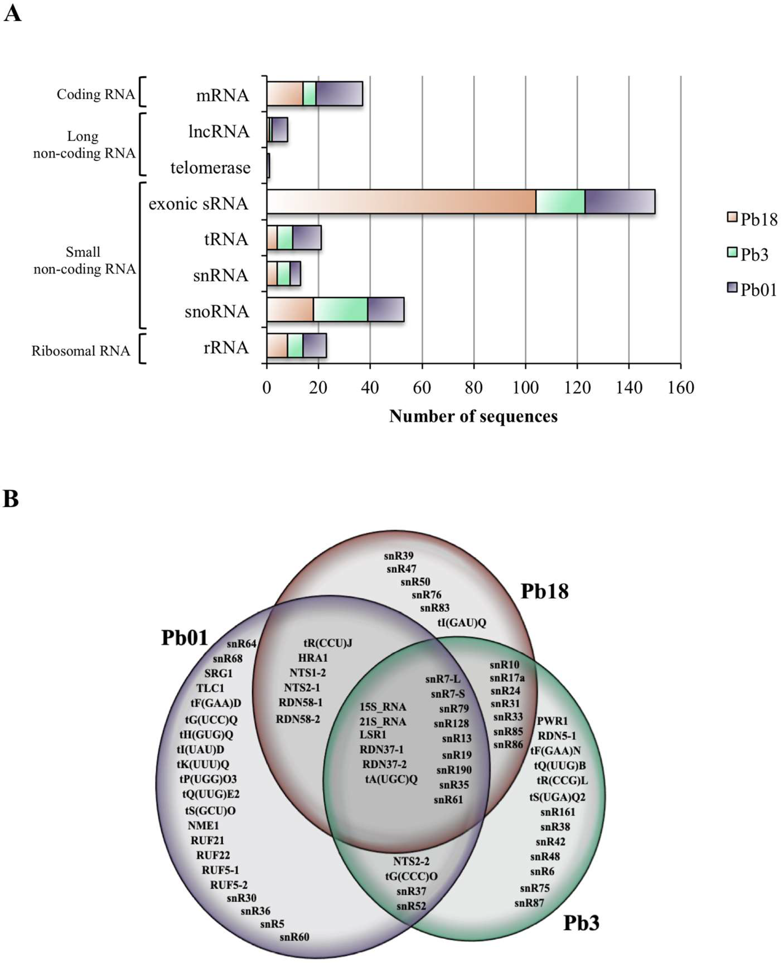
3.2. sRNA Sequences Aligning to mRNA Exons (Exonic sRNA)

The EV RNA reads detected in the < 200-nt fraction were initially aligned with each Paracoccidioides isolate-specific genome at NCBI. We noticed that this fraction was composed mostly of short 25-nt sequences in average that aligned to a specific region of a particular mRNA exon. In our previous work we observed that over 80% of the reads found in the <200-nt RNA fraction from EVs mapped to exons not only in P. brasiliensis Pb18, but also in C. albicans and S. cerevisiae, however not in C. neoformans [21]. In fungi, exonic short interfering RNAs (ex-siRNA) have been described to match to unique exon sites in either the reverse or the forward directions. They originate directly from single-strand RNA via an RNA-dependent RNA polymerase to generate double-stranded RNA (dsRNA) and are then converted to interfering RNA by Dicer [26]. Therefore, we presently deepened our investigation of the reads that had high depth of coverage in exonic regions. Each high-coverage coding sequence was manually accessed to evaluate the alignment position of the reads. Considering the reads generated by the sequencing of both < and > 200-nt fractions, 160 mRNA sequences showed high values of depth of coverage. Among them, only 30 had reads covering more than 50% of the entire mRNA, as detailed earlier. The remaining 130 were represented by small sequences that aligned at unique sites. We therefore called them exonic sRNA. We found a total of 104 (Pb18), 19 (Pb3), and 27 (Pb01) exonic sRNA in EVs that corresponded to a specific exonic region (5′, 3′or middle) of a single mRNA (Table 3 and Figure 1A). For Pb18 EVs, 53% of those sequences were only in the forward orientation (F), while 68% and 56% were only in the reverse (R) direction in Pb3 and Pb01 EVs, respectively.
Four EV exonic sRNA sequences mapped to a common transcript in all Paracoccidioides isolates: calcium calmodulin-dependent protein kinase (PADG_07652), nucleotide binding (PADG_03535), transcription factor tfiiib complex subunit brf1 (PADG_00916), and a hypothetical protein (PADG_11439). As seen in Table 3, most of the exonic sRNAs were specific to EVs from Pb18 (89) and Pb01 (21), whereas only four were exclusive to Pb3 EVs. Ten fragments mapped to homologous transcripts in Pb18 and Pb3 EVs. Most of the target sequences have unknown functions, while the others are related to carbohydrate/protein/DNA metabolism, translation, oxidation-reduction, and the signaling process (Table 3).
In conclusion, although the number of putative exonic sRNA was 4 to 5-fold higher in Pb18 EVs, 10 target sequences seemed to be characteristic of P. brasiliensis, whereas 21 were exclusive to P. lutzii. These results reveal a previously unknown diversity in the composition of fungal EVs at the genus level.

3.3. Comparison of EV ncRNA Classes in Paracoccidioides EVs

The different classes of ncRNA contained in Pb18, Pb3, and Pb01 EVs were analyzed by aligning the <200-nt reads with the ncRNA database from the Saccharomyces Genome Database. The results revealed the presence of 71 different sequences of ncRNA in Paracoccidioides EVs, from which 15 were common to all isolates and 17 were shared by two of them (Figure 1B). The most abundant class of ncRNA found in Paracoccidiodes EVs was the small nucleolar (sno)RNAs (33), followed by tRNAs (16), rRNAs (10), long ncRNAs (7), and small nuclear (sn)RNAs (4), as seen in Figure 1A. The snoRNA and tRNA were also the most abundant ncRNA populations described in our previous work not only for P. brasiliensis, but also for C. albicans, C. neoformans, and S. cerevisiae [21].
The number of total ncRNA detected in EVs varied with the isolate, being slightly more abundant in P. lutzii Pb01 (45) than in P. brasiliensis Pb3 (39) or Pb18 (35). The class profiles also differed between species. For P. brasiliensis EVs, snoRNA represented about 55% of the total, followed by rRNA (23% in Pb18 and 15% in Pb3), and 11 to 15% tRNA and snRNA (Figure 1A). For P. lutzii EVs, we found similar proportions (20 to 31%) of snoRNA, rRNA, and tRNA, followed by 9 to 13% of snRNA and other ncRNAs (Figure 1A).

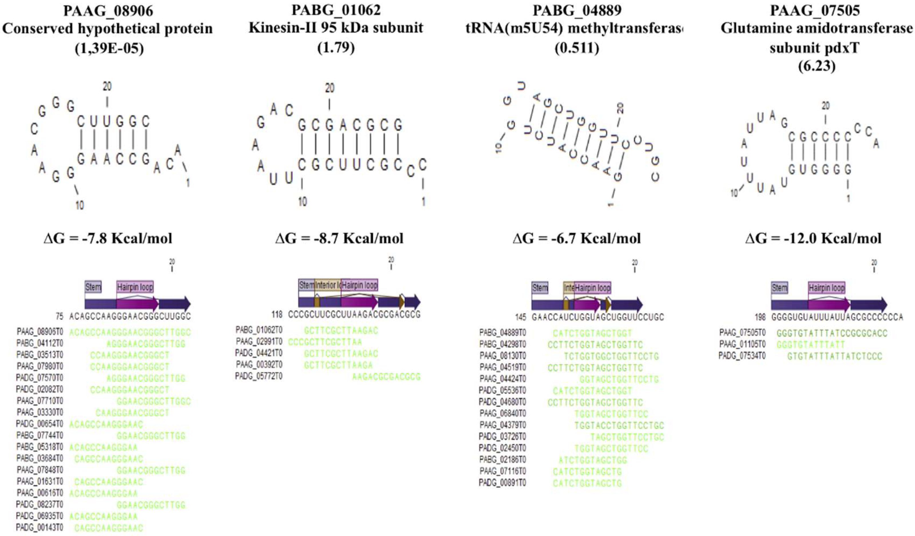
3.4. Secondary Structure in the EVs.

In our previous publication [21], we identified putative miRNA-like (milRNA) sequences in fungal EVs that aligned with mature miRNA following a search in the miRNA database of all organisms (http://www.mirbase.org). In the present work, we searched for sRNA sequences (up to 50 nt) bearing secondary structure in order to have a more precise view of the presence of milRNA in Paracoccidioides EVs. We applied the secondary structure analysis to RNA fragments detected in EVs at high abundance (RPKM > 5000) and for further characterization we selected 42 RNA sequences bearing secondary structures with free energy values below −3.0 Kcal/mol. The sequences with the most negative values are shown in Figure 2. We performed an alignment with these 42 sequences to infer their function and identify in which strand they would align (Supplemental Table S1). Two were present in all EV samples, while 23 were found in both Pb3 and Pb01 EVs. Interestingly, 50% of the structured RNAs aligned at the complementary strand of the RNA, thus suggesting that they could act as miRNA or the transcripts (Supplemental Table S1). Most of the structured RNAs localize to transcripts that code for hypothetical proteins, however we could also identify RNAs that align to transcripts of kinesin-II, glutamine amidotransferase and lysine methyltransferase (Figure 2).

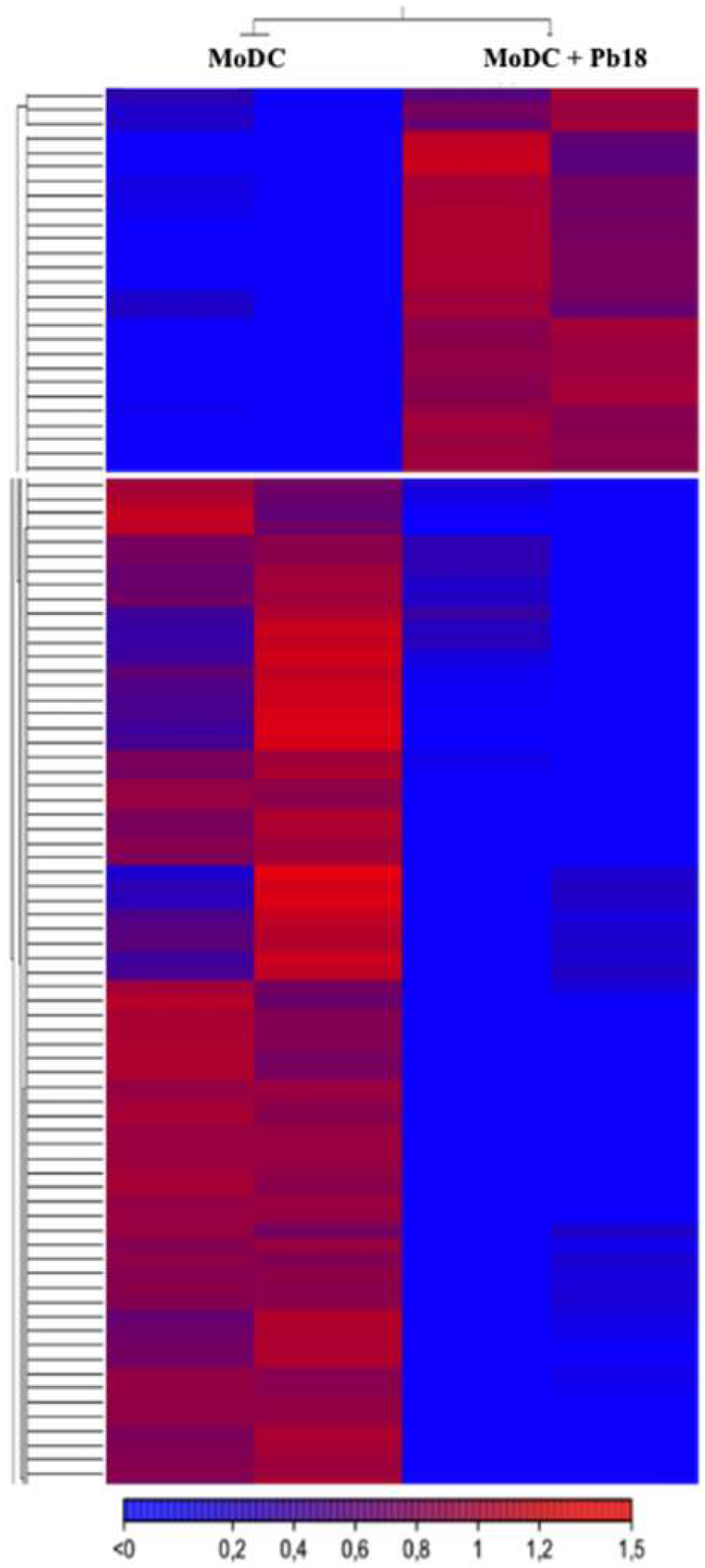
3.5. Paracoccidioides EVs Might Modulate the Transcriptome of Dendritic Cells

Dendritic cells make the bridge between the innate and adaptative immune responses, which ultimately define the course of the fungal infections [27]. The disease progression in paracoccidioidomycosis depends on dominant Th1 or Th2 types of immune response. The Th1-driven inflammatory immune response is responsible for protection against the disease and the IL-12 expressed by dendritic cells is detrimental to stimulate this type of response [28]. In this context, we investigated if extracellular fractions containing EVs produced by P. brasiliensis Pb18 would affect gene expression in recipient dendritic cells. In order to do that, we characterized the transcriptome of murine monocyte-derived CD11c+ (MoDC) cells co-cultivated for 48 h with the Pb18 yeast cells in a transwell system. We compared the sequences with those of the controls cultivated in the absence of fungal cells. In the transwell system, we had the fungal (upper compartment) and MoDC cell (bottom compartment) cultures communicating by a porous membrane that allowed the transit to the bottom well of soluble molecules and EVs up to 0.4 µm in size.
We observed that the indirect co-culture with Pb18 led to a slight alteration in the levels of mRNA expressed by MoDC cells when compared to the control (Figure 3 and Table S2). We detected 20 upregulated and 28 downregulated mRNAs (Figure 3). We chose to follow highly stringent criteria in this analysis (similar levels of expression in both replicates, FDR ≤ 5% and log FC ≥ 2), considering we had duplicates, but not triplicates. Among the upregulated transcripts, three code for membrane proteins (Ankar, Unc13c, and Smim7) and two code for mitochondrial proteins associated to translation elongation (mt-Te and mt-Tv) (Table S2). Among the downregulated transcripts, there was 25% enrichment for transcripts related to gene expression regulation (transcription and translation). When we applied the hypergeometric test to the annotated transcripts for the gene set enrichment analysis, the p-value of the gene expression group was 2.40 × 10⁵, thus reinforcing the significance of this enrichment (Table S2). It was interesting to notice that three transcripts that code for transcription factors (Gabpb2, Pknox1, and Zfp575) are among the downregulated MoDC mRNAs in indirect co-cultures with Pb18 (Table S2), suggesting that sRNAs from the fungal EVs were potentially able to modulate the gene expression of the recipient cell. It is important to note that we have preliminary cytometry data showing that Pb18 yeast cells can actively release EVs under our transwell experimental conditions and that MoDC cells can internalize fungal EVs, as depicted in Figure S1.

4. Discussion

In the present work we have shown that the features of mRNA and sRNA sub-populations contained in EVs from P. brasiliensis Pb18, Pb3, and P. lutzii Pb01 differ considerably between species and also between representative isolates of the same species. The results suggest that Paracoccicioides extracellular fractions containing EVs can modulate the transcription profile of dendritic cells. By using in vitro translation, we also reported for the first time in pathogenic fungi that the EV mRNAs are active and can be translated.
The mRNA sequences presently detected in the >200-nt RNA fraction of Paracoccidoides EVs varied in number with the isolate and only five orthologs were common to EVs from more than one isolate. We have recently compared the characteristics of mRNA sequences from EVs exported by two H. capsulatum isolates, specifically G186AR and G217B [29]. The latter isolate lacks cell wall alpha-1,3-glucan, which is a virulence factor [30]. The number of EV mRNA sequences varied from 93 in G186AR (mostly related to metabolic processes) to 31 in G217B (related to transport pathways possibly requiring vesicles, oxidation-reduction, and translation mechanisms). In the present analysis for P. brasiliensis, which is genetically related to Histoplasma [31], transcripts associated with DNA metabolism/biogenesis and protein modification were enriched in all samples, while those related with translation and transport prevailed, respectively, in Pb18 and Pb01.
While there are several pieces of evidence suggesting the role of fungal EVs on host cells, specially by interfering with the course of the immune response [32,33,34,35,36], it has recently been demonstrated that EV-like liposomal particles can cross the fungal cell wall inwards [37], thus opening the possibility that EVs can be uptaken and signal fungal cells. It has recently been demonstrated that EV-associated plant sRNA can silence virulence genes in a fungal pathogen, which agrees with the hypothesis that EVs mediate trans-kingdom regulation of gene expression [38]. On the other hand, Bielska et al. (2018) demonstrated that EVs produced by virulent Cryptococcus gattii were essential to signal quiescent strains within phagolysosomes located in distant body sites, which then became virulent; that phenomenon was apparently mediated by EV protein and RNA [39]. We have here detected active mRNA within Paracoccidioides EVs and we can envision that they might be uptaken by other neighboring or distant fungal cells and play a role in the host-fungal relationship by delivering virulence transcripts. Table 1 includes, for e.g., heat shock proteins like Hsp 70 (PAAG_08003, in Pb01 EVs) and Hsp 90-like (PADG_07715, in Pb18 EVs) that have a role in virulence of dimorphic fungi [40].
On the other hand, among the siRNA described in fungi, the exonic ex-siRNAs promote gene silencing in Mucor circinelloides and Fusarium graminearum [26,41,42]. In these species, the ex-siRNAs correspond to sense and antisense siRNAs converted into double-stranded RNA (dsRNA) via RNA-dependent RNA polymerase and Dicer. They regulate translation of the same mRNA that originated it. The presence of sense and antisense exonic sRNA-like in Paracoccidioides EVs suggests that these molecules could be involved in gene silencing via dsRNA. In the present work, we can point out the finding of exonic sRNA-like sequences, in Pb18 EVs, that map to α-amylase (PADG_04422), which is essential to the synthesis of the virulence factor α-glucan [30,43], and β-glucanase (PADG_04922), that cleaves dectin-1 ligand β-glucan present in the cell wall. We propose that they could have a role in regulating the expression of virulence genes in other fungal cells.
The comparison of the small ncRNA subtypes in Paracoccidioides EVs showed clear inter-species diversity. In P. lutzii, the percentage of snoRNA was about half that in P. brasiliensis, whereas for tRNA and other ncRNA it was, respectively, 2- and 4-fold higher. Consequently, 20 sequences were exclusive of P. lutzii EVs (mostly tRNA) versus six for Pb18 and thirteen for Pb3. In our experimental conditions, H. capsulatum EVs transport almost exclusively rRNA and tRNA [29]. The tRNA sequences prevailed in the EVs from isolate G186AR and most of them were not detected in EVs from the G217R strain. The fragments of tRNAs (the tRFs) are implicated in diverse processes in the cell, from regulation of cell viability, protein synthesis, apoptosis, to RNA metabolism, including turnover and stability [44]. The tRFs present in the EVs form Trypanosoma cruzi, the causing agent of Chagas disease, can be transferred to other parasites and promote cell communication and/or to host cells to modulate gene expression or facilitate infection [45].
The gene-silencing mechanism known as RNA interference (RNAi) is prompted by small noncoding (s)RNAs averaging 25 nt that act at either a post-transcriptional or post-translational level. The RNAi-related sRNAs are short interfering RNAs (siRNA), microRNAs (miRNA), and piwi-interacting RNAs (piRNA). In fungi, the conventional miRNA pathway has only been demonstrated in C. neoformans [46], while alternative miRNA-like (mil-RNA) pathways have been reported in, for ex., Fusarium oxysporum [47], Penicillium marneffei [48], and Neurospora crassa [49].
We have previously detected 145 sequences in EVs from P. brasiliensis Pb18 that matched those of mature miRNA deposited in the miRNA database (mirbase). More recently, Curcio et al. [50] searched for matches of mil-RNAs already described in fungi, discarded those located in genes and looked for pre-miRNA secondary structure in the genome by considering their flanking nucleotides. They found that 11 mil-RNAs previously reported in fungal EVs matched the criteria for mil-RNA in the P. brasiliensis Pb18 genome. Besides, the authors found that in Pb18 the paralogous genes for Argonaute (1 and 2) and Dicer (1 and 2) (PADG_00716; PADG_03108; PADG_11946; PADG_07189) seem to be induced in the pathogenic yeast phase of the fungus, suggesting that mechanisms involving RNAi can be functional in this species [50]. We have presently shown partial secondary RNA structures in sRNA sequences from EVs that was differentially represented in the Paracoccidioides isolates. These sequences aligned to exons, notably in the reverse position. We found similar results for H. capsulatum [29], but the role of these molecules has to be investigated further. It was interesting to find, in Histoplasma EVs, a series of RNA-binding proteins and one of them, Snd1, is a component of the RNA-induced silencing complex (RISC) that is part of the RNAi machinery [51]. In Pb18 EVs [52] we have not found RNA-binding proteins that matched those found in Histoplasma EVs (data not shown).
Modulation of the host immune system by EV components has been described in C. neoformans, C. albicans, Malassezia sympodialis, P. brasiliensis and Sporothrix brasiliensis. The fungal EVs activate immune cells in vitro inducing the release of pro-inflammatory mediators, suggesting a role of fungal EVs in activating the immune system to respond to the fungal infection [32,33,34,35,36]. Importantly, in all studies performed so far, it was uncertain if the density of EVs used to stimulate host cells corresponded to that observed during physiologic and/or pathogenic conditions. Therefore, we opted for a transwell-based experimental system where the amount of fungal EVs corresponded to that physiologically produced by P. brasiliensis, even with the limitation that molecules not related to EVs may participate in the interaction with host cells. We observed that the Pknox1 and Gbpb2 transcription factors were highly downregulated (65-fold and 30-fold, respectively) in MoDC cells upon indirect co-coculture with P. brasiliensis in the transwell experiment. Pknox1 (Pbx/knotted 1 homeobox) belongs to the HOX family of transcription factors and is critical for the immune system homeostasis as it regulates the expression of IL-10 in macrophages and dendritic cells [53]. Gbpb2 (GA-binding protein subunit beta 2) is critical for T cell development and the expression regulation of IL-7R alpha [54]. IL-7 is associated with improved immune response against bacterial and viral infections, and also in fungal sepsis [55] and Pneumocystis infection [56]; however, a direct correlation of IL-7 and paracoccidioidomycosis has not been evaluated yet. On the other hand, IL-10 is a potent anti-inflammatory cytokine that is generally associated with increased infection in paracoccidioidomycosis [57]. Our experimental design using a transwell system did not allow distinguishing between the biological effects of EVs from other secreted molecules. However, considering our data suggesting that Paracoccidioides Pb18 produce EVs that are uptaken by MoDC in a transwell system (Figure S1), we feel entitled to assume that EVs from P. brasiliensis Pb18 might help to modulate the MoDC response, possibly favoring the infection at the early stages of interaction with cells of the innate immune system. This modulation could be related to regulatory sRNA carried by the fungal EVs or even by surface ligands like DC-SIGN mannose ligands exposed in the EV surface [58].
Considering that sRNA classes are involved in transcription/translation modulation in a variety of systems and potentially also in Paracoccidioides, our general results may indicate that differences in virulence among fungal isolates could be related to their distinct EV RNA content. That hypothesis will hopefully be experimentally tested.

Supplementary Materials

The following files are available online, Figure S1: Uptake of EVs from Pb18 by MoDC cells in a transwell system, Table S1: List of genes from Pb18, Pb3, and Pb01 matching partially structured sRNA, as indicated. Separated lists contain those found exclusively in one isolate or shared by two or three of them, Table S2: MoDC genes differentially expressed (upregulated in green and downregulated in pink) after incubation with Pb18 cells in a transwell system, as analyzed using the RNA-seq strategy.

Author Contributions

Conceptualization, R.P.d.S., M.L.R., S.G., L.R.A. and R.P.; Data curation, R.P.D.S., L.R.A., L.G.V.L., J.P.C.D.C., T.J.P.S.; Formal analysis, R.P.d.S., J.P.C.D.C., L.R.A. and R.P.; Funding acquisition, R.P., M.L.R. and S.G.; Methodology, R.P.d.S., J.P.C.D.C., H.F., S.G. and L.R.A; Supervision, R.P.; Writing—original draft, R.P.d.S., L.R.A., and R.P.; Writing—review & editing, R.P.d.S., L.G.V.L., M.L.R., S.G., L.R.A. and R.P.

Funding

This work was supported by Fundação de Amparo à Pesquisa (grant 13/25950-1 and scholarship 10/19410-9). Sequencing was carried out in the plataforms of Universidade Federal do Paraná (through INCT). MLR was supported by grants from the Brazilian agency Conselho Nacional de Desenvolvimento Científico e Tecnológico (CNPq, grants 405520/2018-2, 440015/2018-9, and 301304/2017-3), and Fiocruz (grants VPPCB-007-FIO-18-2-57 and VPPIS-001-FIO-18-66).

Conflicts of Interest

The authors declare no conflict of interest.

Abbreviations

EVsExtracellular vesicles
GM-CSFGranulocyte macrophage colony-stimulating factor
miRNAMicro RNA
ncRNANon-coding RNA
PCMparacoccidioidomycosis
sRNASmall RNA
snoRNAsSmall nucleolar RNA
tRNATransporter RNA

References

  1. Martinez, R. New trends in paracoccidioidomycosis epidemiology. J. Fungi 2017, 3, 1. [Google Scholar] [CrossRef] [PubMed]
  2. Bocca, A.L.; Amaral, A.C.; Teixeira, M.M.; Sato, P.K.; Shikanai-Yasuda, M.A.; Soares Felipe, M.S. Paracoccidioidomycosis: Eco-epidemiology, taxonomy and clinical and therapeutic issues. Future Microbiol. 2013, 8, 1177–1191. [Google Scholar] [CrossRef] [PubMed]
  3. Teixeira, M.M.; Theodoro, R.C.; Nino-Vega, G.; Bagagli, E.; Felipe, M.S. Paracoccidioides species complex: Ecology, phylogeny, sexual reproduction, and virulence. PLoS Pathog. 2014, 10, e1004397. [Google Scholar] [CrossRef] [PubMed]
  4. Turissini, D.A.; Gomez, O.M.; Teixeira, M.M.; McEwen, J.G.; Matute, D.R. Species boundaries in the human pathogen Paracoccidioides. Fungal Genet. Biol. 2017, 106, 9–25. [Google Scholar] [CrossRef] [PubMed]
  5. Matute, D.R.; Sepulveda, V.E.; Quesada, L.M.; Goldman, G.H.; Taylor, J.W.; Restrepo, A.; McEwen, J.G. Microsatellite analysis of three phylogenetic species of Paracoccidioides brasiliensis. J. Clin. Microbiol. 2006, 44, 2153–2157. [Google Scholar] [CrossRef] [PubMed]
  6. Carvalho, K.C.; Ganiko, L.; Batista, W.L.; Morais, F.V.; Marques, E.R.; Goldman, G.H.; Franco, M.F.; Puccia, R. Virulence of Paracoccidioides brasiliensis and gp43 expression in isolates bearing known pbgp43 genotype. Microbes Infect. 2005, 7, 55–65. [Google Scholar] [CrossRef] [PubMed]
  7. Munoz, J.F.; Farrer, R.A.; Desjardins, C.A.; Gallo, J.E.; Sykes, S.; Sakthikumar, S.; Misas, E.; Whiston, E.A.; Bagagli, E.; Soares, C.M.; et al. Genome diversity, recombination, and virulence across the major lineages of paracoccidioides. mSphere 2016, 1, e00213-16. [Google Scholar] [CrossRef] [PubMed]
  8. Cocucci, E.; Racchetti, G.; Meldolesi, J. Shedding microvesicles: Artefacts no more. Trends Cell Biol. 2009, 19, 43–51. [Google Scholar] [CrossRef] [PubMed]
  9. Kalra, H.; Drummen, G.P.; Mathivanan, S. Focus on extracellular vesicles: Introducing the next small big thing. Int. J. Mol. Sci. 2016, 17, 170. [Google Scholar] [CrossRef]
  10. Raposo, G.; Stoorvogel, W. Extracellular vesicles: Exosomes, microvesicles, and friends. J. Cell Biol. 2013, 200, 373–383. [Google Scholar] [CrossRef] [Green Version]
  11. Rodrigues, M.L.; Franzen, A.J.; Nimrichter, L.; Miranda, K. Vesicular mechanisms of traffic of fungal molecules to the extracellular space. Curr. Opin. Microbiol. 2013, 16, 414–420. [Google Scholar] [CrossRef] [PubMed]
  12. Thery, C.; Witwer, K.W.; Aikawa, E.; Alcaraz, M.J.; Anderson, J.D.; Andriantsitohaina, R.; Antoniou, A.; Arab, T.; Archer, F.; Atkin-Smith, G.K.; et al. Minimal information for studies of extracellular vesicles 2018 (misev2018): A position statement of the international society for extracellular vesicles and update of the misev2014 guidelines. J. Extracell. Vesicles 2018, 7, 1535750. [Google Scholar] [CrossRef] [PubMed]
  13. Schorey, J.S.; Harding, C.V. Extracellular vesicles and infectious diseases: New complexity to an old story. J. Clin. Investig. 2016, 126, 1181–1189. [Google Scholar] [CrossRef] [PubMed]
  14. Hosseini-Beheshti, E.; Grau, G.E.R. Extracellular vesicles as mediators of immunopathology in infectious diseases. Immunol. Cell Biol. 2018. [Google Scholar] [CrossRef]
  15. Volgers, C.; Benedikter, B.J.; Grauls, G.E.; Hellebrand, P.H.M.; Savelkoul, P.H.M.; Stassen, F.R.M. Effects of n-acetyl-l-cysteine on the membrane vesicle release and growth of respiratory pathogens. FEMS Microbiol. Lett. 2017, 364. [Google Scholar] [CrossRef] [PubMed]
  16. Mateescu, B.; Kowal, E.J.; van Balkom, B.W.; Bartel, S.; Bhattacharyya, S.N.; Buzas, E.I.; Buck, A.H.; de Candia, P.; Chow, F.W.; Das, S.; et al. Obstacles and opportunities in the functional analysis of extracellular vesicle rna - an isev position paper. J. Extracell. Vesicles 2017, 6, 1286095. [Google Scholar] [CrossRef] [PubMed]
  17. Valadi, H.; Ekstrom, K.; Bossios, A.; Sjostrand, M.; Lee, J.J.; Lotvall, J.O. Exosome-mediated transfer of mrnas and micrornas is a novel mechanism of genetic exchange between cells. Nat. Cell Biol. 2007, 9, 654–659. [Google Scholar] [CrossRef]
  18. Pegtel, D.M.; Cosmopoulos, K.; Thorley-Lawson, D.A.; van Eijndhoven, M.A.; Hopmans, E.S.; Lindenberg, J.L.; de Gruijl, T.D.; Wurdinger, T.; Middeldorp, J.M. Functional delivery of viral mirnas via exosomes. Proc. Natl. Acad. Sci. USA 2010, 107, 6328–6333. [Google Scholar] [CrossRef] [PubMed]
  19. Bayraktar, R.; Van Roosbroeck, K.; Calin, G.A. Cell-to-cell communication: Micrornas as hormones. Mol. Oncol. 2017, 11, 1673–1686. [Google Scholar] [CrossRef] [PubMed]
  20. Fatima, F.; Nawaz, M. Vesiculated long non-coding rnas: Offshore packages deciphering trans-regulation between cells, cancer progression and resistance to therapies. Non-Coding RNA 2017, 3, 10. [Google Scholar] [CrossRef]
  21. Peres da Silva, R.; Puccia, R.; Rodrigues, M.L.; Oliveira, D.L.; Joffe, L.S.; Cesar, G.V.; Nimrichter, L.; Goldenberg, S.; Alves, L.R. Extracellular vesicle-mediated export of fungal rna. Sci. Rep. 2015, 5, 7763. [Google Scholar] [CrossRef] [PubMed]
  22. Vallejo, M.C.; Matsuo, A.L.; Ganiko, L.; Medeiros, L.C.; Miranda, K.; Silva, L.S.; Freymuller-Haapalainen, E.; Sinigaglia-Coimbra, R.; Almeida, I.C.; Puccia, R. The pathogenic fungus Paracoccidioides brasiliensis exports extracellular vesicles containing highly immunogenic α-galactosyl epitopes. Eukaryot. Cell 2011, 10, 343–351. [Google Scholar] [CrossRef] [PubMed]
  23. Mortazavi, A.; Williams, B.A.; McCue, K.; Schaeffer, L.; Wold, B. Mapping and quantifying mammalian transcriptomes by RNA-seq. Nat. Methods 2008, 5, 621–628. [Google Scholar] [CrossRef] [PubMed]
  24. Sükösd, Z.; Knudsen, B.; Kjems, J.; Pedersen, C.N.S. Ppfold 3.0: Fast rna secondary structure prediction using phylogeny and auxiliary data. Bioinformatics 2012, 28, 2691–2692. [Google Scholar] [CrossRef] [PubMed]
  25. Williams, L.M.; Fu, Z.; Dulloor, P.; Yen, T.; Barron-Casella, E.; Savage, W.; Van Eyk, J.E.; Casella, J.F.; Everett, A. Hemoglobin depletion from plasma: Considerations for proteomic discovery in sickle cell disease and other hemolytic processes. Proteom. Clin. Appl. 2010, 4, 926–930. [Google Scholar] [CrossRef]
  26. Nicolas, F.E.; Moxon, S.; de Haro, J.P.; Calo, S.; Grigoriev, I.V.; Torres-Martinez, S.; Moulton, V.; Ruiz-Vazquez, R.M.; Dalmay, T. Endogenous short rnas generated by dicer 2 and rna-dependent rna polymerase 1 regulate mrnas in the basal fungus Mucor circinelloides. Nucleic Acids Res. 2010, 38, 5535–5541. [Google Scholar] [CrossRef] [PubMed]
  27. Gow, N.A.; Hube, B. Importance of the Candida albicans cell wall during commensalism and infection. Curr. Opin. Microbiol. 2012, 15, 406–412. [Google Scholar] [CrossRef] [PubMed]
  28. Thind, S.K.; Taborda, C.P.; Nosanchuk, J.D. Dendritic cell interactions with Histoplasma and Paracoccidioides. Virulence 2015, 6, 424–432. [Google Scholar] [CrossRef]
  29. Alves, L.R.; Peres da Silva, R.; Sanchez, D.A.; Zamith-Miranda, D.; Rodrigues, M.L.; Goldenberg, S.; Puccia, R.; Nosanchuk, J.D. Extracellular vesicle-mediated rna release in Histoplasma capsulatum. mSphere 2019, 4, e00176-19. [Google Scholar] [CrossRef]
  30. Rappleye, C.A.; Eissenberg, L.G.; Goldman, W.E. Histoplasma capsulatum α-(1,3)-glucan blocks innate immune recognition by the beta-glucan receptor. Proc. Natl. Acad. Sci. USA 2007, 104, 1366–1370. [Google Scholar] [CrossRef]
  31. Jiang, Y.; Dukik, K.; Muñoz, J.F.; Sigler, L.; Schwartz, I.S.; Govender, N.P.; Kenyon, C.; Feng, P.; van den Ende, B.G.; Stielow, J.B.; et al. Phylogeny, ecology and taxonomy of systemic pathogens and their relatives in Ajellomycetaceae (onygenales): Blastomyces, Emergomyces, Emmonsia, Emmonsiellopsis. Fungal Divers. 2018, 90, 245–291. [Google Scholar] [CrossRef]
  32. da Silva, T.A.; Roque-Barreira, M.C.; Casadevall, A.; Almeida, F. Extracellular vesicles from Paracoccidioides brasiliensis induced m1 polarization in vitro. Sci. Rep. 2016, 6, 35867. [Google Scholar] [CrossRef] [PubMed]
  33. Gehrmann, U.; Qazi, K.R.; Johansson, C.; Hultenby, K.; Karlsson, M.; Lundeberg, L.; Gabrielsson, S.; Scheynius, A. Nanovesicles from Malassezia sympodialis and host exosomes induce cytokine responses--novel mechanisms for host-microbe interactions in atopic eczema. PLoS ONE 2011, 6, e21480. [Google Scholar] [CrossRef] [PubMed]
  34. Oliveira, D.L.; Freire-de-Lima, C.G.; Nosanchuk, J.D.; Casadevall, A.; Rodrigues, M.L.; Nimrichter, L. Extracellular vesicles from Cryptococcus neoformans modulate macrophage functions. Infect. Immun. 2010, 78, 1601–1609. [Google Scholar] [CrossRef] [PubMed]
  35. Vargas, G.; Rocha, J.D.; Oliveira, D.L.; Albuquerque, P.C.; Frases, S.; Santos, S.S.; Nosanchuk, J.D.; Gomes, A.M.; Medeiros, L.C.; Miranda, K.; et al. Compositional and immunobiological analyses of extracellular vesicles released by Candida albicans. Cell Microbiol. 2014. [Google Scholar] [CrossRef]
  36. Ikeda, M.A.K.; de Almeida, J.R.F.; Jannuzzi, G.P.; Cronemberger-Andrade, A.; Torrecilhas, A.C.T.; Moretti, N.S.; da Cunha, J.P.C.; de Almeida, S.R.; Ferreira, K.S. Extracellular vesicles from Sporothrix brasiliensis are an important virulence factor that induce an increase in fungal burden in experimental sporotrichosis. Front. Microbiol. 2018, 9, 2286. [Google Scholar] [CrossRef] [PubMed]
  37. Walker, L.; Sood, P.; Lenardon, M.D. The viscoelastic properties of the fungal cell wall allow traffic of ambisome as intact liposome vesicles. MBio 2018, 9. [Google Scholar] [CrossRef]
  38. Cai, Q.; Qiao, L.; Wang, M.; He, B.; Lin, F.M.; Palmquist, J.; Huang, S.D.; Jin, H. Plants send small rnas in extracellular vesicles to fungal pathogen to silence virulence genes. Science 2018, 360, 1126–1129. [Google Scholar] [CrossRef]
  39. Bielska, E.; Sisquella, M.A.; Aldeieg, M.; Birch, C.; O’Donoghue, E.J.; May, R.C. Pathogen-derived extracellular vesicles mediate virulence in the fatal human pathogen Cryptococcus Gattii. Nat. Commun. 2018, 9, 1556. [Google Scholar] [CrossRef] [PubMed]
  40. Cleare, L.G.; Zamith-Miranda, D.; Nosanchuk, J.D. Heat shock proteins in Histoplasma and Paracoccidioides. Clin. Vaccine Immunol. 2017, 24, e00221-17. [Google Scholar] [CrossRef]
  41. Nicolas, F.E.; Ruiz-Vazquez, R.M. Functional diversity of rnai-associated srnas in fungi. Int. J. Mol. Sci. 2013, 14, 15348–15360. [Google Scholar] [CrossRef]
  42. Son, H.; Park, A.R.; Lim, J.Y.; Shin, C.; Lee, Y.W. Genome-wide exonic small interference rna-mediated gene silencing regulates sexual reproduction in the homothallic fungus Fusarium graminearum. PLoS Genet. 2017, 13, e1006595. [Google Scholar] [CrossRef]
  43. Camacho, E.; Sepulveda, V.E.; Goldman, W.E.; San-Blas, G.; Nino-Vega, G.A. Expression of paracoccidioides brasiliensis amy1 in a Histoplasma capsulatum amy1 mutant, relates an alpha-(1,4)-amylase to cell wall alpha-(1,3)-glucan synthesis. PLoS ONE 2012, 7, e50201. [Google Scholar] [CrossRef]
  44. Kumar, P.; Kuscu, C.; Dutta, A. Biogenesis and function of transfer rna-related fragments (trfs). Trends Biochem. Sci. 2016, 41, 679–689. [Google Scholar] [CrossRef]
  45. Garcia-Silva, M.R.; Cabrera-Cabrera, F.; das Neves, R.F.; Souto-Padron, T.; de Souza, W.; Cayota, A. Gene expression changes induced by Trypanosoma cruzi shed microvesicles in mammalian host cells: Relevance of trna-derived halves. Biomed Res. Int. 2014, 2014, 305239. [Google Scholar] [CrossRef]
  46. Jiang, N.; Yang, Y.; Janbon, G.; Pan, J.; Zhu, X. Identification and functional demonstration of mirnas in the fungus Cryptococcus neoformans. PLoS ONE 2012, 7, e52734. [Google Scholar] [CrossRef]
  47. Chen, R.; Jiang, N.; Jiang, Q.; Sun, X.; Wang, Y.; Zhang, H.; Hu, Z. Exploring microrna-like small rnas in the filamentous fungus Fusarium oxysporum. PLoS ONE 2014, 9, e104956. [Google Scholar] [CrossRef]
  48. Lau, S.K.; Chow, W.N.; Wong, A.Y.; Yeung, J.M.; Bao, J.; Zhang, N.; Lok, S.; Woo, P.C.; Yuen, K.Y. Identification of microrna-like rnas in mycelial and yeast phases of the thermal dimorphic fungus Penicillium marneffei. PLoS Negl. Trop. Dis. 2013, 7, e2398. [Google Scholar] [CrossRef]
  49. Lee, H.C.; Li, L.; Gu, W.; Xue, Z.; Crosthwaite, S.K.; Pertsemlidis, A.; Lewis, Z.A.; Freitag, M.; Selker, E.U.; Mello, C.C.; et al. Diverse pathways generate microrna-like rnas and dicer-independent small interfering rnas in fungi. Mol. Cell 2010, 38, 803–814. [Google Scholar] [CrossRef]
  50. Curcio, J.S.; Batista, M.P.; Paccez, J.D.; Novaes, E.; Soares, C.M.A. In silico characterization of micrornas-like sequences in the genome of Paracoccidioides brasiliensis. Genet. Mol. Biol. 2019, 42, 95–107. [Google Scholar] [CrossRef]
  51. Santhekadur, P.K.; Das, S.K.; Gredler, R.; Chen, D.; Srivastava, J.; Robertson, C.; Baldwin, A.S., Jr.; Fisher, P.B.; Sarkar, D. Multifunction protein staphylococcal nuclease domain containing 1 (snd1) promotes tumor angiogenesis in human hepatocellular carcinoma through novel pathway that involves nuclear factor kappab and mir-221. J. Biol. Chem. 2012, 287, 13952–13958. [Google Scholar] [CrossRef]
  52. Vallejo, M.C.; Nakayasu, E.S.; Matsuo, A.L.; Sobreira, T.J.; Longo, L.V.; Ganiko, L.; Almeida, I.C.; Puccia, R. Vesicle and vesicle-free extracellular proteome of Paracoccidioides brasiliensis: Comparative analysis with other pathogenic fungi. J. Proteome Res. 2012, 11, 1676–1685. [Google Scholar] [CrossRef]
  53. Chung, E.Y.; Liu, J.; Homma, Y.; Zhang, Y.; Brendolan, A.; Saggese, M.; Han, J.; Silverstein, R.; Selleri, L.; Ma, X. Interleukin-10 expression in macrophages during phagocytosis of apoptotic cells is mediated by homeodomain proteins pbx1 and prep-1. Immunity 2007, 27, 952–964. [Google Scholar] [CrossRef]
  54. Yu, S.; Zhao, D.M.; Jothi, R.; Xue, H.H. Critical requirement of gabpalpha for normal T cell development. J. Biol. Chem. 2010, 285, 10179–10188. [Google Scholar] [CrossRef]
  55. Unsinger, J.; Burnham, C.A.; McDonough, J.; Morre, M.; Prakash, P.S.; Caldwell, C.C.; Dunne, W.M., Jr.; Hotchkiss, R.S. Interleukin-7 ameliorates immune dysfunction and improves survival in a 2-hit model of fungal sepsis. J. Infect. Dis. 2012, 206, 606–616. [Google Scholar] [CrossRef]
  56. Ruan, S.; Samuelson, D.R.; Assouline, B.; Morre, M.; Shellito, J.E. Treatment with interleukin-7 restores host defense against Pneumocystis in cd4+ t-lymphocyte-depleted mice. Infect. Immun. 2016, 84, 108–119. [Google Scholar] [CrossRef]
  57. Calich, V.L.; da Costa, T.A.; Felonato, M.; Arruda, C.; Bernardino, S.; Loures, F.V.; Ribeiro, L.R.; de Cassia Valente-Ferreira, R.; Pina, A. Innate immunity to Paracoccidioides brasiliensis infection. Mycopathologia 2008, 165, 223–236. [Google Scholar] [CrossRef]
  58. Peres da Silva, R.; Heiss, C.; Black, I.; Azadi, P.; Gerlach, J.Q.; Travassos, L.R.; Joshi, L.; Kilcoyne, M.; Puccia, R. Extracellular vesicles from Paracoccidioides pathogenic species transport polysaccharide and expose ligands for dc-sign receptors. Sci. Rep. 2015, 5, 14213. [Google Scholar]
Figure 1. (A) Graph showing the number of sequences in each class of RNA identified in EV preparations from Pb18, Pb3 and Pb01. (B) Venn diagram showing all ncRNA sequences found in EV preparations from Pb18, Pb3 and Pb01.
Cells 08 00765 g001
Figure 2. Secondary structures with the lowest deltaG among 42 sRNA sequences with values below −3 Kcal/mol. The Blast search result for these sequences is shown.
Cells 08 00765 g002
Figure 3. Heat map of transcripts from MoDC cells that were modulated over 2-fold and had a p-value < 0.05 when co-cultivated indirectly with Pb18 in a transwell system.
Cells 08 00765 g003
Table 1. Proteins corresponding to validated mRNA sequences (RPKM > 100) found in Paracoccidioides EVs. The sequences are grouped according with their functions. Coverage used for validation is indicated, as well as the gene access codes for each isolate.
Pb18 Feature IDPb3 Feature IDPb01 Feature IDSequence DescriptionCoverageGO
Protein Modification
PAAG_08003hsp70-like protein100%ion binding
PAAG_05980ubiquitin-conjugating enzyme e2-16 kda80%ligase activity
PADG_07715 hsp90-like protein70%protein folding, response to stress
PADG_01605PABG_03078PAAG_07080polyubiquitin70%protein modification process
PADG_11111 nuclear transport factor 275%protein targeting
PAAG_05679ATP-dependent molecular chaperone hsc8290%nucleic acid binding transcription factor activity, protein folding
Carbohydrate Metabolism
PADG_02145 glycogen phosphorylase70%carbohydrate metabolic process
Translation
PADG_05025 60s ribosomal protein l2670%translation
PADG_00692 PAAG_11418elongation factor 1-alpha100%translation
PADG_03326 40s ribosomal protein s980%translation
PABG_05744 pre-mrna splicing factor70%translation
Oxidation-Reduction
PAAG_03216thiol-specific antioxidant90%oxidoreductase activity
Transport
PAAG_11262hsp7-like protein100% + MRtransmembrane transport
PAAG_03058high-affinity methionine permease100% + 5′Rtransmembrane transport
PAAG_07634gtp-binding protein rhoa70%vesicle-mediated transport
DNA Metabolism or Biogenesis
PADG_00873PABG_02444PAAG_07099histone h3100%chromosome organization
PABG_05588PAAG_08917histone h2a90%chromosome organization
PAAG_08918histone h2b70%chromosome organization
PADG_06568 tctp family protein50%cell differentiation
PABG_03449PAAG_08247calmodulin70%ion binding
Other/Unknown Function
PADG_02280 hypothetical protein75%Unknown
PADG_02399 calcium-binding protein70%Unknown
PADG_04049 hypothetical protein60%Unknown
PADG_08402 hypothetical protein100%Unknown
PADG_12385 ser thr protein phosphatase family protein60%Unknown
PAAG_00340conserved hypothetical protein85%unknown
PAAG_12435hypothetical protein80%unknown
PAAG_12692ATP synthase subunit beta95% reverseunknown
PAAG_12694plant senescence-associated protein100% reverseunknown
PAAG_02087kelch-like protein 3870%unknown
Table 2. Protein sequences detected by in vitro translation of total mRNA extracted from Pb18 EVs. The mean RPKM corresponds to that found in the RNA-seq analysis.
Feature IDNameRPKM Mean
PADG_00648conserved hypothetical protein 7
PADG_0405614-3-3 protein epsilon 25
PADG_04810 GTP-binding nuclear protein GSP1/Ran 37
PADG_06159sulfate transporter 12
PADG_07715Hsp90-like protein 127
Table 3. Target mRNA of exonic sRNA sequences found in EVs from Pb18, Pb3, and Pb01 isolates. The position of sRNA in the exon is indicated as 5′, 3′, and M (middle), as well as the direction (F, forward; R, reverse). The sequences are grouped according with their functions.
Pb18
Feature ID
AlignmentPb3
Feature ID
AlignmentPb01
Feature ID
AlignmentSequence DescriptionGO
Protein modification
PADG_013655′R/3′F disulfide isomeraseprotein folding
PADG_03114MR phospho-2-dehydro-3-deoxyheptonate aldolaseamino acid metabolic process
PADG_040923′F peptidyl-prolyl cis-trans isomerase bprotein modification process
PADG_05011MF peptidyl-prolyl cis-trans isomerase-like 3protein modification process
PADG_055603′F 26s proteasome regulatory subunit rpn-1small molecule metabolic process
PADG_057313′F hypothetical proteinamino acid metabolic process
PADG_072415′F dihydroxy-acid dehydrataseamino acid metabolic process
PADG_07550MRPABG_04093MR microsomal signal peptidase subunitpeptidase activity
PAAG_059625′Fproteasome regulatory particle subunitcatabolic process
Carbohydrate Metabolism
PADG_021455′FPABG_068015′F glycogen phosphorylasecarbohydrate metabolic process
PADG_03169MR alpha-glucan synthase ags2carbohydrate metabolic process
PADG_04432MF alpha-amylasecarbohydrate metabolic process
PADG_049223′F cell wall glucanasecarbohydrate metabolic process
PADG_058705′R/5′F glucan synthesis regulatory proteincarbohydrate metabolic process
Lipid Metabolism
PADG_124305′FPABG_072953′F amp-bindinglong-chain fatty acid metabolic process
Oxidation-Reduction
PADG_044193′RPABG_010643′R proline oxidasecatabolic process
PAAG_05378MFd-3-phosphoglycerate dehydrogenaseoxidoreductase activity
PAAG_019373′Fduf887 domain-containing proteinbiosynthetic process
PAAG_11262mRNA/MRhsp7-like proteintransmembrane transpor
PADG_050805′F pyridoxamine phosphate oxidase family proteinion binding
PADG_061813′F c-5 sterol desaturaselipid metabolic process
PADG_074315′R/3′F/3R chloroperoxidase-like proteinhomeostatic process
PADG_122145′R alcohol dehydrogenaseoxidoreductase activity
Translation
PADG_00995MF ubiquitin-40s ribosomal protein s27atranslation
PADG_02452MF rna polymerase rpb1 c-terminal repeat domain-containing protein
PADG_02484MF valyl-trna synthetasetRNA metabolic process
PADG_05025MF 60s ribosomal protein l26translation
PADG_060823′F pre-mrna splicing factortranslation
PADG_06160MF eukaryotic translation initiation factor 2 alpha subunittranslation
PADG_061915′FPABG_069643′R trna isopentenyltransferasetRNA metabolic process
PADG_065225′F glycine--trna ligase
PADG_068335′F ATP-dependent rna helicase drs1ribosome biogenesis
PADG_086055′F 40s ribosomal protein s28translation
PADG_01891MF translation initiation factor rli1translation
PADG_023173′R translation machinery-associated protein 17
Signaling Process
PADG_054473′F vacuolar membrane-associated protein iml1signal transduction
PADG_066425′F ste ste7 protein kinaseresponse to stress
PADG_08337*3′F gtp-binding protein rhoasignal transduction
Transport
PADG_00326MF adp-ribosylation factor-like protein 1vesicle-mediated transport
PADG_013033′F abc transporterbiosynthetic process
PADG_015673′R sorting nexin 3vesicle-mediated transport
PADG_050845′R high affinity copper transportertransmembrane transport
PADG_058215′F importin
PADG_069823′F ncs1 family nucleobase:cation symporter-1transmembrane transport
PADG_081013′F mrna cleavage factor complex component pcf11transport
PADG_03535MRPABG_01859MRPAAG_01288MRnucleotide bindingion binding, ligase activity
PABG_116603′F endoplasmic reticulum vesicle protein 25vesicle-mediated transport
PAAG_03058mRNA/5′Rhigh-affinity methionine permeasetransmembrane transport
PAAG_03479*3′Rmfs multidrugtransmembrane transport
PAAG_116823′Rduf1903-domain-containing proteintransport
PAAG_121343′Fmfs drug transportertransmembrane transport
DNA Metabolism or Biogenesis
PADG_00916MRPABG_024945′RPAAG_071535′Rtranscription factor tfiiib complex subunit brf1biosynthetic process
PADG_032513′F c6 finger domain protein acr-biosynthetic process
PADG_054755′R dna-directed rna polymerases and iii 145 kda polypeptidebiosynthetic process
PADG_06799MR camk camkl kin4 protein kinasecell division
PADG_07652*MRPABG_06307*MRPAAG_05737*MRcalcium calmodulin-dependent protein kinasecytoskeleton organization, cell division
PADG_112683′F tyrosine recombinase -likeDNA metabolic process
PADG_11500MF fungal specific transcription biosynthetic process
PADG_123433′F serine threonine protein kinaseregulation of transcription
PABG_009845′FPAAG_039685′Fc6 transcriptionbiosynthetic process
PABG_033563′R homeobox transcriptionbiosynthetic process
Other/Unknown Function
PADG_000693′R hypothetical protein
PADG_001385′R hypothetical protein
PADG_006395′R/5′F hypothetical protein
PADG_011275′R rna-binding proteinion binding
PADG_011983′F vps9 domain
PADG_01219MF/MR hypothetical protein
PADG_014763′F PAAG_069443′Fc2h2 finger domainion binding
PADG_017393′R
PADG_01808MF hypothetical protein
PADG_018803′F u-box domain-containing protein
PADG_018923′R formin binding protein
PADG_02119MRPABG_03545MR protein
PADG_021815′R had superfamilyisomerase activity
PADG_027645′F disulfide bond formation protein d
PADG_028713′R cfem domain-containing protein
PADG_029263′R tam domain methyltransferase
PADG_031033′R phytasephosphatase activity
PADG_031623′R domain protein
PADG_03436MF 3 exoribonuclease family protein
PADG_03788MF polyadenylation factor subunit 64
PADG_040495′R/5′F hypothetical protein
PADG_041575′R cellobiose dehydrogenase
PADG_044173′F hypothetical protein
PADG_044485′F polarized growth protein
PADG_04473MR duf647 domain-containing protein
PADG_046293′R protein
PADG_047605′F multiple myeloma tumor-associated protein 2 like
PADG_04828MF/MR adenylosuccinate lyasebiosynthetic process
PADG_05226MF protein
PADG_053525′R ubiquitin carboxyl-terminal hydrolase 19
PADG_053785′F protein
PADG_05589MR protein
PADG_056033′R increased rdna silencing protein 4ion binding
PADG_060443′F ankyrin repeat containing protein
PADG_062403′F hypothetical protein
PADG_064493′R phosphotransferase enzyme family protein
PADG_072055′F protein
PADG_07675MF/MR cellular morphogenesis protein
PADG_078975′R/5′F/5′NS hypothetical protein
PADG_079883′F/3′R conserved hypothetical portein
PADG_07990MF tam domain methyltransferase
PADG_086173′F/3′ RPABG_077343′R hypothetical protein
PADG_110345′R/5′FPABG_118275′R/5′F protein
PADG_110353′F protein
PADG_112775′R protein
PADG_114395′RPABG_001265′RPAAG_119265′Rhypothetical protein
PADG_114735′R hypothetical protein
PADG_11562MF hypothetical protein
PADG_116135′R hypothetical protein
PADG_116523′RPABG_016753′R kh domain rna binding proteinRNA binding
PADG_117583′R/3′F/3′NS hypothetical protein
PADG_11762MF hypothetical protein
PADG_120013′F hypothetical protein
PADG_123855′R/5′FPABG_065063′R ser thr protein phosphatase family protein
PABG_069433′R hypothetical protein
PABG_12403MR/MF/MNS hypothetical protein
PAAG_013765′Rhypothetical protein
PAAG_014245′F/MFiron-sulfur cluster assembly accessory proteinbiosynthetic process
PAAG_01967MRhypothetical protein
PAAG_03361MFpredicted protein
PAAG_04613MRvacuolar protein sorting-associated protein 51
PAAG_050893′F
PAAG_076003′F
PAAG_078773′Fprotein
PAAG_117505′F/5′Rhypothetical protein
PAAG_122913′Rcamp-dependent protein kinase pathway proteinkinase activity
PAAG_12405MRhypothetical protein
PAAG_125345′Rmitochondrial 37s ribosomal protein nam9rRNA binding
PAAG_12681MRhypothetical proteinion binding

