Article

Phenotypic and Gene Expression Profiles of Embryo Development of the Ascidian Ciona robusta Exposed to Dispersants

by
Maria Concetta Eliso
1,*,
Ilaria Corsi
2,
Loredana Manfra
3,4,*,† and
Antonietta Spagnuolo
1,†
1
Department of Biology and Evolution of Marine Organisms, Stazione Zoologica Anton Dohrn, 80121 Naples, Italy
2
Department of Physical, Earth and Environmental Sciences, University of Siena, 53100 Siena, Italy
3
Department of Marine Biotechnology, Stazione Zoologica Anton Dohrn, 80121 Naples, Italy
4
Institute for Environmental Protection and Research (ISPRA), 00144 Rome, Italy
*
Authors to whom correspondence should be addressed.
These authors contributed equally to this work.
Submission received: 15 March 2022 / Revised: 6 May 2022 / Accepted: 9 May 2022 / Published: 11 May 2022
(This article belongs to the Special Issue Aquatic Ecotoxicity Assessment)

Abstract

:
Within EU approval policies, most dispersant ecotoxicity testing considers lethal concentrations for marine adult species, overlooking the embryotoxicological effects. Here we studied the ecotoxicity of two commercial dispersant formulations (dispersant A and B) on the embryogenesis of the ascidian Ciona robusta. Embryotoxicity and phenotypic alterations stated that dispersant B resulted more toxic than A (EC50 value of 44.30 and 160 μg mL−1, respectively) and induced severe larvae malformations at lower concentrations. Furthermore, the analysis of genes involved in different cellular response pathways indicated that those belonging to biotransformation were upregulated by dispersant A treatment, likely related to the presence of hydrocarbons. Instead, dispersant B induced cas8 gene downregulation, probably as a result of the prolonged exposure to mixture components. Our preliminary findings support the use of the C. robusta embryotoxicity test as a valuable tool for dispersant approval procedures, by providing sub-lethal responses on marine invertebrates closely related to vertebrates.

1. Introduction

Accidental oil spill events and the consequent pollution of the sea still represent a major environmental threat with detrimental consequences on marine wildlife and entire ecosystem functioning and services [1]. To mitigate the impact of oil slicks, chemical dispersants, which are mixtures of anionic and non-ionic surfactants in water-soluble solvents (e.g., hydrocarbons, glycols or light petroleum distillate solvents), have been employed to clean and disperse crude oil into the water column at very low concentrations [2]. Although dispersants are considered among the most reliable tools to counteract oil spills’ impact on the marine environment upon their application, toxicity cannot be ruled out due to their capacity to adsorb and penetrate the cell membranes of exposed marine species. For example, once in contact with cells, dispersants may in turn affect their functioning and metabolism by inducing electrolyte imbalance, loss of osmotic permeability and cell lysis [3,4,5,6]. The toxic effects of dispersants have been widely studied in various marine species ranging from zooplankton to fish [7]. Most of the existing laboratory-based dispersant-only toxicity data showed that a good number of dispersants fall within the range that is considered to be moderately toxic (1–10 mg L−1), but field dispersant concentrations should be well below toxic thresholds [7]. Ecotoxicological information is required for regulatory approval of dispersant products and authorization for their use in the sea [8]. Test species (crustaceans, mollusks, rotifers, bacteria and fish) and related protocols differ among countries; furthermore, most countries (France, Norway, Spain, Greece and Italy) test the toxicity of the dispersant alone, while only three countries (United Kingdom, United States and Australia) assay the toxicity of dispersed oil (oil/dispersant mixture) [9]. One or two species are often included in dispersant toxicity testing, but a battery of at least three trophic levels is required under OSPAR guidelines [10] and applied only in Italy (algae, crustaceans and fish), Spain (bacteria, rotifers and crustaceans) and Australia (algae, mollusks, crustaceans and fish). Lethality is the main endpoint in the existing procedures, while the effects on embryos and their development, including the investigation of sub-lethal endpoints, have been overlooked [11]. The few contributions on the impact on embryonic development of marine species revealed deleterious effects, as, for instance, in the oyster Crassostrea virginica, in the brackish fish, Menidia beryllina, and in the capelin Mallotus villosus, with skeletal malformations, reduced hatching and impaired survival [12,13,14]. Recently, DeMiguel-Jimenez et al. [15] and Barron et al. [16] showed that third-generation dispersants were toxic for sea urchin larvae of Paracentrotus lividus and Arbacia punctulata. Since embryo development represents the most sensitive life stage of a marine species and perturbation of embryogenesis may lead to lethality, with consequences on performances at the populations level, the toxicity of dispersants on the fertilization success and embryo development should be further investigated [17]. More sensitive sub-lethal responses on ecotoxicologically promising species, with respect to regulatory existing ones, could be considered within the dispersant approval procedures. The invertebrate Urochordate Ciona robusta (formerly Ciona intestinalis type A) is a marine sessile benthic organism distributed worldwide, which, for more than a century, has been considered an excellent model system for molecular and developmental biology studies, thanks to several advantages. These include the easy management in the laboratory, the numerous gametes, the rapid development, the resemblance to vertebrates, the low risk of ethical issues and the genomic and genetic resources developed over the years [18,19,20,21]. These features have encouraged the adoption of Ciona as a model also for ecotoxicological studies, such as the embryotoxicity evaluation of several legacy pollutants (e.g., heavy metals, pesticides, organic compounds) [22,23,24,25] and, more recently, emerging contaminants (as polystyrene nanoparticles as proxy for nanoplastics) [26]. Our previous study examined the effects of two dispersant formulations on the survival of C. robusta juveniles, promoting the potential use of this species as a replacement for vertebrates based on the similarity in sensitivity with what was observed in European sea bass [27]. Based on our previous findings [28], the current study aimed at investigating the morphological and molecular effects exerted by two commercial dispersant formulations (named A and B) on C. robusta embryogenesis. Here we aimed to: (i) evaluate the dispersant ecotoxicological profile using C. robusta as the biological model system potentially helpful for approval procedures; (ii) offer a panel of molecular as well as phenotypic information not previously considered in the species used for dispersant approval purposes; (iii) compare the response between C. robusta and the Italian regulatory species (algae, crustaceans and fish). These data will help to shed light on the physiological processes affected by those formulations, giving important information, which can be of a greater support to the ecotoxicity assessment provided for the dispersant approval procedures and useful for more complex organisms, given the close relationship to vertebrates.

2. Materials‘ and Methods

2.1. Dispersants Formulation and Preparation

Two commercial third-generation dispersant formulations, here named as A and B, have been selected for the study. Dispersant A is a mixture of anionic surfactant (20–25%), hydrocarbons (C11–C14), n-alkanes, isoalkanes, cyclics and aromatics (15–20%), (2-methoxymethylethoxy)propanol (15–20%) and 2-aminoethanol (0–1%); dispersant B is a mixture of anionic surfactant (10–30%), non-ionic surfactant (5–15%) and 2-butoxyethanol (<5%). Surfactant components of both dispersants meet European biodegradability requirements (oxygen consumption greater than 60% of theoretical oxygen demand). Dispersants were kindly provided by RAMOGE Executive Secretariat funding the two projects “Dispersant approval procedures in France and Italy: a comparative study” (2016) and “Harmonization of protocols and criteria for evaluation and classification of dispersant ecotoxicity” (2018). Stock solutions of 1 mg mL−1 of A and B in filtered natural seawater (NSW, 0.22 µm) were prepared. The final concentration range for the embryotoxicity assay was identified on the basis of followed considerations: (i) our preliminary range-finding test results; (ii) same product test concentrations reported by Manfra et al. [28]; (iii) concentrations used in other studies testing oil dispersants [29,30]; and (iv) the lowest (10 mg L−1), the intermediate (100 mg L−1) and the highest concentrations (10,000 mg L−1) reported by the EU dispersant toxicity classification criteria [9].

2.2. Animal and Gametes Collection and In Vitro Fertilization
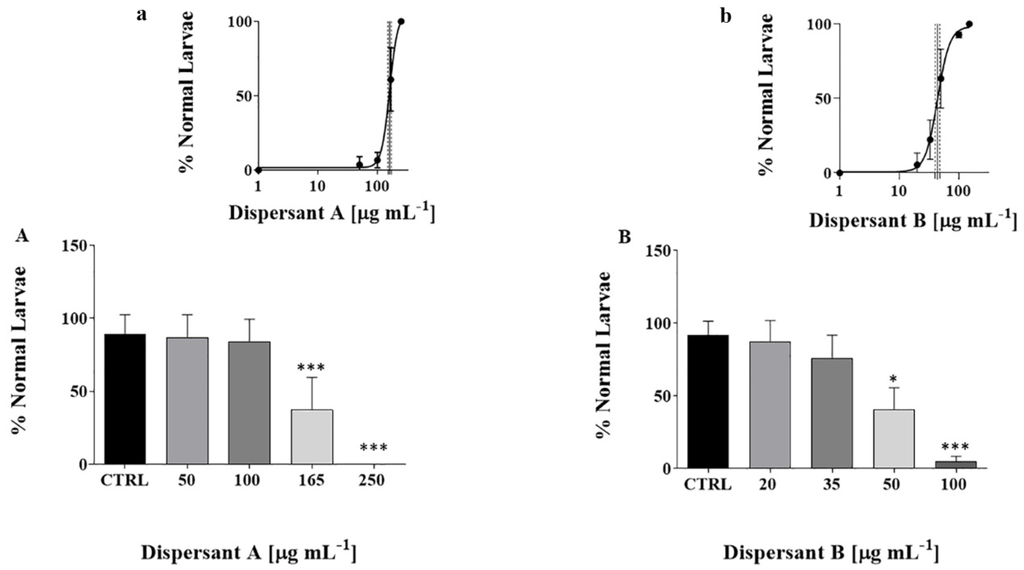
Adult specimens of the ascidian C. robusta were collected in the Gulf of Taranto (Italy) by local fishermen between November 2018 and March 2019. The organisms were transported in cool boxes, within a few hours, to the aquarium facility of the Zoological Station Anton Dohrn of Naples (Italy) in plastic bags filled with NSW (salinity 40‰, pH 8). Before experiments, animals were acclimated for 7 days in flow-through circulating aquarium in NSW (filtered 0.45 µm) (T18 ± 1 °C, salinity 40 ± 1‰, dissolved O2 7 mg L−1 and pH of 8.1) and under constant aeration and continuous light to stimulate gametes maturation and to avoid spawning [31]. Animals were fed ad libitum every 48 h with a mix of marine algae (Shellfish Diet 1800®, Reed Mariculture, Campbell, CA, USA). Gametes were obtained from each specimen by dissecting the gonoducts with a scalpel. To avoid self-fertilization, oocytes and sperms were collected by distinct individuals. The oocytes were rinsed twice in 0.22 µm filtered NSW while dry sperm was pooled and stored on ice until fertilization. Sperm was diluted 100X in NSW and then added to the egg’s suspension for fertilization. After 10 min of incubation on a rotating shaker, the fertilized eggs were transferred to tissue culture plates (Falcon® 100 mm × 15 mm, Singapore) and further rinsed in 0.22 µm filtered NSW.

2.3. Embryotoxicity

Embryotoxicity assay was carried out following the protocol reported in Bellas et al. [32]. Sixty embryos (~two-cell stage, about 1 h post-fertilization (hpf)) were added to 6-well plates (Thermo Scientific™ 6 Well Plate) and exposed to increasing concentrations of both dispersants formulations as follows: A (50; 100; 165; 250 μg mL−1) and B (20; 35; 50; 100 μg mL−1). Embryos were incubated under dark static conditions at 18 °C until the free-swimming larva stage was reached (22 hpf). The toxicity of both dispersants (A and B) was evaluated as a percentage of normal hatched larvae and morphological alterations at 22 hpf compared to controls, as described below. Larvae were first fixed in 4% paraformaldehyde and then washed twice in 1X PBS. A larva was recorded as normal when it presented a good general embryo morphology, with proper trunk and palps formation, as well as tail elongation following the Four-dimensional Ascidian Body Atlas Ver. 2 (https://www.bpni.bio.keio.ac.jp/chordate/faba2/top.html, accessed on 15 March 2019). Larvae phenotypes were examined by using the microscope Zeiss Axio Imager M1 and classified for simplicity in Mild I, Mild II, Intermediate, Severe and Not Developed. The assay was run at least three times and considered valid when controls (only in NSW) showed a percentage of normal hatched larvae ≥80% at 22 hpf.

2.4. RNA Extraction and cDNA Synthesis

About 180 embryos were exposed to 160 μg mL−1 of dispersant A and 44 μg mL−1 of dispersant B (~EC50 value of each dispersant). Control samples were run in parallel. At 22 hpf, the swimming larvae were collected by centrifugation at 3000 rcf for 3 min, frozen in liquid nitrogen and kept at −80 °C. Total RNA was extracted using RNAqueous-micro kit (Ambion) according to the manufacturer’s instructions. The RNA extracted was quantified measuring the absorbance at 260 nm (ND-1000 Spectrophotometer; NanoDrop Technologies, Wilmington, DE, USA) and the integrity was evaluated by agarose gel electrophoresis. For each sample, 1 µg of total RNA was retro-transcribed using QuantiTect Reverse Transcription Kit (Qiagen, Germantown, MD, USA) following the manufacturer’s instruction.

2.5. Gene Expression by Real Time-Quantitative PCR (RT-qPCR)

The variation of expression of the genes Cu, Zn superoxide dismutases (soda, sodb), manganese superoxide dismutase (MnSod), glutathione peroxidase (gpx), Heat Shock Proteins (hsp60, hsp70), Cytochrome P450 (cyp450), glutathione S-transferase (gst), glutathione reductase (GluR), p38 mitogen-activated protein kinases (p38 MAPK), cytochrome B (cytB) and caspase 8 (cas8), involved in stress response, detoxification and cell survival, was analyzed for each condition by RT-qPCR. The entire coding sequence of the 12 genes was obtained from NCBI (https://0-www-ncbi-nlm-nih-gov.brum.beds.ac.uk/, accessed on 12 December 2019) and Aniseed (https://www.aniseed.cnrs.fr/ accessed on 12 December 2019). Specific primers were designed with the help of Primer 3 software (Table S1), and then the selected amplicons were amplified. The reaction mix contained 1X Fast Start SYBR Green Master Mix (Roche), 1 μL of cDNA template (1:100 dilution) and 0.7 pmol μL−1 for each primer. PCR amplifications were performed in MicroAmp Optical 384-Well reaction plate with Optical Adhesive Covers (Applied Biosystems) in a ViiATM 7 Real Time PCR System (Applied Biosystems, Monza, Italy) thermal cycler using the following thermal profile: 95 °C for 20 s, 40 cycles of 95 °C for 1 s and 60 °C for 20 s, 1 cycle for melting curve analysis (from 60 to 95 °C, reading every 0.5 °C) to verify the presence of a single product. All the reactions were carried out in triplicate, and each assay included three negative controls with no template for each primer pair. Expression levels of target genes were normalized using, as reference gene, cytoskeletal actin (GenBank ID: NM_001032502.1, [33]). Actin-specific primers were as follows: sense primer, 5′-CCCAAATCATGTTCGAAACC-3′; antisense primer, 5′-ACACCATCACCACTGTCGAA-3′. Fluorescence was analyzed with ViiA™ 7 Real-Time PCR software (Life Technologies) and then quantified according to the comparative Ct method (2−ΔΔCt) based on Ct values of each gene, and the Ct average of the selected reference gene, in order to calculate the relative mRNA expression level. The expression levels of the selected genes were evaluated in a number-fold increase relative to the control condition that has been assigned as “1”.

2.6. Statistical Analysis

All the statistical analyses were performed using Graphpad Prism 6. Shapiro-Wilk’s test was performed to study normality of variances of the datasets. All data were expressed as mean ± standard deviation (SD). The median effective concentration (EC50), corresponding to a 50% reduction of normal hatched larvae, was calculated using a sigmoidal dose−response model according to the Equation: y = b + (a − b)/1 + 10 (Log EC50 − x), where y is response, b response minimum, a response maximum, x the logarithm of effect concentration and EC50 the concentration of effect giving 50% of maximum effect. Data were normalized to the control mean percentage of larval abnormality using Abbot’s formula:
P = (Pe − Pc/100 − Pc) × 100
where Pc and Pe are the control and the experimental percentages of response, respectively.
Data from the embryotoxicity assay and for the analyses of the morphological alterations are representative of at least three independent experiments and were analyzed using the non-parametric Kruskal-Wallis test followed by Dunn’s post hoc test. The statistical analyses of RT-qPCR were performed using GraphpadPRISM 6 software. Significance of the relative 2−ΔΔCt of each group (biological replicates, n = 3), compared to the controls, was determined using ‘unpaired parametric t-test’.

3. Results and Discussion

The massive use of dispersants for oil pollution remediation in the marine environment has drawn the attention of many scientists and legislators due to the potential detrimental impact on marine ecosystems and, in particular, on marine species [7,11]. Nowadays, their toxicity has been widely studied at different trophic levels, and often, only lethality has been evaluated as the main endpoint for dispersants, based on regulatory requirements for final approval procedures in their use. For refining risk, it would be important to perform additional tests, based on sub-lethal endpoints as a further step [7]. However, a variety of sub-lethal responses, which can occur in marine organisms, particularly during the embryogenesis, should be taken into consideration, such as assessment of gene expression and monitoring of organism physiology [34,35]. This study, aimed at inspecting the impact of two dispersants formulations (A and B) on Ciona embryogenesis, showed that both A and B affected the normal larval development in a dose-dependent manner, with the EC50 values of 160 μg mL−1 (150.4 to 171.2) for A and 44.30 μg mL−1 (39.92 to 49.15) for B, thus showing higher toxicity of B compared to A (Figure 1).
Our previous findings, obtained on a multi-trophic battery from algae to fish (Phaeodactylum tricornutum, Tigriopus fulvus and Artemia franciscana, Dicentrarchus labrax), tested according to the Italian Decree Law 2/25/2011, indicated a higher toxicity of dispersant A compared to B, with E(L)C50 value ranging from 1.60 to 16.19 μg mL−1 for A and from 49.66 to 82.70 μg mL−1 for B [28]. Thus, based on E(L)C50, dispersant A results 10–100 times less toxic to Ciona, while the data on dispersant B are almost comparable to those obtained in this study (see Table S2). In accordance with Fingas [36], the different effects exerted by these compounds could depend on test species, exposure time, physical-chemical parameters (especially temperature and salinity) and dispersant composition. The discrepancy on dispersant A effects, identified in this study, could be related to various factors, including the chemical composition of the dispersant itself. An important aspect to consider is the species-specific sensitivity, the biological model and the life stage used for the assays. Indeed, previous studies already showed that the E(L)C50 of third-generation dispersants may vary in a range of 4–105 mg L−1, depending on the diverse sensitivity of marine organisms [15,37]. In this regard, it should be pointed out that the lower sensitivity of Ciona embryos could be due to the presence of the external egg envelope formed by a cellular layer, named vitelline coat (or chorion), and two populations of maternally supplied cells, the follicle cells and the test cells. This complex structure protects Ciona embryos from external insults and may provide a stronger shielding role towards some chemicals present in the dispersant formulations [38].

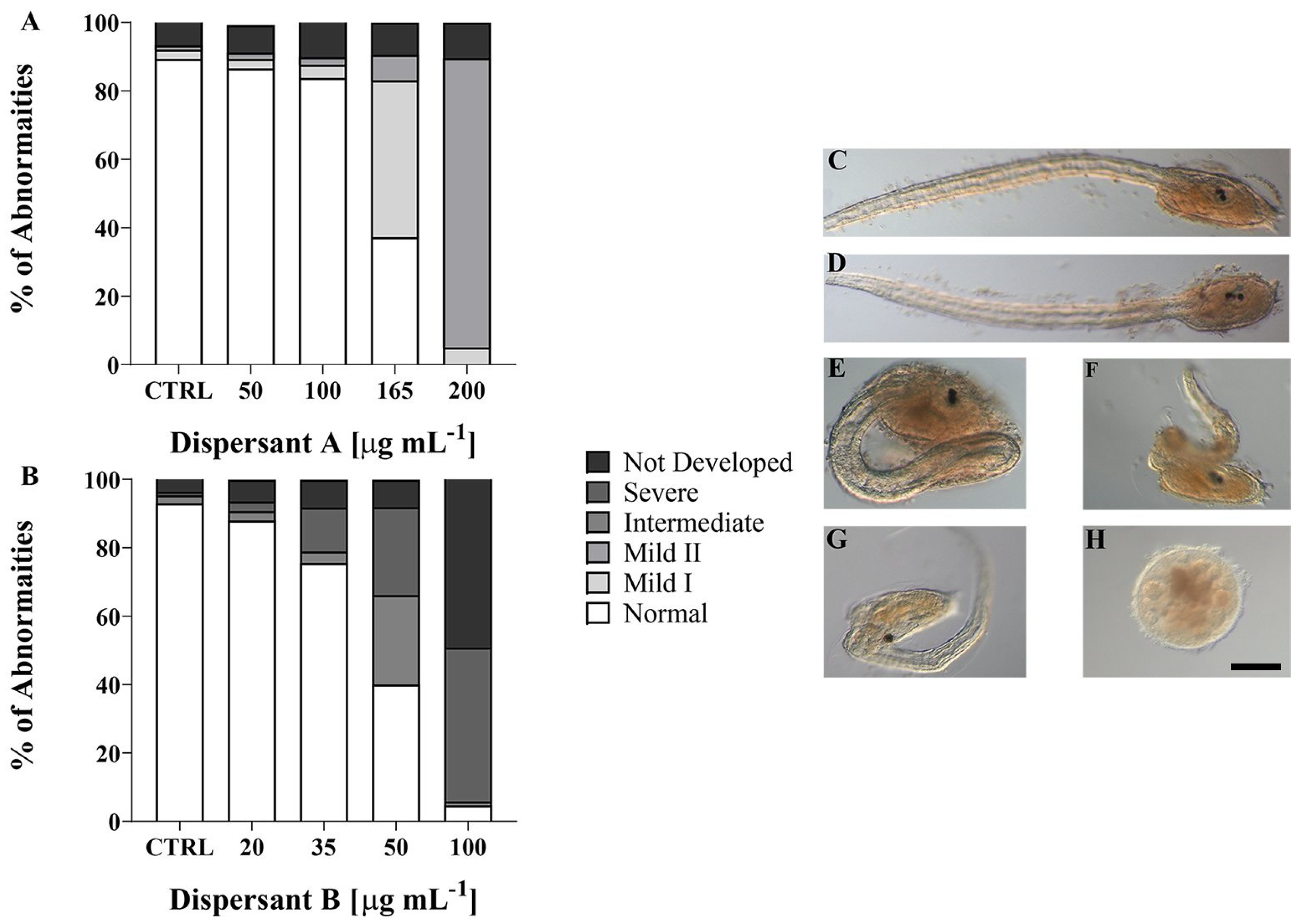
3.1. Larvae Phenotypes

Treatment with dispersants A and B resulted also in different phenotypes on Ciona larvae, as revealed by microscopy observation (Figure 2). The phenotypes were classified as follows: (i) Mild I: larvae with a shorter trunk and normal tail, (ii) Mild II: larvae like Mild I but unable to hatch, (iii) Intermediate: larvae with a proper trunk, but with a shorter, kinked and disorganized tail, (iv) Severe: larvae with a shorter, kinked and disorganized tail and malformed trunk, (v) Not developed: embryos stopped in development (Figure 2C–H). In particular, as shown in Figure 2A, the appearance of Mild I phenotype (46%) has been observed at 165 µg mL−1 of dispersant A, and the effects were stronger at 250 µg mL−1, since around 84% of larvae showed a Mild II phenotype, being able to twitch the tail weakly but becoming unable to break the chorion and hatch. Notably, the Mild I and Mild II larvae remained blocked at the larval stage and were unable to continue their life cycle. Interestingly, exposure to dispersant A did not lead to embryos with Intermediate and/or Severe phenotypes and Not Developed. Treatment with dispersant B resulted in different and stronger effects on embryonic development since, already at 50 µg mL−1, the larvae showed the Intermediate (26%) and Severe (25%) phenotypes, and the percentage of Severe increased up to 50% at 100 µg mL−1 with larvae showing strong aberrations at both trunk and tail levels. Furthermore, at 100 µg mL−1, the not developed embryos peaked to almost 50%, indicating a deleterious effect exerted by this dispersant on embryo survival (Figure 2B). Thus, both the embryo-toxicological and morphological evaluations indicate the highest toxicity and teratogenicity of dispersant B compared to A (Figure 2A,B).
In accordance with the literature, the exposure to various dispersant formulations in shorter periods and low concentrations does not cause deleterious effects on phenotypic endpoints and gene expressions in fish embryos [39,40,41]. However, their chemical composition varies among formulations and exposure times. The higher toxicity of dispersant B, compared to A, could be related to the high presence in the mixture of anionic surfactants (10–30%), known to break protein/protein interactions, and non-ionic surfactants (5–15%), which potentially dissolve unipolar compounds as bilayer membranes [42,43,44]. It is conceivable that the combined action of these substances could induce destabilization within the egg envelopes which surround and protect Ciona embryos, thus exposing the developing embryos directly to the toxic action of both surfactants and to the 2-butoxyethanol (2BE). It has been demonstrated that, in zebrafish embryo toxicity tests, 2BE induces general teratogenicity, such as pericardial edema and yolk sac edema [45]. As for zebrafish, one can suppose that 2BE, which has clearly lipophilic character and, therefore, is more likely to diffuse within the embryo cells, induces teratogenic effects in Ciona embryos, further amplified by the presence of surfactants, thus resulting in almost 50% of not developed embryos at the highest concentration of dispersant B (100 µg mL−1). The severe phenotype we detected, at 50 (25%) and even more at 100 µg mL−1 (50%) dispersant B treatment, includes, besides tail malformations, alterations in the brain vesicle internalization. It is known that the main metabolic pathway for the aliphatic alcohols, as 2BE, is the conversion, by specific enzymes, into aldehyde and then into acetate products. However, when in large excess, other enzymes are recruited in the process, as the ones related to all-trans-retinoic acid [45]. Retinol metabolism into retinoic acid is fundamental during embryonic development and any perturbation of RA concentration results in embryo malformations, including impaired closure of the neural tube as demonstrated also in Ciona embryos [46]. One can assume that imbalances of RA concentrations is one of the toxicity pathways elicited by 2BE in Ciona. However, this hypothesis is very speculative and will be clarified by further studies, aimed at inspecting the effective involvement of RA metabolism after dispersant B treatment. Concerning dispersant A, the absence of non-ionic surfactants might have had a role in preventing the preliminary effects at the membrane level, although the presence in the formulation of carcinogenic toxic compounds, as hydrocarbons, should suggest a higher toxic potential.

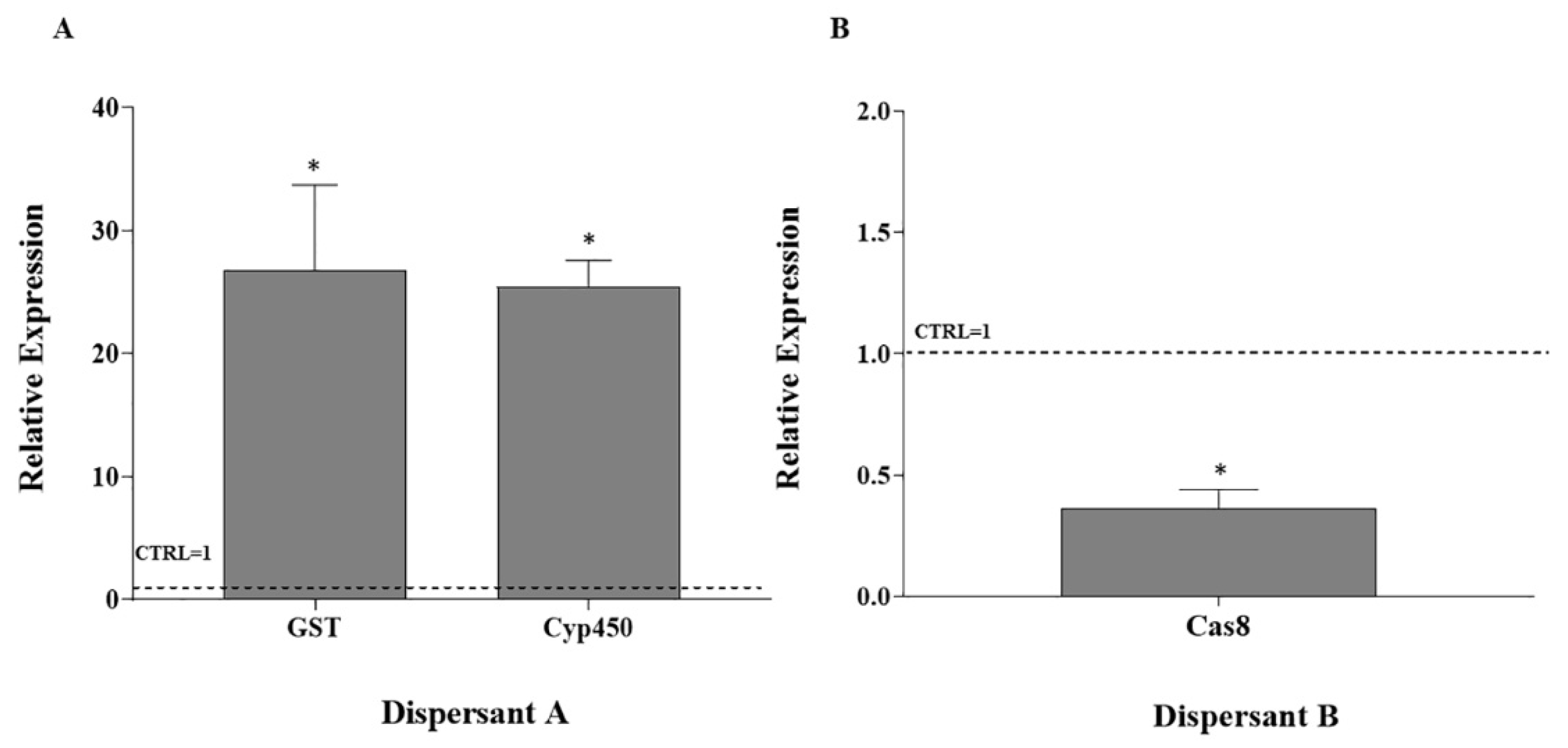
3.2. RT-qPCR on Stress Response Genes

As previously mentioned, regulatory requirements often include only lethality assays for the evaluation of toxic substances. However, sub-lethal tests can provide information on the possible mode of action of contaminants and, in turn, may help to identify the compound of the mixture that causes toxicity [34,47]. In this perspective, here we have evaluated the expression of genes involved in detoxification, stress response and cell survival, based on previous studies showing that some of these pathways can be affected by dispersant exposure [34]. Larvae treated with dispersant A showed a significant upregulation of gst (p = 0.0005) and cyp450 (p = 0.003) genes at 160 µg mL−1 compared to the controls (Figure 3A). The genes cyp450 and gst are involved in the detoxification mechanisms of xenobiotics; in particular, cyp450 is involved in xenobiotics oxidation during phase I [48], followed by phase II which involves enzymes as gst that conjugates the phase I metabolite to small polar moieties [49]. The upregulated gene machinery involving cytochrome P450 biotransformation phase I and phase II could be related to the presence of hydrocarbons in dispersant A formulation. One can suppose that the P450 metabolism could protect embryos from further detrimental outcomes, through the activation of pathways induced by the exposure to hydrocarbons (e.g., Aryl hydrocarbon pathway (AhR), pregnane X receptor (PXR)), thus suggesting that detoxification is taking place in Ciona embryos exposed to dispersant A [50,51]. It has been reported in the literature that hyperactivation of the genes (as P450), related to these signaling pathways, upon prolonged exposure or in the presence of high concentrations of hydrocarbons, may, in turn, induce developmental abnormalities, hatching failure and lethality [50,52,53]. Notably, in Ciona, exposure to high concentrations of dispersant A resulted in larvae showing abnormalities at the trunk level (Mild I), that, in the Mild II phenotype, become unable to hatch. Differently from dispersant A, dispersant B was not able to elicit a significant deregulation of genes involved in stress response and detoxification. Dispersant B exposure, indeed, caused a downregulation of cas8 (p = 0.01) at 44 µg mL−1 (Figure 3B). Cas8 gene takes part in the extrinsic pathway, or death receptor pathway of apoptosis, in which the ligation of death receptors on the cell surface leads to caspase activation [54]. One component of dispersant B mixture, 2BE, as other ethylene glycol ethers, is known to induce oxidative stress in the rat brain by inhibiting the total antioxidant activity [55]. One can suppose that in the Ciona model, the action of 2BE is achieved through an interference, rather than an inhibition, with the mobilization of the defense mechanism machinery. As a result, the high and prolonged exposure to stress conditions could induce a decreased transcription of cas8 gene, involved in the activation of apoptosis. This, in turn, would result in the observed aberrations in the phenotype, which include Intermediate and Severe phenotypes and Not Developed embryos. However, we cannot exclude that other molecular mechanisms are involved in the toxic activity of both dispersants, and further studies are required to better understand the complex molecular responses evoked by dispersant treatment, acting upstream and downstream of the genes analyzed in this study.

4. Conclusions

To the best of our knowledge, this study is the first contribution to the understanding of embryotoxicity of commercial dispersant formulations on the marine invertebrate C. robusta. Our data show a dose-response effect relationship for both dispersants with dispersant B (44.30 μg mL−1) being more toxic compared to A (160 μg mL−1).
Colinearly, the analysis of Ciona larvae phenotypes indicates a high percentage of larvae with Intermediate, Severe malformation (problem at the trunk and tail levels) and Not Developed embryos induced by dispersant B treatment compared to A. The different phenotypic outputs and toxic effects of these treatments could be related to the data on the expression levels of genes involved in stress response, detoxification and cell survival, analyzed in this study and previously discussed. Our study underlines the importance of investigating the effects of dispersants on the embryogenesis of marine species. Embryonic development represents the most delicate and important life stage of an organism since it is crucial for ensuring the fitness of the species. A compromised embryonic development, especially for the organisms with pelagic life stages, could influence the survival of a species, due to the strong vulnerability of this life stage to the exposure to different chemicals. Thus, the embryotoxicity test, coupled with gene expression analyses, could strengthen the dispersant toxicity evaluation, as part of the approval procedures for the use of these products at sea. Under this perspective, Ciona embryos represent the ideal model, given the ease of manipulation in the laboratory, the fast embryonic development, the possibility of easily scoring the phenotypes at the larval stage and the close phylogenetic relationship with vertebrates.

Supplementary Materials

The following supporting information can be downloaded at: https://0-www-mdpi-com.brum.beds.ac.uk/article/10.3390/w14101539/s1, Figure S1: Expression of soda, sodb, gpx, gst, GluR, Cyp450, hsp60, hsp70, MnSod, cytB, p38 MAPK and cas8 genes by comparative RT q-PCR with total RNA isolated from control and exposed embryos to dispersant A (A) and dispersant B (B) after 22 hpf; Table S1: Accession number and/or Gene Model ID, sequences and length of PCR fragments are listed for the analyzed genes; Table S2: E(L)C50 values (μg mL−1) calculated for dispersant A and B.

Author Contributions

Conceptualization, M.C.E., L.M. and A.S.; methodology, M.C.E.; validation, I.C.; formal analysis, M.C.E.; investigation, M.C.E.; writing—original draft preparation, M.C.E.; writing—review and editing, I.C., L.M. and A.S.; visualization, M.C.E.; supervision, A.S.; funding acquisition, I.C. and A.S. All authors have read and agreed to the published version of the manuscript.

Funding

This study was performed in the framework of the Ph.D. project entitled “Ciona robusta (formerly Ciona intestinalis type A) as model system for ecotoxicological studies” co-funded by the University of Siena (Italy) and the Zoological Station Anton Dohrn of Naples.

Institutional Review Board Statement

Ethical review and approval were waived for this study since Ciona robusta is not protected species by any law in Italy.

Informed Consent Statement

Not applicable.

Data Availability Statement

All data is contained within this article and Supplementary Materials.

Acknowledgments

Student Maria Concetta Eliso, Ph.D. School in Geological, Environmental and Polar sciences and technologies, Department of Physical, Earth and Environmental Sciences, University of Siena (Italy). The authors are indebted to the RAMOGE Executive Secretariat for providing the dispersant formulations.

Conflicts of Interest

The authors declare no conflict of interest.

References

  1. Lee, K.; Nedwed, T.; Prince, R.C.; Palandro, D. Lab Tests on the Biodegradation of Chemically Dispersed Oil Should Consider the Rapid Dilution That Occurs at Sea. Mar. Pollut. Bull. 2013, 73, 314–318. [Google Scholar] [CrossRef] [PubMed]
  2. Lessard, R.R.; DeMarco, G. The Significance of Oil Spill Dispersants. Spill Sci. Technol. Bull. 2000, 6, 59–68. [Google Scholar] [CrossRef]
  3. National Research Council (USA). Committee on Effectiveness of Oil Spill Dispersants. In Using Oil Spill Dispersants on the Sea; National Academy Press: Cambridge, MA, USA, 1989; ISBN 9780309038829. [Google Scholar]
  4. Committee on Understanding Oil Spill Dispersants: Efficacy and Effects; Ocean Studies Board; Division on Earth and Life Studies; National Research Council. Oil Spill Dispersants: Efficacy and Effects; National Academies Press: Cambridge, MA, USA, 2005; ISBN 9780309095624. [Google Scholar]
  5. Singer, M.M.; Smalheer, D.L.; Tjeerdema, R.S.; Martin, M. Effects of Spiked Exposure to an Oil Dispersant on the Early Life Stages of Four Marine Species. Environ. Toxicol. Chem. 1991, 10, 1367–1374. [Google Scholar] [CrossRef]
  6. Singer, M.M.; George, S.; Jacobson, S.; Lee, I.; Weetman, L.L.; Tjeerdema, R.S.; Sowby, M.L. Comparison of Acute Aquatic Effects of the Oil Dispersant Corexit 9500 with Those of Other Corexit Series Dispersants. Ecotoxicol. Environ. Saf. 1996, 35, 183–189. [Google Scholar] [CrossRef] [Green Version]
  7. Bejarano, A.C. Critical Review and Analysis of Aquatic Toxicity Data on Oil Spill Dispersants. Environ. Toxicol. Chem. 2018, 37, 2989–3001. [Google Scholar] [CrossRef] [Green Version]
  8. International Petroleum Industry Environmental Conservation Association-International Association of Oil & Gas Producers (IPIECA-OGP). Regulatory Approval of Dispersants and Authorization for Their Use; IPIECA-OGP: London, UK, 2014. [Google Scholar]
  9. European Maritime Safety Agency. In Overview of National Dispersant Testing and Approval Policies in the EU; European Maritime Safety Agency: Lisbon, Portugal, 2016.
  10. Oil Spill Prevention, Administration and Response (OSPAR). Guidelines for Completing the Harmonised Offshore Chemical Notification Format (HOCNF); OSPAR Agreement: 2012-05; OSPAR Commission: London, UK, 2015. [Google Scholar]
  11. Wise, J.; Wise, J.P., Sr. A Review of the Toxicity of Chemical Dispersants. Rev. Environ. Health 2011, 26, 281–300. [Google Scholar] [CrossRef]
  12. Vignier, J.; Donaghy, L.; Soudant, P.; Chu, F.L.E.; Morris, J.M.; Carney, M.W.; Lay, C.; Krasnec, M.; Robert, R.; Volety, A.K. Impacts of Deepwater Horizon Oil and Associated Dispersant on Early Development of the Eastern Oyster Crassostrea Virginica. Mar. Pollut. Bull. 2015, 100, 426–437. [Google Scholar] [CrossRef] [Green Version]
  13. Adeyemo, O.K.; Kroll, K.J.; Denslow, N.D. Developmental Abnormalities and Differential Expression of Genes Induced in Oil and Dispersant Exposed Menidia Beryllina Embryos. Aquat. Toxicol. 2015, 168, 60–71. [Google Scholar] [CrossRef]
  14. Beirão, J.; Baillon, L.; Litt, M.A.; Langlois, V.S.; Purchase, C.F. Impact of Crude Oil and the Dispersant CorexitTM EC9500A on Capelin (Mallotus Villosus) Embryo Development. Mar. Environ. Res. 2019, 147, 90–100. [Google Scholar] [CrossRef]
  15. DeMiguel-Jiménez, L.; Etxebarria, N.; Lekube, X.; Izagirre, U.; Marigómez, I. Influence of Dispersant Application on the Toxicity to Sea Urchin Embryos of Crude and Bunker Oils Representative of Prospective Oil Spill Threats in Arctic and Sub-Arctic Seas. Mar. Pollut. Bull. 2021, 172, 112922. [Google Scholar] [CrossRef]
  16. Barron, M.G.; Bejarano, A.C.; Conmy, R.N.; Sundaravadivelu, D.; Meyer, P. Toxicity of Oil Spill Response Agents and Crude Oils to Five Aquatic Test Species. Mar. Pollut. Bull. 2020, 153, 110954. [Google Scholar] [CrossRef] [PubMed]
  17. Pineda, M.C.; McQuaid, C.D.; Turon, X.; López-Legentil, S.; Ordóñez, V.; Rius, M. Tough Adults, Frail Babies: An Analysis of Stress Sensitivity across Early Life-History Stages of Widely Introduced Marine Invertebrates. PLoS ONE 2012, 7, e46672. [Google Scholar] [CrossRef] [PubMed] [Green Version]
  18. Dehal, P.; Satou, Y.; Campbell, R.K.; Chapman, J.; Degnan, B.; De Tomaso, A.; Davidson, B.; Di Gregorio, A.; Gelpke, M.; Goodstein, D.M.; et al. The Draft Genome of Ciona Intestinalis: Insights into Chordate and Vertebrate Origins. Science 2002, 298, 2157–2167. [Google Scholar] [CrossRef] [PubMed] [Green Version]
  19. Zega, G.; De Bernardi, F.; Groppelli, S.; Pennati, R. Effects of the Azole Fungicide Imazalil on the Development of the Ascidian Ciona Intestinalis (Chordata, Tunicata): Morphological and Molecular Characterization of the Induced Phenotype. Aquat. Toxicol. 2009, 91, 255–261. [Google Scholar] [CrossRef] [PubMed]
  20. Mizotani, Y.; Itoh, S.; Hotta, K.; Tashiro, E.; Oka, K.; Imoto, M. Evaluation of Drug Toxicity Profiles Based on the Phenotypes of Ascidian Ciona Intestinalis. Biochem. Biophys. Res. Commun. 2015, 463, 656–660. [Google Scholar] [CrossRef]
  21. Satou, Y.; Imai, K.S. Gene Regulatory Systems That Control Gene Expression in the Ciona Embryo. Proc. Japan Acad. Ser. B 2015, 91, 33–51. [Google Scholar] [CrossRef] [Green Version]
  22. Bellas, J.; Vázquez, E.; Beiras, R. Toxicity of Hg, Cu, Cd, and Cr on Early Developmental Stages of Ciona Intestinalis (Chordata, Ascidiacea) with Potential Application in Marine Water Quality Assessment. Water Res. 2001, 35, 2905–2912. [Google Scholar] [CrossRef]
  23. Bellas, J.; Beiras, R.; Mariño-Balsa, J.C.; Fernández, N. Toxicity of Organic Compounds to Marine Invertebrate Embryos and Larvae: A Comparison between the Sea Urchin Embryogenesis Bioassay and Alternative Test Species. Ecotoxicology 2005, 14, 337–353. [Google Scholar] [CrossRef]
  24. Bellas, J. Comparative Toxicity of Alternative Antifouling Biocides on Embryos and Larvae of Marine Invertebrates. Sci. Total Environ. 2006, 367, 573–585. [Google Scholar] [CrossRef]
  25. Beiras, R.; Bellas, J. Inhibition of Embryo Development of the Mytilus Galloprovincialis Marine Mussel by Organic Pollutants; Assessment of Risk for Its Extensive Culture in the Galician Rias. Aquaculture 2008, 277, 208–212. [Google Scholar] [CrossRef]
  26. Eliso, M.C.; Bergami, E.; Manfra, L.; Spagnuolo, A.; Corsi, I. Toxicity of Nanoplastics during the Embryogenesis of the Ascidian Ciona Robusta (Phylum Chordata). Nanotoxicology 2020, 14, 1415–1431. [Google Scholar] [CrossRef] [PubMed]
  27. Eliso, M.C.; Manfra, L.; Savorelli, F.; Tornambè, A.; Spagnuolo, A. New Approaches on the Use of Tunicates (Ciona Robusta) for Toxicity Assessments. Environ. Sci. Pollut. Res. Int. 2020, 27, 32132–32138. [Google Scholar] [CrossRef] [PubMed]
  28. Manfra, L.; Tornambè, A.; Guyomarch, J.; Le Guerrogue, P.; Kerambrun, L.; Rotini, A.; Savorelli, F.; Onorati, F.; Magaletti, E. Dispersant Approval Procedures in France and Italy: A Comparative Ecotoxicity Study. Ecotoxicol. Environ. Saf. 2017, 143, 180–185. [Google Scholar] [CrossRef] [PubMed]
  29. Durier, G.; Nadalini, J.-B.; Saint-Louis, R.; Genard, B.; Comeau, L.A.; Tremblay, R. Sensitivity to Oil Dispersants: Effects on the Valve Movements of the Blue Mussel Mytilus Edulis and the Giant Scallop Placopecten Magellanicus, in Sub-Arctic Conditions. Aquat. Toxicol. 2021, 234, 105797. [Google Scholar] [CrossRef]
  30. Scarlett, A.; Galloway, T.S.; Canty, M.; Smith, E.L.; Nilsson, J.; Rowland, S.J. Comparative Toxicity of Two Oil Dispersants, Superdispersant-25 and Corexit 9527, to a Range of Coastal Species. Environ. Toxicol. Chem. 2005, 24, 1219–1227. [Google Scholar] [CrossRef]
  31. Lambert, C.C.; Brandt, C.L. The effect of light on the spawning of Ciona Intestinalis. Biol. Bull. 1967, 132, 222–228. [Google Scholar] [CrossRef]
  32. Bellas, J.; Beiras, R.; Vázquez, E. A Standardisation of Ciona Intestinalis (Chordata, Ascidiacea) Embryo-Larval Bioassay for Ecotoxicological Studies. Water Res. 2003, 37, 4613–4622. [Google Scholar] [CrossRef]
  33. Fujikawa, T.; Munakata, T.; Kondo, S.-I.; Satoh, N.; Wada, S. Stress Response in the Ascidian Ciona Intestinalis: Transcriptional Profiling of Genes for the Heat Shock Protein 70 Chaperone System under Heat Stress and Endoplasmic Reticulum Stress. Cell Stress Chaperones 2010, 15, 193–204. [Google Scholar] [CrossRef] [Green Version]
  34. Colvin, K.A.; Lewis, C.; Galloway, T.S. Current Issues Confounding the Rapid Toxicological Assessment of Oil Spills. Chemosphere 2020, 245, 125585. [Google Scholar] [CrossRef]
  35. International Maritime Organization. Manual on Oil Pollution, Combating Oil Spills. In Chemical Dispersion; Section IV; International Maritime Organization: London, UK, 2011; Chapter 7. [Google Scholar]
  36. Fingas, M. Visual Appearance of Oil on the Sea. J. Mar. Sci. Eng. 2021, 9, 97. [Google Scholar] [CrossRef]
  37. Thompson, H.; Shimeld, S.M. Transmission and Scanning Electron Microscopy of the Accessory Cells and Chorion During Development of Ciona Intestinalis Type B Embryos and the Impact of Their Removal on Cell Morphology. Zoolog. Sci. 2015, 32, 217–222. [Google Scholar] [CrossRef] [PubMed]
  38. Edmunds, R.C.; Gill, J.A.; Baldwin, D.H.; Linbo, T.L.; French, B.L.; Brown, T.L.; Esbaugh, A.J.; Mager, E.M.; Stieglitz, J.; Hoenig, R.; et al. Corresponding Morphological and Molecular Indicators of Crude Oil Toxicity to the Developing Hearts of Mahi Mahi. Sci. Rep. 2015, 5, 17326. [Google Scholar] [CrossRef] [PubMed]
  39. Esbaugh, A.J.; Mager, E.M.; Stieglitz, J.D.; Hoenig, R.; Brown, T.L.; French, B.L.; Linbo, T.L.; Lay, C.; Forth, H.; Scholz, N.L.; et al. The Effects of Weathering and Chemical Dispersion on Deepwater Horizon Crude Oil Toxicity to Mahi-Mahi (Coryphaena Hippurus) Early Life Stages. Sci. Total Environ. 2016, 543, 644–651. [Google Scholar] [CrossRef] [PubMed]
  40. Greer, J.B.; Pasparakis, C.; Stieglitz, J.D.; Benetti, D.; Grosell, M.; Schlenk, D. Effects of Corexit 9500A and Corexit-Crude Oil Mixtures on Transcriptomic Pathways and Developmental Toxicity in Early Life Stage Mahi-Mahi (Coryphaena Hippurus). Aquat. Toxicol. 2019, 212, 233–240. [Google Scholar] [CrossRef] [PubMed]
  41. Parsi, K. Interaction of Detergent Sclerosants with Cell Membranes. Phlebology 2015, 30, 306–315. [Google Scholar] [CrossRef] [PubMed]
  42. Pedersen, A.F.; Meyer, D.N.; Petriv, A.-M.V.; Soto, A.L.; Shields, J.N.; Akemann, C.; Baker, B.B.; Tsou, W.-L.; Zhang, Y.; Baker, T.R. Nanoplastics Impact the Zebrafish (Danio Rerio) Transcriptome: Associated Developmental and Neurobehavioral Consequences. Environ. Pollut. 2020, 266, 115090. [Google Scholar] [CrossRef]
  43. Aguirre-Ramírez, M.; Silva-Jiménez, H.; Banat, I.M.; Díaz De Rienzo, M.A. Surfactants: Physicochemical Interactions with Biological Macromolecules. Biotechnol. Lett. 2021, 43, 523–535. [Google Scholar] [CrossRef]
  44. Van der Ven, L.T.M.; Schoonen, W.G.; Groot, R.M.; den Ouden, F.; Heusinkveld, H.J.; Zwart, E.P.; Hodemaekers, H.M.; Rorije, E.; de Knecht, J. The Effects of Aliphatic Alcohols and Related Acid Metabolites in Zebrafish Embryos-Correlations with Rat Developmental Toxicity and with Effects in Advanced Life Stages in Fish. Toxicol. Appl. Pharmacol. 2020, 407, 115249. [Google Scholar] [CrossRef]
  45. Nagatomo, K.-I.; Ishibashi, T.; Satou, Y.; Satoh, N.; Fujiwara, S. Retinoic Acid Affects Gene Expression and Morphogenesis without Upregulating the Retinoic Acid Receptor in the Ascidian Ciona Intestinalis. Mech. Dev. 2003, 120, 363–372. [Google Scholar] [CrossRef]
  46. Hylland, K.; Burgeot, T.; Martínez-Gómez, C.; Lang, T.; Robinson, C.D.; Svavarsson, J.; Thain, J.E.; Vethaak, A.D.; Gubbins, M.J. How Can We Quantify Impacts of Contaminants in Marine Ecosystems? Mar. Environ. Res. 2017, 124, 2–10. [Google Scholar] [CrossRef] [Green Version]
  47. Nebert, D.W.; Dalton, T.P. The Role of Cytochrome P450 Enzymes in Endogenous Signalling Pathways and Environmental Carcinogenesis. Nat. Rev. Cancer 2006, 6, 947–960. [Google Scholar] [CrossRef] [PubMed]
  48. Chen, C.-H. Activation and Detoxification Enzymes: Functions and Implications; Springer: Berlin/Heidelberg, Germany, 2011; ISBN 9781461410492. [Google Scholar]
  49. Portnoy, D.S.; Fields, A.T.; Greer, J.B.; Schlenk, D. Genetics and Oil: Transcriptomics, Epigenetics, and Population Genomics as Tools to Understand Animal Responses to Exposure Across Different Time Scales. In Deep Oil Spills: Facts, Fate, and Effects; Springer: Berlin/Heidelberg, Germany, 2019. [Google Scholar]
  50. Murawski, S.A.; Ainsworth, C.H.; Gilbert, S.; Hollander, D.J.; Paris, C.B.; Schlüter, M.; Wetzel, D.L. (Eds.) Deep Oil Spills: Facts, Fate, and Effects; Springer International Publishing: Cham, Germany, 2020; pp. 515–532. ISBN 9783030116057. [Google Scholar]
  51. Kumagai, T.; Suzuki, H.; Sasaki, T.; Sakaguchi, S.; Miyairi, S.; Yamazoe, Y.; Nagata, K. Polycyclic Aromatic Hydrocarbons Activate CYP3A4 Gene Transcription through Human Pregnane X Receptor. Drug Metab. Pharmacokinet. 2012, 27, 200–206. [Google Scholar] [CrossRef] [PubMed] [Green Version]
  52. Jenny, M.J.; Karchner, S.I.; Franks, D.G.; Woodin, B.R.; Stegeman, J.J.; Hahn, M.E. Distinct Roles of Two Zebrafish AHR Repressors (AHRRa and AHRRb) in Embryonic Development and Regulating the Response to 2,3,7,8-Tetrachlorodibenzo-P-Dioxin. Toxicol. Sci. 2009, 110, 426–441. [Google Scholar] [CrossRef] [PubMed] [Green Version]
  53. Whitehead, A.; Dubansky, B.; Bodinier, C.; Garcia, T.I.; Miles, S.; Pilley, C.; Raghunathan, V.; Roach, J.L.; Walker, N.; Walter, R.B.; et al. Genomic and Physiological Footprint of the Deepwater Horizon Oil Spill on Resident Marsh Fishes. Proc. Natl. Acad. Sci. USA 2012, 109, 20298–20302. [Google Scholar] [CrossRef] [Green Version]
  54. Tummers, B.; Green, D.R. Caspase-8: Regulating Life and Death. Immunol. Rev. 2017, 277, 76–89. [Google Scholar] [CrossRef] [Green Version]
  55. Pomierny, B.; Krzyżanowska, W.; Smaga, I.; Pomierny-Chamioło, L.; Stankowicz, P.; Budziszewska, B. Ethylene Glycol Ethers Induce Oxidative Stress in the Rat Brain. Neurotox. Res. 2014, 26, 422–429. [Google Scholar] [CrossRef] [Green Version]
Figure 1. Percentage (%) of normal hatched larvae of C. robusta upon exposure to dispersant A (A) and dispersant B (B) in NSW for 22 h. Bars represent mean ± SD (dispersant A n = 2700; dispersant B n = 2700). Asterisks indicate values that are significantly different compared to the control (Kruskal-Wallis test, Dunn’s post hoc test, * p < 0.05, *** p < 0.001). In the upper part of the graphs (a,b), curves represent the nonlinear regression of normal larval development data (sigmoidal) with the best fit for EC50 values (dashed lines) as well as the relative 95% CIs (dotted lines). Error bars represent standard deviation.
Water 14 01539 g001
Figure 2. (A) Percentages (%) of total abnormalities identified upon exposure to dispersant A; (B) percentages (%) of total abnormalities identified upon exposure to dispersant B. Bars represent mean ± SD (n = 1080); light microscopy images of the different phenotypes obtained upon exposure to dispersant A and dispersant B. (C) Control; (D) Mild I phenotype; (E) Mild II phenotype; (F) Intermediate; (G) Severe; (H) Not Developed embryo. Scale bar: 50 µm.
Water 14 01539 g002
Figure 3. Expression of gst, cyp450 and cas8 genes by comparative RT q-PCR with total RNA isolated from control and exposed embryos to dispersant A (A) and dispersant B (B) after 22 hpf. Results are expressed as fold increase compared to controls assumed as 1, using cytoskeletal actin as reference gene for normalization. Each bar represents the mean of three independent experiments ± SEM. * indicates a significant difference (t-test, p < 0.05) with respect to controls. This is the graphical representation of only significantly deregulated genes. The results of the expression of all the selected genes are reported in Figure S1.
Water 14 01539 g003
Publisher’s Note: MDPI stays neutral with regard to jurisdictional claims in published maps and institutional affiliations.

