Article

Enterococcus Species and Their Antimicrobial Resistance in an Urban Watershed Affected by Different Anthropogenic Sources

by
Abasiofiok M. Ibekwe
1,*,
Amarachukwu C. Obayiuwana
2 and
Shelton E. Murinda
3
1
US Salinity Laboratory, USDA-ARS, 450 West Big Springs Road, Riverside, CA 92507, USA
2
Department of Biological Sciences, Augustine University Ilara-Epe, Lagos 106101, Lagos State, Nigeria
3
Animal and Veterinary Sciences Department, California State Polytechnic University, Pomona, CA 91768, USA
*
Author to whom correspondence should be addressed.
Submission received: 1 December 2023 / Revised: 22 December 2023 / Accepted: 26 December 2023 / Published: 28 December 2023

Abstract

:
Different anthropogenic sources can have a significant influence on bacterial populations and their antimicrobial activities. In this study, the impact of anthropogenic activities on Enterococcus species was studied in an urban watershed in southern California affected by concentrated animal feeding operations (CAFOs), recreational activities, wastewater treatment plants (WWTPs), urban runoff, and control sites. Water samples were collected quarterly for two years for the enumeration of Enterococcus species based on the Enterolert most probable-number (MPN) assay. Concentrations of enterococci were higher in the sediment compared to surface water (4.5 × 10⁶ CFU/g of sediment vs. 2.3 × 10⁵ MPN/100 mL of water). The species diversity was dominated by E. mundtii (32%), E. faecalis (27%), and E. faecium (25%). E. faecium exhibited the highest antibiotic-resistant phenotype. Resistances were mostly to ciprofloxacin, erythromycin, and tetracycline. Tetracycline and erythromycin resistance genes, encoded by tet (C, K, O, S) and ermB, respectively, were more common in isolates from sediment (42.9%) compared to water (12.7%). E. mundtii was sensitive to ampicillin, chloramphenicol, gentamicin, and high levels of vancomycin. A significant percentage of E. faecalis were also resistant to these antibiotics. E. faecium and E. faecalis exhibited resistance to multiple antibiotics. Our data suggest that resistant Enterococcus species within the watershed might provide some useful data to determine pollutant types and sources in that watershed. Therefore, the widespread occurrence and abundance of E. faecium and E. faecalis, and their resistance genes associated with multiple antibiotics may potentially pose risks to the local populations exposed to these water sources during recreational activities.

Graphical Abstract

1. Introduction

Enterococcus are known to carry antibiotic resistance genes (ARGs), transmit these genes into the environment and cause infections in humans. In a mixed watershed like the one used in this study, Enterococcus species are relatively widespread and are sometimes used as indicators for fecal contamination across a One-Health continuum [1]. Enterococci are common bacteria in the gastrointestinal tract of vertebrates [2] and cause community- and hospital-acquired infections, such as bacteremia and endocarditis, urinary tract infections, and neonatal sepsis [3,4,5]. They are Gram-positive, catalase-negative, and can survive in a wide range of environments such as soils, sediment, surface water, and aquatic and terrestrial plants [6]. They are very abundant in human and animal fecal samples, easily cultured, and are used as fecal indicator bacteria in water quality monitoring [7]. Humans may contact Enterococcus species through eating crops grown with contaminated water or soil, as well as drinking contaminated water, and these bacteria can spread their ARGs to the human microbiome through horizontal gene transfer (HGT) [8]. The major health effect of the infection is a complicated treatment process due to the development of multidrug-resistant (MDR) Enterococcus species, and this is a major public health problem worldwide [9]. It is well known that enterococci are resistant to a wide range of antimicrobials, such as β-lactams, and aminoglycosides, and can easily acquire resistance to many antimicrobial drugs and disseminate antimicrobial resistance (AR) determinants via HGT [9]. Significantly, ready-to-eat dishes have been shown to enhance the HGT of genes encoding resistance to aminoglycosides, tetracyclines, and macrolides in Enterococcus strains [10]. These authors noted that the transfer of resistance frequency to tetracyclines ranged from 1.3 × 10⁻⁶ to 8.7 × 10⁻⁷ transconjugants/donor, for macrolides from 3.2 × 10⁻⁶ to 2.4 × 10⁻⁸ transconjugants/donor, and for aminoglycosides from 1.7 × 10⁻⁶ to 3.2 × 10⁻⁸ transconjugants/donor. Therefore, the high number of food-borne enterococci carrying resistance genes may significantly reduce the effectiveness of antibiotic therapy in intestinal infections [11,12]. This is critical in a large urban watershed with various sources of antimicrobial residues impacting surface water and where continuous interactions between humans, animals, and the environment are constantly changing.
In a recent surveillance study for a One-Health investigation of antimicrobial resistance (AMR) in Enterococcus species, 8430 isolates were collected from different pollutant sources for two years [1]. These authors concluded that AMR profiles among isolates were correlated with antimicrobial use practices in each sector of the One-Health continuum. Data in a large watershed with different sources of contaminants impacting enterococci and their AMR are limited. Monitoring Enterococcus species and their AMR in a large watershed impacted by different pollutant sources may provide critical insight into the potential transfer of antibiotic resistance determinants to other bacteria in the same niche. This is because antibiotic-resistant Enterococcus in the aquatic environment may pose a risk to human health, as they can be ingested by humans and animals through drinking water, recreational activities, and consumption of food irrigated with contaminated water. Considering the One Health approach, it is important to understand the abundance and distribution of antibiotic-resistant Enterococcus in any urban watershed environment, as their presence in surface waters is a public health concern. It should be noted that the food chain is the key site where resistance is transmitted between the environment and humans.
Overall, the aquatic environment plays an important role as a reservoir and in the dissemination of antibiotic resistance necessitating further studies to elucidate key aspects of this process. Surface water constantly receives pathogenic and non-pathogenic bacteria from diverse sources including wastewater treatment plants (WWTPs), septic systems, wildlife, and agriculture. Antibiotics of human origin can enter the environment through wastewater effluent and agricultural activities. In most cases, the wastewater effluent is released as run-off directly into the receiving surface water bodies [13]. Similarly, the antimicrobials can enter the environment through animal manure or wash water from dairy cattle farms and end up in fields, groundwater, or other aquatic environments. To gain insight into the impact of these anthropogenic activities, we focused our study on understanding the impact of different pollutant sources on Enterococcus species and their antimicrobial activities. The overall significance of this study is to use this data to design a cost-effective strategy within the watershed to minimize the disease burden associated with Enterococcus infections.
This study was conducted in the Santa Ana River watershed in southern California which is impacted by some of the highest concentrations of cattle in the United States and over 2 million people. In fact, between 1998 and 2005 there were about 200,000 cattle in an area of about 50,000 ha2 [14,15,16]. The river is the main source of water used in the prevention of saltwater intrusion from the Pacific Ocean to the domestic water supply of Orange County, California. However, the middle Santa Ana River is undergoing drastic changes as most of the cattle are being relocated to other parts of the state while some farmers are increasing their animal populations. At the same time, developers are using the available land to build new single-family homes and businesses. The watershed is impacted by agriculture, urban, recreational, and industrial developments, eleven wastewater treatment plants, and a significant flow along segments of the middle portion of the river is comprised mostly of treated effluent. In this study, we investigated the abundance and distribution of antibiotic-resistant Enterococcus species in the watershed as well as changes in diversity and prevalence due to anthropogenic impact and seasonal changes/other environmental variables. To understand the human health risk, studies are needed to investigate the impact of the different pollutant sources on the watershed and antimicrobial activities of Enterococcus. We hypothesized that selection pressure from the different pollutant sources would create different antimicrobial resistance levels in different Enterococcus species throughout the watershed. The overall objective of this study was to examine the distribution of Enterococcus species and their AMR in the different environmental matrices within the watershed and to assess factors that may be contributing to AMR in the Enterococcus species (fecal indicator bacteria) within the watershed and relate these to other watersheds globally.

2. Materials and Methods

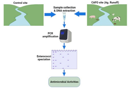
2.1. Study Area, Sampling Sites, and Sample Collection

This study was conducted in the middle Santa Ana River (MSAR) watershed (Figure S1; Table S1) that covers approximately 1264 km2 and lies largely in the southwestern corner of San Bernardino County and the northwestern corner of Riverside County [13,14,15,16,17,18]. Sampling points are shown in Figure 1, collected from agricultural runoff, urban runoff, recreational area, control, and wastewater treatment plants. Land use in the MSAR watershed includes urban, agricultural, and open space. Agricultural activities are mainly dairying, while the urban area is mainly residential and commercial properties. Water samples were collected in duplicates to determine salinity, pH, temperature, turbidity, and dissolved oxygen from each sample location using standard methods [19] as previously discussed [17,18]. Concentrations (mM) of Na+, K+, Ca2+, Mg2+, P, and S in sediment water extracts were determined using an Optima-3300 DV ICP-OES spectrometer (Perkin-Elmer, Waltham, MA, USA) that was calibrated with certified standards. Concentrations of Enterococcus species in surface water were determined within six hours of sample collection according to EPA Method 1600 for Enterococcus species [20], with slight modification for Enterolert (IDEXX Laboratories, Westbrook, ME, USA) quantification using Quanti-Trays and incubated for 48 h at 37 °C. Sediment samples from 0 to 15 cm depth were taken from the creek or riverbanks using ethanol-disinfected core tubes and stored in Whirl-Pak bags as previously reported [18].
Samples were transported to the laboratory for analysis in coolers maintained between 2 °C–10 °C using ice packs, and were at 4 °C until processed; usually within 24–48 h. Enterococcus species counts in sediment samples (10 g) were determined using a serial dilution method in a 1:9 sediment: sterile phosphate buffered saline (PBS; 0.0425 g/L KH2PO4 and 0.4055 g/L MgCl2 in distilled deionized water) solution. In brief, 90 mL of PBS was added to the sediment and shaken for 15 min. Ten mL of suspension was added to an Enterolert vessel, diluted 1:10 and mixed. One mL from the 1:10 dilution was transferred to another vessel and was further diluted 1:1000, and an aliquot was added to the media, mixed, then sealed in Quanti-Trays and incubated at 37 °C for 48 h. Samples were processed following the manufacturer’s protocol as stated above. Thereafter, 100 uL samples from positive wells were spread plated on Enterococcosel Agar (Becton Dickinson & Co., Sparks, MD, USA) for colony isolation. Individual colonies of pure cultures that were isolated were stored at −80 °C (in sterile 30% sterile glycerol) for further characterization.

2.2. Speciation of Enterococcus Species

Isolates were identified using genus- and species-specific multiplex PCR for enterococci [21] and were typed to the species level using multiplex PCR and sequencing of the 16S rRNA and rpoA gene.

2.3. Antimicrobial Resistance Analysis

Antimicrobial susceptibility tests (phenotypes) of Enterococcus species against eight antimicrobials, were assessed using disk diffusion assays (Figure S2) following CLSI standard methods [22]. Mueller-Hinton II agar (Difco, Sparks, MD, USA) was used, and cells were harvested from the surface of the medium with a cotton swab after 24 h growth at 37 °C. Antimicrobial agents were tested with BD BBL Sensi-Disc antimicrobial susceptibility test discs (Becton Dickinson & Co., Sparks, MD, USA) with the breakpoints (μg mL−1) indicated below. The panel of antimicrobials and breakpoints for classification as resistant were as follows: chloramphenicol (≥32 µg mL−1), ciprofloxacin (≥4 µg mL−1), erythromycin (≥8 µg mL−1), gentamicin (≥500 µg mL−1), penicillin (≥16 µg mL−1), tetracycline (≥16 µg mL−1), and vancomycin (≥32 µg mL−1). Enterococcus faecalis ATCC 29212, Staphylococcus aureus ATCC 29213, E. coli ATCC 25922 and Pseudomonas aeruginosa ATCC 27853 were employed as quality control strains for antibiotic susceptibility tests.

2.4. PCR Screening of Virulence and Antibiotic Resistance Genes

The genomic DNA of isolates was extracted using the QIAGEN extraction kit (Qiagen, Valencia, CA, USA) according to the manufacturer’s instructions. DNA from samples was quantified using a fluorometer Qubit 2.0. Amplified PCR products were separated by electrophoresis on a 1.5% agarose gel and visualized by staining with ethidium bromide. Isolates were screened by multiplex and single PCR reactions for the presence of genes encoding for virulence factors, including aggregation substance (asa1), cytolysin (cylA), enterococcal surface protein (esp), and gelatinase (gelE) (Table S2). Multiplex PCRs were performed to detect genes mediating resistance to vancomycin, tetracycline, and erythromycin. For vancomycin resistance presence of vanA, vanB, vanC1 and vanC2 genes was tested. Furthermore, the presence of genes encoding for tetracycline resistance genes, i.e., tet (A), tet (C), tet (Q), tet (K), tet (O), and tet (S) was evaluated, whereas for erythromycin resistance genes, presence of the erm (B) gene was tested. Each reaction mixture consisted of 25 μL Master mix (Promega, Madison, WI, USA), 4 mM MgCl2 (group I) or 3 mM MgCl2 (group II), and 3 μL of supernatant from freshly boiled cells as described previously [10].

2.5. Statistical Analysis

All of the statistical modeling and analysis for this study were performed using the GLM and MIXED procedures in the SAS STAT software package, version 9 [23]. Additionally, adjusted F and t-tests for all parameter estimates and/or contrasts of interest were computed using the Kenward-Roger adjustment technique [20]. p values of ≤0.05 were considered significant. Sampling sites were grouped into four zones based on site similarity and geographical location. Sites S13 and S14 were classified into their separate zone since the effluent water from each treatment plant was sampled immediately at the discharge outlet (located off the Chino Creek tributary). Likewise, site S1 was classified into its own zone, given its’ location in the base of the foothills and land use is open space (S1). This location (S1) represents a control site. The primary factors of interest in this study were the geographical locations of the sample sites (i.e., a site effect) and the temporal conditions surrounding the sampling location based on stream flow and the surrounding landscape. Thus, an analysis of variance (ANOVA) model using both site and surface flow classification effects represents a statistical model for analyzing the (log10-transformed) density of Enterococcus species. Four additional field parameters, pH, salinity (EC), turbidity, and temperature of the surface water were also measured at each sample point. Variations in each of these water quality parameters are known to affect bacterial concentrations. Therefore, the field parameters were measured as possible covariates in the subsequent statistical analyses. Given the primary factors of interest and additional covariate effects, the following analysis of covariance (ANOCOVA) model was initially specified as a plausible model for modeling each specific set of bacteria samples [24]:
ln ( y i j ) = μ + τ i + δ k + β 1 ( p H i j ) + β 2 ( ln [ E C i j ] ) + β 3 ( ln [ t B i j ] ) + β 4 ( T i j ) + ε i j .
In the equation, y i j represents the average bacteria count at the ith site during the jth sampling period, the τ i parameters quantify the 13 distinct site effects, the δ k parameters quantify the three (temporally dependent) water flow conditions, the four β parameters quantify the four water quality covariate effects (water pH, salinity [EC], turbidity [tB], and temperature [T]), and the ε i j residual errors are assumed to be normally distributed, but possibly temporally and/or spatially correlated. Bacterial data were log-transformed to induce approximate normality in the residual error distribution. Additionally, the salinity and turbidity covariate readings were also log-transformed in order to reduce the influence of a few large covariate readings (i.e., both covariate distributions appeared to follow approximate lognormal distributions).

3. Results

3.1. Identification of Covariance Structures for Major Physical and Chemical Parameters

Major physical and chemical parameters were quantified in water and sediment samples retrieved from different locations. These included temperature, turbidity, salinity, dissolved oxygen (DO), salinity (EC), pH, NO3, NO2, NH4, Ca, Na, Mg, P, S, and K as previously summarized [23]. The enterococci count for the watershed throughout the sampling season was based on univariate summary statistics [23] that were computed using the Shapiro–Wilk test for residual normality and were summarized to log10 transformed data of about 3.6 × 10⁵ CFU mL−1. Further analysis of data throughout the watershed showed that during the sampling period, enterococci count frequently exceeded the applicable water quality objectives for the Environmental Protection Agency’s (EPA’s) recommended water quality criteria except in the control site [20]. Our study showed that enterococci count in surface water was significantly correlated with pH (p < 0.01), NO2 (p < 0.03), and NH4N (p < 0.005) and in the sediment with NO3 (p < 0.015).

3.2. Distribution of Environmental Enterococcus spp. in the Watershed

The relative abundances of the Enterococcus isolates (n = 1917) from the Santa Ana River watershed are summarized in Figure 2. These included 524 E. faecalis (27.33%), 492 E. faecium (25.67%), and 625 E. mundtii (32.60%), which were the three dominant species of Enterococcus in this watershed. The rest of the species identified were (Figure 2): E. avium (1); E. casseliflavus (60); E. cecorum (2); E. durans (25); E. hirae (73); E. gallinarum (18); E. raffinosus (53) and unknowns (44). The identities of the 44 unknowns after sequencing (Figure 3) were; E. termitis (27), Vagococcus lutrae (2), Camobacterium (1), while some were unresolved (14). The following species were screened for but not found: E. asini, E. columbae, E. dispar, E. flavescens, E. gilvus, E. malodoratus, E. palens, E. porcinus, E. pseudoavium, E. saccharolyticus, E. seriolicida, E. solitarius, E. sulfureus.

3.3. Antimicrobial Resistance in Surface Water and Sediment

Enterococcal isolates from surface water were tested against a panel of eight antimicrobials. Isolates were from wastewater treatment plant effluent, control site, urban runoff, recreational runoff, and agricultural runoff (Table 1). Enterococcal isolates from different sites responded differently to the test antimicrobials. For instance, 21/24 (87%) of E. faecium in the WWTP were resistant to erythromycin, whereas 48/58 (83%) of E. mundtii were resistant to this antibiotic. However, in the urban runoff, the highest level of resistance was shown by E. faecium to tetracycline (109/181; 60%), ciprofloxacin (78/181; 43%), and erythromycin (45/181; 25%). E. faecalis demonstrated lower levels of resistance to the three antibiotics. In the recreational and agricultural runoffs, E. faecium also exhibited the highest level of resistance (Table 1). It is interesting to note that none of the isolates from this watershed were resistant to ampicillin, gentamicin, and vancomycin. Resistance of isolates in sediments to the different antibiotics followed almost the same pattern as observed in surface water with few exceptions. For instance, none of the isolates were resistant to ampicillin and vancomycin; however, 10% E. faecium, 2% E. mundtti, and 6% E. faecalis from the agricultural runoff and the control site were resistant to chloramphenicol (Table 2). Most of the isolates were resistant to ciprofloxacin, erythromycin, and tetracycline following the same pattern as in surface water. Whereas, in the urban runoff sediment, E. faecium was resistant to tetracycline (30/43; 70%), ciprofloxacin (33/43; 77%), and erythromycin (9/43; 21%). E. faecalis and E. mundtti showed some levels of resistance to the same three compounds though at a lower percentage level. In the recreational area, E. faecium was resistant to mostly tetracycline (31/61; 51%), ciprofloxacin (50/61; 82%), and erythromycin (33/61; 54%). In the agricultural runoff sediment, 100% of E. faecium was resistant to tetracycline.

3.4. Antibiotics Resistance and Virulence Factor Genes in Enterococci Isolated from Sediment and Surface Water Samples

Eleven resistance genes and four virulent factor encoding (VFE) genes were tested among E. faecium and E. faecalis isolates in surface water and sediment samples (Table 3 and Table 4). We used E. faecium and E. faecalis in this part of the study because they showed the most resistant phenotypes. None of the E. faecium or E. faecalis isolates showed resistance to any of the vancomycin resistance genes and erythromycin resistance gene (ermB) in surface water collected from WWTP. However, E. faecalis from urban runoff, recreational water, and agricultural runoff showed the presence of van (C1) and van (C2) resistance genes. A total of 10%, 5%, 10%, and 1% of E. faecalis showed the presence of tet (C, K, O, and S) resistance genes, respectively, whereas 6%, 1%, 1%, and 1% of E. faecium from WWTP showed the presence of the tet (C, K, O, and S) resistance genes. None of the isolates had tet (A, Q), or erm (B) resistance genes. In general, no isolate was resistant to tet (A, Q) in all the surface water samples collected from urban runoff, recreational runoff, agricultural runoff, and the control site. However, 48%, 10%, 35%, and 49% of E. faecalis from urban runoff water had tet (C, K, O, and S) resistance genes, whereas 1% were positive for erm (B) genes. This was contrary to E. faecium from the same source which was devoid of the five genes. In the recreational water and agricultural runoff E. faecium had higher levels of tet (C, K, O) resistance genes, while E. faecalis had higher levels of tet (S) and erm (B) genes than E. faecium (Table 3).
The ratio of phenotype to genotype in isolates from surface water was examined based on antibiotic sensitivities and genes that encoded resistance to tetracycline, vancomycin, and erythromycin. E. faecium was resistant to tetracycline and erythromycin in WWTP, agricultural runoff, and urban runoff, but not in recreational water and the control site (Table 1). However, the presence of tet (C, K, O, S) genes was only observed in agricultural runoff and WWTP, but not in urban runoff (Table 3). E. faecalis from all the samples except the control samples were resistant to tetracycline and they showed the presence of the four tet genes above in all the samples except tet (K) in the control sites. Although none of the isolates were resistant to vancomycin, van (C1) and van (C2) genes were detected in E. faecalis from urban runoff, recreational water, and agricultural runoff. E. faecium from surface water did not show the presence of any of the van resistance genes. E. faecium from all the samples except the control were resistant to erythromycin. However, resistance to erm (B) was only shown in urban runoff, agricultural runoff, and recreational water, while E. faecalis that were positive for erm (B) were only isolated from urban and agricultural runoffs. VFE genes of E. faecalis and E. faecium from surface water were detected (Table 3). The results showed that the asa1, cylA, esp, and gelE VFE genes of E. faecalis and E. faecium were detected in most of the sampling sites. asa1 and gelE were detected in all samples except E. faecium in recreational water and these two VFE genes were the most dominant in all the samples. The four VFE genes were detected in E. faecalis from recreational water.
However, E. faecalis was resistant to ciprofloxacin, erythromycin, and tetracycline, in urban runoff, recreational water, agricultural runoff, and the control sites at different percentage levels as stated above. tet (C, K, O, and S) genes were detected in urban runoff, agricultural runoff, and the control samples, but not in recreational samples. Only 20% of E. faecalis in urban runoff were positive for the erm (B) gene (Table 4). E. faecium was positive for the four genes, whereas only tet (O) and tet (K, O) were, respectively, found in urban runoff and control isolates. None of the sediment samples were positive for van genes in comparison to surface water samples. Virulence genes of E. faecalis and E. faecium were detected in sediment samples (Table 4). The results demonstrated the presence of asa1, cylA, esp, and gelE VFE genes in E. faecalis from most of the sampling sites, whereas, asa1 and gelE were detected in E. faecium from recreational water samples.
MDR, defined as resistance to three or more antimicrobial classes and resistance to multiple antimicrobials is summarized in Table 5. Isolates from different sampling sites were resistant to three different antimicrobial classes. In the recreational sediment samples, 26 isolates of E. faecium, were resistant to three antimicrobials (CipEryTet) and three antimicrobial classes. In the urban runoff sediment, nine isolates of E. faecium were also resistant to three antimicrobials (CipEryTet) from three antimicrobial classes. The same patterns were observed in agricultural runoff sediment, with two isolates, recreation water with eight isolates, and one isolate from WWTP. However, one isolate from the recreational water showed resistance to the same antimicrobials (CipPenTet). In the urban runoff water, 15 isolates were resistant to three antimicrobials (CipEryTet) and one was resistant to CipPenTet. For E. faecalis, only two isolates from agricultural sediments and one isolate from the control were resistant to three antimicrobials (CipChlTet), and 15 isolates were resistant to CipEryTet.

4. Discussion

4.1. Enterococcus Species in the Watershed

In this study, we used a culture-based method to investigate the Enterococcus population in surface water and sediment that may potentially be in contact with humans in an urban watershed. Our results agree with our previous study which suggested that microbial community compositions were influenced by several environmental factors, and pH, NO2, and NH4 were the major environmental factors driving fecal indicator bacteria in urban river runoff water based on canonical correspondence analysis, while NO3 was the only factor in sediment [14]. Therefore, the high numbers of Enterococcus may be associated with the presence of nitrogen and other nutrient sources as they correlate significantly with Enterococcus species as previously reported [23]. This is critical because Enterococcus species above a certain threshold limit in an urban watershed may be a health risk to humans living within the watershed especially those using the water for recreational activities. The health effects in humans can be grouped into gastrointestinal, respiratory, eye, ear, nose, skin rashes, etc., and some of these may require hospitalization. It should be noted that some of the bodies of water within the watershed are used for noncontact recreational purposes and high densities of bacteria can result in immediate closure for public use. As seen in this study, the log10 transformed data of about 3.6 × 10⁵ CFU mL−1 enterococci count throughout the watershed has exceeded the applicable water quality objectives for the Environmental Protection Agency’s (EPA’s) recommended water quality criteria except in the control site [20]. Therefore, most sites along urban and agricultural runoffs were in violation of local and US EPA water quality standards.
Different watersheds may contain different Enterococcus species depending on the input to the watershed. As previously shown, the Upper Oconee watershed identified E. casseliflavus as the dominant species in that watershed [25,26]. The Upper Oconee watershed is represented by a range of land uses, such as forest, and agriculture that is dominated by poultry production, residential, recreational, and industrial. In contrast to the Upper Oconee watershed, the Middle Santa Ana River watershed is highly populated (≥2 million people), with over 200,000 cattle in a 50,000 ha2, and large recreational and industrial/warehouse settings throughout the watershed. This watershed is also highly regulated in terms of input and other forms of discharge.

4.2. Antimicrobial Resistance in Surface Water and Sediment

Higher occurrence of MDR Enterococcus species in the urban/recreational areas than in agricultural samples were observed in this study. This agrees with our previous study on E. coli which showed greater numbers of E. coli with MDRs from urban runoff sources than agricultural sources [13]. Most Enterococcus isolates were resistant to erythromycin, tetracycline, ciprofloxacin, and penicillin (Table 1 and Table 3). The resistance by isolates from these sites to the four antimicrobials may suggest that these antimicrobials were naturally present throughout the watershed since they were the most common AMRs detected in our surface water samples.
It should be noted that ampicillin and tetracycline are old antimicrobials, but none of the isolates showed resistance to ampicillin. Tetracycline is used less frequently in humans than in animals, and the high rates of tetracycline resistance among Enterococcus isolates from urban runoff were unexpected. It is interesting to note that a strong correlation was observed between certain phenotypes and genotypes in this study, suggesting that the resistance to a given antimicrobial was likely caused in some cases by a single gene. In some instances, the antimicrobial resistance phenotypes and genotypes correlated very well, whereas in others they did not. For instance, we detected no phenotypic and genotypic resistance to vancomycin in isolates from the sediment of all the sampling sites in this study, but this was not the case with surface water samples. In this instance, while no phenotypic resistance was observed in surface water, a few isolates were positive for vanC1 and vanC2 genes (Table 3) but not vanA and vanB. Enterococci are also a major nosocomial pathogen due to their resistance to several antimicrobials and ability to acquire and disseminate AR determinants [27,28]. The most important factor in the outbreak of hospital vancomycin-resistant enterococci is the colonization of the excretory system, which almost always precedes bacteremia and is the main reservoir from which the spread of microorganisms in the hospital environment takes place.
Tetracycline resistance was by far the most common type of resistance observed in Enterococcus isolates in this study. No significant differences in resistance to tetracycline were found in isolates from urban runoff and agricultural runoff samples. As noted above, the agricultural runoff received input from CAFOs where tetracycline is often used as a first-line antimicrobial in disease prevention and growth promotion in food animals. Resistance to tetracyclines has been found in strains isolated from sausage, cheese, fish, and fish products [29]. Another antimicrobial that was very prevalent was erythromycin as it was as common as tetracycline. Previous studies have also reported high tetracycline and erythromycin resistance in Enterococcus isolates from food-producing animals [27]. In our study, the most common resistance genes found in the isolates were tetC, tetK, tetO, and tetS for tetracycline resistance and ermB for erythromycin resistance. These genes can be transferred from environment to clinical as previously reported by Jahan et al. [30] with the transfer of E. faecium and E. faecalis strains from food to clinical strains by HGT from pathogenic enterococci to strains of commensals and other species of bacteria constituting the physiological microflora of the gastrointestinal tract [31].
Genetic determinants that may enhance the virulence of Enterococcus isolates may include a variety of VF genes [28]. The ARGs and VF genes can be transferred to other bacteria, which pose a serious threat to public health through the food chain [26]. Our study found the presence of asa1, cylA, esp, and gelE VF genes. The gelE gene is responsible for the production of gelatinase, which is a metalloproteinase hydrolyzing casein, hemoglobulin, insulin, fibrinogen, collagen, and gelatin, as well as various proteins or peptides. The participation of this VFG in pathogenesis has also been observed [32,33], and the main role of these proteins is to provide nutrients to bacteria by breaking down the host tissue and in biofilm formation [34], translocation of E. faecalis across human enterocytes, and facilitates microbial invasion [35]. The esp gene contributes to the colonization and persistence of enterococci in infections [27]. The high prevalence of gelE and esp has previously been reported in bovine and genes encoding adhesion factors such as esp and asa1 are highly prevalent among E. faecalis and E. faecium [36]. In food-borne strains, cyl, gel, and hyl are detected but with much lower frequency compared to clinic enterococci [37,38]. In our study cyl was also observed at low frequency. One limitation of this study is that we did not do whole genome sequencing to determine the correlation between phenotypic resistance and resistance genes to analyze the prevalence and locations of different ARGs. Also, we did not determine antimicrobial residues (antibiotics) in sediment and surface water to correlate with the genes. This approach was used in a recent study where 26 antibiotics were measured, and almost all water samples (98.7%) had detectable levels of antibiotics [39].
Enterococcus faecium is part of the ESKAPE pathogens (Enterococcus faecium, Staphylococcus aureus, Klebsiella pneumoniae, Acinetobacter baumannii, Pseudomonas aeruginosa, and Enterobacter spp.) which are the leading cause of nosocomial infections [40]. Their multidrug resistance phenotypes and the clinical and economic burdens of the infections have made them the AR “priority pathogens” by WHO since they represent a global threat to human health [39,41]. As shown in Table 5, MDR ESKAPE E. faecium was detected in our watershed, suggesting that this pathogen is present outside the clinical environment in the natural environment. It should be noted that this watershed contains eleven WWTPs and water samples were collected directly from the effluent of two of these plants to investigate if WWTPs are a source of AR in the natural environment. Many studies have shown that WWTPs only partially removed ARB, ARGs, and antibiotics, and the final effluents may contain high levels of contaminants [42,43,44,45]. These contaminants present in the effluents may end up in rivers and streams and potentially affect the indigenous bacterial populations within the receiving waters [11,12,46,47,48]. Wastewater treated by WWTPs has also been widely reused for different purposes, including agriculture landscape irrigation and aquaculture. Hence, wastewater effluents should be treated further to prevent them from spreading into the environment, and one of the strategies that we have developed in our work is to use a system of layered environmental media (consisting of gravel, sand, soil, and soil + biochar) through which antibiotic-laden water was pumped through the system to polish wastewater for the removal of the contaminants in the final effluent [49]. This study showed that the overall removal efficiencies of the antibiotics amoxicillin, cefalexin, sulfadiazine, and tetracycline were 81, 91, 51, and 98%, respectively.

5. Conclusions

This study showed diverse species of Enterococcus in the watershed that are impacted by different anthropogenic sources. This has resulted in the detection of high levels of AMR and MDR isolates throughout the watershed. Resistant species such as E. faecium exhibited the highest resistant phenotype and genotype. The highest resistance was observed for ciprofloxacin in WWTP effluent, recreational area, and control sediment. Resistance of Enterococcus species to tetracycline was highest in agricultural sediment, and this was applicable to all the antibiotics used in this study. Overall, this study endeavored to provide an overall picture of the Enterococcus population in a mixed-use urban watershed. The current study shows that Enterococcus are prevalent and diverse in the middle Santa Ana River freshwater environment and are resistant to antibiotics used for human and veterinary purposes, medically important antibiotics, as well as the genes associated with resistance to these antibiotics. The wide dissemination and abundance of Enterococcus may potentially pose health concerns to the populations exposed to these water sources. Therefore, the continuous surveillance of Enterococcus in aquatic environments, identification of MDR Enterococcus hotspots, and preventive interventions may be some of the critical tools for reducing the burden and transmission of MDR Enterococcus species to ensure public health.

Supplementary Materials

The following supporting information can be downloaded at: https://0-www-mdpi-com.brum.beds.ac.uk/article/10.3390/w16010116/s1, Figure S1: Sampling locations in the watershed used for the study; Figure S2: Disk diffusion assay displaying antibiotics resistance; Table S1: Sampling Locations for MSAR Enterococcus Evaluation Study; Table S2: Primers and PCR conditions used in this study.

Author Contributions

Conceptualization, A.M.I. and S.E.M.; methodology, A.M.I. and A.C.O.; software, A.M.I.; validation, A.M.I., S.E.M. and A.C.O.; formal analysis, A.M.I.; investigation, A.M.I., S.E.M. and A.C.O.; resources, A.M.I.; data curation, A.M.I.; writing—original draft preparation, A.M.I.; writing—review and editing, A.M.I., S.E.M. and A.C.O.; visualization, A.M.I.; supervision, A.M.I.; project administration, A.M.I.; funding acquisition, A.M.I. All authors have read and agreed to the published version of the manuscript.

Funding

This research was supported by USDA-ARS 2036-32000-005-00D and 2036-12130-011-00D (NP 212). Mention of trade names or commercial products in this publication is solely for the purpose of providing specific information and does not imply recommendation or endorsement by the U.S. Department of Agriculture. The U.S. Department of Agriculture (USDA) prohibits discrimination in all its programs and activities based on race, color, national origin, age, disability, and where applicable, sex, marital status, familial status, parental status, religion, sexual orientation, genetic information, political beliefs, reprisal, or because all or part of an individual’s income is derived from any public assistance program.

Data Availability Statement

Data are contained within the article and Supplementary Materials.

Conflicts of Interest

The authors declare no conflicts of interest.

References

  1. Zaheer, R.; Cook, S.R.; Barbieri, R.; Goji, N.; Cameron, A.; Petkau, A.; Polo, R.O.; Tymensen, L.; Stamm, C.; Song, J.; et al. Surveillance of Enterococcus spp. reveals distinct species and antimicrobial resistance diversity across a One-Health continuum. Sci. Rep. 2020, 10, 3937. [Google Scholar] [CrossRef] [PubMed]
  2. Makowska, N.; Bresa, K.; Koczura, R.; Philips, A.; Nowis, K.; Mokracka, J. Urban wastewater as a conduit for pathogenic Gram-positive bacteria and genes encoding resistance to β-lactams and glycopeptides. Sci. Total Environ. 2021, 765, 144176. [Google Scholar] [CrossRef] [PubMed]
  3. Goldstein, R.E.R.; Micallef, S.A.; Gibbs, S.G.; George, A.; Claye, E.; Sapkota, A.; Joseph, S.W.; Sapkota, A.R. Detection of vancomycin-resistant enterococci (VRE) at four U.S. wastewater treatment plants that provide effluent for reuse. Sci. Total Environ. 2014, 466–467, 404–411. [Google Scholar] [CrossRef] [PubMed]
  4. Bhardwaj, P.; Ziegler, E.; Palmer, K.L. Chlorhexidine induces VanA-type vancomycin resistance genes in enterococci. Antimicrob. Agents Chemother. 2016, 60, 2209–2221. [Google Scholar] [CrossRef] [PubMed]
  5. Karimi, F.; Samarghandi, M.R.; Shokoohi, R.; Godini, K. Prevalence and removal efficiency of enterococcal species and vancomycin-resistant enterococci of a hospital wastewater treatment plant. Avicenna J. Environ. Health Eng. 2016, 3, e8623. [Google Scholar] [CrossRef]
  6. Byappanahalli, M.N.; Nevers, M.B.; Korajkic, A.; Staley, Z.R.; Harwood, V.J. Enterococci in the Environment. Microbiol. Mol. Biol. Rev. 2012, 76, 685–706. [Google Scholar] [CrossRef] [PubMed]
  7. Jang, J.; Hur, H.-G.; Sadowsky, M.; Byappanahalli, M.; Yan, T.; Ishii, S. Environmental Escherichia coli: Ecology and public health implications—A review. J. Appl. Microbiol. 2017, 123, 570–581. [Google Scholar] [CrossRef] [PubMed]
  8. Bager, F. DANMAP: Monitoring antimicrobial resistance in Denmark. Int. J. Antimicrob. Agents 2000, 14, 271–274. [Google Scholar] [CrossRef]
  9. Zainab, S.M.; Junaid, M.; Xu, N.; Malik, R.N. Antibiotics and antibiotic resistant genes (ARGs) in groundwater: A global review on dissemination, sources, interactions, environmental and human health risks. Water Res. 2020, 187, 116455. [Google Scholar] [CrossRef]
  10. Chajęcka-Wierzchowska, W.; Zadernowska, A.; Zarzecka, U.; Zakrzewski, A.; Gajewska, J. Enterococci from ready-to-eat food—Horizontal gene transfer of antibiotic resistance genes and genotypic characterization by PCR melting profile. J. Sci. Food Agric. 2019, 99, 1172–1179. [Google Scholar] [CrossRef]
  11. Choi, J.-M.; Woo, G.-J. Transfer of Tetracycline Resistance Genes with Aggregation Substance in Food-Borne Enterococcus faecalis. Curr. Microbiol. 2015, 70, 476–484. [Google Scholar] [CrossRef] [PubMed]
  12. Haug, M.C.; Tanner, S.A.; Lacroix, C.; Stevens, M.J.; Meile, L. Monitoring horizontal antibiotic resistance gene transfer in a colonic fermentation model. FEMS Microbiol. Ecol. 2011, 78, 210–219. [Google Scholar] [CrossRef] [PubMed]
  13. Ibekwe, A.M.; Murinda, S.E.; Graves, A.K. Genetic diversity and antimicrobial resistance of Escherichia coli from human and animal sources uncovers multiple resistances from human sources. PLoS ONE 2011, 6, e20819. [Google Scholar] [CrossRef] [PubMed]
  14. Ibekwe, A.M.; Ma, J.; Murinda, S.E. Bacterial community composition and structure in an Urban River impacted by different pollutant sources. Sci. Total Environ. 2016, 566–567, 1176–1185. [Google Scholar] [CrossRef] [PubMed]
  15. SAWPA: Santa Ana Watershed Project Authority. Middle Santa Ana River bacterial indicator TMDL 2013 Dry Season Report. Prepared by CDM Smith on behalf of SAWPA and the Middle Santa Ana River Watershed TMDL Task Force. July 2013. Available online: https://sawpa.gov/wp-content/uploads/2018/04/2013-Triennial-Report_Tier-1-Source-Evaluation-Final.pdf (accessed on 1 July 2023).
  16. RWQCB, Regional Water Quality Control Board. Resolution amending the water quality control plans for the Santa Ana River Basin to incorporate bacterial indicator total maximum daily loads (TMDLs) for middle Santa Ana River Watershed Water bodies. Regional Board Resolution R8-2005-0001. 2005. Available online: https://www.waterboards.ca.gov/board_decisions/adopted_orders/resolutions/2006/rs2006_0030attach.pdf (accessed on 1 July 2023).
  17. Ibekwe, A.M.; Leddy, M.; Murinda, S.E. Potential human pathogenic bacteria in a mixed urban watershed as revealed by pyrosequencing. PLoS ONE 2013, 8, e79490. [Google Scholar] [CrossRef] [PubMed]
  18. Ibekwe, A.M.; Leddy, M.B.; Bold, R.M.; Graves, A.K. Bacterial community composition in low-flowing river water with different sources of pollutants. FEMS Microbiol. Ecol. 2012, 79, 155–166. [Google Scholar] [CrossRef]
  19. American Public Health Association. Standard Methods for the Examination of the Examination of Water and Wastewater; American Public Health Association: Washington, DC, USA, 1995. [Google Scholar]
  20. USEPA. Review of Zoonotic Pathogens in Ambient Waters, 822-R-09-002; USEPA Office of Water, Ed.; Office of Water, U.S. Environmental Protection Agency: Washington, DC, USA, 2009.
  21. Jackson, C.R.; Fedorka-Cray, P.J.; Barrett, J.B.; Ladely, S.R. Effects of tylosin use on erythromycin resistance in enterococci isolated from swine. Appl. Environ. Microbiol. 2004, 70, 4205–4210. [Google Scholar] [CrossRef]
  22. CLSI. Performance Standards for Antimicrobial Susceptibility Testing; Clinical and Laboratory Standards Institute: Wayne, PA, USA, 2018. [Google Scholar]
  23. Ibekwe, A.M.; Lesch, S.M.; Bold, R.M.; Leddy, M.B.; Graves, A.K. Variations of indicator bacteria in a large urban watershed. Trans. ASABE 2011, 54, 2227–2236. [Google Scholar] [CrossRef]
  24. Kenward, M.G.; Roger, J.H. Small sample inference for fixed effects from restricted maximum likelihood. Biometrics 1997, 53, 983–997. [Google Scholar] [CrossRef]
  25. Cho, S.; Hiott, L.M.; McDonald, J.M.; Barrett, J.B.; McMillan, E.A.; House, S.L.; Adams, E.S.; Frye, J.G.; Jackson, C.R. Diversity and antimicrobial resistance of Enterococcus from the Upper Oconee Watershed, Georgia. J. Appl. Microbiol. 2019, 128, 1221–1233. [Google Scholar] [CrossRef]
  26. Cho, S.; Jackson, C.; Frye, J. The prevalence and antimicrobial resistance phenotypes of Salmonella, Escherichia coli and Enterococcus sp. in surface water. Lett. Appl. Microbiol. 2020, 71, 3–25. [Google Scholar] [CrossRef] [PubMed]
  27. Diarra, M.S.; Rempel, H.; Champagne, J.; Masson, L.; Pritchard, J.; Topp, E. Distribution of antimicrobial resistance and virulence genes in Enterococcus spp. and characterization of isolates from broiler chickens. Appl. Environ. Microbiol. 2010, 76, 8033–8043. [Google Scholar] [CrossRef] [PubMed]
  28. Cho, S.; Barrett, J.B.; Frye, J.G.; Jackson, C.R. Antimicrobial resistance gene detection and plasmid typing among multidrug resistant enterococci isolated from freshwater environment. Microorganisms 2020, 8, 1338. [Google Scholar] [CrossRef]
  29. Thumu, S.C.R.; Halami, P.M. Acquired Resistance to Macrolide-Lincosamide-Streptogramin Antibiotics in Lactic Acid Bacteria of Food Origin. Indian J. Microbiol. 2012, 52, 530–537. [Google Scholar] [CrossRef] [PubMed]
  30. Jahan, M.; Zhanel, G.G.; Sparling, R.; Holley, R.A. Horizontal transfer of antibiotic resistance from Enterococcus faecium of fermented meat origin to clinical isolates of E. faecium and Enterococcus faecalis. Int. J. Food Microbiol. 2015, 199, 78–85. [Google Scholar] [CrossRef] [PubMed]
  31. Peters, J.; Mac, K.; Wichmann-Schauer, H.; Klein, G.; Ellerbroek, L. Species distribution and antibiotic resistance patterns of enterococci isolated from food of animal origin in Germany. Int. J. Food Microbiol. 2003, 88, 311–314. [Google Scholar] [CrossRef] [PubMed]
  32. Del Papa, M.F.; Hancock, L.E.; Thomas, V.C.; Perego, M. Full activation of Enterococcus faecalis gelatinase by a C-terminal proteolytic cleavage. J. Bacteriol. 2007, 189, 8835–8843. [Google Scholar] [CrossRef] [PubMed]
  33. Thurlow, L.R.; Thomas, V.C.; Narayanan, S.; Olson, S.; Fleming, S.D.; Hancock, L.E. Gelatinase contributes to the pathogenesis of endocarditis caused by Enterococcus faecalis. Infect. Immun. 2010, 78, 4936–4943. [Google Scholar] [CrossRef]
  34. Ali, L.; Goraya, M.U.; Arafat, Y.; Ajmal, M.; Chen, J.L.; Yu, D. Molecular mechanism of quorum-sensing in Enterococcus faecalis: Its role in virulence and therapeutic approaches. Int. J. Mol. Sci. 2017, 18, 960. [Google Scholar] [CrossRef]
  35. Zeng, J.; Teng, F.; Murray, B.E. Gelatinase is important for translocation of Enterococcus faecalis across polarized human enterocyte-like T84 cells. Infect. Immun. 2005, 73, 1606–1612. [Google Scholar] [CrossRef]
  36. Dapkevicius, M.d.L.E.; Sgardioli, B.; Câmara, S.P.A.; Poeta, P.; Malcata, F.X. Current trends of enterococci in dairy products: A comprehensive review of their multiple roles. Foods 2021, 10, 821. [Google Scholar] [CrossRef] [PubMed]
  37. Spirli, H.; Demirbaş, F.; Dertli, E. Characterization of functional properties of Enterococcus spp. isolated from Turkish white cheese. LWT Food Sci. Technol. 2017, 75, 358–365. [Google Scholar] [CrossRef]
  38. Zkan, E.R.; Demirci, T.; Akın, N. In vitro assessment of probiotic and virulence potential of Enterococcus faecium strains derived from artisanal goatskin casing Tulum cheeses produced in central Taurus Mountains of Turkey. LWT 2021, 141, 110908. [Google Scholar]
  39. Iweriebor, B.C.; Obi, L.C.; Okoh, A.I. Virulence and antimicrobial resistance factors of Enterococcus spp. isolated from fecal samples from piggery farms in Eastern Cape, South Africa. BMC Microbiol. 2015, 15, 136. [Google Scholar] [CrossRef] [PubMed]
  40. Chajęcka-Wierzchowska, W.; Zadernowska, A.; Łaniewska-Trokenheim, Ł. Virulence factors, antimicrobial resistance and biofilm formation in Enterococcus spp. isolated from retail shrimps. LWT Food Sci. Technol. 2016, 69, 117–122. [Google Scholar] [CrossRef]
  41. Yang, F.; Zhang, S.; Shang, X.; Wang, X.; Yan, Z.; Li, H.; Li, J. Short communication: Antimicrobial resistance and virulence genes of Enterococcus faecalis isolated from subclinical bovine mastitis cases in China. J. Dairy Sci. 2019, 102, 140–144. [Google Scholar] [CrossRef]
  42. Cho, S.; Hiott, L.M.; Read, Q.D.; Damashek, J.; Westrich, J.; Edwards, M.; Seim, R.F.; Glinski, D.A.; McDonald, J.M.B.; Ottesen, E.A.; et al. Distribution of antibiotic resistance in a mixed-use watershed and the impact of wastewater treatment plants on antibiotic resistance in surface water. Antibiotics 2023, 12, 1586. [Google Scholar] [CrossRef]
  43. Mulani, M.S.; Kamble, E.E.; Kumkar, S.N.; Tawre, M.S.; Pardesi, K.R. Emerging Strategies to Combat ESKAPE Pathogens in the Era of Antimicrobial Resistance: A Review. Front. Microbiol. 2019, 10, 539. [Google Scholar] [CrossRef]
  44. Tacconelli, E.; Carrara, E.; Savoldi, A.; Harbarth, S.; Mendelson, M.; Monnet, D.L.; Pulcini, C.; Kahlmeter, G.; Kluytmans, J.; Carmeli, Y.; et al. Discovery, research, and development of new antibiotics: The WHO priority list of antibiotic-resistant bacteria and tuberculosis. Lancet Infect. Dis. 2018, 18, 318–327. [Google Scholar] [CrossRef]
  45. Xu, J.; Xu, Y.; Wang, H.; Guo, C.; Qiu, H.; He, Y.; Zhang, Y.; Li, X.; Meng, W. Occurrence of antibiotics and antibiotic resistance genes in a sewage treatment plant and its effluent-receiving river. Chemosphere 2015, 119, 1379–1385. [Google Scholar] [CrossRef]
  46. Rodriguez-Mozaz, S.; Chamorro, S.; Marti, E.; Huerta, B.; Gros, M.; Sànchez-Melsió, A.; Borrego, C.M.; Barceló, D.; Balcázar, J.L. Occurrence of antibiotics and antibiotic resistance genes in hospital and urban wastewaters and their impact on the receiving river. Water Res. 2015, 69, 234–242. [Google Scholar] [CrossRef] [PubMed]
  47. An, X.-L.; Su, J.-Q.; Li, B.; Ouyang, W.-Y.; Zhao, Y.; Chen, Q.-L.; Cui, L.; Chen, H.; Gillings, M.R.; Zhang, T.; et al. Tracking antibiotic resistome during wastewater treatment using high throughput quantitative PCR. Environ. Int. 2018, 117, 146–153. [Google Scholar] [CrossRef] [PubMed]
  48. Alexander, J.; Hembach, N.; Schwartz, T. Evaluation of antibiotic resistance dissemination by wastewater treatment plant effluents with different catchment areas in Germany. Sci. Rep. 2020, 10, 8952. [Google Scholar] [CrossRef] [PubMed]
  49. Ashworth, D.J.; Ibekwe, A.M. System of multi-layered environmental media for the removal of antibiotics from wastewater. J. Environ. Chem. Eng. 2020, 8, 104206. [Google Scholar] [CrossRef]
Figure 1. Details of sample types and sources of Enterococcus species investigated for species distribution and antimicrobial activities in the Santa Ana River watershed. Sample locations and sites consist of both sediment and surface water collected from agricultural runoff, urban runoff, recreational area, control, and wastewater treatment plants.
Water 16 00116 g001
Figure 2. Analysis of Enterococcus isolates in the Santa Ana River watershed.
Water 16 00116 g002
Figure 3. Identification of the 44 unknown isolates after sequencing. Y-axis are percentages of Enterococcus species recovered.
Water 16 00116 g003
Table 1. Spatial distribution of antimicrobial resistance phenotypes in enterococci isolated from surface water samples. Number of resistant isolates (%).
SitesSpeciesAmp *ChlGenPenCipEryTetVan
WWTPE. faecalis (28)0000001 (3)0
E. faecium (24) 00002 (8)21 (87)3 (12)0
E. mundtii (58)0000048 (83)00
Urban runoffE. faecalis (116)01 (1)0010 (8)4 (3)44 (38)0
E. faecium (181) 0003 (2)78 (43)45 (25)109 (60)0
E. mundtii (116)0000011 (9)3 (2)0
Recreational runoffE. faecalis (29)01(3)0010 (34)04 (14)0
E. faecium (89) 00013 (15)42 (47)22 (25)00
E. mundtii (39)0000001 (2)0
Ag. runoffE. faecalis (48)00003 (6)3 (6)10 (21)0
E. faecium (61) 000036 (59)16 (26)30 (49)0
E. mundtii (65)00000000
ControlE. faecalis (60)0008 (13)2 (3)000
E. faecium (0)00000000
E. mundtii (0)00000000
Note: * Antimicrobials: ampicillin (Amp), gentamicin (Gen), ciprofloxacin (Cip), Erythromycin (Ery), chloramphenicol (Chl), penicillin (Pen), tetracycline (Tet), vancomycin (Van).
Table 2. Spatial distribution of antimicrobial resistance phenotypes in enterococci isolated from sediment samples. Number of resistant isolates (%).
SitesSpeciesAmp *ChlGenPenCipEryTetVan
WWTPE. faecalis (0)00000000
E. faecium (0) 00000000
E. mundtii (19)0000019 (100)00
Urban runoffE. faecalis (113)00003 (2)8 (7)24 (21)0
E. faecium (43) 0001 (2)33 (77)9 (21)30 (70)0
E. mundtii (126)0000038 (30)2 (1)0
Recreational runoffE. faecalis (0)00000000
E. faecium (61) 0001 (2)50 (82)33 (54)31 (51)0
E. mundtii (80)0000036 (45)00
Ag. runoffE. faecalis (86)09 (10)1 (1)04 (5)1 (1)18 (21)0
E. faecium (11) 00000011(100)0
E. mundtii (102)02 (2)00005 (5)0
ControlE. faecalis (35)02 (6)002 (6)1 (3)19 (54)0
E. faecium (22)00012 (55)22 (100)000
E. mundtii (20)00000000
Note: * Antimicrobials: ampicillin (Amp), gentamicin (Gen), ciprofloxacin (Cip), Erythromycin (Ery), chloramphenicol (Chl), penicillin (Pen), tetracycline (Tet), vancomycin (Van).
Table 3. Antibiotic resistance genes and virulence factor encoding genes in enterococci isolated from surface water samples. Number of resistant isolates (%).
SitesSpeciestet (A)tet (C)tet (Q)tet (K)tet (O)tet (S)van (A)van (B)van (C1)van (C2)erm (B)asa1cylAespgelE
WWTPE. faecalis056 (10)028 (5)56 (10)83 (15)0000078 (14)0078 (14)
E. faecium086 (6)014 (1)14 (1)14 (1)0000029 (20)0029 (2)
Urban runoffE. faecalis079 (48)016 (10)57 (35)80 (49)002 (1)08 (5)46 (28)0097 (59)
E. faecium 00009 (9)3 (3)005 (5)
Recreational runoffE. faecalis022 (5)04 (1)9 (2)87 (20)000013 (3)022 (5)4 (1)4 (1)87 (20)
E. faecium098 (55)02 (1)20 (11)45 (25)00005 (3)0000
Ag. runoffE. faecalis078 (28)0017 (6)83 (30)0003 (1)3 (1)17 (6)019 (7)94 (34)
E. faecium095 (40)05 (2)19 (8)45 (19)00005 (2)2 (1)002 (1)
ControlE. faecalis055 (11)00095 (19)00005 (1)70 (14)00100 (20)
E. faecium
Table 4. Antibiotic resistance genes and virulence factor encoding genes in enterococci isolated from sediment samples. Number of resistant isolates (%).
SitesSpeciestet (A)tet (C)tet (Q)tet (K)tet (O)tet (S)van (A)van (B)van (C1)van (C2)erm (B)asa1cylAespgelE
WWTPE. faecalis
E. faecium
Urban runoffE. faecalis090 (45)06 (3)40 (20)88 (44)00004 (2)38 (19)010 (5)100 (50)
E. faecium075 (15)025 (5)050 (10)000000000
Recreational runoffE. faecalis
E. faecium097 (28)003 (1)62 (18)000007 (2)003 (1)
Ag. runoffE. faecalis083 (33)025 (10)50 (20)65 (26)0000065 (26)33 (13)48 (19)78 (31)
E. faecium0100 (8)063 (50)25 (2)38 (3)000000000
E. mundtii
ControlE. faecalis057 (8)021 (3)7 (1)50 (7)0000086 (12)043 (6)100 (14)
E. faecium0100 (6)000100 (6)000000000
Note: E. faecium and E. faecalis from the WWTP sediment were not resistant to any of the antibiotics and were negative for the resistance encoding genes that were tested.
Table 5. Multidrug resistance patterns of E. faecium and E. faecalis in sediment and surface water.
Sample NameSpeciesNo. IsolatesAmpGenCipEryChlPenTetVan
Recreation sediment E. faecium26SSRRSSRS
Urban runoff sediment E. faecium9SSRRSSRS
Agricultural water E. faecium2SSRRSSRS
Recreation water E. faecium1SSRSSRRS
Recreation waterE. faecium8SSRRSSRS
WWTP E. faecium1SSRRSSRS
Urban Runoff water E. faecium1SSRISRRS
Urban Runoff waterE. faecium15SSRRSSRS
E. faecalis
Agricultural sediment E. faecalis2SSRIRSRI
Control E. faecalis1SSRIRSRI
Urban Runoff sediment E. faecalis15SSRRSSRS
Note: Antimicrobials: ampicillin (Amp), gentamicin (Gen), ciprofloxacin (Cip), Erythromycin (Ery), chloramphenicol (Chl), penicillin (Pen), tetracycline (Tet), vancomycin (Van).
Disclaimer/Publisher’s Note: The statements, opinions and data contained in all publications are solely those of the individual author(s) and contributor(s) and not of MDPI and/or the editor(s). MDPI and/or the editor(s) disclaim responsibility for any injury to people or property resulting from any ideas, methods, instructions or products referred to in the content.

