Case Report

Surgical-Orthodontic Diagnosis and Treatment Planning in an Asymmetric Skeletal Class III Patient—A Case Report

by
Francisco Vale
1,*,
Catarina Nunes
1,
Adriana Guimarães
1,
Anabela Baptista Paula
1,2,3,4,5 and
Inês Francisco
1
1
Faculty of Medicine, Institute of Orthodontics, University of Coimbra, Área de Medicina Dentária—Av. Bissaya Barreto, Bloco de Celas, 3000-075 Coimbra, Portugal
2
Faculty of Medicine, Institute of Integrated Clinical Practice, University of Coimbra, 3000-075 Coimbra, Portugal
3
Faculty of Medicine, Area of Environment Genetics and Oncobiology (CIMAGO), Coimbra Institute for Clinical and Biomedical Research (iCBR), University of Coimbra, 3000-075 Coimbra, Portugal
4
Centre for Innovative Biomedicine and Biotechnology (CIBB), University of Coimbra, 3000-075 Coimbra, Portugal
5
Clinical Academic Center of Coimbra (CACC), 3000-075 Coimbra, Portugal
*
Author to whom correspondence should be addressed.
Submission received: 8 June 2021 / Revised: 22 June 2021 / Accepted: 24 June 2021 / Published: 27 June 2021
(This article belongs to the Special Issue Symmetry in Dentistry: From the Clinic to the Lab)

Abstract

:
The skeletal Class III pattern is characterized by a sagittal intermaxillary mesial discrepancy. This discrepancy may have an unfavorable impact on function and aesthetics, which can be aggravated by the presence of facial asymmetries. This case report describes the diagnosis and treatment planning of a 19-year-old male patient with a skeletal Class III, maxillary hypoplasia, anterior crossbite, and mandibular asymmetry. When the patient reached skeletal maturity at the end of puberty, the definitive diagnosis was skeletal Class III with hyperdivergent profile and mandibular asymmetry, and a surgical-orthodontic treatment was proposed. At the end of the treatment, bimaxillary surgical correction allowed a skeletal Class I with mandibular symmetry, improving the function of the stomatognathic system and facial aesthetics.

1. Introduction

Nowadays, dentofacial aesthetics of the smile and face symmetry are two factors considered in social comparisons [1], i.e., a symmetrical smile is considered more attractive [2]. Additionally, orthodontic treatment has been demonstrated to increase oral health parameters [3]. Facial asymmetry can manifest as a part of disharmony of the craniofacial complex, altering the proportions between the facial thirds [4], having skeletal, dental, and soft tissue involvement [5]. According to Thiesen et al., the prevalence of facial asymmetry varied from 11 to 37% [6], being 61% more prevalent in skeletal Class III patients [7] than skeletal Class II [4].
The assessment of facial asymmetry is usually performed by comparing the distance between gnation or ment points and the facial midline [4]. The etiology is not entirely known, but several genetic and environmental risk factors are identified [4,5]. Most cases of facial asymmetry have a mandibular origin, resulting in a deviation of the lower third of the face [6]. Additionally, the mandible has a longer growth period, which increases the probability of being more affected than other structures. Haraguchi et al. reported that the longer growth and the mobility of the mandible promotes a more asymmetrical growth than the maxilla, which is connected to the adjacent skeletal structures [8]. Moreover, Severt and Proffit reported a prevalence of 85% of lateral excursion to the left in patients with dentofacial deformities with mandibular deviation [9].
Orthognathic surgery aims to correct asymmetry to align the maxillary midline and chin with the facial midline, leveling the labial commissures and the occlusal plane [10]. The correction of the intermaxillary discrepancy contributes to the patient’s well-being and social life, which will improve their psychological condition [11]. Generally, these patients may have a malocclusion associated with asymmetric facial growth. The skeletal Class III pattern is characterized by sagittal intermaxillary mesial discrepancy. This can result from a mandibular prognathism, maxillary retrusion, or a combination of both [12,13], but, in most cases, it involves the mandibular component [14,15].
The etiology of this skeletal discrepancy is multifactorial [16], resulting from a combination of genetic or intrinsic factors with environmental or external factors [13]. In a recent systematic review of Dehesa-Santos et al., it was demonstrated that skeletal Class III condition has polygenic characteristics, probably affected by ethnicity and epigenetic factors, namely nutrition, muscle strength, and basal metabolism [16].
Regarding worldwide prevalence, skeletal Class III ranges between 0 and 26.7% [13]. Zere et al. found that Class III malocclusion is more prevalent in the Hispanic group than in the Caucasian and African groups. The European continent has a skeletal Class III prevalence of 4.88% [13].
There are three treatment options for skeletal Class III patients with mandibular asymmetry. On growing patients, interceptive orthodontics through facial mask allows the maxillary protraction and changes craniofacial growth direction. In the adulthood stage, patients with moderated skeletal Class III, with or without medium craniofacial asymmetry with acceptable facial morphology [17,18], can be treated only by orthodontic treatment. Although camouflage allows dental correction, the aesthetic ideal may not be reached [19,20]. When the severity of Class III malocclusion makes camouflage unfeasible, surgical-orthodontic treatment is the best treatment option [21]. This treatment improves the functional component (chewing and phonetics) and allows the correction of the position of the skeletal base’s relation to the cranium base anterior portion. These enable the cutaneous profile correction and a more symmetric and pleasant facial aesthetic, increasing the patient’s self-esteem and quality of life [19,22]. The clinical exam, cephalometric analysis, patient’s age, and malocclusion severity are decision-makers for the treatment plan [20,23]. Surgical-orthodontic treatment is the only one that allows the changing of facial aesthetics, correcting asymmetry with greater predictability of the treatment.
Surgical-orthodontic treatment is divided into three phases. The first one, the presurgical orthodontic treatment, can last between one and two years, and involves retro inclination of the upper incisors and proinclination of the lower incisors and loss of lower anchorage until reaching the correct inclination with maxillary planes [22,24,25]. The second phase culminates with orthognathic surgery, which is typically bimaxillary: Le Fort I osteotomy with maxillary advancement, and bilateral sagittal splint osteotomy (BSSO) of mandibular setback and probably a genioplasty [24,26]. The third and last phase is the postsurgical orthodontic treatment [24], which involves the use of intermaxillary elastics to stabilize the new occlusal position, prevent relapse, and serve as a guide for the reestablishment of the new functional pattern [27].
This case report presents the diagnosis and a successfully orthodontic-surgical treatment of skeletal Class III with hyperdivergent profile and mandibular asymmetry.

2. Materials and Methods
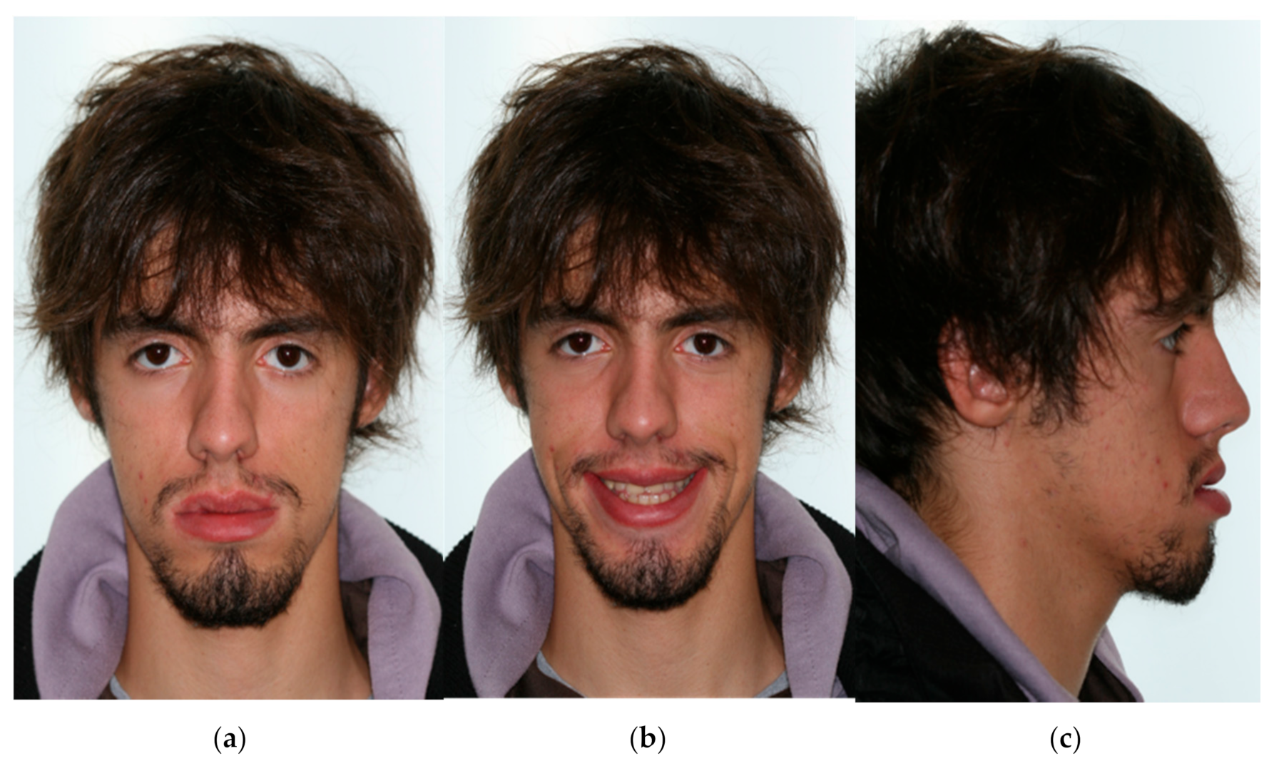
A 19-year-old male attended the Institute of Orthodontics, Faculty of Medicine University of Coimbra, with the main complaint of a very advanced and deflected chin to the left. The patient reported the use of an Eschler appliance since 10 years of age. However, the patient’s growth revealed the failure of this treatment.
The patient’s medical history was collected, in which he reported trauma to the chin in childhood, which may explain his mandibular deviation. The facial analysis evidenced maxillary hypoplasia, malar deficiency, concave profile, and increased lower third of the face, and facial asymmetry due to mandibular deviation to the left. The upper dental midline was centered with the facial midline, and the lower dental midline was centered with the ment. The intraoral photographs, taken with a Canon EOS T100 body camera with a Canon EF 100 mm f/2.8 Macro lens and Canon ring lite MR 14EX ii, show an angle Class III malocclusion, an anterior crossbite with a negative overjet of 4 mm, an overbite of 3 mm, a noncoincident dental midline, and a slight crowding in the lower arch. Figure 1 and Figure 2 show extra- and intraoral pretreatment records, respectively.
The orthopantomogram (Figure 3a) showed an asymmetrical condylar morphology, with a smaller condyle on the right side, and the impaction of the four third molars was also observed. Root resorptions, dental restorations, or endodontic treatments were not observed, and bone level was normal.
The lateral cephalometric analysis (Figure 3b,c, and Table 1) showed a severe Class III skeletal pattern (ANB, −5°), maxillary hypoplasia (SNA, 82°), and slight protrusion of the mandible (SNB, 82.5°). The upper incisors presented a slight pro-inclination (U1 to palatal plan, 115.8°), and the lower incisors presented a decreased axial inclination to the mandibular plane (L1 to Go–Gn, 90.3°). The soft tissue profile had a retrusion of the upper lip and protrusion of the lower lip.
Considering all diagnostic elements, the patient was diagnosed as having a severe skeletal Class III, with a mandibular deviation to the left and a bilateral molar and canine Class III relation.
The treatment objectives for this patient were: (1) to align and level the arches with lower crowding through stripping; (2) to correct the axial inclination of the incisors with the skeletal planes (dental decompensation); (3) to correct the skeletal base relationship to the SN plane and mandibular asymmetry through orthognathic surgery (Le Fort I osteotomy with maxillary advancement and BSSO of mandibular setback, with repositioning to correct asymmetry); (4) to achieve an angle Class I; (5) to obtain a correct overjet and overbite; (6) to improve the function of the stomatognathic system and facial aesthetic with a functional occlusion.

3. Results

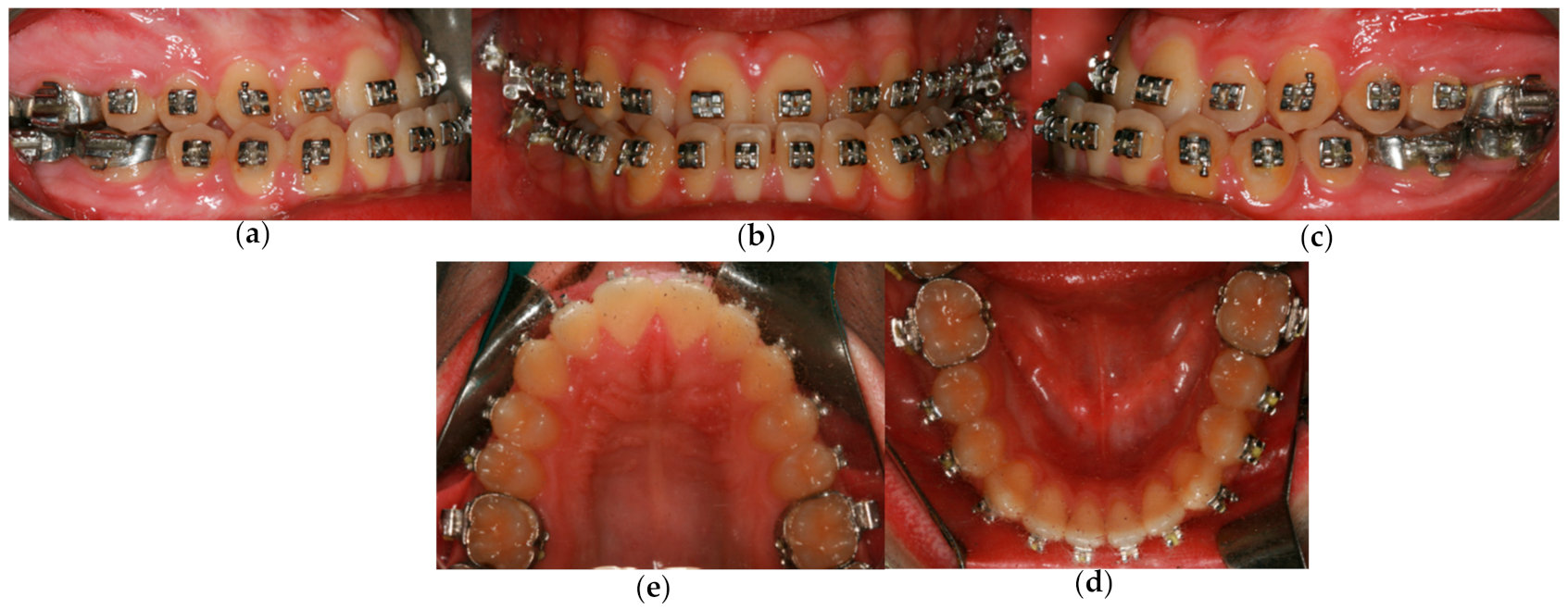
3.1. The Presurgical Orthodontic Treatment

Initially, the fixed orthodontic appliances (Roth prescription slot 0.018 equilibrium®2 Dentaurum (Turnstrabe 31, Ispringen, Germany)) were placed in both arches. The arches were levelled with continuous arch wires, starting with 0.014 nickel–titanium (NiTi) (Rematitan® LITE Idealbogen/ideal arches (Turnstrabe 31-Ispringen-germany)) and finishing with 0.017 × 0.025 stainless steel (Remanium® Dentaurum Stangendraht/straight wire Turnstrabe 31-Ispringen-germany) to control torque movement. Stripping was performed to obtain space to align and level the mandibular dental arch. Plaster models were obtained periodically to check if satisfactory occlusion was obtained to perform the surgery. The lower third molars were extracted 6 months before surgery to allow bone formation in the extraction wounds, since these teeth are located in the osteotomy area. Figure 4 shows the presurgical orthodontic preparation, where it is possible to observe the alignment and dental levelling.
The patient presented a correct inclination of the lower incisors in relation to the mandibular plane (L1–GoGn,90.3°), as well as the upper incisors in relation to the maxillary plane (U1–Mx Base, 115.8°), having obtained an ideal dental decompensation (Figure 5a). Subsequently, surgical hooks were placed on the stainless steel archwires in all inter-bracket spaces.

3.2. Orthognathic Surgery

According to a facial analysis by Dolphin Image (software version 11.9 (Dolphin Image & Management Solutions®, Chatsworth, CA, USA), a surgical simulation was planned, allowing the preparation of surgical guides. The surgical treatment plan included the following movements: Le Fort I with a 5 mm advance and BSSO mandibular setback of 4 mm with repositioning to correct asymmetry (Figure 5b). The combination of these movements allows a reduction of the lower one-third of the face due to the decrease of the angle of the mandibular plane.

3.3. Postsurgical Orthodontic Treatment

After orthognathic surgery, the patient returned for orthodontic finishing for obtaining an angle Class I relationship, normal overjet, and overbite and coincident midlines. After completing 12 months of active postsurgical orthodontic treatment, the fixed appliances were removed, a fixed retainer was bonded from canine to canine in the lower arch, and a Hawley retainer was placed in the upper arch. The patient was advised to use the Hawley 24 h a day for the first three months, and every night after that.

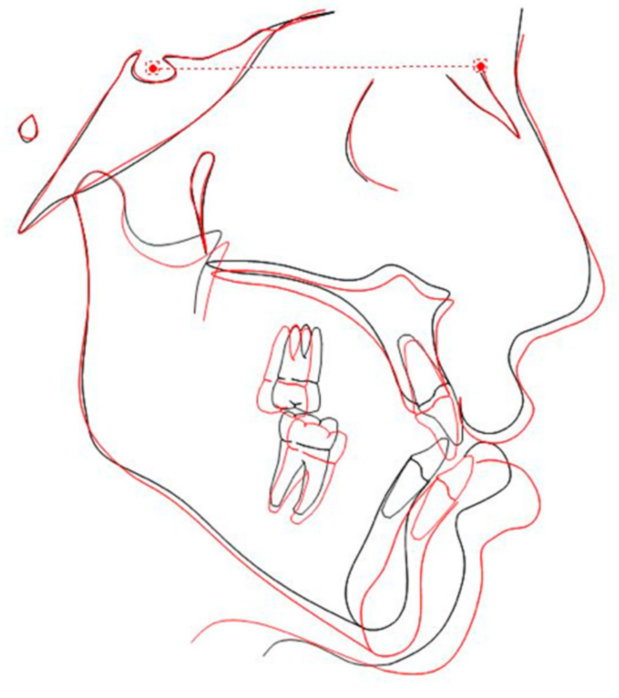
3.4. Treatment Results

At the end of treatment, a bilateral Class I canine and a slight Class III molar with normal overjet and overbite were achieved (Figure 6). Normal lateral and protrusive excursions were also verified. Despite there being a slight deviation of the lower midline with the upper midline, a coincidence of the facial midline and chin was observed (Figure 7). Therefore, maxillary hypoplasia and mandibular prognathism and asymmetry were correct, improving the stomatognathic system function and facial aesthetics.
The post-treatment cephalometric analysis (Table 1 and Figure 8b,c) showed: a substantial increase in ANB value, from −5° to 0°, which allowed a skeletal Class I to be achieved, mostly due to the increase of SNA value; the upper incisor increased the axial inclination from 115.8° to 126.8° relative to the maxillary plane, and the lower incisor increased the axial inclination from 90.3° to 99.1° relative to the mandibular plane.
The cephalometric measurements also revealed an improvement in the patient’s profile. The major changes occurred in the lip projection in relation to the E line. The position of the lower lip varied from 4.7 mm to 1.6 mm, reaching normal values. Additionally, a gentle improvement of the upper lip, from −4.3 mm to −2.5 mm, keeping slightly back to the E plane, was observed.
The post-treatment panoramic radiograph (Figure 8a) revealed no significant root resorption, but the root parallelism of teeth 12 and 22 could be improved. No changes in the temporomandibular joint hard structures were observed. A complete osteointegration of the fixing plates was also verified.
The lateral cephalometric superimposition revealed the changes that occurred with the surgical-orthodontic treatment (Figure 9). The patient was really satisfied with the final facial aesthetic results.

4. Discussion

In the current times, facial beauty has a growing relevance in society, becoming an essential feature to determine physical attraction. Malocclusion has a quite significant influence on the measurement of facial beauty and, so, it can cause a negative impact on the cutaneous profile, especially on severe skeletal Class III cases [9].
In skeletal Class III cases, Benyahia et al. mentioned that the facial aesthetic is a crucial factor in the therapy decision [23], adding that the facial profile is what upsets most of these patients [19]. On the other hand, in borderline Class III cases, or in cases where patients refuse orthognathic surgery, the therapeutic decision is not so linear, and the other parameters’ evaluation becomes essential.
The patient’s dissatisfaction with his facial appearance, the severity of skeletal discrepancy between the jaws, and the mandibular asymmetry to the left make the orthodontic-surgical treatment the best treatment option in this clinical case. Nevertheless, if the aesthetics were not a problem for the patient or if he had declined orthognathic surgery, orthodontic camouflage could be advised, although, the patient should be aware that the results of this option would always fall far short of the first option [28]. Orthodontic camouflage involves the extraction of the lower first premolars with lower maximum anchorage, distalization of the lower canines, and retraction of the lower incisor sector [28]. This treatment option does not correct the angle Class III molar relationship, the axial inclination of the incisors to the skeletal planes, the sagittal bone base position, or the asymmetry. So, the patient’s Class III facial appearance remains, and only a functional occlusion with a positive overjet is achieved [28].
Martinez et al. confirmed that Wits appraisal is an ideal parameter to establish the difference between susceptible patients to orthodontic camouflage and patients with orthognathic surgery needed [17]. Eslami et al. mentioned that a Wits value lower than 5.8 mm is an indicator for orthognathic surgery [20]. Considering the results of these studies, the decision to do an orthognathic surgery in this patient was well succeeded because they had a Wits value of −9.4 mm. Furthermore, Philippe et al. suggested that individuals with ANB values less than −4° are more likely to perform orthognathic surgery [29]. The patient presented in this case report had an ANB value of −5°, reinforcing a good therapeutic decision. The Holdaway angle [20,23] can also be used as a decisive factor. Benyahia et al. reported that this angle is the most conclusive parameter [23]. According to Rabie et al., a Holdaway angle lower than 12° is an indication of surgical-orthodontic treatment [30]. In comparison to Benyahia et al. and Eslami et al. (which corresponds to 7.2° and 10.3°, respectively) [20,23], this value is relatively high. Despite the difference across the aformentioned studies, the Holdaway angle of the patient report in this study was 4.9°, staying below the limit values of all authors. It also confirms the decision for surgical-orthodontic treatment.
At the treatment end, there was an improvement in the cephalometric values regarding the soft tissues, which met previous results of Bou Wadi et al. that demonstrated that surgical-orthodontic treatment allows chin prominence reduction, shaping the contour of the lips and chin more favorably [19]. Besides that, it was possible to achieve a correct position of the upper jaw relative to the anterior skull base (SNA, 82.2°) and a neutral intermaxillary relation with a skeletal Class I (ANB, 0°). The results obtained in this case report are above the average of published cases. Martinez et al. showed that only a third of surgical patients achieve an ideal SNA value, and only 36.7% of patients achieve an ideal ANB value [17].
Regarding the angle of the incisors in relation to the bone bases, in the presurgical phase, dento-alveolar decompensation is crucial to maximize the surgical outcomes and normalize the stomatognathic function [7]. Thus, this decompensation will allow a greater magnitude of surgical movements, being a major factor in the success of the treatment [31]. Any failure at this stage may compromise the quality and quantity of orthognathic surgical correction [31]. Martinez et al. confirmed that, when previous dental decompensation was not performed, the skeletal correction fails in 52% of cases [17].
The mandibular asymmetry was corrected by aligning the chin with the facial midline. According to Sandor et al. [32] and Hwang et al. [33], the precise correction of facial asymmetry is a challenge, even when an orthodontic-surgical approach is instituted. Even if skeletal symmetry is obtained after the intervention, the asymmetric soft tissue development over the years may not be corrected in surgery. This explained the cases that ended with stable occlusion but a suboptimal facial aesthetic result. In this case, both occlusion and ideal facial aesthetic were obtained.
Regarding treatment retention, the removable retainer device used for the upper arch was a Hawley retainer and, in the lower arch, a fixed retainer was bonded canine-to-canine. Nonetheless, a thermoplastic/essix retainer could be used instead of a Hawley retainer. Despite both devices being equally good in terms of treatment retention, the thermoplastic retainer over time becomes less resistant [34].
The main limitation of this clinical case is that, at the end of the treatment, the angulation values of the maxillary and mandibular incisors relatively to the respective skeletal bases ended slightly high. This agrees with the study carried out by Bou Wadi et al. that demonstrated that skeletal Class III patients treated with surgery usually end up with slightly proinclination of maxillary incisors [19]. To avoid this, stripping was not enough, and extraction of the first upper premolars should be done in the presurgical phase [35].

5. Conclusions

A correct diagnosis and orthodontic-surgical treatment allowed the correction of skeletal Class III malocclusion and mandibular asymmetry, improving the function of the stomatognathic system and facial aesthetics. Facial asymmetry can be assessed by the mandibular chin point deviation. The camouflage orthodontic treatment should be recommended only if the asymmetry had an acceptable facial morphology.
This clinical case ended successfully with the correction of asymmetry, and the patient reported being satisfied with the result.

Author Contributions

Conceptualization, F.V.; methodology, F.V. and I.F.; formal analysis, A.B.P. and C.N.; data curation, A.G. and C.N.; writing—original draft preparation, C.N.; writing—review and editing, A.B.P. and I.F.; supervision, F.V. All authors have read and agreed to the published version of the manuscript.

Funding

This research received no external funding.

Institutional Review Board Statement

The study was conducted according to the guidelines of the Declaration of Helsinki, and the informed consent was obtain from the patient.

Informed Consent Statement

The patient gave his informed consent for inclusion before his participation in the study. The study was conducted in accordance with the Declaration of Helsinki.

Data Availability Statement

The data presented in this study are available on request from the corresponding author. The data are not publicly available due to ethical concerns.

Conflicts of Interest

The authors declare no conflict of interest.

References

  1. Da Pozzo, F.; Gibelli, D.; Beltramini, G.A.; Dolci, C.; Giannì, A.B.; Sforza, C. The Effect of Orthognathic Surgery on Soft-Tissue Facial Asymmetry: A Longitudinal Three-Dimensional Analysis. J. Craniofac. Surg. 2020, 31, 1578–1582. [Google Scholar] [CrossRef] [PubMed]
  2. Xue, Z.; Wu, L.; Qiu, T.; Li, Z.; Wang, X.; Liu, X. Three-Dimensional Dynamic Analysis of the Facial Movement Symmetry of Skeletal Class III Patients With Facial Asymmetry. J. Oral Maxillofac. Surg. 2020, 78, 267–274. [Google Scholar] [CrossRef] [PubMed]
  3. Sfondrini, M.F.; Zampetti, P.; Luscher, G.; Gandini, P.; Gandía-Franco, J.L.; Scribante, A. Orthodontic Treatment and Healthcare Goals: Evaluation of Multibrackets Treatment Results Using PAR Index (Peer Assessment Rating). Healthcare 2020, 8, 473. [Google Scholar] [CrossRef] [PubMed]
  4. Thiesen, G.; Gribel, B.F.; Kim, K.B.; Pereira, K.C.R.; Freitas, M.P.M. Prevalence and Associated Factors of Mandibular Asymmetry in an Adult Population. J. Craniofac. Surg. 2017, 28, e199–e203. [Google Scholar] [CrossRef]
  5. Yu, J.; Hu, Y.; Huang, M.; Chen, J.; Ding, X.; Zheng, L. A three-dimensional analysis of skeletal and dental characteristics in skeletal class III patients with facial asymmetry. J. X-ray Sci. Technol. 2018, 26, 449–462. [Google Scholar] [CrossRef]
  6. Thiesen, G.; Gribel, B.F.; Kim, K.B.; Freitas, M.P.M. Maxillofacial Features Related to Mandibular Asymmetries in Skeletal Class III Patients. J. Oral Maxillofac. Surg. 2017, 75, 1015–1025. [Google Scholar] [CrossRef]
  7. Chen, Y.-J.; Yao, C.-C.; Chang, Z.-C.; Lai, H.-H.; Yeh, K.-J.; Kok, S.-H. Characterization of facial asymmetry in skeletal Class III malocclusion and its implications for treatment. Int. J. Oral Maxillofac. Surg. 2019, 48, 1533–1541. [Google Scholar] [CrossRef]
  8. Haraguchi, S.; Takada, K.; Yasuda, Y. Facial asymmetry in subjects with skeletal Class III deformity. Angle Orthod. 2002, 72, 28–35. [Google Scholar]
  9. Severt, T.R.; Proffit, W.R. The prevalence of facial asymmetry in the dentofacial deformities population at the University of North Carolina. Int. J. Adult Orthodon. Orthognath. Surg. 1997, 12, 171–176. [Google Scholar]
  10. Lin, C.-W.; Wang, Y.-C.; Chen, Y.-H.; Ko, E.W.-C. Dentoskeletal parameters related to visual perception of facial asymmetry in patients with skeletal class III malocclusion after orthognathic surgery. Int. J. Oral Maxillofac. Surg. 2018, 47, 48–56. [Google Scholar] [CrossRef]
  11. Kurabe, K.; Kojima, T.; Kato, Y.; Saito, I.; Kobayashi, T. Impact of orthognathic surgery on oral health-related quality of life in patients with jaw deformities. Int. J. Oral Maxillofac. Surg. 2016, 45, 1513–1519. [Google Scholar] [CrossRef]
  12. Akan, S.; Kocaderelı, I.; Tuncbılek, G. Long-term stability of surgical-orthodontic treatment for skeletal Class III malocclusion with mild asymmetry. J. Oral Sci. 2017, 59, 161–164. [Google Scholar] [CrossRef] [Green Version]
  13. Zere, E.; Chaudhari, P.K.; Saran, J.; Dhingra, K.; Tiwari, N. Developing Class III malocclusions: Challenges and solutions. Clin. Cosmet. Investig. Dent. 2018, 10, 99–116. [Google Scholar] [CrossRef] [Green Version]
  14. Doraczynska-Kowalik, A.; Nelke, K.H.; Pawlak, W.; Sasiadek, M.M.; Gerber, H. Genetic Factors Involved in Mandibular Prognathism. J. Craniofac. Surg. 2017, 28, e422–e431. [Google Scholar] [CrossRef]
  15. Rizzatto, S.M.D.; Macedo de Menezes, L.; da Cunha Filho, J.J.; Allgayer, S. Conventional surgical-orthodontic approach with double-jaw surgery for a patient with a skeletal Class III malocclusion: Stability of results 10 years posttreatment. Am. J. Orthod. Dentofac. Orthop. 2018, 154, 128–139. [Google Scholar] [CrossRef]
  16. Dehesa-Santos, A.; Iber-Diaz, P.; Iglesias-Linares, A. Genetic factors contributing to skeletal class III malocclusion: A systematic review and meta-analysis. Clin. Oral Investig. 2021, 25, 1587–1612. [Google Scholar] [CrossRef]
  17. Martinez, P.; Bellot-Arcís, C.; Llamas, J.M.; Cibrian, R.; Gandia, J.L.; Paredes-Gallardo, V. Orthodontic camouflage versus orthognathic surgery for class III deformity: Comparative cephalometric analysis. Int. J. Oral Maxillofac. Surg. 2017, 46, 490–495. [Google Scholar] [CrossRef]
  18. Lee, G.-C.; Yoo, J.-K.; Kim, S.-H.; Moon, C.-H. Lip line changes in Class III facial asymmetry patients after orthodontic camouflage treatment, one-jaw surgery, and two-jaw surgery: A preliminary study. Angle Orthod. 2017, 87, 239–245. [Google Scholar] [CrossRef] [Green Version]
  19. Bou Wadi, M.N.; Freitas, K.M.S.; Freitas, D.S.; Cançado, R.H.; de Oliveira, R.C.G.; de Oliveira, R.C.G.; Janson, G.; Valarelli, F.P. Comparison of Profile Attractiveness between Class III Orthodontic Camouflage and Predictive Tracing of Orthognathic Surgery. Int. J. Dent. 2020, 2020, 1–9. [Google Scholar] [CrossRef]
  20. Eslami, S.; Faber, J.; Fateh, A.; Sheikholaemmeh, F.; Grassia, V.; Jamilian, A. Treatment decision in adult patients with class III malocclusion: Surgery versus orthodontics. Prog. Orthod. 2018, 19, 28. [Google Scholar] [CrossRef] [Green Version]
  21. Quast, A.; Santander, P.; Leding, J.; Klenke, D.; Moser, N.; Schliephake, H.; Meyer-Marcotty, P. Orthodontic incisor decompensation in orthognathic therapy—Success and efficiency in three dimensions. Clin. Oral Investig. 2021, 25, 4001–4010. [Google Scholar] [CrossRef] [PubMed]
  22. Hwang, H.-S.; Oh, M.-H.; Oh, H.-K.; Oh, H. Surgery-first approach in correcting skeletal Class III malocclusion with mandibular asymmetry. Am. J. Orthod. Dentofac. Orthop. 2017, 152, 255–267. [Google Scholar] [CrossRef] [PubMed]
  23. Benyahia, H.; Azaroual, M.F.; Garcia, C.; Hamou, E.; Abouqal, R.; Zaoui, F. Treatment of skeletal class III malocclusions: Orthognathic surgery or orthodontic camouflage? How to decide. Int. Orthod. 2011, 9, 196–209. [Google Scholar] [CrossRef] [PubMed]
  24. Soverina, D.; Gasparini, G.; Pelo, S.; Doneddu, P.; Todaro, M.; Boniello, R.; Azzuni, C.; Grippaudo, C.; Saponaro, G.; D’Amato, G.; et al. Skeletal stability in orthognathic surgery with the surgery first approach: A systematic review. Int. J. Oral Maxillofac. Surg. 2019, 48, 930–940. [Google Scholar] [CrossRef]
  25. Seifi, M.; Matini, N.-S.; Motabar, A.-R.; Motabar, M. Dentoskeletal Stability in Conventional Orthognathic Surgery, Presurgical Orthodontic Treatment and Surgery-First Approach in Class-III Patients. WORLD J. Plast. Surg. 2018, 7, 283–293. [Google Scholar] [CrossRef] [Green Version]
  26. Ming, Y.; Hu, Y.; Wang, T.; Zhang, J.; Li, Y.; Xu, W.; Tan, H.; Ye, H.; Zheng, L. Three-dimensional evaluation of skeletal and dental changes in patients with skeletal class III malocclusion and facial asymmetry after surgical-orthodontic treatment. J. X-ray Sci. Technol. 2020, 28, 783–798. [Google Scholar] [CrossRef]
  27. Dietrich, L.; Costa, M.; Ferreira, N.; Andrade, C.; Martins, V.; Mesquita, A. Planejamento E Tratamento Ortodôntico Para cirurgia ortognática. Rev. Odontol. Contemp. 2021, 2, 10–20. [Google Scholar]
  28. Park, J.H.; Emamy, M.; Lee, S.H. Adult skeletal Class III correction with camouflage orthodontic treatment. Am. J. Orthod. Dentofac. Orthop. 2019, 156, 858–869. [Google Scholar] [CrossRef]
  29. Philippe, F.; Mona, S. Surgical Treatment of a Borderline Skeletal Class III Patient: An Interdisciplinary Approach. Med. Arch. 2021, 75, 69. [Google Scholar] [CrossRef]
  30. Rabie, A.-B.M.; Wong, R.W.K.; Min, G.U. Treatment in Borderline Class III Malocclusion: Orthodontic Camouflage (Extraction) Versus Orthognathic Surgery. Open Dent. J. 2008, 2, 38–48. [Google Scholar] [CrossRef] [Green Version]
  31. Troy, B.A.; Shanker, S.; Fields, H.W.; Vig, K.; Johnston, W. Comparison of incisor inclination in patients with Class III malocclusion treated with orthognathic surgery or orthodontic camouflage. Am. J. Orthod. Dentofac. Orthop. 2009, 135, 146.e1–146.e9. [Google Scholar] [CrossRef]
  32. Sándor, G.K.B.; McGuire, T.P.; Ylikontiola, L.P.; Serlo, W.S.; Pirttiniemi, P.M. Management of Facial Asymmetry. Oral Maxillofac. Surg. Clin. N. Am. 2007, 19, 395–422. [Google Scholar] [CrossRef]
  33. Hwang, H.-S.; Hwang, C.H.; Lee, K.-H.; Kang, B.-C. Maxillofacial 3-dimensional image analysis for the diagnosis of facial asymmetry. Am. J. Orthod. Dentofac. Orthop. 2006, 130, 779–785. [Google Scholar] [CrossRef]
  34. Vaida, L.L.; Bud, E.S.; Halitchi, L.G.; Cavalu, S.; Todor, B.I.; Negrutiu, B.M.; Moca, A.E.; Bodog, F.D. The Behavior of Two Types of Upper Removable Retainers—Our Clinical Experience. Children 2020, 7, 295. [Google Scholar] [CrossRef]
  35. Kim, C.-H.; Joh, B.; Lim, H.J.; Park, J.H.; Kook, Y.-A.; Kim, Y. A conservative approach for an adult patient with a fractured tooth and crowding: Autotransplantation at the fracture site. Am. J. Orthod. Dentofac. Orthop. 2021, 159, 234–245. [Google Scholar] [CrossRef]
Figure 1. Pretreatment extraoral photographs: (a) frontal view; (b) frontal with smile view; (c) profile. (Camera settings: 1/200 s, f/6.3, ISO 100).
Symmetry 13 01150 g001
Figure 2. Pretreatment intraoral photographs: (a) right side; (b) frontal; (c) left side; (d) superior arch; (e) inferior arch. (Camera settings: 1/200 s, f/22, ISO 100).
Symmetry 13 01150 g002
Figure 3. Pretreatment radiographs: (a) panoramic radiograph; (b) lateral cephalogram, and (c) cephalometric tracing.
Symmetry 13 01150 g003
Figure 4. Progress intraoral photographs (before surgery): (a) right side; (b) frontal; (c) left side; (d) superior arch; (e) inferior arch. (Camera settings: 1/200 s, f/22, ISO 100).
Symmetry 13 01150 g004
Figure 5. Pre-surgery: (a) cephalometric tracing after presurgical orthodontic treatment; (b) cephalometric tracing after surgery—simulation on Dolphin.
Symmetry 13 01150 g005
Figure 6. Post-treatment intraoral photographs: (a) right side; (b) frontal; (c) left side; (d) superior arch; (e) inferior arch. (Camera settings: 1/200 s, f/22, ISO 100).
Symmetry 13 01150 g006
Figure 7. Post-treatment facial photographs: (a) frontal view; (b) frontal with smile view; (c) profile (Camera settings: 1/200 s, f/6.3, ISO 100).
Symmetry 13 01150 g007
Figure 8. Post-treatment radiographs: (a) panoramic radiograph; (b) lateral cephalogram, and (c) cephalometric tracing.
Symmetry 13 01150 g008
Figure 9. Lateral cephalometric superimposition. Pretreatment situation: red line; post-treatment situation: black line.
Symmetry 13 01150 g009
Table 1. Cephalometric measurement.
VariableNormPretreatmentPost-Treatment
Skeletal
SNA (°)8277.582.2
SNB (°)8082.582.2
ANB (°)2−50
Wits appraisal (mm)−1−9.4−3.4
Dental
Interincisal Angle (U1–L1) (°)130124.9112.8
U1–NA (°)2230.736.6
U1–NA (mm)48.49.9
U1–Mx Base (°)110115.8126.8
L1–NB (°)2529.430.5
L1–NB (mm)47.66.1
L1–GoGn (°)9290.399.1
Soft tissue
Maxillary lip to E-line (mm)−2−4.3−2.5
Mandibular lip to E-line (mm)04.71.6
Holdaway angle (NB to H-line) (°) 4.910.7
(SNA angle (°): the position of the maxilla, in the posteroanterior direction, in relation to the anterior base of the skull (sella–nasion–A-point); SNB angle (°): position of the mandible, in the posteroanterior direction, in relation to the anterior base of the skull (sella–nasion–B-point); ANB angle (°): maxilla–mandible relationship in the anteroposterior direction; Wits appraisal (mm): projection of points A and B perpendicularly to the occlusal plane and measuring the distance between them; interincisal angle (°): angle formed by the intersection of the long axis of the upper and lower incisors; U1–NA (°): inclination of the long axis of the maxillary incisor in relation to the nasion–point-A line; U1–NA (mm): distance from the most anterior part of the maxillary incisor to the nasion–point-A line; U1–Mx base (°): inclination of the long axis of the maxillary incisor in relation to the maxillary plane; L1–NB (°): inclination of the long axis of the mandibular incisor in relation to the nasion–point-B line; L1–NB (mm): distance from the most anterior part of the mandibular incisor to the nasion–point-B line; maxillary lip to E-line (mm): position of the upper lip in relation to the Ricketts’ aesthetic line; mandibular lip to E-line (mm): position of the lower lip in relation to the Ricketts’ aesthetic line; Holdaway angle (°): soft-tissue anteroposterior relationship).
Publisher’s Note: MDPI stays neutral with regard to jurisdictional claims in published maps and institutional affiliations.

