Article

Analysis of Lower Facial Third and Dental Proportions to Predict Maxillary Anterior Teeth Width in the Pakistani Population

1
Prosthodontics Unit, School of Dental Sciences, Health Campus, Universiti Sains Malaysia, Kota Bharu 16150, Malaysia
2
Department of Prosthodontics, Altamash Institute of Dental Medicine, Karachi 75500, Pakistan
3
Conservative Dentistry Unit, School of Dental Sciences, Health Campus, Universiti Sains Malaysia, Kota Bharu 16150, Malaysia
4
Department of Prosthodontics, College of Dentistry, King Khalid University, Abha 61421, Saudi Arabia
5
Department of Oral and Maxillofacial Surgery, Dow Dental College, Dow University of Health Sciences, Karachi 74200, Pakistan
6
Department of Periodontics and Community Dental Sciences, College of Dentistry, King Khalid University, Abha 61321, Saudi Arabia
7
Department of Oral and Maxillofacial Surgery, College of Dentistry, King Khalid University, Abha 61421, Saudi Arabia
8
Department of Preventive Dentistry, College of Dentistry, Jouf University, Sakaka 72345, Saudi Arabia
9
Center for Transdisciplinary Research (CFTR), Saveetha Institute of Medical and Technical Sciences, Saveetha Dental College, Saveetha University, Chennai 600077, India
10
Department of Public Health, Faculty of Allied Health Sciences, Daffodil International University, Dhaka 1207, Bangladesh
*
Author to whom correspondence should be addressed.
Submission received: 11 February 2022 / Revised: 23 March 2022 / Accepted: 26 March 2022 / Published: 2 April 2022
(This article belongs to the Special Issue Symmetry in Dentistry: From the Clinic to the Lab)

Abstract

:
Obtaining the size of the maxillary anterior teeth when performing an esthetic smile rehabilitation can be a difficult task. Metrics based on dental proportions to assist in the process are required. This study aimed to evaluate the lower facial third proportions i.e., the interalar, interphiltral, and intercommisural distance with dental proportions in predicting maxillary anterior teeth width in Pakistani citizens. This analytical study was conducted on 230 participants. Front face and retracted smile photographs were captured for all the participants, followed by maxillary impression making. The cast was then converted to 3D models for analysis. The data were entered into SPSS-25. Descriptive statistics were carried out for frequency, mean, standard deviation, and percentage calculation of gender, teeth widths, horizontal mid facial proportions, and age of the participants. Independent t-test was applied for analysis of gender and arch side disparity. Regression analyses were performed to analyze the relationship between independent variables (gender, age, weight, and height) and dependent variables (horizontal facial proportion, dental proportion). A p-value of ≤0.05 was considered statistically significant. The interphiltral distance (IPLD) modified with Preston proportion (PRP) showed no significant difference with combined central incisor width, whereas a significant difference was found with golden proportion (GP), 70% recurrent esthetic dental (RED) proportion, and golden percentage (GM) modification. However, the interalar (IAD) and intercommisural distance (ICoD) modified with dental proportions showed a significant difference with maxillary anterior teeth width. The width of maxillary anterior teeth determined by plaster dental cast and 3D dental cast showed no significant difference. The ICoD, IAD, and IPLD could not be used to determine combined central incisor and intercanine width directly. The interphiltral distance modified with Preston proportion is a reliable method to predict combined central incisor width in the population studied. There was a significant difference in gender disparity when ICD, IAD, and IPLD were modified with dental proportions, except in the case of IPLD by the Preston proportion group. The golden proportion, 70% RED proportion, and golden percentage by lower facial third facial proportions are not reliable methods to predict maxillary anterior teeth width.

1. Introduction

The appearance of the face is one of the important features that is seen with subjective senses and interpreted by oneself and others as well, which has an important psychological effect [1]. Facial analysis is a method utilized to check the shape, volume, appearance, symmetry of the face, and harmony with teeth. The features of the face and smile play a vital role in esthetics. The maxillary anterior teeth contribute the most to the natural smile of an individual and pose a great challenge to the clinician in its restoration [1].
The restorative dentist must determine the size of anterior teeth to produce optimal esthetic results [2]. The incorporation of accurate anterior teeth width that is in harmony with facial dimensions is necessary in smile design [3]. In this regard, to evaluate the lower facial third region, various horizontal anatomic dimensions such as intercommisural distance (ICoD), interalar distance (IAD), and interphiltral distance (IPLD) have been proposed in previous studies [4].
“In terms of dental proportions, golden proportion (GP) is one of the most commonly used tools to evaluate anterior teeth width [2]. The Preston proportion, golden percentage, and recurring esthetic dental (RED) proportion are other methods that have been developed over time, to determine the maxillary anterior teeth width [5,6,7]. Golden proportion states that the width of the maxillary lateral incisor should be 62% of maxillary central incisors and the width of maxillary canines should be 62% of maxillary lateral incisors when viewed from the front [8]. Golden percentage (GM) was proposed by Snow in 1999 which states that the width of maxillary central incisors should be 25%, lateral incisor 15%, and canine teeth 10%, of the intercanine distance when viewed from the front [9]. The Preston proportion (PRP) proposes that the width of the maxillary lateral incisor should be 66% of the maxillary central incisor and maxillary canine should be 55% of the maxillary lateral incisors when viewed from the front [10]. The recurring esthetic dental proportion is based on the different widths and height of the maxillary anterior teeth that no other dental proportion has previously described” [7,11].
For the prediction of maxillary anterior teeth width, one can divide the anterior six teeth analysis into two segments: the combined central incisor width, and intercanine distance [12,13]. In the past, ICoD and IAD have been recommended in several studies to determine the intercanine width, but it has been concluded that these methods cannot be used to predict anterior teeth width without modification in edentulous patients [14,15,16]. The dental proportions, such as GP, PRP, GM, and 70% RED proportion, are assumed to produce the most harmonious composition of the maxillary anterior teeth; however, studies carried out in different populations around the globe report their absence in esthetically pleasing smiles [3]. The facial measurement modified with these dental proportions can be utilized to determine the anterior teeth width, when the pre-extraction records of the patients are not available [17].
This study aimed to evaluate the horizontal lower facial third proportions i.e., the in-teralar, interphiltral, and intercommisural distance with dental proportions in predicting maxillary anterior teeth width in Pakistani citizens.

2. Materials and Methods

2.1. Study Setting and Sample Size

“This analytical study was carried out in the Altamash Institute of Dental Medicine, Pakistan. A non-probability convenience sampling technique was used to recruit participants in this study. The sample size was calculated with the public service creative research systems survey software (Creative research systems, version 9, Petaluma, CA, United States). Considering the mean value of ICoD, 30.48 ± 2.01 mm [18], the estimated sample size at 5% margin of error with 95% confidence interval, 230 individuals with intact natural maxillary anterior teeth were invited to participate in this study from the population of 10,000,000” [7].

2.2. Participant Recruitment and Ethical Consideration

“Ethical permission was obtained from the ethical review board of Altamash Institute of Dental Medicine, Pakistan (AIDM/EC/06/2019/06) and Universiti Sains Malaysia (USM/JEPeM/19060380). Participants signed the informed consent. The form number, nationality, age, gender, height, the weight of participants, and contact details were noted in a proforma. The intraoral and extraoral examination was carried out to eliminate facial malformation, asymmetry, deviation in the temporomandibular joint, and difficulty in mouth opening. The participants were also screened for dental caries, any restoration in anterior teeth, malalignment of teeth, gingival inflammation, and history of orthodontic treatment. The weight of participants was also recorded in kilogram (kg) with a digital weighing machine (Seca digital flat weighing machine). The height of participants was measured in centimeters (cm) with a stadiometer (Seca 224 conventional meter). Two hundred and fifty participants were initially screened to be included in the study. Later on, 20 participants were excluded based on inclusion and exclusion criteria (malalignment of teeth, facial asymmetry, restored teeth i.e., composite restoration, crown and bridgework subjects with blur/unclear photographs, impression making errors, and broken or destroyed dental casts in the process of fabrication)” [7,19].

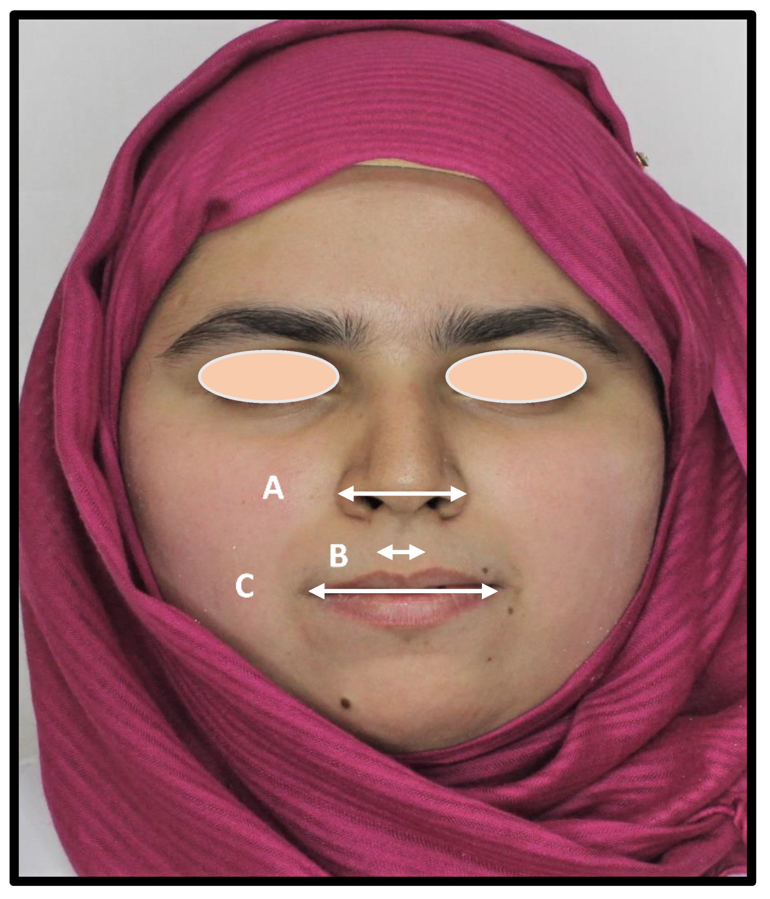
2.3. Capturing Full-Face Frontal 2D Photographs

“A digital camera (Canon EOS, DSLR Camera, CMOS, 18 MP,1920 × 1080p/30fps) was used to capture the extraoral and intraoral photographs. The 1:1 macro setting was used for close-up photography of teeth to includes all four maxillary incisors and canine teeth. The 1:10 setting was used to capture the full-face photographs with focus set on the subject’s nose tip. The camera was set at the 12o’clock position, mounted on a tripod with a standardized focus and distance of 1.5 m from the participants to ensure distortion-free images. The surrounding lighting was standardized for all the photographs. A ring flashlight source system (LED-FD, 480II, Medike Photo and Video Co., Ltd., Yidoblo, Guangdong, China) was used, and its configuration consisted of a light unit that was mounted next to the camera lens. A full-face and anterior teeth 2D photograph for assessment of our subjects were obtained from the front. The participants were seated upright with shoulders and head held straight and facing forward. The head position was guided by the investigator to assist the participants in assuming their natural head position. The natural head position was standardized along with both horizontal and vertical axes. The height of the camera lens was adjusted on the tripod to match the eye level of the subject in full face and at the level of the incisors for a retracted smile. For intraoral 2D photograph, the upper and lower lips were retracted in all photographs to clearly display the maxillary anterior teeth” [7].

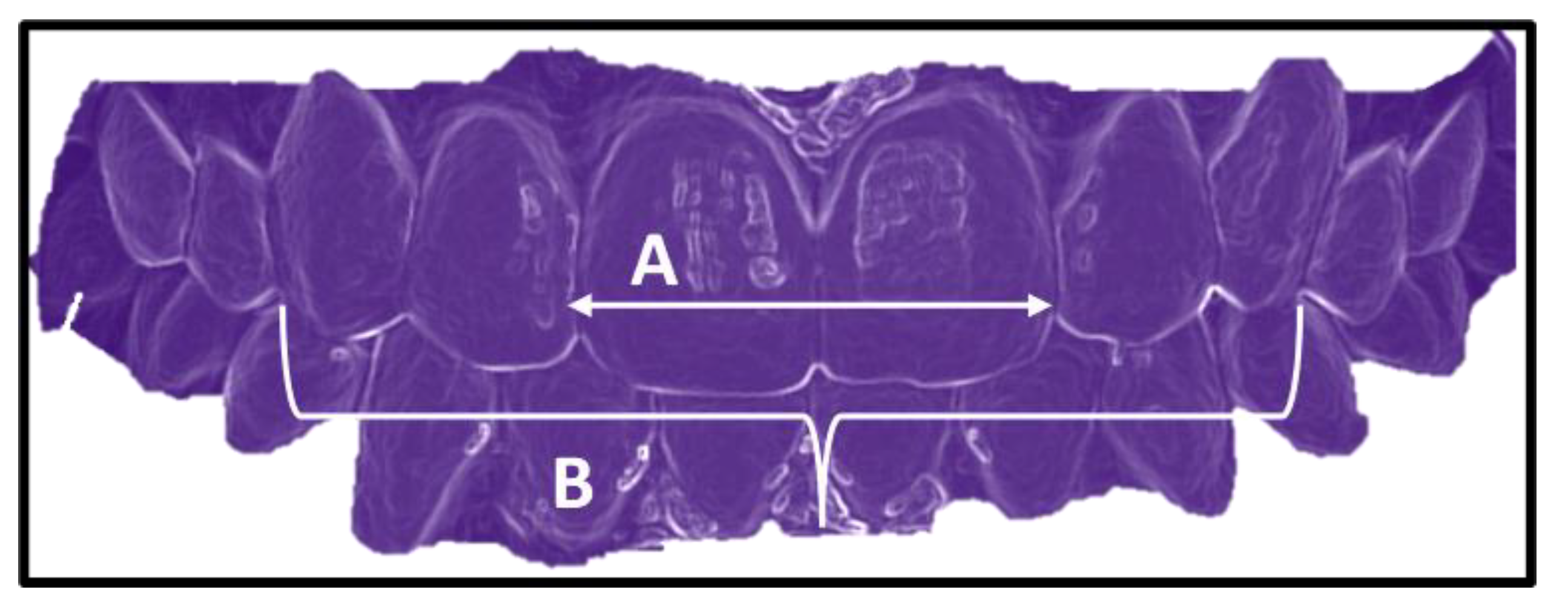
2.4. Maxillary Impression and Dental Cast Making

“The fabrication of the maxillary cast was carried out with an impression made in a perforated type of stainless-steel tray that covers the hamular notches and fovea palatine. A space of 3–4 mm was ensured uniformly for the impression material irreversible hydrocolloid impression material (Fast setting alginate hydrogum, Zhermack SpA, Badia Polesine, Italy) and the borders of the tray were extended up to the functional sulcus depth without causing physical discomfort to the subjects. The impressions were poured with Type IV dental stone (ISO Type 3, Elite Rock Zhermack SpA, Badia Polesine, Italy). The bases of casts were created with soft plaster using standardized base formers. In order to obtain a 3D model, the cast was scanned by UP3D Dental Laboratory Scanner (UP360+, 300 × 300 × 400 mm, 3D scanner, Shenzhen, China). The scanner was equipped with 2.0 MP cameras for high precision. The full arch 3D scan was obtained, and the model was displayed on a compatible dental design software (UPCAD, UP3D, Shenzhen, China), then transferred via USB to store in a personal computer” [7].

2.5. Plaster and 3D Dental Models Analysis

“The mesiodistal width of the maxillary anterior teeth of the 3D dental model was recorded with a measuring tool in millimeters through Photoshop software (Adobe, version 21.0.2, San Jose, CA, USA). Additionally, the plaster dental cast widths were calculated with a sharp-tipped digital Vernier caliper, that can measure to the nearest (0.02 mm). The mesiodistal widths of central incisors, lateral incisors and canine teeth were measured from the facial side using outer edges of Vernier caliper positioned between the contact points Figure 1. The information of teeth measurements obtained from both sources was then recorded on a Performa. The front face photograph obtained through standardized photography was transferred to (Adobe, version 21.0.2, San Jose, CA, USA). Then, by utilizing its measuring tool, the horizontal lower facial third proportions (ICoD, IAD, and IPLD) between anatomical reference points were measured and rec-orded” [7]. As shown in Figure 2.

2.6. Validity and Reliability Assessment

“The data collection was performed by a single operator (N.A.). For calibration purposes, another measurement was performed by a senior colleague (J.S.). Then, the data between the two examiners were subjected to correlation analysis, and a strong correlation value was found (0.739). Furthermore, 20% of the 2D photographs and dental models were then re-assessed after a period of 2 weeks by the same operator. The data were analyzed later by the Dahlberg formula to detect intra operator reliability through correlation statistics. For validity purposes, 20% of the dental models and photograph data that were measured with a vernier caliper were compared with the 3D dental model’s measurements. The intraclass correlation coefficient test (ICC) was carried out to obtain an association between the two sets of measurements. A strong correlation value of (0.816) was found. To minimize the photographic error, the actual width of maxillary anterior teeth obtained from the dental cast was divided by perceived width from photographs to obtain a conversion factor [13]. The perceived teeth widths were multiplied by the conversion factor, to overcome magnification error, and achieve the true width (clean width) captured in the photographs” [7].

2.7. Statistical Analysis

“The data were analyzed with the Statistical Package for the Social Sciences Software (Statistics, version 25, Chicago, IL, USA). The distribution of data was analyzed with normality plots and testing (Shapiro–Wilk and Kolmogorov–Smirnov). Descriptive analysis of categorical (gender) and continuous (age, height, weight, teeth widths, facial proportions) variables was performed to calculate the frequency, percentage, mean, and standard deviation. Moreover, mean values of dependent (maxillary anterior teeth, horizontal facial widths) and independent (age, gender, height, and weight) variables were compared using regression analysis, independent and paired t-tests. A p-value ≤ 0.05 was considered statistically significant” [7].

2.8. Predicting Intercanine Distance and Combined Central Incisor Width with Dental Proportions

The SPSS data through “compute variable” were processed to determine the predicted ITCD through facial and dental proportion analysis. The multiplication analysis was carried out for the following combinations: the IAD value was multiplied with DP (62%GP by IAD, 70%RED by IAD, PRP by IAD, and GM by IAD). Similarly, ICoD and IPLD were multiplied with DP.
Furthermore, for predicting the CIW the lower facial proportion dimensions were multiplied with DP. The ICoD, IAD, and IPLD were multiplied with 70% RED proportion, 0.5% GM, 1.618% GP, and 1.32% PRP.
The predictions for intercanine distance and combined central incisors width using different lower facial third proportions by dental proportions were evaluated with specific metrics mentioned in Table 1.

3. Results

This analytical study consisted of 230 participants. The dropout rate of participants in this study was 0.08%. The age range of participants was 18 to 30 years. The mean age of participants was 24.210 ± 3.541. The height of participants was 168 ± 14.844 cm. The weight of participants was 65.932 ±13.058 kg. There were 112 (48.7%) male and 118 (51.3%) female participants in this study.
“The mean width of maxillary anterior teeth obtained through 2D photographs, plaster dental cast and 3D dental models is shown in Table 2. There was no significant difference between combined six anterior teeth distance (ITCD) obtained from plaster dental cast and 3D dental models (p = 0.0736); however, a significant difference (p < 0.001) was found between the plaster cast, 3D models, and 2D photographic width. To evidence the errors of the 2D photographs when determining the teeth dimensions, the clean width (width of teeth after photographic error assessment) of maxillary anterior teeth was obtained. As presented in Table 3. There was a significant difference (p < 0.001) between the mean values of photographic and clean widths of maxillary anterior teeth” [19].
The comparison of the 3D dental model mesiodistal width for anterior teeth in both sexes has shown a significant difference (p = 0.022) between the right lateral incisor teeth. The t-value (−2.305) was small, which indicates that the mean difference (−0.105) between the values of this tooth was less. However, there was no significant difference between the mean values of the right central incisor (p = 0.138) and canine teeth (p = 0.502).
Furthermore, the mean mesiodistal width of teeth on the left side of the arch showed no significant difference (p ˃ 0.05). Similarly, the intercanine distance showed no significant difference (p = 0.531) in both sexes, Table 4.
The comparison of maxillary anterior teeth’ clean width in both sexes is described in Table 5. There was a significant difference between the widths of the right lateral incisor (p = 0.043) and canine teeth (p = 0.004). This was supported by a small t-value, which indicates that the mean difference between the values of these teeth is less. However, the width of right central incisor teeth was similar in both sexes. Likewise, on the left side of the arch, the width of lateral incisor teeth was similar in both sexes. A significant difference (p = 0.001) was found in left canine tooth. The difference was indicated by a t-value of (−3.582). Whereas no significant difference (p ˃ 0.05) was found between the widths of left central incisor teeth. This was supported by a small t-value of (−1.945). Additionally, a significant difference (p = 0.001) was also found between the ITCD values in both sexes. The intercanine distance was larger in females.
The lower facial third proportion values obtained from 2D photographs is shown in Table 6. The ICoD value was larger than intercanine distance (ITCD), (p = 0.049), whereas IAD was smaller (p = 0.038). The values of both ICoD and IAD were significantly (p = 0.027), (p = 0.019) larger than CIW. The IPLD value was significantly (p = 0.021), (p = 0.001) smaller than CIW and ITCD.
The analysis of gender disparity in mean horizontal facial third proportion revealed no significant difference (p = 0.069) between the mean values of ICoD in both sexes, which is supported by a small t-value (−1.828). Similarly, there was no significant difference between the mean values of IAD (p = 0.650), indicated by a small t-value (−0.454), and also in the case of IPLD (p = 0.341), supported by a small t-value (0.954) in both sexes, Table 7.
The lower facial third proportions were modified with 70% RED proportion, Preston proportion, and golden proportion to predict CIW and ITCD. The mean values of mod-ified IAD, ICoD, and IPLD are shown in Table 8. The mean predicted values of ICoD and IAD were larger than the CIW. However, the (RED and GP by IPLD) values were significantly smaller than CIW. The mean value of IPLD by PRP was found similar to the combined central incisors width.
Furthermore, the mean predicted values of (PRP and GM by ICoD) were larger than ITCD. However, the predicted values of IAD and IPLD with all the dental pro-portions were smaller than intercanine distance.
The mean values of lower facial third proportion modified with dental proportions to predict CIW and ITCD in males are presented in Table 9. The mean predicted values of ICoD and IAD were larger than the CIW. While the IPLD with 70% RED, GP, and GM values were smaller than CIW. There was an exact match of IPLD value mod-ified by PRP with combined central incisors width.
The predicted mean values of ICoD, and IAD were smaller than ITCD. However, the values of ICoD by PRP were larger than intercanine distance.
The mean values of lower facial third proportion modified with dental proportions to predict CIW and ITCD in females are shown in Table 10. The mean predicted values of ICoD, and IAD was larger than the CIW. The IPLD with PRP value was similar to the combined width of central incisors. However, the IPLD value with 70% RED proportion, golden proportion, and the golden percentage was smaller than the combined width of central incisors.
The mean predicted values of ICoD and IAD with PRP and GM were larger than ITCD. Whereas the values of ICoD and IAD with 70% RED, and GP were smaller than the intercanine distance.
Furthermore, Table 11 is representing a regression analysis for the lower facial proportions without modification, with independent variables (age, gender, height, and weight) of participants. The outcome of the study showed that a weak correlation between facial measurements and independent variables existed. The regression model analysis for ICD to independent variables showed a constant for R-Squared (R2) = 0.041 and adjusted R-Squared (AR2) was 0.0241; however, under the influence of external variables, only height presented a significant difference (p = 0.028). The ICoD to height beta (B) value was statistically significant (B = −0.147), which indicates that height had a 0.028 relationship with ICoD, whereas on average, the effect of height on ICoD was (B0 = −0.016) in this study.
The (R2) value of IAD was 0.008 and AR2 was (−0.011). Additionally, in the case of IPLD, the constant for R-Squared (R2) = 0.127 and adjusted R-Squared (AR2) value was 0.016. The IAD and IPLD showed no significant correlation (p ˃ 0.05) with the age, gender, height, and weight of participants.

4. Discussion

In this study, we investigated the lower facial third proportions with dental ratios to determine the width of maxillary anterior teeth. For this purpose, the golden proportion, 70% recurring esthetic dental proportion, Preston proportion, and golden percentage were evaluated with ICoD, IAD, and IPLD. The dominant teeth in a pleasing smile are primarily maxillary central incisor and canine, their morphology plays a vital role in dental esthetics [20]. Therefore, one should wisely select the width of maxillary anterior teeth. The width of anterior teeth varies according to arch size, and face form, it also shows a difference in respect to sex, race, and ethnicity of the population [15,16].
In terms of variations in gender regarding horizontal facial proportions, no significant differences in interalar, intercommisural, and interphiltral distance were found. However, these findings were different from a study by Saurabh et al. [17]. The study was carried out in the Indian population and consisted of 200 participants (164 females, 36 males). A similar methodology except for the use of 3D models was adopted. The outcome was well supported by figures and illustrations. They concluded differences in the lower facial third proportions in both sexes. Furthermore, as part of the study, the facial values obtained were significantly different from the Japanese and Caucasian populations. The reason could be the unequal participation of both genders in the study.
The mean age of participants in this study was 24.210 ± 3.541 years and belonged to different ethnic backgrounds. The majority of the participants were from a younger age group of 18 to 25 years. The reason for including participants above the age of 18 years was due to the completion of the growth spurt by this stage. The facial dimensions become stable and mature following puberty. A similar concern and finding was reported by Kurien et al. [20]. The study consisted of 300 participants from a 20 to 50 years age group. The similarity in findings could be due to the anthropometric methods adopted in their study.
In this study, no difference was seen when the 3D dental casts and clean mesiodistal width of maxillary anterior teeth in both sexes were compared. These results, however, were in disagreement with studies carried out by Leung et al. [21] and Horvath et al. [22], where differences were noted between both sexes. This can be due to differences in ethnicities where variations in the size of the teeth were noted.
Regarding the mean predicted values of ICoD, IPLD, and IAD without modification, it was concluded that the ICoD value was larger than maxillary anterior teeth width. The IAD and IPLD values were smaller than both CIW and ITCD. This finding was in contrast to studies carried out by Gomes et al. [18] and Kurien et al. [20], where IAD and ICoD were recommended to be used as a predictor of intercanine distance. The reason for dissimilar results could be due to the methodology adopted in both the studies, which consisted of direct facial measurements, no photographic error assessment being carried out, a lack of biasness control, and no racial difference of the population studied.
When selecting lower facial third measurement data to analyze maxillary anterior teeth width, measuring IAD modified with 70% RED proportion are recommended in evaluating CIW [23]. The IPLD, and ICoD measurements need to be evaluated. and these three facial dimensions could determine the CIW and ITCD by using the GP, GM, PRP, and RED proportion. Valid metrics to assist in this process are needed.
In the current study, ICoD and IAD mean predicated values differed from the CIW when evaluated with 70% RED proportion, golden percentage, and golden proportion, so interalar distance and intercommisural distance cannot be used to determine the combined width of central incisors. Moreover, the literature recommends IPLD as one of the determinants to predict the combined width of central incisors [19]. However, IPLD when modified with these dental proportions cannot be used to determine the combined width of maxillary anterior teeth as reported in this study. This contradicts the study by Liao P [23], where the values of IAD were reported as similar to the combined width of the central incisors. This paradigm and unique study were based on secondary data published. The results were not comparable due to differences in research design and methodology. There are no other comparative studies available in the literature on ICoD and IPLD modified by dental proportions.
The Preston proportion has been used extensively for the determination of maxillary anterior teeth ratios [24,25,26,27]. In our study, IPLD modified by Preston proportion was the only combination that is concluded to be a reliable tool to predict the combined width of central incisors.
Furthermore, the predicted mean values of ICoD were larger than intercanine distance when modified with PRP and GM. Additionally, the mean values of IAD were smaller than intercanine distance. However, these findings contradict a study by Isa et al. [28] where IAD was analyzed directly, though did correlate with the widths of maxillary anterior teeth. The findings are incomparable due to differences in methodologies adopted.
Although golden proportion, golden percentage, and 70% RED proportion have been developed and highlighted extensively in the literature [29,30,31], we concluded in our study that these notions cannot be used successfully in determining the combined width of central incisors and intercanine distance. The findings correspond with numerous studies in the literature where these dental proportions when studied in the width of natural teeth differed in reaching the ideal maxillary anterior teeth width [32,33]. Most of the studies available in the literature are carried out in anterior teeth directly i.e., not investigated with lower facial dimensions. Due to a lack of evidence, the results could not be compared.
When age, gender, height, and weight correlated with lower facial horizontal proportion, a positive impact of height was found with intercommisural distance. These results, however, contrast with Esan et al. [34], who report no significant differences between intercommisural distance and height of participants. This could be due to differences in ethnicities of the population in the study.
Despite the strengths of this study, such as including a large sample size, the proposal of valid metrics, and comparative analysis to predict the width of maxillary anterior teeth, there are some limitations. Firstly, there can be selection biasness since the non-probability convenience sampling technique was used, which questions the reproducibility of the data. The study was based on a specific geographical region; hence, the outcome best suits the population studied. The findings were not compared within different dental arch classifications. Lastly, the inclusion and comparison of population on an ethnic basis would increase the scope of this study. Therefore, we recommend that patients’ and clinicians’ opinions on the application of dental proportions for esthetic restorative outcomes in clinical cases are critical. The opinion should be determined based on a diverse population from different regions so that it comprehensively covers various beauty standards across the globe. Future studies which assess the clinical impact of the different dental proportions with patient opinions are recommended.
The modified horizontal facial third measurements are recommended to determine the maxillary anterior teeth width. The interphiltral distance modified by the Preston proportion can be used to predict the combined width of central incisors. The current study indicates that the predicted combined width of central incisors using the Preston proportion showed an exact match with maxillary anterior teeth width. This provides substantial scientific evidence for evaluation. The metrics could be adopted by clinicians to construct an esthetically pleasing smile. The metrics could also be used by digital programmers in rapid prototyping machines, computer-aided design, and computer-aided manufacturing (CAD–CAM).

5. Conclusions

Within the limitation of this study the following conclusions were drawn:
  • The ICoD, IAD, and IPLD could not be used to determine the combined central incisors and intercanine width without modification.
  • The IPLD modified by the Preston proportion is a reliable method to predict CIW in the studied population.
  • The study reached conclusions based on a specific population and results from GP, GM, and 70% RED proportions, which had been reported for other regions and are not reliable methods to predict CIW and ITCD.
  • There was a significant difference in gender disparity when ICoD, IAD, and IPLD were modified by DP, except in the case of IPLD with PRP.

Author Contributions

Conceptualization, N.A., A.L., M.S.H. and Z.A.-G.; methodology, A.L., M.S.H. and N.A.; software, N.A., G.D. and M.K.A.; validation, M.K.A., N.A. and Z.A.-G.; formal analysis, A.L. and N.A.; investigation, N.A., A.L., M.S.H. and J.S; resources, M.K.A., A.A.G.K., G.D. and S.S.; data curation, J.S. and N.A.; writing—original draft preparation, N.A., M.S.H. and A.L.; writing—review and editing, N.A., M.S.H., Z.A.-G. and G.D.; visualization, J.S., M.S.H., Z.A.-G. and N.A.; supervision. M.S.H., Z.A.-G. and J.S.; project administration, N.A., M.S.H., Z.A.-G. and J.S.; funding acquisition, M.K.A., G.D., S.S. and A.A.G.K. All authors have read and agreed to the published version of the manuscript.

Funding

This research was funded by the Deanship of Scientific Research, King Khalid University, Abha-Asir, Kingdom of Saudi Arabia under the Short Research Group, by the grant number RGP.1/336/42.

Institutional Review Board Statement

The study was conducted according to the guidelines of the Declaration of Helsinki, and approved by the Institutional Review Board of Universiti Sains Malaysia (USM/JEPeM/19060380) and Altamash Institute of Dental Medicine, Paksitan (AIDM/EC/06/2019/06).

Informed Consent Statement

Informed consent was obtained from all subjects involved in the study.

Data Availability Statement

The data presented in this study are available on request from the corresponding author.

Acknowledgments

The authors thank all participants, the School of Dental Sciences, Health Campus, Universiti Sains Malaysia, and Altamash Institute of Dental Medicine, Pakistan, for the support and facilitation in this study. The authors also thankfully acknowledge the Deanship of Scientific Research, King Khalid University, Abha-Asir, Kingdom of Saudi Arabia for funding this research work under the Short Research Group, by the grant number RGP.1/336/42.

Conflicts of Interest

The authors declare no conflict of interest.

References

  1. Kantrong, N.; Traiveat, K.; Wongkhantee, S. Natural upper anterior teeth display an increasing proportion in mesiodistal direction. J. Clin. Exp. Dent. 2019, 11, 890–897. [Google Scholar]
  2. Kaya, K.S.; Türk, B.; Cankaya, M.; Seyhun, N.; Coşkun, B.U. Assessment of Facial Analysis Measurements by Golden Proportion. Braz. J. Otorhinolaryngol. 2019, 85, 494–501. [Google Scholar] [CrossRef]
  3. Rosential, S.F.; Ward, D.H.; Rashid, R.G. Dentist’s preferences of anterior tooth proportion—A web-based study. J. Prosthodont. 2000, 9, 123–136. [Google Scholar]
  4. Ghoddousi, H.; Elder, R.; Haers, P.; Wertheim, D.; Greenhill, D. Comparison of three methods of facial measurement. Int. J. Oral Maxillofac. Surg. 2007, 36, 250–258. [Google Scholar] [CrossRef] [PubMed]
  5. Shetty, S.; Pitti, V.; Satish Babu, C.S.; Kumar, G.S.; Jnanadev, K. To evaluate the validity of recurring esthetic dental proportion in natural dentition. J. Conserv. Dent. 2011, 14, 314–317. [Google Scholar] [PubMed]
  6. Ahmed, N.; Halim, M.S.; Khalid, S.; Ab Ghani, Z.; Jamayet, N.B. Evaluation of golden percentage in natural maxillary anterior teeth width: A systemic review. J. Prosthet. Dent. 2021. [Google Scholar] [CrossRef]
  7. Ahmed, N.; Halim, M.S.B.; Ghani, Z.A.; Khan, Z.A.; Abbasi, M.S.; Jamayet, N.B.; Alam, M.K. A 2D photographic and 3D digital dental model analysis of golden percentage in maxillary anterior teeth. Biomed. Res. Int. 2021, 17, 1–7. [Google Scholar] [CrossRef]
  8. Levin, E.I. Dental esthetics and the golden proportion. J. Prosthet. Dent. 1978, 40, 244–252. [Google Scholar] [CrossRef]
  9. Mahshid, M.; Khoshvaghti, A.; Varshosaz, M.; Vallaei, N. Evaluation of “golden proportion” in individuals with an esthetic smile. J. Esthet. Restor. Dent. 2004, 16, 185–192. [Google Scholar] [CrossRef] [PubMed]
  10. Preston, J.D. The golden proportion revisited. J. Esthet. Restor. Dent. 1993, 5, 247–251. [Google Scholar] [CrossRef]
  11. Ward, D.H. Using the RED proportion to engineer the perfect smile. Dent. Today 2008, 27, 112, 114–117. [Google Scholar] [PubMed]
  12. Parrini, S.; Rossini, G.; Castroflorio, T.; Fortini, A.; Deregibus, A.; Debernardi, C. Laypeople’s perceptions of frontal smile esthetics: A systematic review. Am. J. Orthod. Dentofac. Orthop. 2016, 150, 740–750. [Google Scholar] [CrossRef]
  13. Deogade, S.; Mantri, S.; Sumathi, K.; Rajoriya, S. The relationship between innercanthal dimension and interalar width to the intercanine width of maxillary anterior teeth in central Indian population. J. Indian Prosthodont. Soc. 2015, 15, 91. [Google Scholar] [CrossRef] [PubMed]
  14. Janini, M.; Jain, A.R. A reliability of sear’s cephalometric index in determination of width of maxillary anterior teeth in south Indian population. Biomed. Pharmacol. J. 2017, 10, 1969–1972. [Google Scholar]
  15. Scandrett, F.R.; Kerber, P.E.; Umrigar, Z.R. A clinical evaluation of techniques to determine the combined width of the maxillary anterior teeth and the maxillary central incisor. J. Prosthet. Dent. 1982, 48, 15–22. [Google Scholar] [CrossRef]
  16. Afrashtehfar, K.I.; Assery, M.K.; Bryant, S.R. Aesthetic Parameters and Patient-Perspective Tools for Maxillary Anterior Single Implants’ Assessment. Int. J. Dent. 2021, 2021, e6684028. [Google Scholar] [CrossRef]
  17. Saurabh, R.; Piyush, B.; Sourabh, B.; Preeti, O.; Trivedi, R.; Vishnoi, P. Assessment of facial golden proportions among central Indian population. J. Ind. Soc. Prev. Community Dent. 2016, 6, 182–186. [Google Scholar] [CrossRef] [Green Version]
  18. Gomes, V.L.; Goncalves, L.C.; do Prado, C.J.; Junior, I.L.; Lucas, B.D.L. Correlation between facial measurement and the mesiodistal width of the maxillary anterior teeth. J. Esthet. Restor. Dent. 2006, 18, 196–205. [Google Scholar] [CrossRef]
  19. Naseer, A.; Halim, M.S.; Aslam, A.; Ghani, Z.A.; Safdar, J.; Alam, M.K. An Analysis of Maxillary Anterior Teeth Crown Width-Height 588 Ratios: A Photographic, Three-Dimensional, and Standardized Plaster Model’s Study. Biomed. Res. Int. 2022, 4695193, 1–10. [Google Scholar]
  20. Kurein, A.; Cherian, K.P.; Mhatre, S.; Tharakan, R.G. A comparative study on the relationship between inter alar width and inter commissural width on circumferential arc width of maxillary anterior teeth in different age groups. J. Indian Prosthodont. Soc. 2014, 14, 352–357. [Google Scholar] [CrossRef]
  21. Leung, E.M.Y.; Yang, Y.; Khambay, B.; Wong, R.W.K.; McGrath, C.; Gu, M. A comparative analysis of tooth size discrepancy between male and female subjects presenting with a class 1 malocclusion. Sci. World J. 2018, 11, 1–6. [Google Scholar] [CrossRef] [PubMed] [Green Version]
  22. Horvath, S.D.; Wegstein, P.G.; Lüthi, M.; Blatz, M.B. The correlation between anterior tooth form and gender—A 3D analysis in humans. Eur. J. Esthet. Dent. 2012, 7, 334–343. [Google Scholar]
  23. Liao, P.; Fan, Y.; Nathanson, D. Evaluation of maxillary anterior teeth width: A systemic review. J. Prosthet. Dent. 2019, 122, 275–281. [Google Scholar] [CrossRef] [PubMed]
  24. Pierrot, K.N.; Paul, S.I.B.J.; Kasiama; Celestin, M.N.; Augustin, M.M.; Joseph, L.P.S.; Bolenge; Fidele, N.B. Determinants factors for the choice of the width prosthetic upper central incisor: Review of the literature. Open J. Stomatol. 2020, 10, 333–340. [Google Scholar] [CrossRef]
  25. de Maldonado, G.J.; Suárez-Vega, D.V.; García-Guevara, V.; Miller-Kobisher, B. Innovative paradigm in aesthetics medicine: Proposal for diagnostic morphological geometric by thirds, semiology in clinical applied to aging facial. J. Cutan. Aesthet. Surg. 2020, 13, 112. [Google Scholar]
  26. Ramirez, L.M.; Ospina, J.D.; Ballesteros, L.E. Mestizo Anterior Teeth’s Proportions. Int. J. Morphol. 2016, 34, 223–231. [Google Scholar] [CrossRef] [Green Version]
  27. Kalia, R. An analysis of the asthetic proportions of anterior maxillary teeth in a UK population. Br. Dent. J. 2020, 228, 449–455. [Google Scholar] [CrossRef]
  28. Isa, Z.M.; Tawfiq, O.F.; Noor, N.M.; Shamsudheen, M.I.; Rijal, O.M. Regression methods to investigate the relationship between the facial measurements and widths of the maxillary anterior teeth. J. Prosthet. Dent. 2010, 103, 182–188. [Google Scholar] [CrossRef] [Green Version]
  29. Mehrdad, B.; Alireza, O.; Nasim, E.E.; Marzieh, Y. Dento-facial proportions analysis of maxillary anterior teeth in khuisf dental students. J. Isfahan Dent. Sch. 2011, 7, 31–38. [Google Scholar]
  30. Hafiza, R.; Oenzil, F.M.M. The difference between the width of the anterior tooth of the upper jaw and the concept of golden proportion in university students of Andalas suku Minang. Andalas. Dent. J. 2015, 3, 92–101. [Google Scholar]
  31. Özdemir, H.; Köseoğlu, M.; Bayindir, F. An investigation of the esthetic indicators of maxillary anterior teeth in young turkish people. J. Prosthet. Dent. 2018, 120, 583–588. [Google Scholar] [CrossRef] [PubMed]
  32. Ahmed, N.; Abbas, M.; Maqsood, A. Evaluation of recurring esthetic dental proportion in natural smile of Pakistani sample. Pak. Oral Dent. 2014, 34, 739–742. [Google Scholar]
  33. Melo, M.; Ata-Ali, F.; Huertas, J.; Cobo, T.; Shibli, J.A.; Galindo-Moreno, P.; Ata-Ali, J. Revisiting the maxillary teeth in 384 subjects reveals a deviation from the classical aesthetic dimensions. Sci. Rep. 2019, 9, 730. [Google Scholar] [CrossRef] [PubMed]
  34. Esan, T.A.; Obzeigbe, O.E.; Onapokya, H.O. Facial approximation: Evaluation of dental and facial proportions with height. Afr. Health Sci. 2012, 12, 63–68. [Google Scholar] [PubMed]
Figure 1. Mesiodistal width measurement of maxillary anterior teeth, (A) CIW (combined central incisors width), mesiodistal width of both maxillary central incisors. (B) ITCD (intercanine distance), distance between distal surfaces of maxillary canines (in straight line).
Symmetry 14 00723 g001
Figure 2. Horizontal lower facial third proportion. (A) IAD (interalar distance), the distance between widest points of alar of nose. (B) IPLD (interphiltral distance) the distance between right and left philtral ridges of upper lip. (C) ICoD (Intercommisural distance), the distance between the right and left corners of the oral aperture.
Symmetry 14 00723 g002
Table 1. Distribution of modification metrics used to predict the intercanine distance and combined width of central incisors.
Dental Proportions (DP)Horizontal Lower Facial Third Proportion Modification Metrics
Recurring esthetic dental proportion ICoD or IAD or IPLD × 0.70
Golden proportion ICoD or IAD or IPLD × 0.62 or 1.618
Preston proportion ICoD or IAD or IPLD × 0.662 (1.32) and 0.842 (1.68)
Golden percentage ICoD or IAD or IPLD × 0.5 or 0.3 or 0.2
ICoD: intercommisural distance; IAD: interalar distance; IPLD: interphiltral distance.
Table 2. Distribution of mean maxillary anterior teeth widths obtained from 2D photographs, 3D, and plaster dental models (n = 230).
Maxillary Anterior Teeth2DPW3DDMWPDCW
Mean (mm)SDMean (mm)SDMean (mm)SD
RCI15.9812.4327.9730.4417.5140.541
RLI12.8814.9566.8430.3457.1510.427
RCa10.7902.8927.1410.2706.9540.256
LCI15.9664.8657.9770.3147.9430.364
LLI12.8030.3186.9460.5136.9820.425
LCa9.9870.9137.9570.3427.9180.384
Intercanine distancef 78.4088.188b 45.6371.115b 44.4621.198
Dissimilar superscript small alphabets denote statistical significance (p ≤ 0.05), SD: standard devi-ation, 2DPW: 2D photographic width, 3DDMW: 3D digital model width, PDCW: plaster dental cast width, RCI: right central incisor, RLI: right lateral incisor, RCa: right canine, LCI: left central incisor, LLI: left lateral incisor, LCa: left canine.
Table 3. Comparison of 2D photographic and clean width of maxillary anterior teeth obtained after photographic error assessment (n = 230).
Variables2D PWCWp-Value
Mean (mm)SDMean (mm)SD
RCI15.9812.4327.9320.514* 0.001
RLI12.8814.9565.8310.721* 0.001
RCa10.7902.8925.9161.471* 0.001
LCI15.9664.8656.8940.726* 0.001
LLI12.8030.3184.8390.851* 0.014
LCa9.9870.9137.9751.424* 0.027
Width of six anterior teeth78.4088.18839.3872.853* 0.001
CW: “Clean width: mesiodistal teeth dimension obtained after photographic error estimation assessment”, * p-Value: p < 0.05 was considered significant, mm: millimeter, 2D: Two dimensional, SD: standard deviation, 2DPW: 2D photographic width, RCI: right central incisor, RLI: right lateral incisor, RCa: right canine, LCI: left central incisor, LLI: left lateral incisor, LCa: left canine.
Table 4. Comparison of 3D dental model mesiodistal width for maxillary anterior teeth in both sexes, independent t-test analysis (male: n = 112; female: n = 118).
Maxillary Anterior TeethGenderMean
(mm)
St. Deviationp-Valuet-ValueMean DifferenceStd. Error Difference
RCIMale8.3420.6160.138−1.490−0.1050.071
Female8.4480.454
RLIMale7.6500.526* 0.022−2.305−0.1660.072
Female7.8160.569
RCaMale8.0600.4080.5020.6720.0340.051
Female8.0250.373
LCIMale8.8010.4330.6510.4530.0250.056
Female8.7760.421
LLIMale7.8630.5540.7000.3860.0310.081
Female7.8310.678
LCaMale8.1860.4770.3610.9150.0560.061
Female8.1300.452
Intercanine distanceMale48.9051.5110.531−0.627−0.1250.199
Female49.0301.510
* Asterisk superscript denotes p-value is ≤0.05, 3D: Three di-mensional, std deviation: Standard deviation, mm: millimeter, RCI: right central incisor, RLI: right lateral incisor, RCa: right canine, LCI: left central incisor, LLI: left lateral incisor, LCa: left canine t-value: measures the size of the difference relative to the variation in sample data, the smaller the t-value, the more similarity exists between the two sample sets, while a large t-score indicates that the groups are different; Mean Difference: the difference between the mean values from two data groups; Std. Error Difference: the standard error of the mean and measures the variability of the sample mean, the smaller the standard error of the mean, the more likely that our sample mean is close to the true participants mean.
Table 5. Comparison of gender disparity in clean mesiodistal width of maxillary anterior teeth, independent t-test analysis (male: n = 112; female: n = 118).
Maxillary Anterior TeethGenderMean
(mm)
St. Deviationp-Valuet-ValueMean DifferenceStd. Error Difference
RCIMale8.0750.7430.261−1.125−0.1060.094
Female8.1820.691
RLIMale6.1170.913* 0.043−2.033−0.2400.118
Female6.3580.880
RCaMale6.3671.404* 0.004−2.872−0.4920.171
Female6.8591.190
LCIMale7.8540.931* 0.053−1.945−0.2160.111
Female8.0710.748
LLIMale5.8850.9600.124−1.545−0.1900.123
Female6.0760.909
LCaMale6.0721.337* 0.001−3.582−0.6080.169
Female6.6801.239
Intercanine distanceMale39.9124.057* 0.001−3.228−1.7060.528
Female41.6193.961
* Asterisk superscript denotes p-value is ≤0.05, Std deviation: standard deviation; clean width: mesiodistal teeth dimension obtained after photographic error estimation assessment; RCI: right central incisor, RLI: right lateral incisor, RCa: right canine, LCI: left central incisor, LLI: left lateral incisor, LCa: left canine t-value: measures the size of the difference relative to the variation in sample data, the smaller the t-value, the more similarity exists between the two sample sets, while a large t-score indicates that the groups are different; Mean Difference: the difference between the mean values from two data group; Std. Error Difference: the standard error of the mean and measures the variability of the sample mean, the smaller the standard error of the mean, the more likely that our sample mean is close to the true participants mean.
Table 6. Comparison of mean horizontal lower facial third proportions measurements with maxillary anterior teeth width without modification (n = 230).
Horizontal Facial ProportionMean (mm) SDβ ITCDp-Valueβ CIWp-Value
Intercommisural distance (ICoD)47.225 ± 1.63445.976 ± 1.760* 0.04916.096 ± 1.438* 0.027
Interalar distance (IAD)37.046 ± 8.146* 0.038* 0.019
Interphiltral distance (IPLD)11.339 ± 7.039* 0.001* 0.021
* Asterisk superscript denotes p-value is ≤0.05, mm: millimeters, β The ITCD and CIW, were obtained from mean values of 3D, clean, and plaster cast teeth width.
Table 7. Comparison of horizontal lower facial third proportion in both sexes, independent t-test analysis (male: n = 112; female: n = 118).
Maxillary Anterior TeethGenderMean
(mm)
St. Deviationp-Valuet-ValueMean DifferenceStd. Error Difference
Intercommisural distanceMale47.0241.6590.069−1.828−0.3920.214
Female47.4161.592
Interalar distanceMale36.7964.6010.650−0.454−0.4881.076
Female37.28410.472
Interphiltral distanceMale11.7949.9580.3410.9540.8860.928
Female10.9081.58728
mm: millimeter, St. Deviation: standard deviation; t-value: measures the size of the difference relative to the variation in sample data, the smaller the t-value, the more similarity exists between the two sample sets, while a large t-score indicates that the groups are different; Mean Difference: the difference between the mean values from two data groups; Std. Error Difference: the standard error of the mean and measures the variability of the sample mean, the smaller the standard error of the mean, the more likely it is that our sample mean is close to the true participant mean.
Table 8. Comparison of predicted combined central incisors width and intercanine distance using dental proportions by horizontal lower facial third proportions with a combined width of central incisors and intercanine distance -mixed group (n = 230).
Horizontal Facial Proporiton* 70% RED Proportion
Mean (mm) SD
* Golden Proportion
Mean (mm) SD
Preston Proportion
Mean (mm) SD
Golden Percentage
Mean (mm) SD
Measured Teeth Width
Mean (mm) SD
PredictedPredictedPredictedPredictedITCDCIW
PCIWPCIWPCIWITCD
ICoD32.916 ± 1.16129.155 ± 1.02962.071 ± 2.19070.536 ± 2.48923.512 ± 0.82947.024 ± 1.65945.976 ± 1.760d 16.096 ± 1.438
IAD25.757 ± 3.22122.813 ± 2.85324.285 ± 3.03739.739 ± 4.96918.398 ± 2.30031.890 ± 3.988
d IPLD8.256 ± 6.9717.312 ± 6.174d 16.568 ± 13.145-----5.897 ± 4.979-----
ITCD: measured intercanine distance, CIW: measured combined width of central incisors, ICoD: Intercommisural distance, IAD: Interalar distance, IPLD: Interphiltral distance, SD: standard deviation, mm: millimeter, PCIW: predicted combined central incisor width value, PITCD: predicted intercanine distance value, d Similar superscript small alphabets denote matched predicted IPLD by Preston proportion value with measured combined central incisors width. * the predicted values by recurrent esthetic dental proportion and golden proportion are for both CIW and ITCD due to their constant ratio.
Table 9. Comparison of predicted central incisors combined width and intercanine distance using dental proportion by horizontal lower facial third proportions with combined width of central incisors and intercanine distance -male group (n = 112).
Horizontal Facial Proporiton* 70%RED Proportion
Mean (mm) SD
* Golden Proportion
Mean (mm) SD
Preston Proportion
Mean (mm) SD
Golden Percentage
Mean (mm) SD
Measured Teeth Width
Mean (mm) SD
PredictedPredictedITCDCIW
PredictedPredictedPCIWPCIWPCIWITCD
ICoD33.057 ± 1.14329.2791 ± 0.01362.337 ± 2.15770.838 ± 2.45123.612 ± 0.81747.22 ± 1.63445.673 ± 1.784b 16.844 ± 0.797
IAD25.932 ± 5.70222.969 ± 5.05024.450 ± 5.37640.010 ± 8.79718.523 ± 4.07332.107 ± 7.060
b IPLD7.937 ± 4.9277.030 ± 4.364b 16.968 ± 9.292-----5.669 ± 3.519-----
ITCD: measured intercanine distance, CIW: measured combined width of central incisors, ICoD: intercommisural distance, IAD: interalar distance, IPLD: interphiltral distance, SD: standard deviation, mm: millimeter, PCIW: predicted combined central incisor width value, PITCD: predicted intercanine distance value, b similar superscript small alphabets denote matched predicted IPLD by Preston proportion value with measured combined central incisors width, * the predicted values by recurrent esthetic dental proportion and golden proportion are for both CIW and ITCD due to their constant ratio.
Table 10. Comparison of predicted central incisors combined width and intercanine distance using dental proportion by horizontal facial proportions with combined width of central incisors and intercanine distance. Female group: n = 118.
Horizontal Facial Proporiton* 70% RED Proportion
Mean (mm) SD
* Golden Proportion
Mean (mm) SD
Preston Proportion
Mean (mm) SD
Golden Percentage
Mean (mm) SD
Measured Teeth Width
Mean (mm) SD
PredictedPredictedITCDITCD
PredictedPredictedPCIWPCIWPCIWITCD
ICoD33.191 ± 1.11529.398 ± 0.98762.589 ± 2.10271.124 ± 2.38923.708 ± 0.79647.416 ± 1.59246.263 ± 1.695f 16.909 ± 0.697
IAD23.116 ± 6.49323.116 ± 6.49324.607 ± 6.91140.267 ± 11.31018.642 ± 5.23632.313 ± 9.076
f IPLD7.63 ± 1.1116.762 ± 0.984f 16.398 ± 2.095------5.454 ± 0.793------
ITCD: measured intercanine distance, CIW: measured combined width of central incisors, ICoD: intercommisural distance, IAD: interalar distance, IPLD: interphiltral distance, SD: standard deviation, PCIW: predicted combined central incisor width value, PITCD: predicted intercanine distance value, f similar superscript small alphabets denote matched predicted IPLD by Preston proportion value with measured combined central incisors width, * the predicted values by recurrent esthetic dental proportion and golden proportion are for both CIW and ITCD due to their constant ratio.
Table 11. Linear regression analysis of age, gender, height, and weight with horizontal lower facial third proportion (n = 230).
VariablesIndependent VariablesUnstandardized CoefficientsStandardized Coefficients
Beta (B)
t-Valuep-Value95% Confidence Interval for BCollinearity
(Bo)Std. ErrorLower BoundUpper BoundToleranceVIF
ICoDAge−0.0030.030−0.007−0.1140.910−0.0630.0560.9931.007
Gender0.3500.2390.1071.4640.145−0.1210.8210.7981.253
Height−0.0160.007−0.147−2.2050.028 *−0.031−0.0020.9631.038
Weight0.0040.0090.0290.4040.687−0.0140.0210.8211.218
IADAge0.1470.1530.0640.9560.340−0.1550.4490.9931.007
Gender0.6561.2090.0400.5430.588−1.7263.0380.7981.253
Height0.0270.0370.0500.7390.461−0.0460.1010.9631.038
Weight0.0010.0460.0010.0140.989−0.0890.0910.8211.218
IPLDAge−0.1520.132−0.076−1.1500.251−0.4120.1080.9931.007
Gender−1.1321.040−0.081−1.0880.278−3.1820.9180.7981.253
Height0.0290.0320.0610.8990.370−0.0340.0920.9631.038
Weight−0.0340.039−0.062−0.8530.394−0.1110.0440.8211.218
ICoD: intercommisural distance; IAD: interalar distance; IPLD: interphiltral distance; B denotes the correlation between dependent and independent variables; B0: unstandardized coefficient i.e., average estimation of age, gender, height, and weight with IAD, ICoD, and IPLD; VIF: variance inflation factor, denotes the amount of multicollinearity in the model; t: test of the regression coefficients; * p-value ≤ 0.05 was considered significant.
Publisher’s Note: MDPI stays neutral with regard to jurisdictional claims in published maps and institutional affiliations.

