
Rb1.66Cs1.34Tb[Si5.43Ge0.57O15]·H2O, a New Member of the OD-Family of Natural and Synthetic Layered Silicates: Topology-Symmetry Analysis and Structure Prediction


Abstract
:1. Introduction
2. Materials and Methods
2.1. Synthesis of Crystals
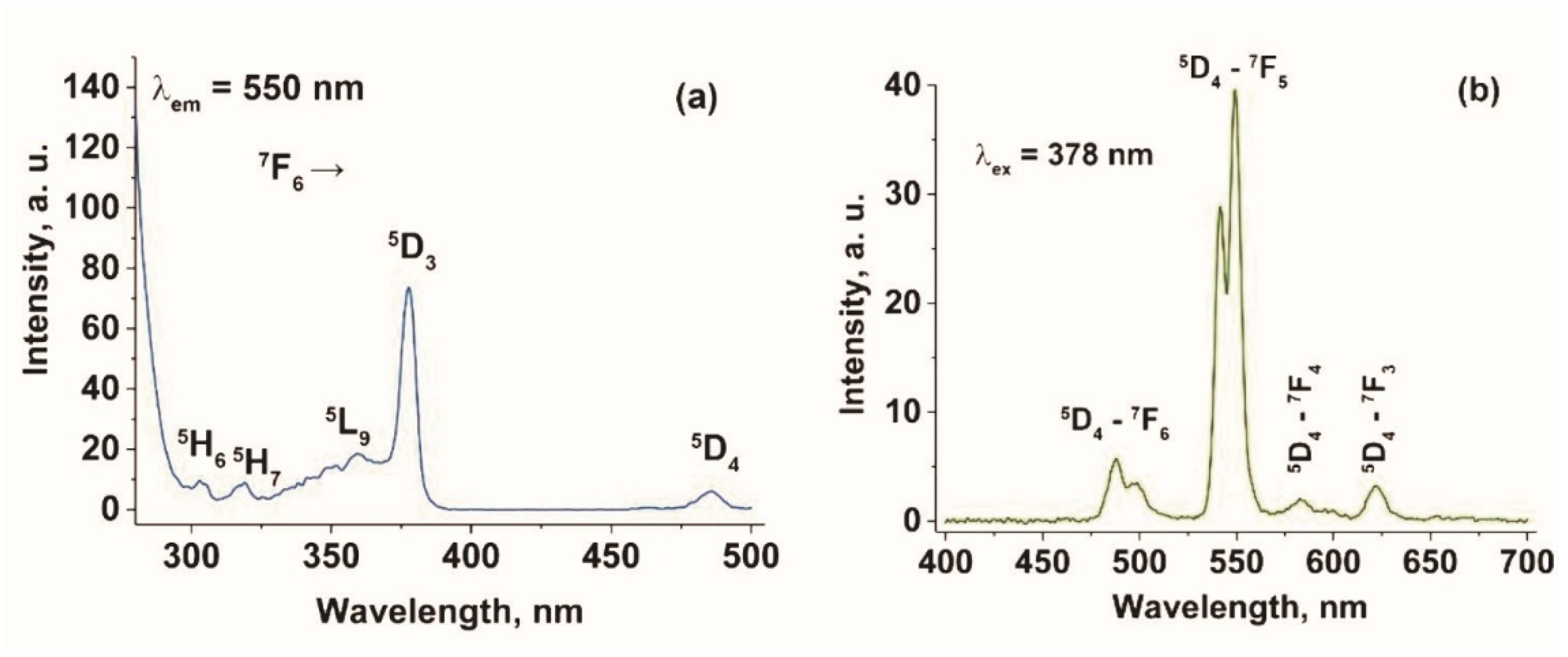
2.2. Luminescence Study
3. Results
Structure Solving and Description
4. Discussion
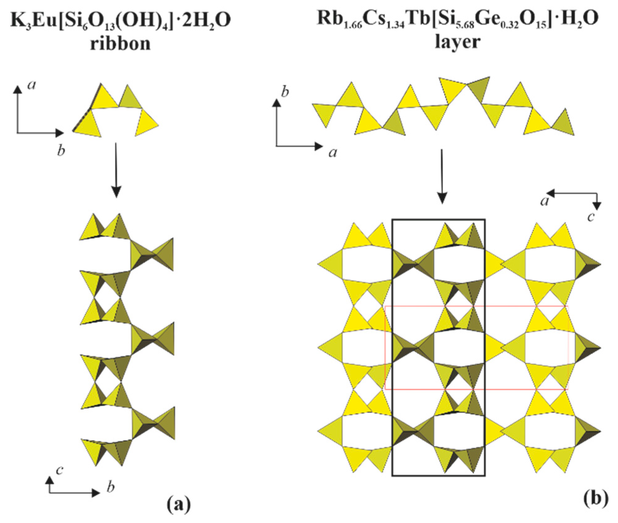
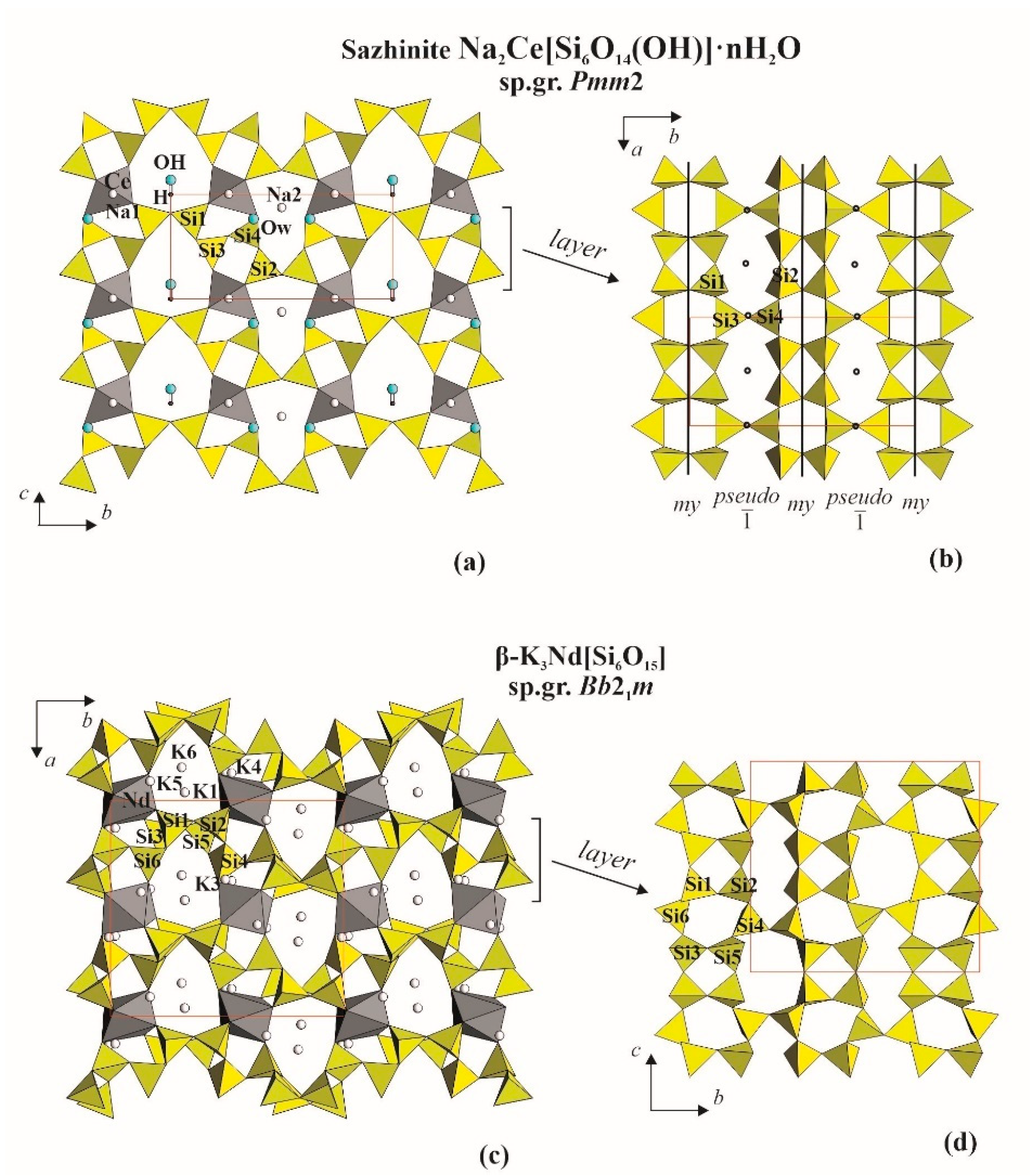
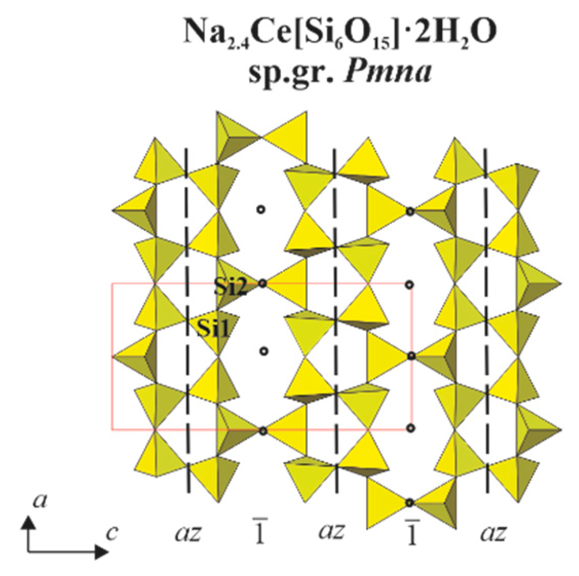
4.1. Structural Comparison with the Related Layered Silicates
4.2. Topology-Symmetry Analysis and Structure Prediction
5. Conclusions
Author Contributions
Funding
Data Availability Statement
Acknowledgments
Conflicts of Interest
References
- Mineralogy Database. Available online: http://www.mindat.org/ (accessed on 20 October 2020).
- ICSD FIZ. Available online: http://www.fiz-karlsruhe.de (accessed on 20 October 2020).
- Crystallography Open Database. Available online: http://www.crystallography.net (accessed on 20 October 2020).
- Garra, W.; Marchetti, F.; Merlino, S. Tb/Na tobermorite: Thermal behaviour and high temperature products. J. Solid State Chem. 2009, 182, 1529–1532. [Google Scholar] [CrossRef]
- Wierzbicka-Wieczorek, M.; Göckeritz, M.; Kolitsch, U.; Lenz, C.; Giester, G. Crystallographic and Spectroscopic Investigations on Nine Metal-Rare-Earth Silicates with the Apatite Structure Type. Eur. J. Inorg. Chem. 2015, 2015, 948–963. [Google Scholar] [CrossRef]
- Latshaw, A.M.; Wilkins, B.O.; Hughey, K.D.; Yeon, J.; Williams, D.E.; Tran, T.T.; Halasyamani, P.S.; Loye, H.-C.Z. A 5 RE 4 X [TO 4] 4 crystal growth and photoluminescence. Fluoride flux synthesis of sodium and potassium rare earth silicate oxyfluorides. CrystEngComm 2015, 17, 4654–4661. [Google Scholar] [CrossRef]
- Fleet, M.E.; Liu, X. Rare earth disilicates R2Si2O7 (R = Gd, Tb, Dy, Ho): Type B. Z. Krist. Cryst. Mater. 2003, 218, 795–801. [Google Scholar] [CrossRef]
- Vidican, I.; Smith, M.D.; Zur Loye, H.-C. Crystal growth, structure determination and optical properties of new potassi-um-rare-earth silicates K3RESi2O7 (RE = Gd, Tb, Dy, Ho, Er, Tm, Yb, Lu). J. Solid St. Chem. 2003, 170, 203–210. [Google Scholar] [CrossRef]
- Sieke, C.; Hartenbach, I.; Schleid, T. Sulfidisch derivatisierte Oxodisilicate der schweren Lanthanide vom Formeltyp M4S3(Si2O7) (M = Gd − Tm). Z. Nat. B J. Chem. Sci. 2002, B57, 1427–1432. [Google Scholar]
- Ananias, D.; Kostova, M.; Paz, F.A.A.; Ferreira, A.; Carlos, L.D.; Klinowski, J.; Rocha, J. Photoluminescent Layered Lanthanide Silicates. J. Am. Chem. Soc. 2004, 126, 10410–10417. [Google Scholar] [CrossRef]
- Fulle, K.; Sanjeewa, L.D.; McMillen, C.D.; Kolis, J.W. Crystal chemistry and the role of ionic radius in rare earth tetrasilicates: Ba2RE2Si4O12F2(RE = Er3+–Lu3+) and Ba2RE2Si4O13(RE = La3+–Ho3+). Acta Crystallogr. Sect. B Struct. Sci. Cryst. Eng. Mater. 2017, 73, 907–915. [Google Scholar] [CrossRef]
- Topnikova, A.P.; Belokoneva, E.L.; Dimitrova, O.V.; Volkov, A.S.; Nelyubina, Yu.V. Na3Tb3[Si6O18]∙H2O, a synthetic analogue of microporous mineral gerenite. Cryst. Rep. 2016, 61, 566–570. [Google Scholar] [CrossRef]
- Lee, C.-S.; Liao, Y.-C.; Hsu, J.-T.; Wang, S.-L.; Lii, K.-H. Rb2REGaSi4O12 (RE = Y, Eu, Gd, Tb): Luminescent Mixed-Anion Double Layer Silicates Containing Chains of Edge-Sharing REO7 Pentagonal Bipyramids. Inorg. Chem. 2008, 47, 1910–1912. [Google Scholar] [CrossRef]
- Bao, X.; Liu, X.; Liu, X. High-pressure synthesis, crystal structure and photoluminescence properties of a new terbium silicate: Na2Tb1.08Ca2.92Si6O18H0.8. RSC Adv. 2017, 7, 50195. [Google Scholar] [CrossRef] [Green Version]
- Wang, G.; Li, J.; Yu, J.; Chen, P.; Pan, Q.; Song, H.; Xu, R. Na3TbSi3O9·3H2O: A New Luminescent Microporous Terbium(III) Silicate Containing HelicalSechserSilicate Chains and 9-Ring Channels. Chem. Mater. 2006, 18, 5637–5639. [Google Scholar] [CrossRef]
- Morrison, G.; Latshaw, A.M.; Spagnuolo, N.R.; Loye, H.-C.Z. Observation of Intense X-ray Scintillation in a Family of Mixed Anion Silicates, Cs3RESi4O10F2(RE = Y, Eu–Lu), Obtained via an Enhanced Flux Crystal Growth Technique. J. Am. Chem. Soc. 2017, 139, 14743–14748. [Google Scholar] [CrossRef]
- Taroev, V.K.; Kashaev, A.A.; Malcherek, T.; Goettlicher, J.; Kaneva, E.V.; Vasiljev, A.D.; Suvorova, L.F.; Suvorova, D.S.; Tauson, V.L. Crystal structures of new potassium silicates and aluminosilicates of Sm, Tb, Gd, and Yb and their relation to the armstrongite (CaZr(Si6O15)∙3H2O) structure. J. Solid State Chem. 2015, 227, 196–203. [Google Scholar] [CrossRef]
- Zhao, X.; Li, J.; Chen, P.; Li, Y.; Chu, Q.; Liu, X.; Yu, J.; Xu, R. New Lanthanide Silicates Based on Anionic Silicate Chain, Layer, and Framework Prepared under High-Temperature and High-Pressure Conditions. Inorg. Chem. 2010, 49, 9833–9838. [Google Scholar] [CrossRef] [PubMed]
- Ananias, D.; Ferreira, A.; Rocha, J.; Ferreira, P.; Rainho, J.P.; Morais, C.; Carlos, L.D. Novel Microporous Europium and Ter-bium Silicates. J. Am. Chem. Soc. 2001, 123, 5735–5742. [Google Scholar] [CrossRef] [PubMed]
- Liebau, F. Structural Chemistry of Silicates: Structure, Bonding, and Classification; Springer: Berlin/Heidelberg, Germany, 1985; 347p. [Google Scholar]
- Pushcharovsky, D.Y. Structural Mineralogy of Silicates and Their Synthetic Analogues; Nedra: Moscow, Russia, 1986; 160p. [Google Scholar]
- Ferraris, G.; Makovicky, E.; Merlino, S. Crystallography of Modular Materials; Oxford University Press (OUP): Oxford, UK, 2008. [Google Scholar]
- Dornberger-Schiff, K. Grundzuege Einer Theorie der OD-Strukturen aus Schichten; Deutsche Akademie der Wissenschaften, Berlin, Abhandlungen, Klasse fur Chemie, Geologie and Biologie: Halle, Germany, 1964; Volume 3, pp. 1–106. [Google Scholar]
- Merlino, S. OD Structures in Mineralogy. Per. Mineral. 1990, 59, 69–92. [Google Scholar]
- Belokoneva, E.L. Borate crystal chemistry in terms of the exnetded OD theory: Topology and symmetry analysis. Cryst. Rev. 2005, 11, 151–198. [Google Scholar] [CrossRef]
- Ivanova, A.G.; Belokoneva, E.L.; Dimitrova, O.V. New condensed acid diborate GdH[B2O5] with chain radical [B2□B2ΔO10]8-]∞: Synthesis and crystal structure; diborates and their structural system in terms of OD theory. Russ. J. Inorg. Chem. 2004, 49, 816–822. [Google Scholar]
- Belokoneva, E.L.; Reutova, O.V.; Dimitrova, O.V.; Volkov, A.S. Germanosilicate Cs2In2[(Si2.1Ge0.9)2O15](OH)2∙H2O with a New Corrugated Tetrahedral Layer: Topological Symmetry-Based Prediction of Anionic Radicals. Crystallogr. Rep. 2020, 65, 566–572. [Google Scholar] [CrossRef]
- Dorenbos, P. Exchange and crystal field effects on the 4fn 15d levels of Tb3. J. Phys. Condens. Matter 2003, 15, 6249–6268. [Google Scholar] [CrossRef]
- Pisarski, W.A.; Zur, L.; Sołtys, M.; Pisarska, J. Terbium-terbium interactions in lead phosphate glasses. J. Appl. Phys. 2013, 113, 143504. [Google Scholar] [CrossRef]
- Berdowski, P.A.M.; Lammers, M.J.J.; Blasse, G. 5D3-5D4 cross-relaxation of Tb3+ in α-GdOF. Chem. Phys. Lett. 1985, 113, 387–390. [Google Scholar]
- Van Uitert, L.G.; Johnson, L.F. Energy Transfer between Rare—Earth Ions. J. Chem. Phys. 1966, 44, 3514. [Google Scholar] [CrossRef]
- Tonooka, K.; Nishimura, O. Spectral changes of Tb3+ fluorescence in borosilicate glasses. J. Lumin. 2000, 87, 679–681. [Google Scholar] [CrossRef]
- Deyneko, D.V.; Morozov, V.A.; Vasin, A.A.; Aksenov, S.M.; Dikhtyar, Y.Y.; Stefanovich, S.Y.; Lazoryak, B.I. The crystal site engineering and turning of cross-relaxation in green-emitting β-Ca3(PO4)2-related phosphors. J. Lumin. 2020, 223, 117196. [Google Scholar] [CrossRef]
- CrysAlis PRO; Agilent Technologies Ltd.: Yarnton, Oxfordshire, UK, 2014.
- Sheldrick, G.M. A short history of SHELX. Acta Cryst. 2008, 64, 112–122. [Google Scholar] [CrossRef] [PubMed] [Green Version]
- Farrugia, L.J. WinGX and ORTEP for Windows: An update. J. Appl. Cryst. 2012, 45, 849. [Google Scholar] [CrossRef]
- Pauling, L. The Nature of the Chemical Bond; Cornell University: Ithaca, NY, USA, 1960; 644p. [Google Scholar]
- Sheldrik, G.M. Crystal structure refinement with SHELXL. Acta Cryst. 2015, 71, 3–8. [Google Scholar]
- Dowty, E. ATOMS; Shape Software: Kingsport, TN, USA, 2006. [Google Scholar]
- Cadoni, M.; Ferraris, J. Polytypic and polymorphic relations between sazhinite and isochemical alkali-REE layer silicates. Eur. J. Mineral. 2011, 23, 85–90. [Google Scholar] [CrossRef]
- Shumyatskaya, N.G.; Voronkov, A.A.; Pyatenko, Yu.A. Sazhinite Na2Ce[Si6O14(OH)]∙nH2O, a new member of crystal chemical family of dalyite. Sov. Phys. Cryst. 1980, 25, 728–734. [Google Scholar]
- Cadoni, M.; Cheah, Y.L.; Ferraris, G. New RE microporous heteropolyhedral silicates containing 41516182 tetrahedral sheets. Acta Crystallogr. Sect. B Struct. Sci. 2010, 66, 158–164. [Google Scholar] [CrossRef] [PubMed]
- Jeong, H.-K.; Chandrasekaran, A.; Tsapatsis, M. Synthesis of a new open framework cerium silicate and its structure determination by single crystal X-ray diffraction. Chem. Commun. 2002, 2398–2399. [Google Scholar] [CrossRef] [PubMed]
- Haile, S.M.; Wuensch, B.J. Structure, phase transitions and ionic conductivity of K3NdSi6O15∙xH2O. II. Structure of β-K3NdSi6O15. Acta Cryst. 2000, 56, 349–362. [Google Scholar] [CrossRef] [Green Version]
- Pushcharovsky, D.Y.; Karpov, O.G.; Pobedimskaya, E.A.; Belov, N.V. Crystal structure of K3NdSi6O15. Dokl. AN SSSR 1977, 234, 1323–1326. [Google Scholar]
- Rastsvetaeva, R.K.; Aksenov, S.M.; Taroev, V.K. Crystal Structures of Endotaxic Phases in Europium Potassium Silicate Having a Pellyite Unit Cell. Crystallogr. Rep. 2010, 55, 1041–1049. [Google Scholar] [CrossRef]
- Cámara, F.; Ottolini, L.; Devouard, B.; Garvie, L.A.J.; Hawthorne, F.C. Sazhinite-(La), Na3LaSi6O15(H2O)2, a new mineral from the Aris phonolite, Namibia: Description and crystal structure. Miner. Mag. 2006, 70, 405–418. [Google Scholar] [CrossRef]
- Karpov, O.G.; Pushcharovsky, D.Y.; Pobedimskaya, E.A.; Burshtein, I.F.; Belov, N.V. Crystal structure of rare earth silicate NaNdSi6O13(OH)2∙nH2O. Dokl. AN SSSR 1977, 236, 593–596. [Google Scholar]
- Haile, S.M.; Wuensch, B.J.; Laudise, R.A.; Maier, J. Structure of Na3NdSi6O15∙2H2O—A Layered Silicate with Paths for Possible Fast-Ion Conduction. Acta Cryst. 1997, 53, 7–17. [Google Scholar] [CrossRef]


| Tb13+ C.N. = 6 0.25* | Tb23+ C.N. = 6 0.25* | Cs1+ C.N. = 11 0.5* | Rb1+ C.N. = 9 0.5* | (Rb,Cs2)+ C.N. = 7 0.5* | T14+ C.N. = 4 0.5* | T24+ C.N. = 4 1.0* | T34+ C.N. = 4 1.0* | T44+ C.N. = 4 0.5* | Σexp | Σtheor | |
|---|---|---|---|---|---|---|---|---|---|---|---|
| O12− 0.5 * | 0.045 | 0.056 | 1.0 | −1.1 | −1.0 | ||||||
| O22− 0.5 * | 0.071 | 0.5 | 0.5 | −1.071 | −1.0 | ||||||
| O32− 1.0 * | 0.125 × 4 | 0.045 × 4 | 0.071 × 2 | 1.0 | −1.825 | −2.0 | |||||
| O42− 1.0 * | 0.125 × 4 | 0.056 × 4 | 1.0 | −1.722 | −2.0 | ||||||
| O52− 1.0 * | 0.056 × 2 | 0.5 × 2 | 1.0 | −2.111 | −2.0 | ||||||
| O62− 1.0 * | 0.071 × 2 | 1.0 | 1.0 | −2.143 | −2.0 | ||||||
| O72− 0.5 * | 0.125 × 2 | 0.045 × 2 | 0.071 | 0.5 | −0.912 | −1.0 | |||||
| O82− 1.0 * | 0.045 × 2 | 1.0 | 0.5 × 2 | −2.091 | −2.0 | ||||||
| O92− 0.5 * | 0.125 × 2 | 0.071 | 0.5 | −0.821 | −1.0 | ||||||
| O102− 0.5 * | 0.045 | 0.056 | 1.0 | −1.1 | −1.0 | ||||||
| O11w2− 0.5 * | 0.045 | 0.056 | −0.1 | 0 | |||||||
| Σ | +0.75 | +0.75 | +0.5 | +0.5 | +0.5 | +2 | +4 | +4 | +2 | 15 | 15 |
| Formula | Rb1.66Cs1.34Tb[Si5.43Ge0.57O15]·H2O |
|---|---|
| formula weight (g/mol) | 930.48 |
| T (K) | 293(2) |
| crystal system | Orthorhombic |
| space group, Z | Pbam, 4 |
| a (Å) | 15.9429(3) |
| b (Å) | 14.8407(3) |
| c (Å) | 7.2781(1) |
| V (Å 3) | 1722.03(6) |
| crystal size (mm) | 0.10 × 0.05 × 0.04 |
| ρcalc (g/cm 3) | 3.532 |
| μ (mm−1) | 12.610 |
| F(000) | 1677 |
| wavelength (Å) | 0.71073 |
| θ range/deg. | 2.75–30.78 |
| limiting indices | −22 ≤ h ≤ 22, −21≤ k ≤ 20, −10 ≤ l ≤ 10 |
| refl. collected/unique | 28316/2756 [Rint = 0.0695] |
| completeness to theta | 99.9 |
| data/restraints/parameters | 2756/0/143 |
| GOF | 1.187 |
| R1, wR21 [I > 2σ(I)] | 0.0541, 0.0885 |
| R1, wR2 (all data) 1 | 0.0670, 0.0926 |
| Δρmax and Δρmin (e Å−3) | 1.726 and −2.318 |
| Atoms | Wyckoff Position, Point Symm. | S.o.f. | X | Y | Z | Ueq |
|---|---|---|---|---|---|---|
| Cs1 | 4g, m | 1.0 | 0.1306(1) | 0.5459(1) | 0 | 0.0255(2) |
| Rb1 | 4h, m | 1.0 | 0.2830(1) | 0.8920(1) | 0.5 | 0.0408(4) |
| (Rb, Cs)2 | 4g, m | 0.66, 0.34 | 0.4222(1) | 0.4033(1) | 0 | 0.0250(2) |
| Tb1 | 2b, 2/m | 1.0 | 0.5 | 0.5 | 0.5 | 0.00832(14) |
| Tb2 | 2d, 2/m | 1.0 | 0 | 0.5 | 0.5 | 0.00750(14) |
| (Si, Ge)1 | 4h, m | 0.93, 0.07 | 0.0874(1) | 0.7829(2) | 0.5 | 0.0083(7) |
| (Si, Ge)2 | 8i, 1 | 0.88, 0.12 | 0.3535(1) | 0.6345(1) | 0.7838(2) | 0.0077(5) |
| (Si, Ge)3 | 8i, 1 | 0.91, 0.09 | 0.0395(1) | 0.3024(1) | 0.2181(2) | 0.0087(5) |
| (Si, Ge)4 | 4h, m | 0.92, 0.08 | 0.2068(1) | 0.6291(2) | 0.5 | 0.0060(7) |
| O(1) | 4g, m | 1.0 | 0.0165(5) | 0.2918(5) | 0 | 0.0192(16) |
| O(2) | 4h, m | 1.0 | 0.1804(4) | 0.7355(4) | 0.5 | 0.0139(15) |
| O(3) | 8i,1 | 1.0 | 0.0526(3) | 0.4042(3) | 0.2791(7) | 0.0150(10) |
| O(4) | 8i,1 | 1.0 | 0.4291(3) | 0.5701(4) | 0.7326(8) | 0.0176(11) |
| O(5) | 8i, 1 | 1.0 | 0.0392(3) | 0.7465(4) | 0.3168(7) | 0.0184(11) |
| O(6) | 8i, 1 | 1.0 | 0.1258(3) | 0.2425(3) | 0.2554(7) | 0.0141(10) |
| O(7) | 4h, m | 1.0 | 0.1292(4) | 0.5630(5) | 0.5 | 0.0134(14) |
| O(8) | 8i, 1 | 1.0 | 0.2653(3) | 0.6115(4) | 0.6809(7) | 0.0189(11) |
| O(9) | 4h, m | 1.0 | 0.0990(4) | 0.8882(5) | 0.5 | 0.0142(15) |
| O(10) | 4g, m | 1.0 | 0.3262(5) | 0.6260(6) | 0 | 0.0187(16) |
| O(11)w | 4g, m | 1.0 | 0.2446(9) | 0.3400(10) | 0 | 0.079(4) |
| Atoms | Bonds (Å) | Atoms | Bonds (Å) |
|---|---|---|---|
| Tb1O6 Octahedron | Tb2O6 Octahedron | ||
| Tb1-O4 x4 | 2.286(5) | Tb2-O7 ×2 | 2.262(7) |
| Tb1-O9 ×2 | 2.290(7) | Tb2-O3 ×4 | 2.304(5) |
| Average | 2.287 | Average | 2.290 |
| (Si,Ge)1O4 Tetrahedron | (Si,Ge)2O4 Tetrahedron | ||
| (Si,Ge)1-O9 | 1.573(7) | (Si,Ge)2-O4 | 1.582(5) |
| (Si,Ge)1-O5 ×2 | 1.632(5) | (Si,Ge)2-O8 | 1.629(5) |
| (Si,Ge)1-O2 | 1.640(7) | (Si,Ge)2-O10 | 1.637(3) |
| Average | 1.619 | (Si,Ge)2-O6 | 1.662(5) |
| (Si,Ge)3O4 Tetrahedron | Average | 1.628 | |
| (Si,Ge)3-O3 | 1.588(5) | (Si,Ge)4O4 Tetrahedron | |
| (Si,Ge)3-O5 | 1.617(5) | (Si,Ge)4-O7 | 1.579(7) |
| (Si,Ge)3-O1 | 1.637(3) | (Si,Ge)4-O8 x2 | 1.634(5) |
| (Si,Ge)3-O6 | 1.660(5) | (Si,Ge)4-O2 | 1.635(7) |
| Average | 1.626 | Average | 1.621 |
| Chemical Formula | Space Group | Unit CellParameters, Å | Reference |
| Rb1.66Cs1.34Tb[Si5.43Ge0.57O15]· H2O | Pbam | a = 15.943 b = 14.841 c = 7.278 | [this work] |
| K3Nd[Si6O15]·2H2O | Pbam | a = 16.008 b = 15.004 c = 7.279 | [44] |
| K3Nd[Si6O15] | Pbam | a = 16.011 b = 14.984 c = 7.276 | [45] |
| K3Eu[Si6O15]·2H2O | P21212 | a = 14.852 b = 15.902 c = 7.243 | [46] |
| Na2Ce[Si6O14(OH)2]·nH2O Ce-sazhinite | Pmm2 | a = 7.500 b = 15.620 c = 7.350 | [41] |
| Na3La[Si6O15]·2H2O La-sazhinite | Pmm2 | a = 7.415 b = 15.515 c = 7.164 | [47] |
| β-K3Nd[Si6O15] | Bb21m | a = 14.370 b = 15.518 c = 14.265 | [44] |
| Na2.4Ce[Si6O15]·2H2O | Pman | a = 7.309 b = 14.971 c = 7.135 | [43] |
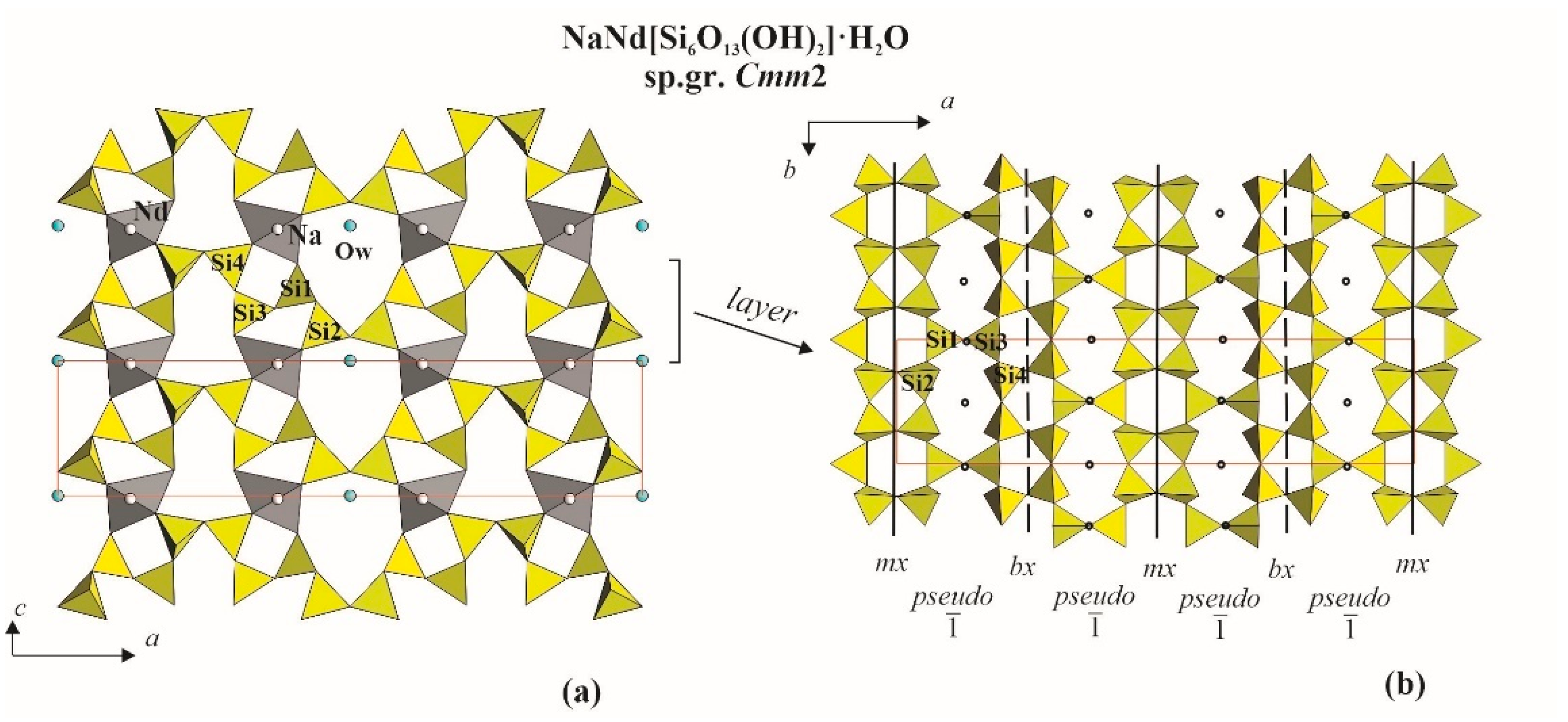
| NaNd[Si6O13(OH)2]·H2O | Cmm2 | a = 30.870 b = 7.387 c = 7.120 | [48] |
| NaNd[Si6O15]·2H2O | Cmm2 | a = 7.385 b = 30.831 c = 7.117 | [49] |
| Na2.74K0.26Ce[Si6O15]·2H2O | Cmm2 | a = 7.413 b = 30.965 c = 7.167 | [42] |
| Na3La[Si6O15]·2.25H2O | Cmm2 | a = 7.415 b = 31.008 c = 7.153 | [42] |
| Na2.72K0.25LaSi6O15·2.25H2O | Cmm2 | a
= 7.422 b = 31.039 c = 7.196 | [42] |
Publisher’s Note: MDPI stays neutral with regard to jurisdictional claims in published maps and institutional affiliations. |
© 2021 by the authors. Licensee MDPI, Basel, Switzerland. This article is an open access article distributed under the terms and conditions of the Creative Commons Attribution (CC BY) license (https://creativecommons.org/licenses/by/4.0/).
Share and Cite
Topnikova, A.; Belokoneva, E.; Dimitrova, O.; Volkov, A.; Deyneko, D. Rb1.66Cs1.34Tb[Si5.43Ge0.57O15]·H2O, a New Member of the OD-Family of Natural and Synthetic Layered Silicates: Topology-Symmetry Analysis and Structure Prediction. Minerals 2021, 11, 395. https://0-doi-org.brum.beds.ac.uk/10.3390/min11040395
Topnikova A, Belokoneva E, Dimitrova O, Volkov A, Deyneko D. Rb1.66Cs1.34Tb[Si5.43Ge0.57O15]·H2O, a New Member of the OD-Family of Natural and Synthetic Layered Silicates: Topology-Symmetry Analysis and Structure Prediction. Minerals. 2021; 11(4):395. https://0-doi-org.brum.beds.ac.uk/10.3390/min11040395
Chicago/Turabian StyleTopnikova, Anastasiia, Elena Belokoneva, Olga Dimitrova, Anatoly Volkov, and Dina Deyneko. 2021. "Rb1.66Cs1.34Tb[Si5.43Ge0.57O15]·H2O, a New Member of the OD-Family of Natural and Synthetic Layered Silicates: Topology-Symmetry Analysis and Structure Prediction" Minerals 11, no. 4: 395. https://0-doi-org.brum.beds.ac.uk/10.3390/min11040395