Article

Study of Ancient Pottery from the Brazilian Amazon Coast by EDXRF, PIXE, XRD, Mössbauer Spectroscopy and Computed Radiography

by
Renato Akio Ikeoka
1,*,
Carlos Roberto Appoloni
1,
Rosa Bernstein Scorzelli
2,
Edivaldo dos Santos
2,
Marcia de Almeida Rizzutto
3 and
Arkley Marques Bandeira
4
1
CCE, Departamento de Física, Universidade Estadual de Londrina—UEL, Caixa Postal 10.011, Londrina 86057-970, Brazil
2
Centro Brasileiro de Pesquisas Físicas, Rua Dr. Xavier Sigaud, 150–Urca, Rio de Janeiro 22290-180, Brazil
3
Laboratório de Análise de Materiais por Feixes Iônicos (LAMFI), Instituto de Física da Universidade de São Paulo, Cidade Universitária, Rua do Matão Travessa R Nr.187, São Paulo 05508-090, Brazil
4
Programa de Pós-Graduação em Cultura e Sociedade, Programa de Pós-Graduação em Desenvolvimento e Meio Ambiente de Ecossistemas Costeiros e, Departamento de Oceanografia e Limnologia, Universidade Federal do Maranhão, Av. dos Portugueses, 1966 Bacanga, São Luís 65080-805, Brazil
*
Author to whom correspondence should be addressed.
Submission received: 25 July 2022 / Revised: 13 October 2022 / Accepted: 14 October 2022 / Published: 16 October 2022

Abstract

:
This paper presents the application of five analytical methods to analyze ceramic fragments excavated in the archaeological sites of Sambaqui do Bacanga (SB), Sambaqui da Panaquatira (SP), and Rabo de Porco (RP) located on the Brazilian Amazon coast. It is a region that presents evidence of one of the oldest ceramic productions in the Americas. Characterization of ceramics from the three sites led to the identification of the production processes, technological acquisition and choices, and the origin of the raw materials. The analytical methods employed in this study were EDXRF, PIXE, Mössbauer spectroscopy, XRD, and computed radiography. It was concluded through the Mössbauer and XRD Spectroscopy analyses that the firing temperature range used on the ceramics was 750–900 °C, performed in an oxidizing environment. Fragments from the three archaeological sites were grouped into two groups through multivariate statistical analysis. The first group was composed of SB and SP samples, and the second of RP samples, indicating that different groups were manufactured with different clays. The result of the cluster analysis indicates that the clay used in the manufacture of the RP site fragments came from the bank of the Bacanga River. The ceramic fragments’ internal structures, observed by computed radiography, revealed the presence of various sizes and types of temper in the fragments, showing different production technologies.

1. Introduction

The Amazonian Equatorial Coast region of Brazil, which comprises a part of the Maranhão Gulf (Golfão Maranhense) area between the mouths of the Mearim River in São Marcos Bay and Itapecuru River in São José Bay, has been presenting archaeological sites, with evidence of ceramic production, that are among the oldest ones in America.
Bandeira [1,2] excavated open-air settlements with lithoceramic occupations and shell mounds that provided enough data for proof of a lifestyle associated with ceramic production, including three archaeological sites: Rabo de Porco (RP), Sambaqui da Panaquatira (SP), and Sambaqui do Bacanga (SB) (Figure 1).
The earliest ceramic production dates for the Sambaqui do Bacanga site demonstrate a strong persistence of this material culture extending from 6600 y BP until around the 14th century of our era. Research at the Sambaqui da Panaquatira site resulted in more recent dates than the Sambaqui do Bacanga site. The production of ceramics started around 5700y BP, extending to around European colonization. At the Rabo de Porco site, ceramics production started between 4870 and 4410 y BP, extending until the 17th century, with the arrival of the first Europeans to the region [2].
Recent research indicates a complex situation in the Late Holocene, marked by groups that produced different cultural materials and a diversified livelihood strategy [3]. The characterization of the ceramic sets excavated at the three sites identifies the production processes, technological choices, and raw material acquisition. In order to understand the complex context of these pre-Columbian peoples’ activities, it is necessary to develop a robust archaeometric approach, using various atomic-molecular and nuclear analytical techniques.
In this work, X-ray dispersive energy fluorescence (EDXRF), particle-induced X-ray emission (PIXE), Mössbauer spectroscopy, X-ray diffraction (XRD), and digital radiography were employed to study a set of ancient ceramic fragments excavated at three sites on the Brazilian Amazon coast. These techniques are becoming more and more important to the study of archaeological artifacts [4,5,6,7,8,9,10].
Due to the scarcity of previous archaeometric studies for the region, the following objectives were outlined in this work.
  • Verify the chemical elements that make up the fragments’ ceramic paste.
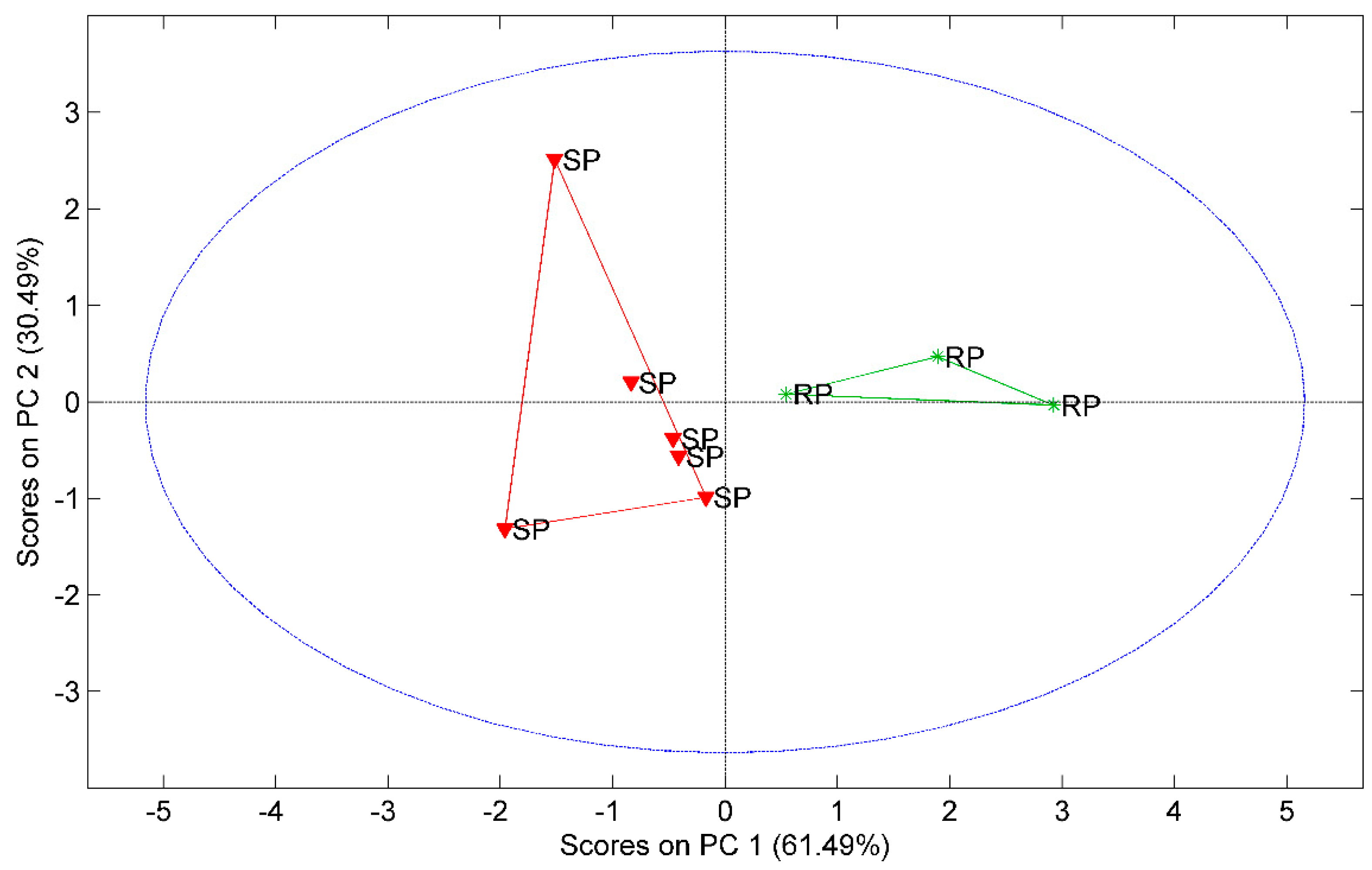
  • Apply multivariate exploratory analysis by principal component analysis (PCA) to study the clustering of fragments with clay sources.
  • Determine the temperature and atmosphere in which these ceramic fragments were burned.
  • Analyze the fragments’ internal structure and use of temper.

2. Materials and Methods

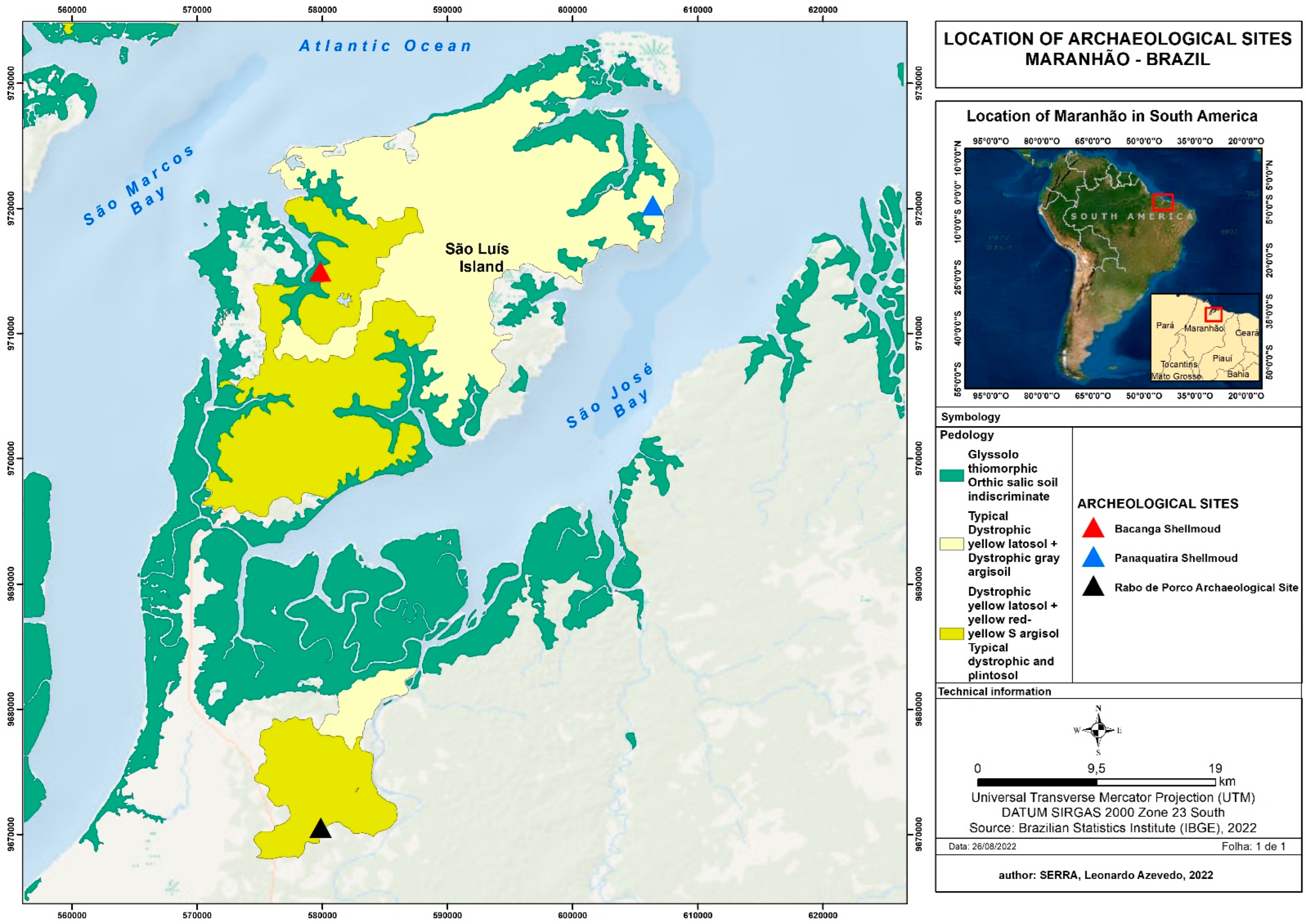
2.1. Archaeological Sites and Samples

The ceramic fragments presented in this article result from archaeological excavations at three archaeological sites in the Maranhão Gulf (Golfão Maranhense) estuary region, which includes a recent geological area covered by the vegetation of the Amazonian coastal region, such as mangroves and sandbank.
In this part of the Amazonian equatorial coast in the Maranhão state, the initial human occupation dates from about 9500 y BP, when the first settlers arrived through São José bay. Such peoples practiced a nomadic hunter–gatherer way of life, and their material culture was associated with splintered stone instruments. The first record of ceramic peoples’ in the region was about six millennia ago, with evidence of ancient pottery production in Sambaqui do Bacanga and Sambaqui da Panaquatira sites at 6600 and 5700 y BP, respectively, and around 4410 y BP at the Rabo de Porco site. The material culture of this period was characterized by a very simple sort of ceramic.
The set of archaeological sites mentioned above forms an arc of ceramic peoples with chronologies among the oldest ceramic production in Brazil, emphasizing the importance of these three settlements. The archaeological site of Sambaqui do Bacanga is located in São Luís at the Bacanga river mouth in São Marcos Bay. The Sambaqui da Panaquatira site is located in the extreme northeast of São Luís Island, in the municipality of São José de Ribamar. The Rabo de Porco site is the furthest from the Maranhão Gulf coast (Golfão Maranhense), located in the region of Itapecuru [1,2].
The archaeologist performed a careful sampling of 63 fragments for the archaeometric analysis, as described in Table 1.
Samples from five clay sources (named A–E) were selected to carry out a fragment cluster analysis of the sites studied. These clay sources were collected around the sites and on the banks of the Bacanga River.
To analyze the temperature and the firing atmosphere of the ceramics, a raw clay source was used to simulate the controlled firing temperature in the laboratory. The firing was carried out in an oxidizing and reducing environment, starting at a temperature of 300 °C, increasing to 1100 °C, at 100 °C intervals for 12 h. The firing was carried out in an electric oven in the sample preparation laboratory of the Brazilian Center for Physical Research.
For the cluster analysis, the raw clays were prepared and fired at 800 °C in an oxidizing atmosphere for 12 h to simulate the firing conditions of the ceramics under study. The firing was carried out in an electric oven at the Ceramics Laboratory of the State University of Londrina (UEL).

2.2. Energy Dispersion X-ray Fluorescence (EDXRF)

EDXRF was conducted using a Shimadzu (Kyoto, Japan) EDX-720 system with an Rh target, a 5–50 kV X-ray tube with a tube current operating range of 1–1000 μA, a 3 mm diameter collimator, and a Si (Li) type solid-state detector. The system is optimized to measure both high and low atomic (Z) number elements. The low Z group (Aluminum to Scandium) was measured with a tube setting of 15 kV and 470 μA, while the high Z group (Titanium to Uranium) was measured with a tube setting of 50 kV and 24 μA. No filters were used in the measurements. The instrument was operated in the air. Thus, the elements above Al were detected.
The measurements were carried out in a non-destructive way. The fragments were positioned vertically in the region delimited by the equipment with the help of a specially designed foam holder. An average of three measurements on the ceramic bulk (flat regions), each with 1000 s acquisition time, was performed for each fragment. The ceramic fragments have an average thickness of 0.8 cm.
The measurements were performed using the “Easy Quali-Quantitative” model, which is already programmed in the equipment software. The results of the values of the average net areas of the elements above three uncertainties were used. During the EDXRF measurements, two different certified materials (IAEA–Soil 7 and Plastic clay–IPT 32) [11,12] were used to test the reproducibility of the measurement.

2.3. Particle-Induced X-ray Emission (PIXE)

The PIXE measurements were performed using an electrostatic Pelletron-tandem accelerator from NEC (National Electronic Corporation, Madison WI, USA), type 5SDH, and an external multi-use analysis station, which allowed the analysis of objects of different shapes and sizes in air. This facility belongs to the Laboratory of Ion Beam Material Analysis (LAMFI-USP) at the University of São Paulo. The external beam setup uses a ~2.4 MeV proton beam and ~10 nA current to analyze the samples.
Three X-ray detectors were used, two Si-PIN detectors and one HPGe, with standard spectrometry electronics; an average of three measurements were performed on the ceramic bulk (flat regions); each spectrum was acquired for 600 s. The measurements were performed in a non-destructive way. An external gamma-ray scintillation detector (NaI(Tl)) was used to determine the ion beam charge to perform normalization between the measurements. The normalization used the beam exit window of an 8 mm aluminum foil and used the gamma rays of the (p, γ) reaction in the exit window foil.
During the PIXE measurements, two different certified materials (Buffalo River and plastic clay) [12,13] were used to test the reproducibility of the measurements and for normalization of the elemental measurements of the ceramic fragments. In this PIXE setup, only elements above Al were detected. The software used to analyze the spectra was Quantitative X-ray Analysis (AXIL), distributed by the International Atomic Energy Agency (IAEA).

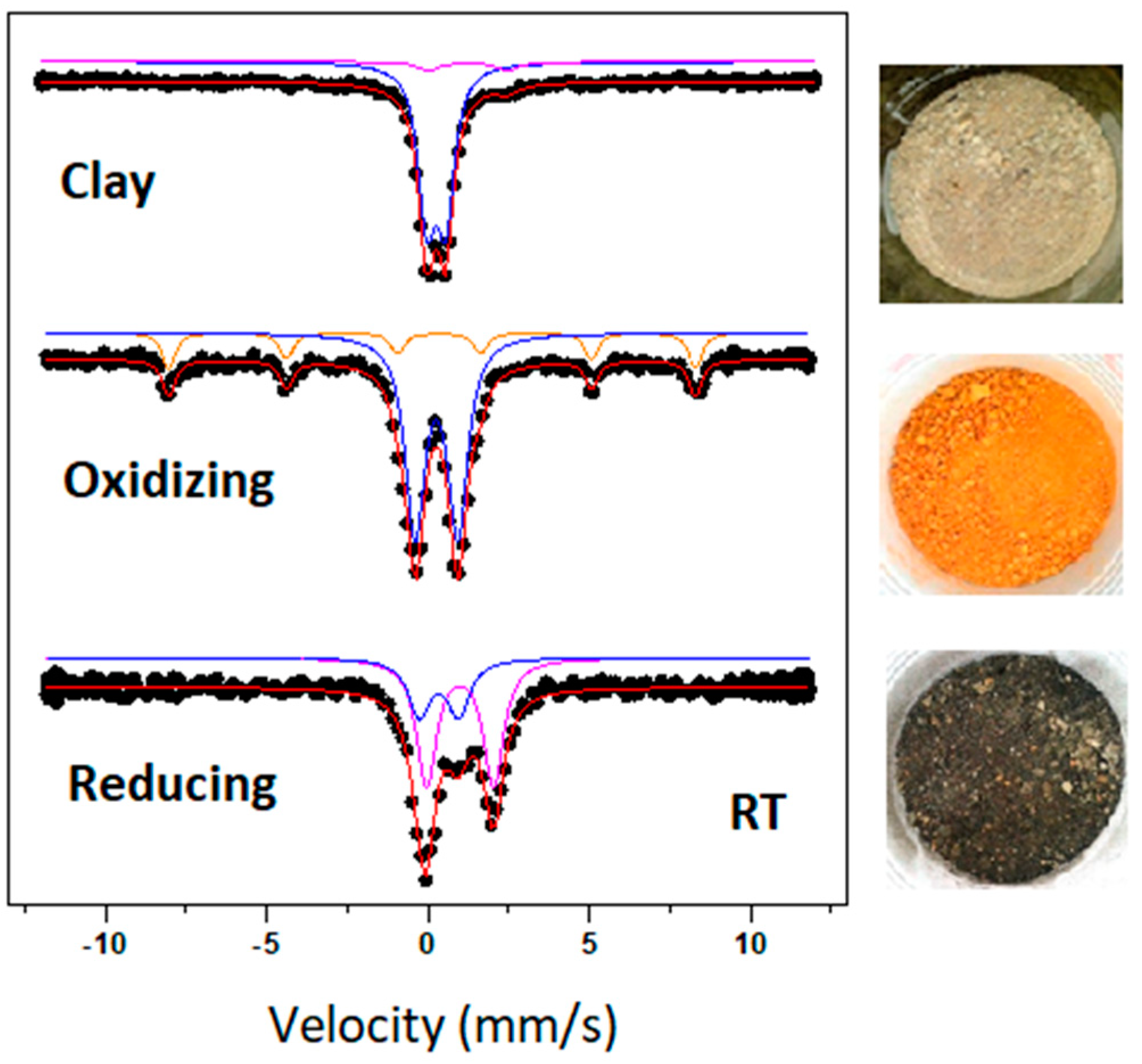
2.4. Mössbauer Spectroscopy

The samples’ Mössbauer spectra were recorded in transmission geometry at room temperature (RT), at a high velocity (12 mm/s), using a 57Co (Rh) source kept at the same temperature as the absorber in a Halder spectrometer with 512 channels. The drive velocity was calibrated with the same source and a metallic iron foil at RT. The average recording time was 15 h per sample. The Mössbauer absorbers were prepared to contain approximately 125 mg/cm2 of the powdered ceramic material, grounded with an agate mortar. The isomer shifts are given about α-Fe. The NORMOS code [14] was employed for the spectrum analysis and fitting with appropriate superpositions of Lorentzian lines, grouped into quadrupole doublets and magnetic sextets.

2.5. X-ray Diffraction

The Panalytical X’PERT PRO diffractometer was employed, with a source of Cu-kα radiation, a scanning step of 0.05°, and a measuring time of 5 s per step. The samples were grounded with an agate mortar and measured in powder form. The software used to analyze the spectra was HighScore Plus (version 3.0) (Panalytical, Malvern, UK).

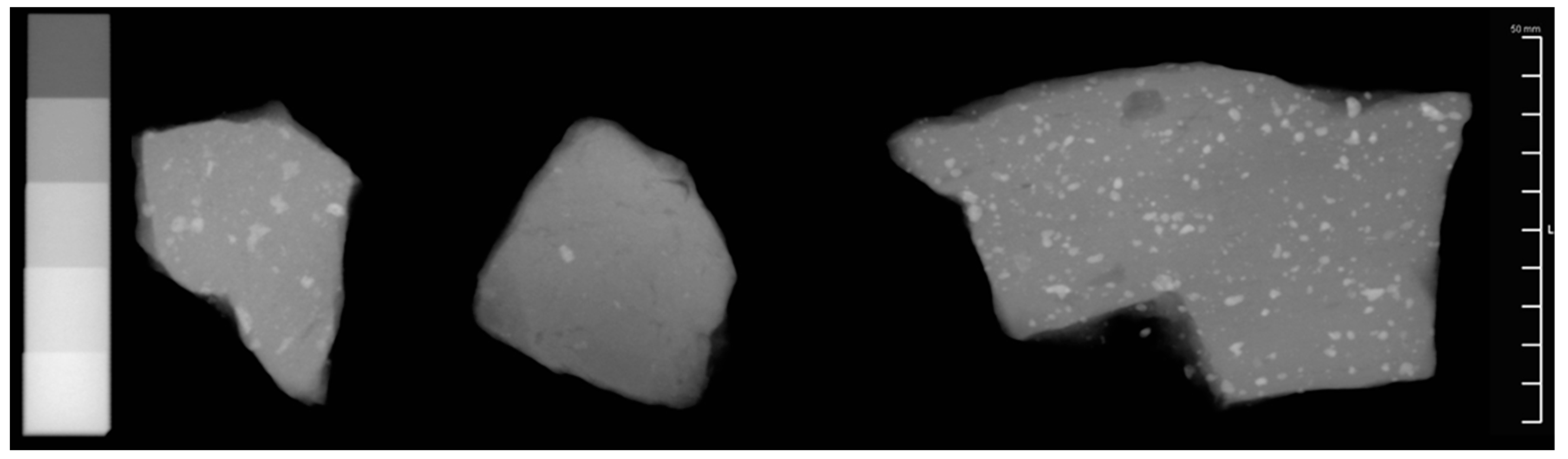
2.6. Computed Radiography

The Computed Radiography system applied to the analysis consists of the X-ray tube Philips MG 450 and scanner CR30-X AGFA. The conditions used for the measurements of fragments were 50 kV, 3 mA, and 10 s of irradiation at a distance of 3.5 m. A small piece of aluminum material with calibrated thicknesses was radiographed together with the ceramic fragments. The calibrated thicknesses of aluminum material varied between 0.2 and 1.0 cm. The images were analyzed using ImageJ (version 1.49) software.

3. Results and Discussion

3.1. EDXRF, PIXE and Cluster Analysis

Utilizing the EDXRF technique, the elements Al, Si, P, S, K, Ca, Ti, Cr, Mn, Fe, Cu, Zn, Rb, Sr, Y, Br, Zr, and Nb were measured with robust statistics in the ceramic fragments and the clay sources, indicating that such elements were present in the clay composition used in the manufacturing process of these ceramics. The clay sources were prepared as described in item 2.1.
Principal Component Analysis (PCA) was applied using the mean Kα X-ray intensities of the elements measured in the ceramic fragments and the clay sources. Thus, it was possible to group the samples according to the elemental chemical similarity of the matrix, contributing to the cluster analysis of the artifacts studied. MATLAB R2021a software was employed to carry out the PCA analysis [15] (Gilat, 2011).
Figure 2 illustrates the PCA results of the fragments of the three archaeological sites and the five clay sources (named A–E) obtained with the EDXRF analysis.
The PCA results shown in Figure 2 present the formation of two groups. The first one is composed of the Bacanga (SB) and Panaquatira (SP) Middens samples, and the second is composed of the Rabo de Porco (RP) site samples. It may also be observed that source D was grouped with the Rabo de Porco (RP) site samples, indicating that this group has the same elemental chemical composition. Therefore, the results of the cluster analysis suggest that the clay used in the manufacture of the Rabo de Porco (RP) site fragments comes from the Bacanga riverbank (source D).
The separation of the two groups occurs due to the higher concentrations of Sr, Ca, and Zn in the SB and SP samples compared to the RP samples. Another important factor for such separation is the presence of Rb, which appeared only in the RP samples.
The fragments of Bacanga (SB) and Panaquatira (SP) Sambaquis were manufactured using clays different to the areas analyzed in this work (sources A to E). It is also observed that the superposition of samples from the two sites suggests the same clay use for both cultures. Historically, potters are believed to have selected clay sources secretly and with difficult access, making it challenging to identify and locate them.
Only a representative subset of these ceramic fragments from the three sites was analyzed by PIXE at the LAMFI laboratory. With good statistics, the elements Si, P, S, K, Ca, Ti, Cr, Mn, Fe, Ni, Cu, and Zn were measured in the different ceramic pastes of the fragments. This indicates that these elements are present in the clay composition used to manufacture these ceramics. The PCA with the mean intensities of the elements measured in the ceramic fragments with PIXE was equivalent to the clusters observed with the EDXRF results, as shown in Figure 3.
The separation between the clusters of the samples occurs due to the higher concentrations of Ni, Ca, and Zn in the SB and SP samples than the RP samples. The agreement in the results obtained by the two analytical techniques validates its potential to analyze the cluster of ceramic fragments.

3.2. Mössbauer Spectroscopy and X-ray Diffraction (XRD)

3.2.1. Firing Atmosphere

The importance of Mossbauer spectroscopy in the study of archaeological ceramics is based on the analysis of the iron chemical state. The hyperfine parameter QS (quadrupole splitting) of Fe3+ is a marker of the maximum temperature mark at which the ceramic was burned. The type of the firing atmosphere of the ceramic is determined by the presence of the hyperfine parameters of Fe2+ and Fe3+ [16,17,18,19,20].
In pursuance of analyzing the firing atmosphere in which the ceramics were subjected in the manufacturing process, a source of raw clay was employed to perform a controlled firing in the laboratory, simulating the oxidizing and reducing atmospheres.
Figure 4 presents the Mössbauer spectra of the clay before and after treatment at the two firing atmospheres (oxidizing or reducing) carried out at 800 °C for 12 h.
With these patterns of firing spectra, it may be observed that in oxidative firing, the presence of the Fe3+ quadrupole doublets and the magnetic sextet of hematite is evident. In the reduction firing, the majority of Fe2+ quadrupole doublets are evident.
In the reduction firing, the black clay shade is related to the carbon black deposited during the process [21]. In oxidative firing at high temperatures, the reddish color is attributed to hematite formed during this firing process.
Six fragments from the Sambaqui da Panaquatira (SP) site and three from the Rabo de Porco (RP) site were sampled to analyze the ceramics firing atmosphere. Figure 5 and Table 2 illustrate the spectra and data of Mössbauer measurements from three samples from SP.
When the relative area (A) values in Table 2 are analyzed, it may be noted that the Panaquatira 145–150 cm fragment was subjected to an oxidative burn, as Fe3+ is the only iron species present.
In the other two fragments, the low concentration of Fe2+ in the ceramic paste indicates that the ceramic structure favored creating a slightly reducing firing environment in their interior, as on the two sides of these ceramics, the firing was totally oxidizing. The other four samples from SP also showed the same behavior as described previously.
The set of RP samples showed a slightly reducing firing in their inner part and totally oxidizing on their sides. A low relative percentage of magnetite was identified in two samples.
Principal Component Analysis was applied using the relative percentages of Fe2+, Fe3+, hematite, and magnetite data from the SP and RP samples.
Two groups are evident in Figure 6 by PCA analyses; a group formed by the SP samples and another formed by the RP samples. The RP sample group differed from the SP sample group due to the presence of magnetite and its higher relative concentration of Fe3+. Such a result suggests that these two sets of samples were subjected to different firing processes, reflecting the difference in the mastery of technology by those who inhabited the sites.

3.2.2. Firing Temperature

In pursuance of analyzing the firing temperature to which the ceramics were subjected in the manufacturing process, a source of raw clay was employed to simulate the firing temperature controlled in the laboratory. The firing was carried out in an oxidizing environment, starting at a temperature of 300 °C, increasing to 1100 °C, at 100 °C intervals for 12 h. Mössbauer spectra and X-ray diffractograms were obtained for each firing interval. Figure 7 illustrates the XRD and Mössbauer measurements of the fired clay from 400 °C to 1000 °C.
The Mössbauer spectra illustrate the formation and increase of the hematite magnetic sextet areas with the firing temperature. The most significant increase in the magnetic sextets occurred at temperatures above 600 °C. In the X-ray diffractograms, it may be observed that mica (muscovite) disappears at a temperature above 800 °C (indicated by the blue arrow). Indeed, as pointed out by [22], temperature decomposition for muscovite ranges from 850 to 940 °C. The diffractograms did not reveal corundum (or another aluminous silicate phase) as a product of muscovite breakdown. The detection of such products is probably hampered by quartz. Hematite formation above 600 °C (red arrows) is also noted. The photographs of the clays show the color change in an oxidizing environment with an increase in the firing temperature. It highlights the increase in hematite with the red hue of the clay.
It is well known that the orientation (i.e., preferred orientation) of clay mineral particles, such as mica (muscovite), affects the XRD intensities of powdered samples [23]. Therefore, it is believed that the orientation effect, combined with the small amount of mica (muscovite) compared to quartz (and other phases), hampers the detection of mica (muscovite) in the XRD pattern of the unfired sample illustrated in Figure 7.
A calibration curve of QS vs temperature was constructed from the Mössbauer spectra hyperfine parameters, as shown in Figure 8. In order to determine the ceramics firing temperature, the same firing processes and methodologies were employed as in the raw clay [16,17,18]. Nine samples were used to determine the firing temperature and atmosphere, where six fragments were from SP and three fragments were from RP.
According to Wagner [24], laboratory firing experiments are tools for determining the firing temperature/processes of ancient pottery. As pointed out by the authors, when the laboratory-fired clays are compared to those of ancient pottery and if the spectra are the same–not only at room temperature but also at other temperatures–one can conclude that the firing conditions in pottery may have been the same as those during the laboratory firing.
Firstly, the samples were measured by XRD, and two types of diffractograms were found in the SP samples. A group of five samples did not show the presence of mica (muscovite), indicating a temperature above 900 °C. The presence of mica (muscovite) was detected in only one sample from the SP (0–5 cm), indicating a firing temperature below 900 °C. In the three RP samples, the presence of mica (muscovite) was detected.
When the clay calibration curve and the SP sample (0–5 cm) data were compared, it was estimated that a temperature of 800 °C was used for the firing of this ceramic, as shown in Figure 8. This result is in accord with the presence of mica (muscovite) in X-ray diffractograms, as it decomposes at 900 °C.
In order to determine the firing temperature of the other five SP samples in the group, the 95–100 cm stratigraphy sample was selected as representative of this group. By comparing the clay calibration curve with this sample data,, a firing temperature of 900 °C was estimated, which agrees with the absence of mica (muscovite) in the XRD spectra.
To determine the Rabo de Porco sample group firing temperature, the stratigraphy sample of 0–10 cm was selected as representative of this group. When the clay calibration curve was compared with the sample data, a firing temperature of 750 °C was estimated, which agrees with the presence of mica (muscovite) in the XRD spectra.
The firing temperature range estimated for these samples was 750–900 °C, performed in an oxidizing environment.

3.3. Computed Radiography

It was possible to analyze and calculate the percentage temper present in samples by digital radiography. Since tempers have a different density (usually higher) than the ceramic paste, the process of binarization was applied to these images, essentially reducing the information contained within the image to a binary image, allowing them to be quantified. The next steps were to calculate the percentage of temper in the samples. Figure 9 illustrates the radiography of three ceramic fragments. An aluminum standard with five calibrated thicknesses (0.2; 0.4; 0.6; 0.8 and 1.0 cm) was also used.
The mean values for the SP, RP and SB site fragment temper percentages are 0.96%, 0.34% and 4.23%, respectively. Due to the highest temper concentration in the SB fragments, two fragments were selected to be analyzed by optical microscopy, performed with an Olympus BX-51 trinocular light microscope. The sample matrices are heterogeneous and formed by the following components: quartz grains, ceramic fragments, and shell fragments. The addition of shells to the ceramic paste is a characteristic of the Mina ceramic tradition, which aims to increase the resistance of the ceramic to shrinking and cracking [2].

4. Conclusions

The characterization of the ceramic sets extracted from the three sites carried out through various analytical techniques led to the identification of different production processes, technological choices, and how raw materials were obtained. Furthermore, it corroborated the technical and typological analyses that resulted in the identification of similar patterns in the way of making ceramics at the Sambaquis do Bacanga (SB) and Panaquatira (SP) sites and the rather different processes at the Rabo de Porco (RP) site.
Through the Mössbauer and XRD analysis, it was estimated that the firing temperature ranged from 750 to 900 °C in an oxidizing environment. Such firing temperature thresholds showed agreement with the data obtained by XRD, indicating the presence or absence of mica (muscovite).
Through digital radiography,, it was possible to estimate the percentage of temper in the fragments for the three archaeological sites studied.
The result of the cluster analysis indicated that the clay used in the Rabo de Porco site fragments manufacture came from Bacanga Riverbank. The fragments of Bacanga and Panaquatira Sambaquis were manufactured using clays originating from different to areas to those analyzed in this work. The groupings obtained by the EDXRF and PIXE techniques are in agreement. They suggest the SB and SP fragments came from similar clay sources and differ from the origin of clay used in the RP fragments.
Such results have considerably supported archaeology in constructing safe parameters in the individualization of the analyzed ceramic sets. Thus, contributing to studying the particularities observed in the characteristic technical elements of almost seven millennia of ceramic production on the Maranhão coast.

Author Contributions

Formal Analysis, Methodology, Writing-original draft, R.A.I.; Formal Analysis, Methodology, Writing-original draft, C.R.A.; Formal Analysis, Methodology, Writing-original draft, R.B.S.; Formal Analysis, Methodology, Writing-original draft, E.d.S.; Formal Analysis, Methodology, Writing-original draft, M.d.A.R.; Formal Analysis, Methodology, Writing-original draft, A.M.B. All authors have read and agreed to the published version of the manuscript.

Funding

This research was funded by the Project INCT-FNA Proc. CNPq No. 464898/2014-5; Project CNPq Proc. No. 306.087/2017-0 and CAPES.

Data Availability Statement

Not applicable.

Acknowledgments

The authors thank Pablo Munayco for help in the Mössbauer spectroscopy and XRD analysis and Tania Cristina Rumi Sugeta for help with preparing the raw clay samples. R. B. Scorzelli, M.A. Rizzutto and C.R. Appoloni acknowledges financial support from CNPq. Edivaldo dos Santos acknowledges FAPERJ.

Conflicts of Interest

The authors declare no conflict of interest.

References

  1. Bandeira, A.M. Ocupações Humanas Pré-Históricas no Litoral Maranhense: Um Estudo Arqueológico Sobre o Sambaqui do Bacanga na Ilha de São Luís-Maranhão. Master’s Thesis, Universidade de São Paulo, São Paulo, Brasil, 2008. [Google Scholar]
  2. Bandeira, A.M. Ocupações Humanas Pré-Coloniais na Ilha de São Luís—MA: Inserção dos Sítios Arqueológicos na Paisagem, Cronologia e Cultura Material Cerâmica. Ph.D. Thesis, Universidade de São Paulo, São Paulo, Brasil, 2013. [Google Scholar]
  3. Colonese, A.C.; Winter, R.; Brandi, R.; Fossile, T.; Fernandes, R.; Soncin, S.; McGrath, K.; Von Tersch, M.; Bandeira, A.M. Stable isotope evidence for dietary diversification in the pre-Columbian Amazon. Sci. Rep. 2020, 10, 16560. [Google Scholar] [CrossRef] [PubMed]
  4. Appoloni, C.R.; Parreira, P.S.; Souza, E.; Nascimento, V.; Gigante, G.E.; Cesareo, R.; Silva, R.M. Estudo de cerâmicas arqueológicas do Paraná por técnicas nucleares não destrutivas. Rev. Mus. Arqueol. Etnol. 1997, 2, 135–149. [Google Scholar] [CrossRef] [Green Version]
  5. Appoloni, C.; Espinoza-Quiñones, F.R.; Aragão, P.; Dos Santos, A.O.; Da Silva, L.M.; Barbieri, P.; Filho, V.D.N.; Coimbra, M. EDXRF study of Tupi-Guarani archaeological ceramics. Radiat. Phys. Chem. 2001, 61, 711–712. [Google Scholar] [CrossRef]
  6. Ikeoka, R.A.; Appoloni, C.R.; Parreira, P.S.; Lopes, F.; Bandeira, A.M. PXRF and multivariate statistics analysis of pre-colonial pottery from northeast of Brazil. X-ray Spectrom. 2012, 41, 12–15. [Google Scholar] [CrossRef]
  7. Ikeoka, R.A.; Appoloni, C.R.; Rizzutto, M.A.; Bandeira, A.M. Computed Radiography, PIXE and XRF analysis of pre-colonial pottery from Maranhão, Brazil. Microchem. J. 2018, 138, 384–389. [Google Scholar] [CrossRef]
  8. Seethaa, D.; Velraj, G. FT-IR, XRD, SEM-EDS, EDXRF and chemometric analyses of archaeological artifacts recently excavated from Chandravalli in Karnataka State, South India. Radiat. Phys. Chem. 2019, 162, 114–120. [Google Scholar] [CrossRef]
  9. Mejia-Bernal, J.R.; Ayala-Arenas, J.S.; Cano, N.F.; Rios-Orihuela, J.F.; Gonzales-Lorenzo, C.D.; Watanabe, S. Dating and determination of firing temperature of ancient potteries from Yumina archaeological site, Arequipa, Peru. Appl. Radiat. Isot. 2020, 155, 108930. [Google Scholar] [CrossRef]
  10. Oliveira, L.; Abreu, C.; Ferreira, F.; Lopes, R.; Almeida, F.; Tamanaha, E.; Belletti, J.; Machado, R.; Rizzutto, M.; Souza, D. Archeometric study of pottery shards from Conjunto Vilas and São João, Amazon. Radiat. Phys. Chem. 2020, 167, 108303. [Google Scholar] [CrossRef]
  11. Pszonicki, L.; Hanna, A.N.; Suschny, O. Report on Intercomparison IAEA/Soil-7 of the Determination of Trace Elements in Soil; International Atomic Energy Agency: Vienna, Austria, 1984; Available online: https://inis.iaea.org/collection/NCLCollectionStore/_Public/15/051/15051886.pdf?r=1 (accessed on 5 February 2021).
  12. IPT32. Padrão Laboratorial Referência 32. Instituto de Pesquisas Técnológicas do Estado de São Paulo, São Paulo. 1980. Available online: http://www.ipt.br/materiais_ref.php?m_tp=2&m_fm=24&familia=24 (accessed on 5 February 2021).
  13. RM8704 Padrão de Sedimento “Buffalo River”. Available online: https://www-s.nist.gov/srmors/certificates/8704.pdf (accessed on 5 February 2021).
  14. Brand, R.A. Normos Mössbauer Fitting Program; Universität Duisburg: Duisburg, Germany, 2007. [Google Scholar]
  15. Gilat, A. MATLAB: An Introduction with Applications, 4th ed.; John Wiley & Sons: Hoboken, NJ, USA, 2011. [Google Scholar]
  16. Shimada, I.; Häusler, W.; Hutzelmann, T.; Riederer, J.; Wagner, U. Early pottery making in Northern Coastal Peru. Part III: Mössbauer study of Sicán Pottery. Hyperfine Interact. 2003, 150, 107–123. [Google Scholar] [CrossRef]
  17. Gebhard, R.; Bott, R.; Distler, N.; Michálek, J.; Riederer, J.; Wagner, F.E.; Wagner, U. Ceramics from the Celtic Oppidum of Manching and Its Influence in Central Europe. Hyperfine Interact. 2004, 154, 199–214. [Google Scholar] [CrossRef]
  18. Cervantes, G.; Shimada, I.; Häusler, W.; Wagner, U.; Wagner, F.E. Mössbauer study of miniature vessels from the Sicán burial site of Huaca Loro. Hyperfine Interact. 2011, 203, 51–57. [Google Scholar] [CrossRef]
  19. Munayco, P.; Latini, R.M.; Bellido, A.V.B.; Scorzelli, R.B. Classification of Archaeological Pottery from Amazon Basin by Mössbauer Spectroscopy and Neutron Activation Analysis. In Proceedings of the 39th International Symposium for Archaeometry, Leuven, Belgium, 28 May–1 June 2012. [Google Scholar]
  20. Munayco, P.; Scorzelli, R.B. Characterization of the firing conditions of archaeological Marajoara pottery by Mössbauer spectroscopy and X-ray diffraction. Hyperfine Interact. 2013, 222 (Suppl. 1), S69–S75. [Google Scholar] [CrossRef]
  21. Shimada, I.; Häusler, W.; Hutzelmann, T.; Wagner, U. Early pottery making in northern coastal Peru. Part I: Mössbauer study of clays. Hyperfine Interact. 2003, 150, 73–89. [Google Scholar] [CrossRef]
  22. Luz, A.B.; Lins, F.A.F. Rochas & Minerais Industriais: Usos e Especificações; CETEM/MCT: Rio de Janeiro, Brazil, 2005. [Google Scholar]
  23. Zhou, X.; Liu, D.; Bu, H.; Deng, L.; Liu, H.; Yuan, P.; Du, P.; Song, H. XRD-based quantitative analysis of clay minerals using reference intensity ratios, mineral intensity factors, Rietveld, and full pattern summation methods: A critical review. Solid Earth Sci. 2018, 3, 16–29. [Google Scholar] [CrossRef]
  24. Wagner, F.E.; Wagner, U. Mössbauer Spectra of Clays and Ceramics. Hyperfine Interact. 2004, 154, 35–82. [Google Scholar] [CrossRef]
Figure 1. The study areas on the Brazilian Amazon coast with the location of the Panaquatira, Bacanga, and Rabo de Porco sites. Identification of the pedology of the region surrounding the archaeological sites, where the clay raw materials were collected, is shown.
Minerals 12 01302 g001
Figure 2. Principal Component Analysis (PCA) with ceramics fragments excavated in three archaeological sites, Sambaqui do Bacanga (SB), Sambaqui da Panaquatira (SP), and Rabo de Porco 1 (RP), together with the five clay sources (A to E).
Minerals 12 01302 g002
Figure 3. Principal Component Analysis (PCA) with pottery fragments excavated in three archaeological sites, Sambaqui do Bacanga (SB), Sambaqui da Panaquatira (SP), and Rabo de Porco (RP).
Minerals 12 01302 g003
Figure 4. Oxidizing and reducing firing environments Mössbauer spectra at 800 °C for 12 h. The blue and pink spectra represent the quadrupole doublets of Fe3+ and Fe2+, respectively. The orange spectrum represents the magnetic sextet of the hematite. The red spectrum is the magnetic components superposition present in the sample.
Minerals 12 01302 g004
Figure 5. Panaquatira samples Mössbauer spectra. The blue and green spectra represent the quadrupole doublets of Fe3+ and Fe2+, respectively. The pink spectrum represents the magnetic sextet of hematite. The red spectrum is the superposition of the magnetic components present in the sample.
Minerals 12 01302 g005
Figure 6. Principal Component Analysis (PCA) with Mössbauer data from the Sambaqui da Panaquatira (SP) and Rabo de Porco (RP) samples.
Minerals 12 01302 g006
Figure 7. Mössbauer and XRD spectra of clay fired from 400 °C to 1000 °C.
Minerals 12 01302 g007
Figure 8. Clay calibration curve and Panaquatira 0–5 cm sample data comparison.
Minerals 12 01302 g008
Figure 9. Aluminum standard and ceramic fragments radiography from the SP, RP, and SB, respectively. The whiter regions of the samples represent the denser parts of the material.
Minerals 12 01302 g009
Table 1. Information about fragments.
SiteQuantity of FragmentsStratigraphic Levels
Sambaqui da Panaquatira26Surface up to 170 cm
Sambaqui do Bacanga17Surface up to 170 cm
Rabo de Porco20Surface up to 105 cm
Total63
Table 2. Hyperfine parameters obtained at room temperature (IS: isomer shift; QS: quadrupole splitting; Bhf: hyperfine field, and A: relative area of the different Fe species).
Fe3+ Fe2+ Hematite
IS (mm/s)QS (mm/s)A
(%)
IS (mm/s)QS (mm/s)A (%)IS (mm/s)QS (mm/s)Bhf
(mm/s)
A (%)
SP
140–145 cm
External0.2570.8954---0.264−0.1950.446
Paste0.2710.79680.9302.3750.240−0.1049.527
Internal0.2360.8256---0.259−0.2150.044
SP
145–150 cm
External0.2520.8752---0.276−0.1749.848
Paste0.2570.7972---0.261−0.1348.628
Internal0.2580.8971---0.238−0.1349.129
SP
170–180 cm
External0.2610.8160---0.265−0.1851.140
Paste0.2650.79671.0202.3070.233−0.1549.326
Internal0.2620.8068---0.231−0.2351.132
Publisher’s Note: MDPI stays neutral with regard to jurisdictional claims in published maps and institutional affiliations.

