
Detection of Enterotoxigenic Escherichia coli and Clostridia in the Aetiology of Neonatal Piglet Diarrhoea: Important Factors for Their Prevention
 , , ,
, , , 
Abstract
:1. Introduction
2. Materials and Methods
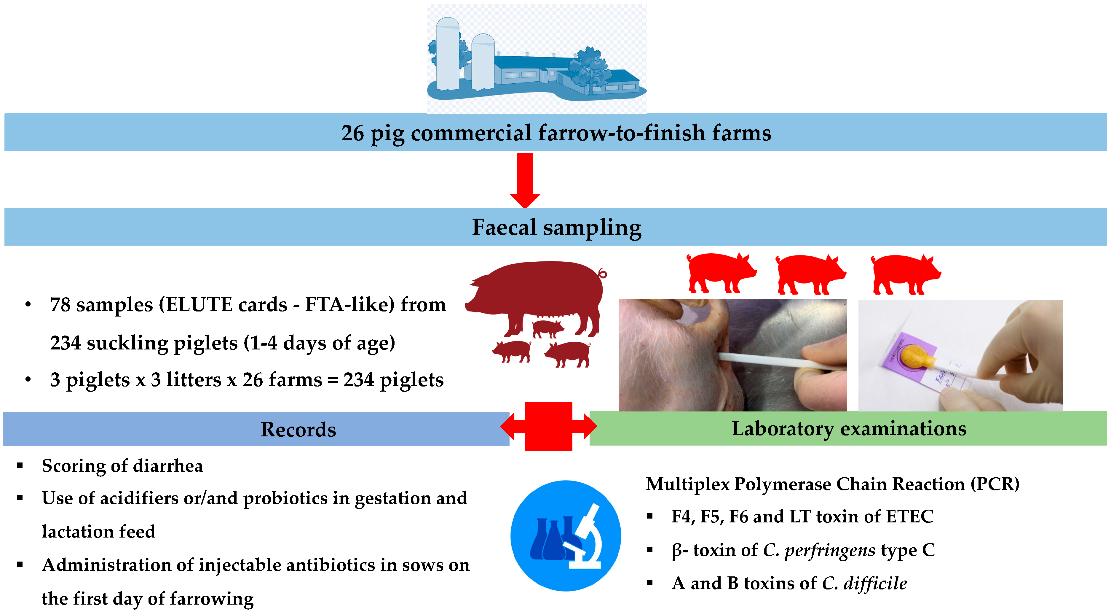
2.1. Study Design
2.1.1. Description of Farms, Criteria for Inclusion, and Study Groups
2.1.2. Sampling and Laboratory Examinations
2.2. Statistical Analysis
3. Results
4. Discussion
5. Conclusions
Supplementary Materials
Author Contributions
Funding
Institutional Review Board Statement
Informed Consent Statement
Data Availability Statement
Acknowledgments
Conflicts of Interest
References
- Chan, G.; Farzan, A.; DeLay, J.; McEwen, B.; Prescott, J.F.; Friendship, R.M. A retrospective study on the etiological diagnoses of diarrhea in neonatal piglets in Ontario, Canada, between 2001 and 2010. Can. J. Vet. Res. 2013, 77, 254–260. [Google Scholar] [PubMed]
- Sjölund, M.; Zoric, M.; Wallgren, P. Financial impact of disease on pig production. Part III. Gastrointestinal disorders. In Proceedings of the 6th European Symposium of Porcine Health Management, Sorrento, Italy, 7–9 May 2014; p. 189. [Google Scholar]
- Kongsted, H.; Pedersen, K.; Hjulsager, C.K.; Larsen, L.E.; Pedersen, K.S.; Jorsal, S.E.; Bækbo, P. Diarrhoea in neonatal piglets: A case control study on microbiological findings. Porc. Health Manag. 2018, 4, 17. [Google Scholar] [CrossRef] [PubMed]
- Dubreuil, J.D.; Isaacson, R.E.; Schifferli, D.M. Animal enterotoxigenic Escherichia coli. EcoSal Plus. 2016, 7. [Google Scholar] [CrossRef]
- Luppi, A.; Gibellini, M.; Gin, T.; Vangroenweghe, F.; Vandenbroucke, V.; Bauerfeind, R.; Bonilauri, P.; Labarque, G.; Hidalgo, A. Prevalence of virulence factors in enterotoxigenic Escherichia coli isolated from pigs with post-weaning diarrhea in Europe. Porc. Health Manag. 2016, 2, 20. [Google Scholar] [CrossRef]
- Ruiz, V.L.A.; Bersano, J.G.; Carvalho, A.F.; Catroxo, M.H.B.; Chiebao, D.P.; Gregori, F.; Miyashiro, S.; Nassar, A.F.C.; Oliveira, T.M.F.S.; Ogata, R.A.; et al. Case–control study of pathogens involved in piglet diarrhea. BMC Res. Notes 2016, 9, 22. [Google Scholar] [CrossRef] [PubMed]
- Uzal, F.A.; Songer, J.G. Clostridial diseases. In Diseases of Swine, 11th ed.; Zimmerman, J., Karriker, L.A., Ramirez, A., Schwartz, K.J., Stevenson, G.W., Eds.; John Wiley & Sons Inc.: Hoboken, NJ, USA, 2019; pp. 792–806. [Google Scholar]
- Vidal, A.; Martín-Valls, G.E.; Tello, M.; Mateu, E.; Martín, M.; Darwich, L. Prevalence of enteric pathogens in diarrheic and non-diarrheic samples from pig farms with neonatal diarrhea in the North East of Spain. Vet. Microbiol. 2019, 237, 108419. [Google Scholar] [CrossRef] [PubMed]
- Holland, R.E. Some infectious causes of diarrhea in young farm animals. Clin. Microbiol. Rev. 1990, 3, 345–375. [Google Scholar] [CrossRef]
- Morin, M.; Turgeon, D.; Jolette, J.; Robinson, Y.; Phaneuf, J.B.; Sauvageau, R.; Beauregard, M.; Teuscher, E.; Higgins, R.; Lariviere, S. Neonatal diarrhea of pigs in Quebec: Infectious causes of significant outbreaks. Can. J. Comp. Med. 1983, 47, 11–17. [Google Scholar]
- Svensmark, B.; Jorsal, S.; Nielsen, K.; Willeberg, P. Epidemiological studies of piglet diarrhoea in intensively managed Danish sow herds. I. Pre-weaning diarrhoea. Acta Vet. Scand. 1988, 30, 43–53. [Google Scholar] [CrossRef]
- Svendsen, J.; Bille, N.; Nielsen, N.; Larsen, J.; Riising, H. Preweaning mortality in pigs. Diseases of the gastrointestinal tract in pigs. Nordisk Veterinär. 1975, 27, 85–101. [Google Scholar]
- Kongsted, H.; Stege, H.; Toft, N.; Nielsen, J.P. The effect of new neonatal porcine Diarrhoea syndrome (NNPDS) on average daily gain and mortality in 4 Danish pig herds. BMC Vet. Res. 2014, 10, 90. [Google Scholar] [CrossRef] [PubMed]
- Johansen, M.; Alban, L.; Kjærsgård, H.D.; Bækbo, P. Factors associated with suckling piglet average daily gain. Prev. Vet. Med. 2004, 63, 91–102. [Google Scholar] [CrossRef] [PubMed]
- Dubreuil, J.D. Escherichia coli STb toxin and colibacillosis: Knowing is half the battle. FEMS Microbiol. Lett. 2008, 78, 137–145. [Google Scholar] [CrossRef] [PubMed]
- Moon, H.W.; Schineider, R.A.; Mosely, S.L. Comparative prevalence of four enterotoxin genes among Escherichia coli isolates from swine. Am. J. Vet. Res. 1986, 47, 210–212. [Google Scholar] [PubMed]
- Toledo, A.; Gómez, D.; Cruz, C.; Carreón, R.; López, J.; Giono, S.; Castro, A.M. Prevalence of virulence genes in Escherichia coli strains isolated from piglets in the suckling and weaning period in Mexico. J. Med. Virol. 2012, 61, 148–156. [Google Scholar] [CrossRef] [PubMed]
- Zajacova, Z.S.; Konstantinová, L.; Alexa, P. Detection of virulence factors of Escherichia coli focused on prevalence of EAST1 toxin in the stool of diarrheic and non-diarrheic piglets and presence of adhesion involving virulence factors in astA positive strains. Vet. Microbiol. 2012, 154, 369–375. [Google Scholar] [CrossRef] [PubMed]
- Nagy, B.; Fekete, P.Z. Enterotoxigenic E. coli (ETEC) in farm animals. Vet. Res. 1999, 30, 259–284. [Google Scholar]
- Vu-Khac, H.; Holoda, E.; Pilipcinec, E.; Blanco, M.; Blanco, J.E.; Dahbi, G.; Mora, A.; López, C.; González, E.A.; Blanco, J. Serotypes, virulence genes, intimin types and PFGE profiles of Escherichia coli isolated from piglets with diarrhoea in Slovakia. Vet. J. 2007, 174, 176–187. [Google Scholar] [CrossRef]
- Fairbrother, J.M.; Gyles, C.L. Colibacillosis. In Diseases of Swine, 10th ed.; Zimmerman, J., Karriker, L.A., Ramirez, A., Schwartz, K.J., Stevenson, G.W., Eds.; WileyBlackwell: Chichester, UK, 2012; pp. 723–749. [Google Scholar]
- Luppi, A. Swine enteric colibacillosis: Diagnosis, therapy and antimicrobial resistance. Porc. Health Manag. 2017, 3, 16. [Google Scholar] [CrossRef]
- Songer, J.G.; Uzal, F.A. Clostridial Enteric Infections in Pigs. J. Vet. Diagn. 2005, 17, 528–536. [Google Scholar] [CrossRef]
- Petit, L.; Gibert, M.; Popoff, M.R. Clostridium perfringens: Toxinotype and genotype. Trends Microbiol. 1999, 7, 104–110. [Google Scholar] [CrossRef] [PubMed]
- Songer, J.G. Clostridial enteric diseases of domestic animals. Clin. Microbiol. Rev. 1996, 9, 216–234. [Google Scholar] [CrossRef] [PubMed]
- Rood, J.I.; Adams, V.; Lacey, J.; Lyras, D.; McClane, B.A.; Melville, S.B.; Moore, R.J.; Popoff, M.R.; Sarker, M.R.; Songer, J.G.; et al. Expansion of the Clostridium perfringens toxin-based typing scheme. Anaerobe 2018, 53, 5–10. [Google Scholar] [CrossRef] [PubMed]
- Gould, L.H.; Limbago, B. Clostridium difficile in food and domestic animals: A new foodborne pathogen? Clin. Infect. Dis. 2010, 51, 577–582. [Google Scholar] [CrossRef] [PubMed]
- Keel, M.K.; Songer, J.G. The comparative pathology of Clostridium difficile-associated disease. Vet. Pathol. 2006, 43, 225–240. [Google Scholar] [CrossRef] [PubMed]
- Keessen, E.C.; van den Berkt, A.J.; Haasjes, N.H.; Hermanus, C.; Kuijper, E.J.; Lipman, L.J.A. The relation between farm specific factors and prevalence of Clostridium difficile in slaughter pigs. Vet. Microbiol. 2011, 154, 130–134. [Google Scholar] [CrossRef]
- Britton, R.A.; Young, V.B. Interaction between the intestinal microbiota and host in Clostridium difficile colonization resistance. Trends Microbiol. 2012, 20, 313–319. [Google Scholar] [CrossRef] [PubMed]
- Lim, S.C.; Knight, D.R.; Riley, T.V. Clostridium difficile and One Health. Clin. Microbiol. Infect. 2020, 26, 857–863. [Google Scholar] [CrossRef]
- Songer, J.G. The emergence of Clostridium difficile as a pathogen of food animals. Anim. Health Res. Rev. 2004, 5, 321–326. [Google Scholar] [CrossRef]
- Debast, S.B.; van Leengoed, L.A.M.G.; Goorhuis, A.; Harmanus, C.; Kuijper, E.J.; Bergwerff, A.A. Clostridium difficile PCR ribotype 078 toxinotype V found in diarrhoeal pigs identical to isolates from affected humans. Environ. Microbiol. 2009, 11, 505–511. [Google Scholar] [CrossRef]
- Goorhuis, A.; Bakker, D.; Corver, J.; Debast, S.B.; Harmanus, C.; Notermans, D.W.; Bergwerff, A.A.; Dekker, F.W.; Kuijper, E.J. Emergence of Clostridium difficile infection due to a new hypervirulent strain, polymerase chain reaction ribotype 078. Clin. Infect. Dis. 2008, 47, 1162–1170. [Google Scholar] [CrossRef]
- Goorhuis, A.; Debast, S.B.; van Leengoed, L.A.M.G.; Harmanus, C.; Notermans, D.W.; Bergwerff, A.A.; Kuijper, E.J. Clostridium difficile PCR ribotype 078: An emerging strain in humans and in pigs? J. Clin Microbiol. 2008, 46, 1157. [Google Scholar] [CrossRef]
- Mylonakis, E.; Ryan, E.T.; Calderwood, S.B. Clostridium difficile–associated diarrhea: A review. Arch. Intern. Med. 2001, 161, 525–533. [Google Scholar] [CrossRef] [PubMed]
- DebRoy, C.; Roberts, E.; Valadez, A.M.; Dudley, E.G.; Cutter, C.N. Detection of Shiga toxin–producing Escherichia coli O26, O45, O103, O111, O113, O121, O145, and O157 serogroups by multiplex polymerase chain reaction of the wzx gene of the O-antigen gene cluster. Foodborne Pathog. Dis. 2011, 8, 651–652. [Google Scholar] [CrossRef]
- Bai, J.; Paddock, Z.D.; Shi, X.; Li, S.; An, B.; Nagaraja, T.G. Applicability of a multiplex PCR to detect the seven major Shiga toxin–producing Escherichia coli based on genes that code for serogroup-specific O-antigens and major virulence factors in cattle feces. Foodborne Pathog. Dis. 2012, 9, 541–548. [Google Scholar] [CrossRef] [PubMed]
- Baker, C.A.; Rubinelli, P.M.; Park, S.H.; Carbonero, F.; Ricke, S.C. Shiga toxin-producing Escherichia coli in food: Incidence, ecology, and detection strategies. Food Control 2016, 59, 407–419. [Google Scholar] [CrossRef]
- Baker, C.A.; Rubinelli, P.M.; Park, S.H.; Ricke, S.C. Immuno-based detection of Shiga toxin-producing pathogenic Escherichia coli in food—A review on current approaches and potential strategies for optimization. Crit. Rev. Microbiol. 2016, 42, 656–675. [Google Scholar] [CrossRef] [PubMed]
- Salvador, J.M.; De Ungria, M.C.A. Isolation of DNA from saliva of betel quid chewers using treated cards. J. Forensic Sci. 2003, 48, 794–797. [Google Scholar] [CrossRef]
- Muthukrishnan, M.; Singanallur, N.B.; Ralla, K.; Villuppanoor, S.A. Evaluation of FTA® cards as a laboratory and field sampling device for the detection of foot-and-mouth disease virus and serotyping by RT-PCR and real-time RT-PCR. J. Virol. Methods 2008, 151, 311–316. [Google Scholar] [CrossRef]
- Linhares, D.C.; Rovira, A.; Torremorell, M. Evaluation of Flinders Technology Associates cards for collection and transport of samples for detection of Porcine reproductive and respiratory syndrome virus by reverse transcription polymerase chain reaction. J. Vet. Diagn. Investig. 2012, 24, 328–332. [Google Scholar] [CrossRef]
- Shalaby, A.G.; Bakry, N.R.; Mohamed, A.; Khalil, A.A. Evaluating Flinders Technology Associates card for transporting bacterial isolates and retrieval of bacterial DNA after various storage conditions. Vet. World. 2020, 13, 2243–2251. [Google Scholar] [CrossRef]
- Stringer, O.W.; Bossé, J.T.; Lacouture, S.; Gottschalk, M.; Fodor, L.; Angen, Ø.; Velazquez, E.; Penny, P.; Lei, L.; Langford, P.R.; et al. Rapid Detection and Typing of Actinobacillus pleuropneumoniae Serovars Directly From Clinical Samples: Combining FTA® Card Technology With Multiplex PCR. Front. Vet. Sci. 2021, 8, 728660. [Google Scholar] [CrossRef]
- Rajendram, D.; Ayenza, R.; Holder, F.M.; Moran, B.; Long, T.; Shah, H.N. Long-term storage and safe retrieval of DNA from microorganisms for molecular analysis using FTA matrix cards. J. Microbiol. Methods. 2006, 67, 582–592. [Google Scholar] [CrossRef] [PubMed]
- Li, J.Y. Current Status and Prospects for in-Feed Antibiotics in the Different Stages of Pork Production—A Review. Asian Austral. J. Anim. 2017, 30, 1667–1673. [Google Scholar] [CrossRef] [PubMed]
- Pamer, E.G. Resurrecting the Intestinal Microbiota to Combat Antibiotic-Resistant Pathogens. Science 2016, 352, 535–538. [Google Scholar] [CrossRef] [PubMed]
- Laird, T.J.; Abraham, S.; Jordan, D.; Pluske, J.R.; Hampson, D.J.; Trott, D.J.; O’Dea, M. Porcine Enterotoxigenic Escherichia Coli: Antimicrobial Resistance and Development of Microbial-Based Alternative Control Strategies. Vet. Microbiol. 2021, 258, 109117. [Google Scholar] [CrossRef]
- Athanasakopoulou, Z.; Reinicke, M.; Diezel, C.; Sofia, M.; Chatzopoulos, D.C.; Braun, S.D.; Reissig, A.; Spyrou, V.; Monecke, S.; Ehricht, R.; et al. Antimicrobial Resistance Genes in ESBL-Producing Escherichia Coli Isolates from Animals in Greece. Antibiotics 2021, 10, 389. [Google Scholar] [CrossRef]
- Tsekouras, N.; Athanasakopoulou, Z.; Diezel, C.; Kostoulas, P.; Braun, S.D.; Sofia, M.; Monecke, S.; Ehricht, R.; Chatzopoulos, D.C.; Gary, D.; et al. Cross-Sectional Survey of Antibiotic Resistance in Extended Spectrum β-Lactamase-Producing Enterobacteriaceae Isolated from Pigs in Greece. Animals 2022, 12, 1560. [Google Scholar] [CrossRef]
- Liu, Y.; Espinosa, C.D.; Abelilla, J.J.; Casas, G.A.; Lagos, L.V.; Lee, S.A.; Kwon, W.B.; Mathai, J.K.; Navarro, D.M.D.L.; Jaworski, N.W.; et al. Non-antibiotic feed additives in diets for pigs: A review. Anim Nutr. 2018, 4, 113–125. [Google Scholar] [CrossRef]
- Papatsiros, V.G.; Billinis, C. The prophylactic use of acidifiers as antibacterial agents in swine. In Antimicrobial Agents; Bobbarala, V., Ed.; InTech: Rijeka, Croatia, 2012; pp. 295–310. [Google Scholar]
- Ji, P.; Li, X.; Liu, Y. Dietary Intervention to Reduce E. coli Infectious Diarrhea in Young Pigs. In E. Coli Infections—Importance of Early Diagnosis and Efficient Treatment (Internet); Rodrigo, L., Ed.; IntechOpen: London, UK, 2020; Available online: https://www.intechopen.com/chapters/71010 (accessed on 24 July 2022). [CrossRef]
- Wellison, A.P.; Franco, S.M.; Reis, I.L.; Mendonça, C.M.N.; Piazentin, A.C.M.; Azevedo, P.O.S.; Tse, M.L.P.; De Martinis, E.C.P.; Gierus, M.; Oliveira, R.P.S. Beneficial effects of probiotics on the pig production cycle: An overview of clinical impacts and performance. Vet. Microbiol. 2022, 269, 109431. [Google Scholar] [CrossRef]
- Kim, Y.Y.; Kil, D.; Oh, H.K.; Han, I. Acidifier as an Alternative Material to Antibiotics in Animal Feed. Asian Australas J. Anim. Sci. 2005, 18, 1048–1060. [Google Scholar] [CrossRef]
- Jacela, J.Y.; DeRouchey, J.M.; Tokach, M.D.; Goodband, R.D.; Nelssen, J.L.; Renter, D.G.; Dritz, S.S. Feed additives for swine: Fact sheets—Acidifi ers and antibiotics. J. Swine Health Prod. 2009, 17, 270–275. [Google Scholar] [CrossRef]
- Roth, F.X.; Kirchgessner, M. Organic acids as feed additives for young pigs: Nutritional and gastrointestinal effects. J. Anim. Feed Sci. 1998, 7, 25–33. [Google Scholar] [CrossRef]
- Papatsiros, V.G.; Katsoulos, P.D.; Koutoulis, K.C.; Karatzia, M.; Dedousi, A.; Christodoulopoulos, G. Alternatives to antibiotics for farm animals. CAB Rev. Perspect. Agric. Vet. Sci. Nutr. Nat. Resour. 2013, 8, 1–15. [Google Scholar] [CrossRef]
- Kantas, D.; Papatsiros, V.G.; Tassis, P.D.; Athanasiou, L.V.; Tzika, E.D. Effect of a natural feed additive (Macleaya cordata), containing sanguinarine, on the performance and health status of weaning pigs. Anim. Sci. J. 2015, 86, 92–98. [Google Scholar] [CrossRef]
- Sayan, H.; Assavacheep, P.; Angkanaporn, K.; Assavacheep, A. Effect of Lactobacillus salivarius on growth performance, diarrhea incidence, fecal bacterial population and intestinal morphology of suckling pigs challenged with F4+ enterotoxigenic Escherichia coli. Asian Australas J. Anim. Sci. 2018, 31, 1308–1314. [Google Scholar] [CrossRef]
- Yue, S.; Li, Z.; Hu, F.; Picimbon, J.F. Curing piglets from diarrhea and preparation of a healthy microbiome with Bacillus treatment for industrial animal breeding. Sci. Rep. 2020, 10, 19476. [Google Scholar] [CrossRef]
- Alexopoulos, C.; Georgoulakis, I.E.; Tzivara, A.; Kritas, S.K.; Siochu, A.; Kyriakis, S.C. Field evaluation of the efficacy of a probiotic containing Bacillus licheniformis and Bacillus subtilis spores, on the health status and performance of sows and their litters. J. Anim. Physiol. Anim. Nutr. 2000, 88, 381–392. [Google Scholar] [CrossRef]
- Böhmer, B.M.; Kramer, W.; Roth-Maier, D.A. Dietary probiotic supplementation and resulting effects on performance, health status, and microbial characteristics of primiparous sows. J. Anim. Physiol. Anim. Nutr. 2006, 90, 309–315. [Google Scholar] [CrossRef]
- Kantas, D.; Papatsiros, V.G.; Tassis, P.D.; Giavasis, I.; Bouki, P.; Tzika, E.D. A feed additive containing Bacillus toyonensis (Toyocerin(®)) protects against enteric pathogens in postweaning piglets. J. Appl. Microbiol. 2015, 118, 727–738. [Google Scholar] [CrossRef]
- Hayakawa, T.; Masuda, T.; Kurosawa, D.; Tsukahara, T. Dietary administration of probiotics to sows and/or their neonates improves the reproductive performance, incidence of post-weaning diarrhea and histopathological parameters in the intestine of weaned piglets. Anim. Sci. J. 2016, 87, 1501–1510. [Google Scholar] [CrossRef] [PubMed]
- Menegat, M.B.; Gourley, K.M.; Braun, M.B.; DeRouchey, J.M.; Woodworth, J.C.; Bryte, J.; Tokach, M.D.; Dritz, S.S.; Goodband, R.D. Effects of a Bacillus-Based Probiotic on Sow Performance and on Progeny Growth Performance, Fecal Consistency, and Fecal Microflora. Kans. Agric. Exp. Stn. Res. Rep. 2018, 4, 9. [Google Scholar] [CrossRef]
- Kritas, S.K.; Marubashi, T.; Filioussis, G.; Petridou, E.; Christodoulopoulos, G.; Burriel, A.R.; Tzivara, A.; Theodoridis, A.; Pískoriková, M. Reproductive performance of sows was improved by administration of a sporing bacillary probiotic (Bacillus subtilis C-3102). J. Anim. Sci. 2015, 93, 405–413. [Google Scholar] [CrossRef] [PubMed]
- Papatsiros, V.G.; Tassis, P.D.; Tzika, E.D.; Papaioannou, D.S.; Petridou, E.; Alexopoulos, C.; Kyriakis, S.C. Effect of benzoic acid and combination of benzoic acid with a probiotic containing Bacillus cereus var. Toyoi in weaned pig nutrition. Pol. J. Vet. Sci. 2011, 14, 117–125. [Google Scholar] [CrossRef] [PubMed]
- Papatsiros, V.; Christodouloupoulos, G.; Filippopoulos, L.C. The use of organic acids in monogastric animals (swine and rabbits). JCAB 2012, 6, 154–159. [Google Scholar] [CrossRef]
- Pearlin, B.V.; Muthuvel, S.; Govidasamy, P.; Villavan, M.; Alagawany, M.; Farag, M.R.; Dhama, K.; Gopi, M. Role of acidifiers in livestock nutrition and health: A review. J. Anim. Physiol. Anim. Nutr. 2020, 104, 558–569. [Google Scholar] [CrossRef]
- Goldstein, M.R.; Kruth, S.A.; Bersenas, A.M.; Holowaychuk, M.K.; Weese, J.S. Detection and characterization of Clostridium perfringens in the feces of healthy and diarrheic dogs. Can. J. Vet. Res. 2012, 76, 161–165. [Google Scholar]
- Pedersen, K.S.; Holyoake, P.; Stege, H.; Nielsen, J.P. Observations of variable inter-observer agreement for clinical evaluation of faecal consistency in pigs. Prev. Vet. Med. 2011, 98, 284–287. [Google Scholar] [CrossRef]
- Quilitis, M.; Lumabiang, J.; Camprodon, A.; Torres, M.; Magcalas, J.; Bautista, C.; Nuestro, F.; Vergel de Dios, R.; Santos, R.; Manuel, R. Control of pre-weaning mortality associated with Escherichia coli using Suiseng® in two Philippine commercial swine farms. In Proceedings of the 6th Asian Pig Veterinary Society Congress, Ho Chi Minh City, Vietnam, 23–25 September 2013. [Google Scholar]
- Kitchodok, R.; Ananratanakul, C.; Kongthong, T. Efficacy and safety of Suiseng in prevention of neonatal diarrhea according to enterotoxigenic E. coli under a mixed infection with PRRSV involved from the field. Thai J. Vet. Med. 2018, 48, 169–170. [Google Scholar]
- Kitchodok, R.; Triyarach, S.; Sutheerakul, K.; Serod, C.; Chompupun, D. Prevalence of Genotypic Fimbrial Antigens of Enterotoxigenic E. Coli Isolated in Thai Pig Herds. In Proceedings of the 20th Khon Kaen Veterinary Annual International Conference, Khon kaen, Thailand, 21–22 March 2019. [Google Scholar]
- R Core Team. R: A language and Environment for Statistical Computing; R Foundation for Statistical Computing: Vienna, Austria, 2021; Available online: https://www.R-project.org/ (accessed on 18 February 2023).
- Levene, H. Contributions to Probability and Statistics: Essays in Honor of Harold Hotelling; Olkin, I., Ghurye, S.G., Hoeffding, W., Madow, W.G., Mann, H.B., Eds.; Stanford University Press: Redwood City, CA, USA, 1960; pp. 278–292. [Google Scholar]
- Kruskal, W.H.; Wallis, W.A. Use of ranks in one-criterion variance analysis. J. Am. Stat. Assoc. 1952, 47, 583–621, Erratum in J. Am. Stat. Assoc. 1952, 48, 907–911. [Google Scholar] [CrossRef]
- Wang, H.; Sanz Garcia, R.; Cox, E.; Devriendt, B. Porcine Enterotoxigenic Escherichia coli Strains Differ in Their Capacity To Secrete Enterotoxins through Varying YghG Levels. Appl. Environ. Microbiol. 2020, 86, e00523-20. [Google Scholar] [CrossRef] [PubMed]
- Dubreuil, J.D. Pig vaccination strategies based on enterotoxigenic Escherichia coli toxins. Braz. J. Microbiol. 2021, 52, 2499–2509. [Google Scholar] [CrossRef] [PubMed]
- Mesonero-Escuredo, S.; Strutzberg-Minder, K.; Casanovas, C.; Segalés, J. Viral and bacterial investigations on the aetiology of recurrent pig neonatal diarrhoea cases in Spain. Porc. Health Manag. 2018, 4, 5. [Google Scholar] [CrossRef] [PubMed]
- Le Dividich, J.; Noblet, J. Colostrum intake and thermoregulation in the neonatal pig in relation to environmental temperature. Biol. Neonate. 1981, 40, 167–174. [Google Scholar] [CrossRef]
- Pereira, D.A.; Vidotto, M.C.; Nascimento, K.A.; Santos, A.C.R.; Mechler, M.L.; Oliveira, L.G. Virulence factors of Escherichia coli in relation to the importance of vaccination in pigs. Cienc. Rural. 2016, 46, 8. [Google Scholar] [CrossRef]
- Pedersen, L.J.; Malmkvist, J.; Kammersgaard, T.; Jorgensen, E. Avoiding hypothermia in neonatal pigs: Effect of duration of floor heating at different room temperatures. J. Anim. Sci. 2013, 91, 425–432. [Google Scholar] [CrossRef]
- Martineau, G.P.; Vaillancourt, J.P.; Broes, A. Principal neonatal diseases. In The Neonatal Pig Development and Survival; Varley, M.A., Ed.; CAB International: Wallingford, UK, 1995; pp. 239–264. [Google Scholar]
- Muirhead, M.R.; Alexander, T.L. Managing Pig Health and the Treatment of Disease: A Reference for the Farm; 5M Enterprises: Sheffield, UK, 1997. [Google Scholar]
- Haesebrouck, F.; Pasmans, F.; Chiers, K.; Maes, D.; Ducatelle, R.; Decostere, A. Efficacy of vaccines against bacterial diseases in swine: What can we expect? Vet. Microbiol. 2004, 100, 255–268. [Google Scholar] [CrossRef]
- Yaeger, M.J.; Kinyon, J.M.; Songer, J.G. A prospective, case control study evaluating the association between Clostridium difficile toxins in the colon of neonatal swine and gross and microscopic lesions. J. Vet. Diagn. Investig. 2007, 19, 52–59. [Google Scholar] [CrossRef]
- Cruz, E.C., Jr.; Salvarani, F.M.; Silva, R.O.S.; Silva, M.X.; Lobato, F.C.F.; Guedes, R.M.C. A surveillance of enteropathogens in piglets from birth to seven days of age in Brazil. Pesqui. Vet. Bras. 2013, 33, 963–969. [Google Scholar] [CrossRef]
- Steele, J.; Feng, H.; Parry, N.; Tzipori, S. Piglet models of acute or chronic Clostridium difficile illness. J. Infect. Dis. 2010, 201, 428–434. [Google Scholar] [CrossRef]
- Arruda, P.H.E.; Madson, D.M.; Ramirez, A.; Rowe, E.; Lizer, J.T.; Songer, J.G. Effect of age, dose and antibiotic therapy on the development of Clostridium difficile infection in neonatal piglets. Anaerobe 2013, 22, 104–110. [Google Scholar] [CrossRef] [PubMed]
- McElroy, M.C.; Hill, M.; Moloney, G.; MacAogain, M.; McGettrick, S. Typhlocolitis associated with Clostridium difficile ribotypes 078 and 110 in neonatal piglets from a commercial Irish pig herd. Ir. Vet. J. 2016, 69, 10. [Google Scholar] [CrossRef] [PubMed]
- Silva, R.O.S.; Salvarani, F.M.; Cruz, E.C.C., Jr.; Pires, P.S.; Santos, R.L.R.; Antunes de Assis, R.; Guedes, R.M.C.; Lobato, F.C.F. Detection of enterotoxin a and cytotoxin B, and isolation of Clostridium difficile in piglets in Minas Gerais, Brazil. Cienc. Rural. 2011, 41, 1430–1435. [Google Scholar] [CrossRef]
- Jonach, B.; Boye, M.; Stockmarr, A.; Jensen, T. Fluorescence in situ hybridization investigation of potentially pathogenic bacteria involved in neonatal porcine diarrhea. BMC Vet. Res. 2014, 10, 68. [Google Scholar] [CrossRef] [PubMed]
- Alvarez-Perez, S.; Alba, P.; Blanco, J.L.; Garcia, M.E. Detection of toxigenic Clostridium difficile in pig feces by PCR. Vet. Med. 2009, 54, 360–366. [Google Scholar] [CrossRef]
- Larsson, J.; Aspan, A.; Lindberg, R.; Grandon, R.; Baverud, V.; Fall, N.; Jacobson, M. Pathological and bacteriological characterization of neonatal porcine diarrhoea of uncertain aetiology. J. Med. Microbiol. 2015, 64, 916–926. [Google Scholar] [CrossRef]
- Yaeger, M.; Funk, N.; Hoffman, L. A survey of agents associated with neonatal diarrhea in Iowa swine including Clostridium difficile and porcine reproductive and respiratory syndrome virus. J. Vet. Diag. 2002, 14, 281–287. [Google Scholar] [CrossRef]
- Farzan, A.; Kircanki, J.; DeLay, J.; Soltes, G.; Songer, J.G.; Friendship, R.; Prescott, J.F. An investigation into the association between cpb2-encoding C. perfringens type A and diarrhea in neonatal piglets. Can. J. Vet. Res. 2013, 77, 45–53. [Google Scholar]
- Gibert, X.; Puig, A.; Sabaté, D.; Vidal-Mas, J.; March, R. Effects of a new vaccine against Clostridioides Difficile and Clostridium Perfigens Type A on the incidence of diarrhoea and antibiotic treatments uder field conditions. In Proceedings of the European Symposium of Porcine Health Management (ESPHM 2021), Bern, Switzerland, 14–16 April 2021. [Google Scholar]
- Dors, A.; Czyżewska-Dors, E.; Wasyl, D.; Pomorska-Mól, M. Prevalence and factors associated with the occurrence of bacterial enteropathogens in suckling piglets in farrow-to-finish herds. Vet. Rec. 2016, 179, 598. [Google Scholar] [CrossRef]
- Estienne, M.J.; Hartsock, T.G.; Harper, A.F. Effects of antibiotics and probiotics on suckling pig and weaned pig performance. Int. J. Appl. Res. Vet. Med. 2005, 4, 303–308. [Google Scholar]
- Szabó, I.; Wieler, L.H.; Tedin, K.; Scharek-Tedin, L.; Taras, D.; Hensel, A.; Appel, B.; Nöckler, K. Influence of a probiotic strain of enterococcus faecium on salmonella enterica serovar Typhimurium DT104 infection in a porcine animal infection model. Appl. Environ. Microbiol. 2009, 96, 219–233. [Google Scholar] [CrossRef] [PubMed]
- Liao, S.F.; Nyachoti, M. Using probiotics to improve swine gut health and nutrient utilization. Anim. Nutr. 2017, 3, 331–343. [Google Scholar] [CrossRef] [PubMed]
- Satora, M.; Magdziarz, M.; Rząsa, A.; Rypuła, K.; Płoneczka-Janeczko, K. Insight into the intestinal microbiome of farrowing sows following the administration of garlic (Allium sativum) extract and probiotic bacteria cultures under farming conditions. BMC Vet. Res. 2020, 16, 442. [Google Scholar] [CrossRef] [PubMed]
- Betancur, C.; Martínez, Y.; Tellez-Isaias, G.; Castillo, R.; Ding, X. Effect of oral administration with Lactobacillus plantarum CAM6 strain on sows during gestation-lactation and the derived impact on their progeny performance. Med. Inflamm. 2021, 2021, 6615960. [Google Scholar] [CrossRef] [PubMed]
- Satora, M.; Rząsa, A.; Rypuła, K.; Płoneczka-Janeczko, K. Field evaluation of the influence of garlic extract and probiotic cultures on sows and growing pigs. Med. Weter. 2021, 77, 21–29. [Google Scholar] [CrossRef]
- Laskowska, E.; Jarosz, Ł.; Grądzki, Z. Effect of multi-microbial probiotic formulation bokashi on pro-and anti-inflammatory cytokines profile in the serum, colostrum and milk of sows, and in a culture of polymorphonuclear cells isolated from colostrum. Probiotics Antimicrob. Proteins 2019, 11, 220–232. [Google Scholar] [CrossRef]
- Tsukahara, T.; Inatomi, T.; Otomaru, K.; Amatatsu, M.; Romero-Pérez, G.A.; Inoue, R. Probiotic supplementation improves reproductive performance of unvaccinated farmed sows infected with porcine epidemic diarrhea virus. Anim. Sci. J. 2018, 89, 1144–1151. [Google Scholar] [CrossRef]
- Partanen, K.H.; Morz, Z. Organic acids for performance enhancement in pig diets. Nutr. Res. Rev. 1999, 12, 117–145. [Google Scholar] [CrossRef]
- Thompson, J.L.; Lawrence, T.L.J. Dietary manipulation of gastric pH in the profilaxis of enteric disease in weaned pigs. Some field observations. Vet. Rec. 1981, 109, 120–122. [Google Scholar] [CrossRef]
- Tanaka, T.; Imai, Y.; Kumagae, N.; Sato, S. The effect of feeding lactic acid to Salmonella typhimurium experimentally infected swine. J. Vet. Med. Sci. 2010, 72, 827–831. [Google Scholar] [CrossRef]
- Ferronato, G.; Prandini, A. Dietary Supplementation of Inorganic, Organic, and Fatty Acids in Pig: A Review. Animals 2020, 10, 1740. [Google Scholar] [CrossRef] [PubMed]
- Sampath, V.; Park, J.H.; Pineda, L.; Han, Y.; Kim, I.H. Impact of synergistic blend of organic acids on the performance of late gestating sows and their offspring. Ital. J. Anim. Sci. 2022, 21, 1334–1342. [Google Scholar] [CrossRef]
- Devi, S.M.; Lee, K.Y.; Kim, I.H. Analysis of the effect of dietary protected organic acid blend on lactating sows and their piglets. Rev. Bras. Zootech. 2016, 45, 39–47. [Google Scholar] [CrossRef]
- Turner, J.L.; Dritz, S.; Minton, J.E. Alternatives to Conventional Antimicrobials in Swine Diets1. PAS 2001, 17, 4. [Google Scholar] [CrossRef]
- Greeff, A.; Schokker, D.; Roubos-van den Hil, P.; Ramaekers, P.; Vastenhouw, S.A.; Harders, F.; Bossers, A.; Smits, M.A.; Rebel, J.M.J. The effect of maternal antibiotic use in sows on intestinal development in offspring. J. Anim. Sci. 2020, 98, skaa181. [Google Scholar] [CrossRef]
- Lessa, F.C.; Gould, C.V.; McDonald, L.C. Current status of Clostridium difficile infection epidemiology. Clin. Infect. Dis. 2012, 55, S65–S70. [Google Scholar] [CrossRef]
- Kociolek, L.K.; Gerding, D.N. Breakthroughs in the treatment and prevention of Clostridium difficile infection. Nat. Rev. Gastroenterol. Hepatol. 2016, 13, 150–160. [Google Scholar] [CrossRef]
- Nale, J.Y.; Redgwell, T.A.; Millard, A.; Clokie, M.R.J. Efficacy of an Optimised Bacteriophage Cocktail to Clear Clostridium difficile in a Batch Fermentation Model. Antibiotics 2018, 7, 13. [Google Scholar] [CrossRef]
- Fry, P.R.; Thakur, S.; Abley, M.; Gebreyes, W.A. Antimicrobial resistance, toxinotype, and genotypic profiling of Clostridium difficile isolates of swine origin. J. Clin. Microbiol. 2012, 50, 2366–2372. [Google Scholar] [CrossRef]
- Alvarez-Perez, S.; Blanco, J.L.; Bouza, E.; Alba, P.; Gibert, X.; Maldonado, J.; Garcia, M.E. Prevalence of Clostridium difficile in diarrhoeic and non-diarrhoeic piglets. Vet. Microbiol. 2009, 137, 302–305. [Google Scholar] [CrossRef]
- Avbersek, J.; Janezic, S.; Pate, M.; Rupnik, M.; Zidaric, V.; Logar, K.; Vengust, M.; Zemljic, M.; Pirs, T.; Ocepek, M. Diversity of Clostridium difficile in pigs and other animals in Slovenia. Anaerobe 2009, 15, 252–255. [Google Scholar] [CrossRef] [PubMed]
- Susick, E.K.; Putnam, M.; Bermudez, D.M.; Thakur, S. Longitudinal study comparing the dynamics of Clostridium difficile in conventional and antimicrobial free pigs at farm and slaughter. Vet. Microbiol. 2012, 157, 172–178. [Google Scholar] [CrossRef] [PubMed]
- Schneeberg, A.; Neubauer, H.; Schmoock, G.; Baier, S.; Harlizius, J.; Nienhoff, H.; Brase, K.; Zimmermann, S.; Seyboldt, C. Clostridium difficile genotypes in piglet populations in Germany. J. Clin. Microbiol. 2013, 51, 3796–3803. [Google Scholar] [CrossRef] [PubMed]


| Region of Greece | Number of Farms | Capacity of Sows per Farm | Number of Faecal Pool Samples | |||
|---|---|---|---|---|---|---|
| <100 | 101–300 | 301–500 | >500 | |||
| Central Greece | 10 | 2 | 3 | 1 | 4 | 30 |
| North Greece | 7 | 0 | 4 | 1 | 2 | 21 |
| West Greece | 5 | 0 | 2 | 0 | 3 | 15 |
| South Greece | 4 | 1 | 1 | 1 | 1 | 12 |
| Total | 26 | 3 | 10 | 3 | 10 | 78 |
| Groups of the Trial Farms Based on Their Routine Program in Sows | |||||||
|---|---|---|---|---|---|---|---|
| Non-Use of AB *, PR **, and AC *** | Injectable AB at 1st Day of Farrowing | Use in Pre-Farrowing Feed | Combination of Injectable AB and PR or/and AC in Pre-Farrowing Feed | ||||
| Group None | Group AB | Group PR | Group AC | Group PR + AC | Group PR + AB | Group AC + AB | Group AB + PR + AC |
| 6 | 8 | 2 | 1 | 4 | 1 | 1 | 3 |
| Analytical Target | % Positive Farms (No Farms) | % Positive Samples (No Pool Samples) |
|---|---|---|
| F4 gene | 69.23% (18) | 53.85% (42) |
| F5 gene | 30.77% (8) | 24.36% (19) |
| F6 gene | 61.54% (16) | 55.13% (43) |
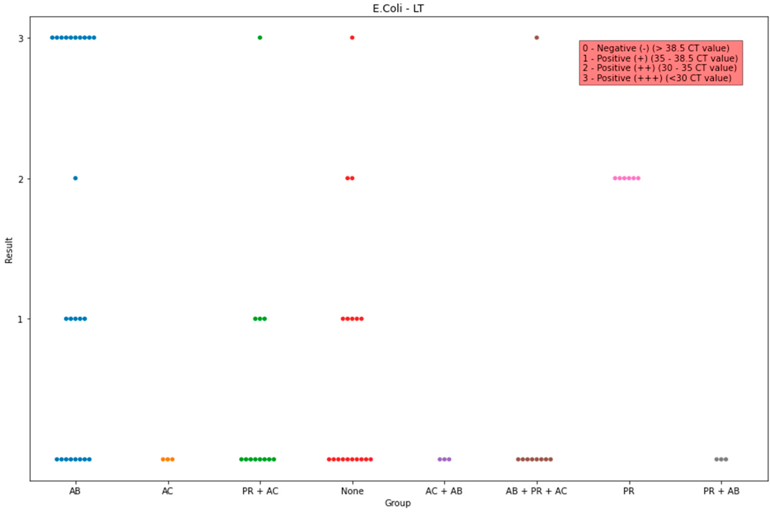
| Enterotoxin LT | 57.69% (15) | 44.88% (35) |
| F4 gene + Enterotoxin LT | 42.31% (11) | 25.64% (20) |
| F5 gene + Enterotoxin LT | 19.23% (5) | 16.67% (13) |
| F6 gene + Enterotoxin LT | 42.31% (11) | 32.05% (25) |
| β-toxin—C. perfringens type C | 0.00% (0) | 0.00% (0) |
| Toxin A—C. difficile | 84.62% (22) | 65.38% (51) |
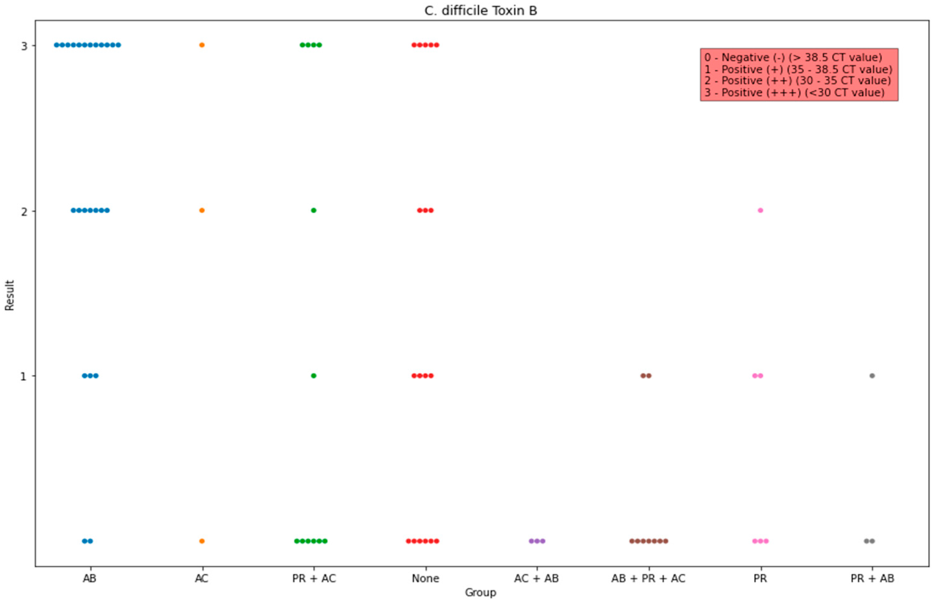
| Toxin B—C. difficile | 88.46% (23) | 61.54% (48) |
Disclaimer/Publisher’s Note: The statements, opinions and data contained in all publications are solely those of the individual author(s) and contributor(s) and not of MDPI and/or the editor(s). MDPI and/or the editor(s) disclaim responsibility for any injury to people or property resulting from any ideas, methods, instructions or products referred to in the content. |
© 2023 by the authors. Licensee MDPI, Basel, Switzerland. This article is an open access article distributed under the terms and conditions of the Creative Commons Attribution (CC BY) license (https://creativecommons.org/licenses/by/4.0/).
Share and Cite
Tsekouras, N.; Meletis, E.; Kostoulas, P.; Labronikou, G.; Athanasakopoulou, Z.; Christodoulopoulos, G.; Billinis, C.; Papatsiros, V.G. Detection of Enterotoxigenic Escherichia coli and Clostridia in the Aetiology of Neonatal Piglet Diarrhoea: Important Factors for Their Prevention. Life 2023, 13, 1092. https://0-doi-org.brum.beds.ac.uk/10.3390/life13051092
Tsekouras N, Meletis E, Kostoulas P, Labronikou G, Athanasakopoulou Z, Christodoulopoulos G, Billinis C, Papatsiros VG. Detection of Enterotoxigenic Escherichia coli and Clostridia in the Aetiology of Neonatal Piglet Diarrhoea: Important Factors for Their Prevention. Life. 2023; 13(5):1092. https://0-doi-org.brum.beds.ac.uk/10.3390/life13051092
Chicago/Turabian StyleTsekouras, Nikolaos, Eleftherios Meletis, Polychronis Kostoulas, Georgia Labronikou, Zoi Athanasakopoulou, Georgios Christodoulopoulos, Charalambos Billinis, and Vasileios G. Papatsiros. 2023. "Detection of Enterotoxigenic Escherichia coli and Clostridia in the Aetiology of Neonatal Piglet Diarrhoea: Important Factors for Their Prevention" Life 13, no. 5: 1092. https://0-doi-org.brum.beds.ac.uk/10.3390/life13051092