Article

Importance of Potential New Biomarkers in Patient with Serouse Ovarian Cancer

by
Aneta Cymbaluk-Płoska
1,*,
Karolina Chudecka
1,
Anita Chudecka-Głaz
1,
Katarzyna Piotrowska
2,
Sebastian Kwiatkowski
3 and
Maciej Tarnowski
2
1
Department and Clinic of Gynaecological Surgery and Gynaecological Oncology of Adults and Adolescents, Pomeranian Medical University, ul. Powstańców Wlkp. 72, 70-111 Szczecin, Poland
2
Department of Physiology, Pomeranian Medical University, Powstańców Wlkp. 72, 70-111 Szczecin, Poland
3
Department of Obstetrics and Gynaecology, Pomeranian Medical University, ul. Powstańców Wlkp. 72, 70-111 Szczecin, Poland
*
Author to whom correspondence should be addressed.
Submission received: 15 May 2021 / Revised: 28 May 2021 / Accepted: 29 May 2021 / Published: 3 June 2021
(This article belongs to the Collection Biomarkers in Medicine)

Abstract

:
Ovarian cancer remains the gynecological cancer with the highest mortality rate. In our study, we compare a number of proteins from different effector pathways to assess their usefulness in the diagnosis of ovarian cancer. The tissue expression of the tested proteins was assessed by two methods: qRT-PCR and an immunohistochemical analysis. A significantly higher level of mRNA expression was found in the ovarian cancer group for YAP and TEAD4 (p = 0.004 and p = 0.003, respectively). There was no statistical significance in the expression of mRNA for SMAD3, and there was borderline statistical significance for SMAD2 between the groups of ovarian cancer patients and other subgroups of patients with simple cysts and healthy ovarian tissue (p = 0.726 and p = 0.046, respectively). Significantly higher levels of transferrin receptor (CD71), H2A.X, and ADH1A gene expression were found in the ovarian cancer group compared to the control group for YAP, and TEAD4 showed strong nuclear and cytoplasmic staining in ovarian carcinoma and weak staining in non-carcinoma ovarian samples, ADH1A1 showed strong staining in the cytoplasm of carcinoma sections and a weak positive reaction in the non-carcinoma section, H2A.X showed strong positive nuclear staining in carcinoma sections and moderate positive staining in non-carcinoma samples, and CD71 showed moderate positive staining in carcinoma and non-carcinoma samples. YAP, TEAD4, and ADH1A proteins appear to be promising biomarkers in the diagnosis of ovarian cancer.

1. Introduction

Epithelial ovarian cancers (EOCs) constitute the majority of malignant ovarian cancers in adult women. The diagnosis of ovarian cancer has not improved over the years. Only a small percentage of patients visit a gynecologist at an early stage of the disease due to the lack of characteristic symptoms. The prognosis of patients with advanced disease is very serious.
Conventional therapies usually include surgery and chemotherapy (platinum and taxol-based, as well as radiotherapy). The literature includes many proteins with their expression assessed in ovarian cancer tissue and compared with healthy ovarian tissue. There are ongoing searches for proteins that could serve as diagnostic, prognostic, or predictive factors in the fight against this cancer. The expression of proteins involved in apoptosis, proliferation, DNA repair, and immune processes are of specific interest in ovarian cancer research.
Many current studies concern the role of SMAD-group proteins. SMADs are proteins that are signal transmitters and transcription modulators that mediate numerous signaling pathways. In ovarian cancer patients, mutations in the SMAD family of proteins are not common, but they have been found in cellular lines and primary cultures [1]. Inactivation of those proteins is associated with a more aggressive course of this type of tumor [2]. In previous studies, we found that the expression of the SMAD4 gene significantly differed in healthy patients compared to those with ovarian cancer [3]. It was also an independent predictive factor. Higher expression of the SMAD4 protein was associated with a shorter overall survival (OS) of patients with ovarian cancer.
The Hippo pathway is a recently discovered signal transduction pathway. YAP promotes cell proliferation, inhibits cell apoptosis, and also promotes the endodermal-mesenchymal transition (EMT) of cells [4]. YAP plays a key role in the development and progression of many types of cancer, including ovarian cancer. However, the impact of YAP on the in vivo development of ovarian cancer and its effects remains uncertain. Xiu et al. were the first to show that high levels of YAP expression were positively correlated with the expression of the TEAD4 gene [5].
TEAD binds to gene promoter sites, which are responsible for inhibiting apoptosis or promoting cell proliferation. Research confirmed that the YAP molecule is required for the full transcriptional activity of this protein, with the YAP molecule always co-precipitating with the TEAD factor [6,7]. Data in the literature confirmed the role of Hippo pathway components in the tumorigenesis of various types of tissues, also showing the complicated relations between Hippo and other functionally similar biochemical pathways [8].
Double-strand breakage (DSB) is one of the first procedures that initiates tumor formation under the influence of both endogenous and exogenous factors [9]. Several years ago, it was discovered that phosphorylation of the histone protein H2A.X in the serine 139 position (after phosphorylation, marked as γ-H2A.X) during DSB formations is important in the formation of a repair complex of double-strand DNA breaks. The increased expression of γ-H2A.X could potentially serve as a biomarker for the transformation of normal tissue into the pre-neoplastic condition and, consequently, neoplastic tissues [10].
ALDH1 is one of the isoenzymes of aldehyde dehydrogenase responsible for the oxidation of acetaldehyde. Its toxic and potentially carcinogenic effects have been described for many years. It has been suggested that neoplastic cells have a high ethanol oxidation capacity but a lower acetaldehyde removal capacity compared to normal tissues [11]. This further intensifies the proliferation process and disturbs the metabolism of some biologically important factors, e.g., retinoic acid. It has been suggested that the determination of changes in serum and tissue levels of ALDH1 isoenzymes could be useful in the diagnosis of some cancers [12].
The transferrin receptor (CD71) is present at a low level in almost all tissues in our body. CD71 is a type II receptor that is located on the outer cell membrane. It transports iron in the form of clarine or dynamin-dependent endosomes and then returns to the surface of the cell [13]. Despite the fact that CD71 is almost ubiquitous in our body, its expression is much stronger in cancerous tissues. In the case of some cancers, it is closely related to tumor advancement or differentiation [14].
Transketolase (TKT) is a key enzyme in the anaerobic pentose phosphate pathway. There are three isoenzymes: TKT, TKTL1, and TKTL2. All of the enzymes belonging to the transketolase family are found in normal tissues as well as in cancerous tissues. Increased glycolysis and the pentose phosphate pathway are characteristic for cancer cells to maintain these cells in homeostasis during oxidative stress and to quickly provide energy for fast division and, hence, for proliferation and metastasis [15,16].

Aim of the Study

We evaluated the differences in the expression of the YAP, TEAD4, SMAD2, SMAD3, H2A.X, ALD1A1, CD71, TKT, and TKTL1 in ovarian cancers and benign cysts. We evaluated the usefulness of YAP, TEAD4, SMAD2, SMAD3, H2A.X, ALD1A1, CD71, TKT and TKTL1 as diagnostic or prognostic markers in patients with ovarian cancer.

2. Materials and Methods

2.1. Tissue Specimens

The specimens were obtained from 79 women who were subject to surgeries in the years 2015–2016 in the Department of Surgical and Oncological Gynecology of the Pomeranian Medical University. The types of surgeries performed providing us with tissues included bilateral adnexectomy or hysterectomy with bilateral adnexectomy. Tissues stored in −70 °C and histopathological specimens that were prepared by fixation in 10% formalin were used in the study. After obtaining complete histopathological diagnoses, the collected specimens were divided into two groups: 41 benign ovarian lesions and 38 ovarian epithelial cancers. Patients with the BRCA 1 mutation, those diagnosed with borderline ovarian tumors, and those with non-epithelial tumors were excluded from the study. Patients with epithelial non-serous types of ovarian cancer were excluded from the study.
The study protocol was approved on 22-02-2011 by the Pomeranian University of Medicine Ethical Committee number: KB-0012/58/11.

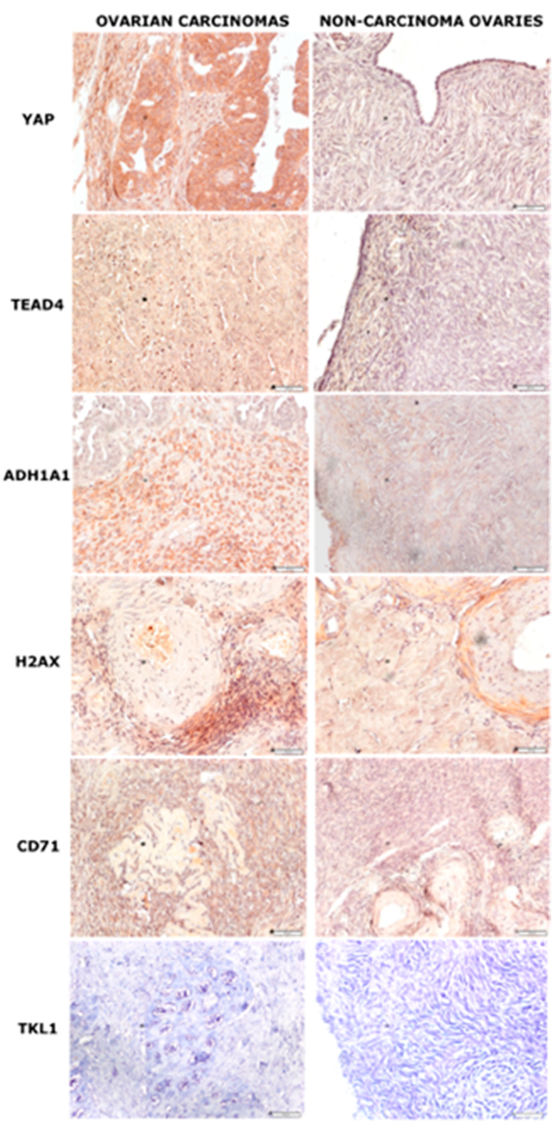
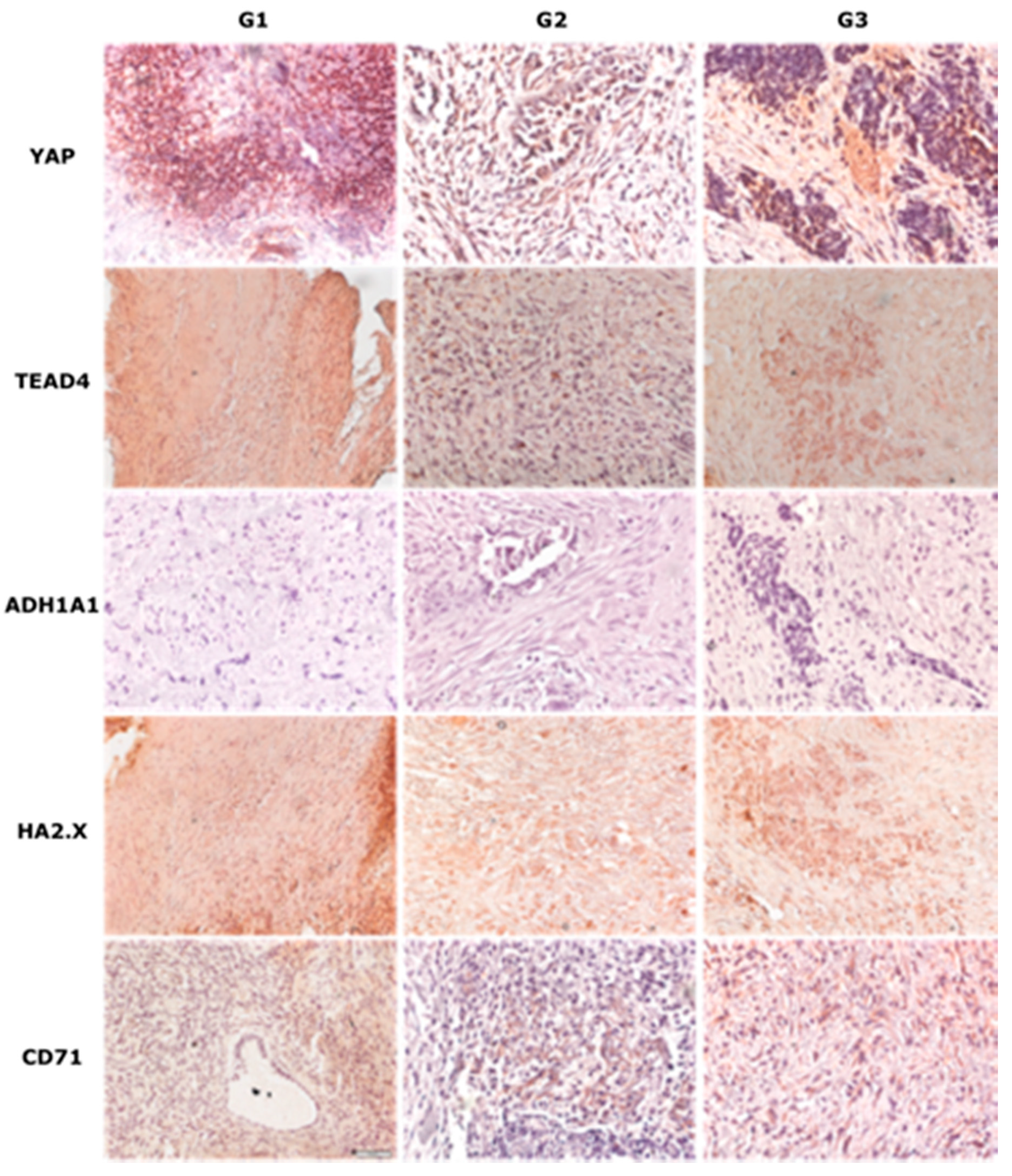
2.2. Immunohistochemical Analysis of Chosen Markers

Deparaffinized sections of ovaries (3 μM thick) were hydrated and heat epitope retrieval was performed using a microwave oven with retrieval solution buffer pH = 6 (DAKO, Dennmark). After cooling to room temperature (RT), the slides were incubated with 0.3% solution of H2O2, washed twice with PBS, and further incubated with 2.5% horse serum (Vector Laboratories, USA). After washing in PBS, the slides were incubated with primary antibodies: rabbit anti-human YAP (ProteinTech, Europe) and rabbit anti-human TEAD4 (Sigma-Aldrich, USA), rabbit anti-human ADH1A1 (ProteinTech, Europe), mouse anti-human CD71 (Invitrogen), and mouse anti-human H2A.X (Invitrogen) for 1h in RT.
After washing in PBS, immunoreactions were visualized with ImmPRESS UNIVERSAL REAGENT and Vector NovaRED Substrate KIT FOR PEROXIDASE (VECTOR LABORATORIES, USA) according to the manufacturer’s protocol. As a negative control, the primary antibody was replaced with PBS on the specimen. Positive staining was defined by visual identification of a yellow/brown pigmentation in the light microscope. Images were collected with an Olympus IX81 inverted microscope (Olympus, Germany) with a color camera and with CellSens image processing software (Olympus, Germany).

2.3. Quantitative Real Time Polymerase Chain Reaction (qRT-PCR)

Quantitative analysis of the mRNA expression of YAP1, SMAD2, SMAD3, CD71, H2A.X, TKTL1, ALDH1A1 and TKT genes was performed by two-step reverse transcription PCR. In our work, we examined the total protein expression. The total RNA was extracted from 50–100 mg tissue samples using a RNeasy Lipid Tissue Mini Kit (Qiagen). cDNA was prepared from 1 μg of total cellular RNA in 20 μL of reaction volume using a FirstStrand cDNA synthesis kit and oligo-dT primers (Fermentas).
Quantitative assessment of the mRNA levels was performed by real-time RT-PCR using an ABI 7500 Fast instrument with Power SYBR Green PCR Master Mix reagent (Applied Biosystems). The conditions were as follows: 95 °C (15 s), 40 cycles at 95 °C (15 s), and 60 °C (1 min). According to melting point analysis, only one PCR product was amplified under these conditions. Each sample was analyzed in two technical replicates, and mean Ct values were used for further analysis. The relative quantity of a target, normalized to the endogenous controls GAPDH gene as internal calibrators, was calculated as the fold difference (2^dCt) and further processed using statistical analysis. The data are presented as the tumor tissue absolute expression.

2.4. Statistical Analysis

In the first stage of statistical analysis, the compliance of all of the obtained results with a Gaussian distribution was verified using the Shapiro–Wilk test. Most of the examined parameters had a non-normal distribution. Descriptive statistical parameters (the arithmetic mean, standard deviation, median, minimum, and maximum value) were calculated. The parameters calculated using semi-quantitative scales are represented by the median, the minimum (min), and the maximum (max) values. The results of the RT-PCR analysis were compared for the study group (A) and the control group (B). The Mann–Whitney U test was used in the case of unrelated variables (non-parametric test for unrelated variables for two groups).
Fisher’s exact test was conducted in order to compare two positive groups and the correlations between the expression of studied proteins and histological types or clinical-pathological features. The Wilcoxon rank-sum test was used for comparison between two immunohistochemical staining point groups.

3. Results

The mean age of the studied patient population was 54.2 years. The age range of the patients was 44–73 years. No age differences were found in the studied groups of patients. These characteristics are presented in Table 1.
After receiving the result of the histopathological examination, two groups of patients were created: the study group and the control group. The study group included 37 patients diagnosed with papillary serous carcinoma. The control group consisted of 41 patients.
Two subgroups were isolated in the group of patients without ovarian cancer: 21 patients with healthy ovaries and 20 patients with simple ovarian cysts. Almost all patients with ovarian cancer who qualified for the study were at the high stage of the clinical development of cancer. The exact characteristics of the patients are shown in Table 2.

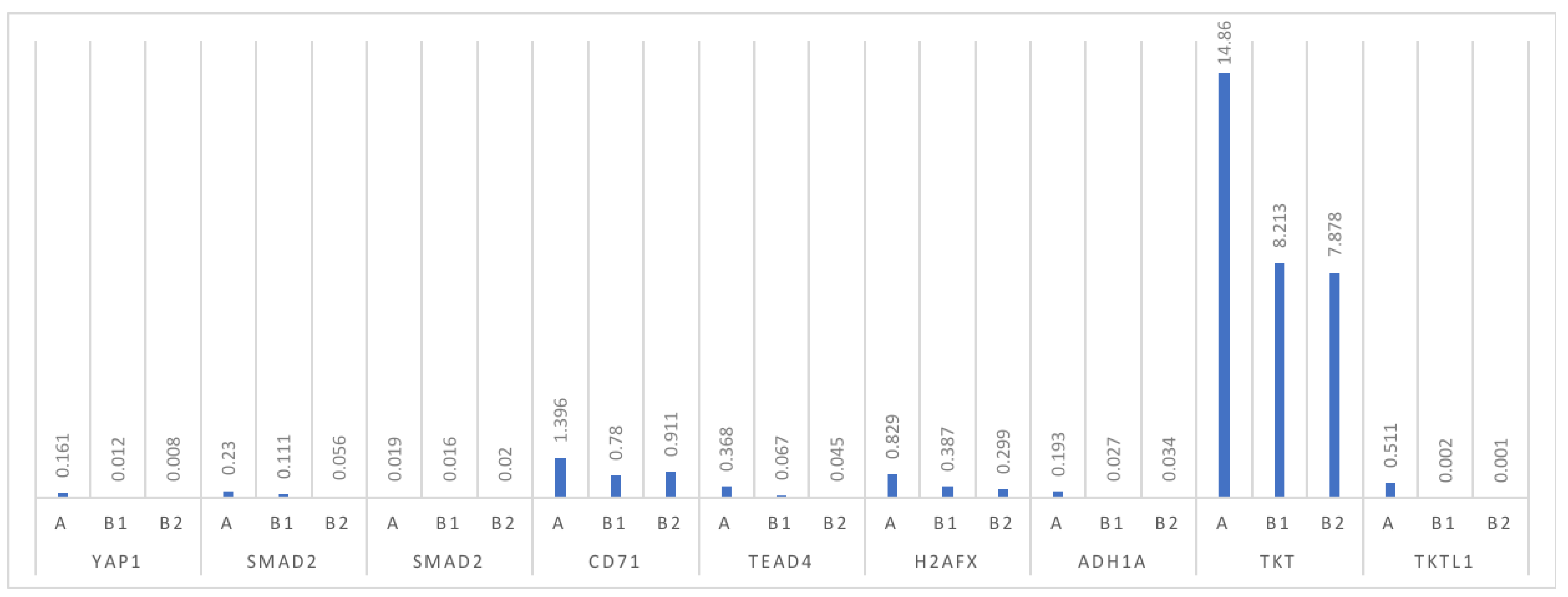
3.1. Analysis of mRNA Expression for Individual Proteins

A significantly higher statistically significant level of mRNA expression was found in the ovarian cancer group for YAP and TEAD4 (p = 0.003 and p = 0.006, respectively). There was no statistical significance in the expression of mRNA for SMAD3, and borderline statistical significance for SMAD2 between the groups of ovarian cancer patients and other subgroups of patients with simple cysts and healthy ovarian tissue (p = 0.709 and p = 0.053, respectively). Moreover, a statistically significantly higher level of mRNA expression was found in the ovarian cancer group compared to the control group for the transferrin receptor (CD71), H2A.X and ADH1A. On the other hand, the differences in the mRNA expression for TKT and TKTL1 were at a statistically significant level, with p = 0.003 and p = 0.016, respectively Table 3 and Table 4, Figure 1.
Immunohistochemical staining was performed on proteins that showed significant differences in expression during the qRT-PCR tests. We immunolocalized the YAP, TEAD4, ADH1A1, CD71, and H2A.X proteins in sections of ovarian tissue. YAP and TEAD4 showed strong nuclear and cytoplasmic staining in ovarian carcinoma and weak staining in non-carcinoma ovarian samples, ADH1A1 showed strong staining in the cytoplasm of carcinoma sections and a weak positive reaction in the non-carcinoma section, H2A.X showed strong positive nuclear staining in carcinoma sections and moderate positive staining in non-carcinoma samples, and CD71 showed moderate positive staining in carcinoma and non-carcinoma samples. The original magnification was 20× objective power, scale bar 50 μM (Figure 2, Table 5).
As shown in Table 6, we found no statistically significant differences in the expression of the tested proteins during the immunohistochemical test depending on age groups and grading (Figure 3). For two proteins, YAP and TEAD4 higher expression was found in the case of higher clinical advancement of the tumor and in the presence of metastases.

3.2. Proteins as a Prognostic Factors

After analysis with the univariate Cox regression model, the strongest relationship with progression-free survival (PFS) and overall survival (OS) was confirmed for grading (respectively: p = 0.049; p = 0.008). We found a statistically significant relationship between longer PFS and protein: YAP, TEAD4, SMAD2 and i concentration for the median (respectively: p = 0.031, p = 0.026, p = 0.048, p = 0.053, and p = 0.015). The relationship between longer overall survival and the YAP, TEAD4, ADH.1, CD71 and TKT median was also respectively significant (p = 0.02, p = 0.017, p = 0.043, p = 0.024, and p = 0.028).
In the multivariate analysis, we found a relationship with both the progression-free survival and overall survival for only one protein. The YAP concentration was determined for the median or 95th percentile and correlated respectively with PFS (p = 0.027/p = 0.046) and OS (p = 0.041/0.039) as shown in Table 7.

4. Discussion

In 2020, there were approximately 313,000 new cases of ovarian cancer and 207,000 deaths due to this disease worldwide. The majority (65–75%) of cases were diagnosed with stage III or IV advanced neoplastic disease according to FIGO. Despite the original good response to chemotherapy treatment in 90% of patients, only about 15% of them did not experience a relapse after several years [4]. The most common histopathological type of ovarian cancer is the serous one. The endometrial, clear cell, or mucinous type is less common. The biology of ovarian cancer is highly heterogeneous and remains unexplained.
The main goal of any ongoing research is to attempt to find markers that would allow the early detection of tumors and more effective treatment, as well as longer patient survival. In this paper, we compared, on both the mRNA and protein level, a number of proteins from different effector pathways involved in apoptosis, proliferation, DNA repair, and immune processes; the aim was to identify those that could be used in the diagnosis or prognosis of ovarian cancer.
One of the pathways that aroused our interest was the Hippo pathway [17], which is considered to be a pathway integrating extracellular and intracellular signals, resulting in the production and maintenance of the correct sizes of internal organs [18]. This pathway regulates organ size by controlling cell proliferation and apoptosis through the YAP protein, which is a transcription co-activator [19].
DNA damage or activation of the FAS death receptor leads to binding of the YAP protein during apoptosis [20]. In our study, the expression of YAP at both the mRNA and protein level was high in ovarian cancer tissue. The observed expression was low in benign cyst tissues. Similar reports were presented in studies by Yan Xia and Xio Wei, suggesting that the YAP protein may be directly associated with the development and progression of ovarian cancer [5,8,21].
The main elements of the Hippo pathway are kinases and adaptor proteins, which act by inhibiting the function of their nuclear effector—the YAP protein [6]. If the pathway ceases to be active, activated YAP migrates to the cell nucleus, where, along with the TEAD-family, transcription factors bind to gene promoter sites that are responsible for inhibiting apoptosis or promoting cell proliferation [22]. Attempts have been made to use the mechanisms of Hippo pathway action by blocking kinases in the regulation of the immune response in cancer immunotherapy [23].
Numerous literature data confirm the role of the components of the Hippo pathway, including both YAP and TEAD in tumorigenesis in various types of tissues: stomach, large intestine, breast, and esophagus [24,25,26]. In our study, we also confirmed the high expression of not only YAP but also TEAD at both the mRNA and protein level in ovarian cancer tissue. The clearly high expression in ovarian cancer tissue in both well-differentiated and poorly-differentiated cancers shows the possibility of using these proteins in an ovarian cancer diagnosis.
Another group of proteins with a prominent role in modern oncology is the SMAD-family proteins. These are responsible for transmitting the signal induced by TGF-beta to the cellular nucleus [27]. Changes to this pathway have been described in many diseases. The reduced expression of SMAD proteins has been observed in cardiovascular diseases, autoimmune diseases, Alzheimer′s disease, and osteoporosis [28]. Mutations of SMAD proteins have also been described in neoplastic diseases, mainly of epithelial origin from the gastrointestinal tract, lungs, and breasts [29].
The data on the role of SMAD proteins in the carcinogenesis of ovarian cancer appear to be inconclusive. Wang et al. noted that TGF-β1 inhibits ovarian tumor growth and improves sensitivity to chemotherapy by promoting the BRCA1/SMAD3 signal pathway [30]. Kennedy et al. emphasized the possibility of excessive proliferation in ovarian cancer caused by abnormalities in the TGF-β and SMAD4 signaling pathway. In vitro studies on cell lines demonstrated that the increased expression of SMAD4 inhibits the migration and proliferation of ovarian cancer cells. The authors suggested that SMAD4 may inhibit the invasion and metastases of human ovarian cancer cells through a pathway that is also mediated by TGF-β [7].
In their studies on SKOV3 and CaOV3 ovarian cancer cell lineages, Dunfield et al. showed the key role of TGF β/SMAD disorders but described SMAD protein roles as uncertain and requiring further studies [2]. In our study, we found no difference in the mRNA expression for SMAD3 and SMAD4 between ovarian cancer tissue and simple cyst tissue. However, we must consider that it is difficult to make a comparison between the patient′s complex body environment, in which cells are present with a relatively “simple” environment with no additional signals, such as with an in vitro culture. Wakahara et al. reported the inhibition of proliferation by SMAD in SKOV3 cells in unstimulated culture [31].

4.1. Selected Factors Inducing DNA Damage

Shigeta et al. presented transferrin as a new biomolecule inducing DNA damage. In our study, we decided to investigate the expression of the transferrin receptor (CD71) in ovarian cancer patients and the expression of phosphorylated H2A.X protein during DNA damage [32].
Double-strand DNA breaks (DSB) are one of the most dangerous forms of DNA damage in the body, leading to either apoptosis or the formation of chromosomal aberrations. The main mediator of H2A.X phosphorylation occurring as a result of double-strand DNA breaks is ATM kinase [33,34].
ATM activation and H2A.X phosphorylation are related to p53 protein function. The phosphorylation of p53 in the Ser15 position causes its direct bonding to the sites of DNA fracture, likely facilitating the DNA repair processes [35]. Cells containing the normal p53 protein usually have an elevated level of constitutive H2A.X phosphorylation compared to cells with a mutated form of this protein. This suggests that p53 facilitates H2A.X phosphorylation, which mobilizes DNA repair systems against the formation of DSB, thus, preventing oxidative damage to DNA [36].
The current prevailing theory of neoplastic lesion formation concerns p53 gene mutation. This mutation leads to pre-neoplastic lesions [37]. The presence of p53 protein mutations and changes in the number of H2A.X gene copies were found in breast cancer; changes in the H2A.X promoter were associated with a higher risk of breast cancer and non-Hodgkin′s lymphoma [38]. Elevated levels of endogenous H2A.X, similarly to other proteins involved in DDR (DNA′s damage repair) were observed in people in pre-neoplastic conditions of the breast [39].
Changes of the DNA damage repair type occur in the entire population of patients, regardless of the presence or absence of germinal mutations. In our study, we found a significant difference in the expression of mRNA for H2A.X, whereas the differences were not so apparent in the immunohistochemical examination of proteins in ovarian tissue. The expression in the ovarian cancer tissue was moderate, while the expression was much weaker in the benign cyst tissue.

4.2. The Effect of Transferrin

Transferrin, through the TRF1 receptor, may cause the formation of double-strand DNA breaks; it also inhibits H2A.X phosphorylation, which stops the helix repair system [32]. Transferrin is one of the main glycoproteins in human serum and is undoubtedly essential for survival. Nevertheless, periodic, recurring exposure to higher levels of transferrin can promote cancer development, including poorly-differentiated ovarian cancer [32].
Our studies confirmed differences in the transferrin receptor mRNA levels between ovarian cancer tissue and benign cyst tissue. However, the differences in the qualitative expression in IHC were no longer as pronounced. In future studies, we intend to investigate the differences in the expression of the H2A.X protein and receptor protein for transferrin in the ovarian tissue of patients with a BRCA1 mutation and healthy controls. Nearly all patients with a BRCA1 mutation develop G3 grade ovarian cancer with poor histopathological differentiation, where differences in the expression of the examined proteins should be even more pronounced.

4.3. Effect of Alcohol

Frequent alcohol consumption can lead to numerous tumors. In particular, these include gastrointestinal tumors of the liver, the pancreas, and large intestine. In our study, we evaluated the expression of class 1 alcohol dehydrogenase (ADH1A) in ovarian cancer tissue and benign and healthy ovary tissue. We found high expression at both the mRNA and protein level in the ovarian cancer tissue, with little or no expression in the benign cyst tissue and healthy ovarian tissue. Similar reports were presented by Orywal et al. [40].
They showed a significant expression of mRNA for ADH1A in ovarian cancer tissue compared to healthy ovarian tissue and benign cysts [41]. However, they noted that the activity of the enzyme responsible for ADH1A degradation was the same in ovarian cancer tissue as in healthy tissue. These results suggest an increased capacity to produce ADH1A by ovarian cancer cells. Acetaldehyde interferes with DNA synthesis and repair by inhibiting methylguanine methyltransferase (MGMT) [42]. The higher expression of ADH1A was described in tissues in other types of neoplastic diseases: colorectal, uterine, and cervical cancer [43], while Jelski et al. described low ADH1A expression in breast cancer [11].
A high expression of ALDH aldehyde dehydrogenase was described in breast cancer, with the foregoing expression correlating with a poor prognosis and the total survival time [44]. Significantly increased expression of ADH1A in ovarian cancer tissue, with disproportionately unincreased levels of aldehyde dehydrogenase (degradation enzyme), may lead to the increased production of acetaldehyde and the support of carcinogenesis in ovarian tissue [45].

4.4. Transketolase Family

We selected two proteins from the transketolase family: TKT and TKTL2. These proteins are thiamine-dependent enzymes and are considered to be key enzymes in the non-oxidant part of the pentose phosphate pathway. This pathway provides over 85% of the ribose for nucleic acid synthesis, including in neoplastic cells. In our study, the differences in mRNA expression were statistically significant for TKT and TKTL1.
Other publications on ovarian cancer confirmed the increased expression of these two proteins in ovarian cancer tissue compared to healthy tissue [46,47]. Studies conducted by Menghuang Zhao et al. not only showed the higher expression of TKT and TKTL1 in ovarian cancer but also the high mRNA expression of the aforementioned transketolase genes, which is associated with a shorter PFS in patients with high clinical grade tumors [48]. A close correlation of TKTL1 expression with the overall survival (OS) was also demonstrated in all ovarian cancer patients.
To summarize our study, in which we traced several pathways involved in cell apoptosis and proliferation, the most promising appeared to be the YAP and TEAD4 complex, as well as alcohol dehydrogenase. The remaining proteins require further studies and clarification of their applications.

5. Conclusions

The YAP, TEAD4, and ADH1A proteins appear to be promising biomarkers in the diagnosis of ovarian cancer. The H2A.X, CD71, SMAD3 and 4, TKT, and TKTL1 proteins require further studies to confirm their suitability for clinical use in ovarian cancer patients. The YAP protein may be considered as a good prognostic factor in patients with ovarian cancer.

Author Contributions

Conceptualization, A.C.-P.; validation, A.C.-P.; investigation, K.C.; resources, K.C.; software, A.C.-G.; writing—original draft preparation, K.P.; visualization, K.P.; formal analysis, S.K.; supervision, M.T.; QRT-PCR methodology, M.T. All authors have read and agreed to the published version of the manuscript.

Funding

This research was funded by statutory activity of the Pomeranian Medical University.

Institutional Review Board Statement

The study protocol was approved was approved on 22 February 2011 by the Pomeranian University of Medicine Ethical Committee number: KB-0012/58/11. Each patient had informed consent to participate in the study.

Informed Consent Statement

Written informed consent was obtained from the patients to publish this paper.

Data Availability Statement

The data sets used during current study are available from the corresponding author on reasonable request.

Conflicts of Interest

The authors declare no conflict of interest.

References

  1. Liu, L.; Liu, X.; Ren, X.; Tian, Y.; Chen, Z.; Xu, X.; Du, Y.; Jiang, C.; Fang, Y.; Liu, Z.; et al. Smad2 and Smad3 have differential sensitivity in relaying TGFβ signal-ing and inversely regulate early lineage specification. Sci. Rep. 2016, 6, 21602. [Google Scholar] [CrossRef] [PubMed] [Green Version]
  2. Roane, B.M.; Arend, R.C.; Birrer, M.J. Review: Targeting the Transforming Growth Factor-Beta Pathway in Ovarian Cancer. Cancers 2019, 11, 668. [Google Scholar] [CrossRef] [Green Version]
  3. Yang, J.; Wang, Y.; Zeng, Z.; Qiao, L.; Zhuang, L.; Gao, Q.; Ma, D.; Huang, X. Smad4 deletion in blood vessel endothelial cells promotes ovarian cancer metastasis. Int. J. Oncol. 2017, 50, 1693–1700. [Google Scholar] [CrossRef] [Green Version]
  4. Reid, B.M.; Permuth, J.B.; Sellers, T.A. Epidemiology of ovarian cancer: A review. Cancer Biol. Med. 2017, 14, 9–32. [Google Scholar] [CrossRef] [Green Version]
  5. Xia, Y.; Chang, T.; Wang, Y.; Liu, Y.; Li, W.; Li, M.; Fan, H.-Y. YAP Promotes Ovarian Cancer Cell Tumorigenesis and Is Indicative of a Poor Prognosis for Ovarian Cancer Patients. PLoS ONE 2014, 9, e91770. [Google Scholar] [CrossRef]
  6. Xie, Q.; Chen, J.; Feng, H.; Peng, S.; Adams, U.; Bai, Y.; Huang, L.; Li, J.; Huang, J.; Meng, S.; et al. YAP/TEAD–Mediated Transcription Controls Cellular Senescence. Cancer Res. 2013, 73, 3615–3624. [Google Scholar] [CrossRef] [Green Version]
  7. Yeh, K.-T.; Chen, T.-H.; Yang, H.-W.; Chou, J.-L.; Chen, L.-Y.; Yeh, C.-M.; Chen, Y.-H.; Lin, R.-I.; Su, H.-Y.; Chen, G.C.-W.; et al. Aberrant TGFβ/SMAD4 signaling contributes to epigenetic silencing of a putative tumor suppressor, RunX1T1, in ovarian cancer. Epigenetics 2011, 6, 727–739. [Google Scholar] [CrossRef] [Green Version]
  8. Zhang, X.; AOCS Study Group; George, J.M.; Deb, S.; Degoutin, J.L.; Takano, E.A.; Fox, S.B.; Bowtell, D.D.L.; Harvey, K.F. The Hippo pathway transcriptional co-activator, YAP, is an ovarian cancer oncogene. Oncogene 2011, 30, 2810–2822. [Google Scholar] [CrossRef] [Green Version]
  9. Turinetto, V.; Giachino, C. Multiple facets of histone variant H2AX: A DNA double-strand-break marker with several bio-logical functions. Nucleic Acids Res. 2015, 43, 2489–2498. [Google Scholar] [CrossRef] [Green Version]
  10. Fernandez-Capetillo, O.; Lee, A.; Nussenzweig, M.; Nussenzweig, A. H2AX: The histone guardian of the genome. DNA Repair 2004, 3, 959–967. [Google Scholar] [CrossRef] [PubMed] [Green Version]
  11. Jelski, W.; Chrostek, L.; Markiewicz, W.; Szmitkowski, M. Activity of alcohol dehydrogenase (adh) isoenzymes and aldehyde dehydrogenase (ALDH) in the sera of patients with breast cancer. J. Clin. Lab. Anal. 2006, 20, 105–108. [Google Scholar] [CrossRef]
  12. Althobiti, M.; El Ansari, R.; Aleskandarany, M.; Joseph, C.; Toss, M.S.; Green, A.R.; Rakha, E.A. The prognostic significance of ALDH1A1 expression in early invasive breast cancer. Histopathology 2020, 77, 437–448. [Google Scholar] [CrossRef]
  13. Shen, Y.; Li, X.; Dong, D.; Zhang, B.; Xue, Y.; Shang, P. Transferrin receptor 1 in cancer: A new sight for cancer therapy. Am. J. Cancer Res. 2018, 8, 916–931. [Google Scholar] [PubMed]
  14. Chan, K.T.; Choi, M.Y.; Lai, K.K.; Tan, W.; Tung, L.N.; Lam, H.Y.; Tong, D.K.; Lee, N.P.; Law, S. Overexpression of transferrin receptor CD71 and its tumorigenic properties in esophageal squamous cell carcinoma. Oncol. Rep. 2014, 31, 1296–1304. [Google Scholar] [CrossRef] [Green Version]
  15. Qin, Z.; Xiang, C.; Zhong, F.; Liu, Y.; Dong, Q.; Li, K.; Shi, W.; Ding, C.; Qin, L.; He, F. Transketolase (TKT) activity and nuclear localization promote hepatocellular carcinoma in a metabolic and a non-metabolic manner. J. Exp. Clin. Cancer Res. 2019, 38, 1–21. [Google Scholar] [CrossRef]
  16. Xu, I.M.-J.; Lai, R.K.-H.; Lin, S.-H.; Tse, A.P.-W.; Chiu, D.K.-C.; Koh, H.-Y.; Law, C.-T.; Wong, C.-M.; Cai, Z.; Wong, C.C.-L.; et al. Transketolase counteracts oxidative stress to drive cancer development. Proc. Natl. Acad. Sci. USA 2016, 113, E725–E734. [Google Scholar] [CrossRef] [Green Version]
  17. Chan, S.W.; Lim, C.J.; Chen, L.; Chong, Y.F.; Huang, C.; Song, H.; Hong, W. The hippo pathway in biological control and cancer development. J. Cell. Physiol. 2011, 226, 928–939. [Google Scholar] [CrossRef]
  18. Allegra, A.; Pioggia, G.; Innao, V.; Musolino, C.; Gangemi, S. New Insights into YES-Associated Protein Signaling Pathways in Hematological Malignancies: Diagnostic and Therapeutic Challenges. Cancers 2021, 13, 1981. [Google Scholar] [CrossRef]
  19. He, C.; Lv, X.; Hua, G.; Lele, S.M.; Remmenga, S.W.; Dong, J.; Davis, J.S.; Wang, C. YAP forms autocrine loops with the ERBB pathway to regulate ovarian cancer initiation and progression. Oncogene 2015, 34, 6040–6054. [Google Scholar] [CrossRef] [Green Version]
  20. Liu-Chittenden, Y.; Huang, B.; Shim, J.S.; Chen, Q.; Lee, S.-J.; Anders, R.A.; Liu, J.O.; Pan, D. Genetic and pharmacological disruption of the TEAD-YAP complex suppresses the oncogenic activity of YAP. Genes Dev. 2012, 26, 1300–1305. [Google Scholar] [CrossRef] [Green Version]
  21. Wei, X.; Jia, Y.; Lou, H.; Ma, J.; Huang, Q.; Meng, Y.; Sun, C.; Yang, Z.; Li, X.; Xu, S.; et al. Targeting YAP suppresses ovarian cancer progression through regulation of the PI3K/Akt/mTOR pathway. Oncol. Rep. 2019, 42, 2768–2776. [Google Scholar] [CrossRef]
  22. Kim, C.-L.; Choi, S.-H.; Mo, J.-S. Role of the Hippo Pathway in Fibrosis and Cancer. Cells 2019, 8, 468. [Google Scholar] [CrossRef] [Green Version]
  23. Moroishi, T.; Hayashi, T.; Pan, W.-W.; Fujita, Y.; Holt, M.V.; Qin, J.; Carson, D.A.; Guan, K.-L. The Hippo Pathway Kinases LATS1/2 Suppress Cancer Immunity. Cell 2016, 167, 1525–1539.e17. [Google Scholar] [CrossRef] [PubMed] [Green Version]
  24. Shen, J.; Cao, B.; Wang, Y.; Ma, C.; Zeng, Z.; Liu, L.; Li, X.; Tao, D.; Gong, J.; Xie, D. Hippo component YAP promotes focal adhesion and tumour aggressiveness via transcriptionally activating THBS1/FAK signalling in breast cancer. J. Exp. Clin. Cancer Res. 2018, 37, 175. [Google Scholar] [CrossRef] [PubMed]
  25. Li, K.; Guo, J.; Wu, Y.; Jin, D.; Jiang, H.; Liu, C.; Qin, C. Suppression of YAP by DDP disrupts colon tumor progression. Oncol. Rep. 2018, 39, 2114–2126. [Google Scholar] [CrossRef] [PubMed] [Green Version]
  26. Li, N.; Feng, Y.; Hu, Y.; He, C.; Xie, C.; Ouyang, Y.; Artim, S.C.; Huang, D.; Zhu, Y.; Luo, Z.; et al. Helicobacter pylori CagA promotes epithelial mesenchymal transition in gastric carcinogenesis via triggering oncogenic YAP pathway. J. Exp. Clin. Cancer Res. 2018, 37, 1–15. [Google Scholar] [CrossRef] [Green Version]
  27. Macias, M.J.; Martin-Malpartida, P.; Massagué, J. Structural determinants of Smad function in TGF-β signaling. Trends Biochem. Sci. 2015, 40, 296–308. [Google Scholar] [CrossRef] [Green Version]
  28. Pardali, E.; Dijke, P.T. TGFβ Signaling and Cardiovascular Diseases. Int. J. Biol. Sci. 2012, 8, 195–213. [Google Scholar] [CrossRef]
  29. Yu, W.; Chai, H. Inhibition of BAMBI reduces the viability and motility of colon cancer via activating TGF-β/Smad pathway in vitro and in vivo. Oncol. Lett. 2017, 14, 4793–4799. [Google Scholar] [CrossRef] [Green Version]
  30. Kennedy, B.A.; Deatherage, D.E.; Gu, F.; Tang, B.; Chan, M.W.Y.; Nephew, K.P.; Huang, T.H.-M.; Jin, V.X. ChIP-seq Defined Genome-Wide Map of TGFβ/SMAD4 Targets: Implications with Clinical Outcome of Ovarian Cancer. PLoS ONE 2011, 6, e22606. [Google Scholar] [CrossRef] [Green Version]
  31. Wakahara, K.; Kobayashi, H.; Yagyu, T.; Matsuzaki, H.; Kondo, T.; Kurita, N.; Sekino, H.; Inagaki, K.; Suzuki, M.; Kanayama, N.; et al. Transforming growth factor-?1-dependent activation of Smad2/3 and up-regulation of PAI-1 expression is negatively regulated by Src in SKOV-3 human ovarian cancer cells. J. Cell. Biochem. 2004, 93, 437–453. [Google Scholar] [CrossRef] [PubMed]
  32. Shigeta, S.; Toyoshima, M.; Kitatani, K.; Ishibashi, M.; Usui, T.; Yaegashi, N. Transferrin facilitates the formation of DNA double-strand breaks via transferrin receptor 1: The possible involvement of transferrin in carcinogenesis of high-grade serous ovarian cancer. Oncogene 2016, 35, 3577–3586. [Google Scholar] [CrossRef] [PubMed]
  33. Helt, C.E.; Cliby, W.A.; Keng, P.C.; Bambara, R.A.; O’Reilly, M.A. Ataxia Telangiectasia Mutated (ATM) and ATM and Rad3-related Protein Exhibit Selective Target Specificities in Response to Different Forms of DNA Damage. J. Biol. Chem. 2005, 280, 1186–1192. [Google Scholar] [CrossRef] [PubMed] [Green Version]
  34. Kastan, M.B. DNA Damage Responses: Mechanisms and Roles in Human Disease. Mol. Cancer Res. 2008, 6, 517–524. [Google Scholar] [CrossRef] [Green Version]
  35. Opławski, M.; Nowakowski, R.; Średnicka, A.; Ochnik, D.; Grabarek, B.; Boroń, D. Molecular Landscape of the Epithelial–Mesenchymal Transition in Endometrioid Endometrial Cancer. J. Clin. Med. 2021, 10, 1520. [Google Scholar] [CrossRef]
  36. Tanaka, T.; Halicka, H.D.; Huang, X.; Traganos, F.; Darzynkiewicz, Z. Constitutive Histone H2AX Phosphorylation and ATM Activation, the Reporters of DNA Damage by Endogenous Oxidants. Cell Cycle 2006, 5, 1940–1945. [Google Scholar] [CrossRef]
  37. Escargueil, A.E.; Soares, D.G.; Salvador, M.; Larsen, A.K.; Henriques, J.A.P. What histone code for DNA repair? Mutat. Res. Mutat. Res. 2008, 658, 259–270. [Google Scholar] [CrossRef]
  38. Novik, K.L.; Spinelli, J.J.; MacArthur, A.C.; Shumansky, K.; Sipahimalani, P.; Leach, S.; Lai, A.; Connors, J.M.; Gascoyne, R.D.; Gallagher, R.P.; et al. Genetic Variation in H2AFX Contributes to Risk of Non–Hodgkin Lymphoma. Cancer Epidemiol. Biomark. Prev. 2007, 16, 1098–1106. [Google Scholar] [CrossRef] [Green Version]
  39. Gorgoulis, V.G.; Vassiliou, L.-V.F.; Karakaidos, P.; Zacharatos, P.; Kotsinas, A.; Liloglou, T.; Venere, M.; Ditullio, R.A., Jr.; Kastrinakis, N.G.; Levy, B.; et al. Activation of the DNA damage checkpoint and genomic instability in human precancerous lesions. Nat. Cell Biol. 2005, 434, 907–913. [Google Scholar] [CrossRef] [PubMed]
  40. Orywal, K.; Jelski, W.; Zdrodowski, M.; Szmitkowski, M. The diagnostic value of alcohol dehydrogenase isoenzymes and aldehyde dehydrogenase measurement in the sera of patients with endometrial cancer. Anticancer Res. 2013, 33, 3725–3730. [Google Scholar]
  41. Orywal, K.; Jelski, W.; Zdrodowski, M.; Szmitkowski, M. The activity of class I, II, III and IV alcohol dehydrogenase isoenzymes and aldehyde dehydrogenase in ovarian cancer and ovarian cysts. Adv. Med. Sci. 2013, 58, 216–220. [Google Scholar] [CrossRef] [Green Version]
  42. Akçay, T.; Dinçer, Y.; Alademir, Z.; Aydınlı, K.; Arvas, M.; Demirkıran, F.; Kösebay, D. Significance of the O6-methylguanine-DNA methyltransferase and glutathione S-transferase activity in the sera of patients with malignant and benign ovarian tumors. Eur. J. Obstet. Gynecol. Reprod. Biol. 2005, 119, 108–113. [Google Scholar] [CrossRef] [PubMed]
  43. Orywal, K.; Szmitkowski, M. Alcohol dehydrogenase and aldehyde dehydrogenase in malignant neoplasms. Clin. Exp. Med. 2017, 17, 131–139. [Google Scholar] [CrossRef] [PubMed] [Green Version]
  44. Ginestier, C.; Hur, M.H.; Charafe-Jauffret, E.; Monville, F.; Dutcher, J.; Brown, M.; Jacquemier, J.; Viens, P.; Kleer, C.G.; Liu, S.; et al. ALDH1 Is a Marker of Normal and Malignant Human Mammary Stem Cells and a Predictor of Poor Clinical Outcome. Cell Stem Cell 2007, 1, 555–567. [Google Scholar] [CrossRef] [PubMed] [Green Version]
  45. Guo, E.; Wei, H.; Liao, X.; Xu, Y.; Li, S.; Zeng, X. Prognostic value of alcohol dehydrogenase mRNA expression in gastric cancer. Oncol. Lett. 2018, 15, 5505–5516. [Google Scholar] [CrossRef] [Green Version]
  46. Bery, A.; Leung, F.; Smith, C.R.; Diamandis, E.; Kulasingam, V. Deciphering the ovarian cancer ascites fluid peptidome. Clin. Proteom. 2014, 11, 13. [Google Scholar] [CrossRef] [Green Version]
  47. Ricciardelli, C.; Lokman, N.; Cheruvu, S.; Tan, I.A.; Ween, M.P.; Pyragius, C.E.; Ruszkiewicz, A.; Hoffmann, P.; Oehler, M.K. Transketolase is upregulated in metastatic peritoneal implants and promotes ovarian cancer cell proliferation. Clin. Exp. Metastasis 2015, 32, 441–455. [Google Scholar] [CrossRef]
  48. Zhao, M.; Ye, M.; Zhou, J.; Zhu, X. Prognostic values of transketolase family genes in ovarian cancer. Oncol. Lett. 2019, 18, 4845–4857. [Google Scholar] [CrossRef] [Green Version]
Figure 1. The expression distribution of the tested proteins.
Diagnostics 11 01026 g001
Figure 2. Immunohistochemical staining in ovarian carcinoma and non-carcinoma ovarian samples. Magnification: 20×, scale bar: 50 μM.
Diagnostics 11 01026 g002
Figure 3. Immunohistochemical examination of YAP, TEAD4, ALDH1A1, HA2.X, and CD71 on specimens in different stages of disease (G1–G3). Magnification: 20×, scale bar 50 μM.
Diagnostics 11 01026 g003
Table 1. The characteristics of each group and subgroup of patients.
Histopathology Typen PatientAge Mean (Years) (SD)p
Papillary serous carcinoma3863.8 (1.6–2.9)NS
Benign changes4150.3 (1.1–1.9)
Simple cysts2144.9 (0.7–1.8)NS
Normal ovary2058.2 (1.4–3.1)
NS: not significant.
Table 2. Descriptive characteristics of patients with ovarian cancer.
n
Patient
Age
Mean (Years) (SD)
Papillary serous adenocarcinoma3863.8 (2.1–3.0)
G1666.6 (1.7–3.2)
G21461.5 (2.8–4.1)
G31862.3 (2.0–2.9)
FIGO I–II260.2 (1.6–3.2)
FIGO III–IV3667.1 (2.4–3.4)
Table 3. Statistic parameters; group A: cancer and group B: non-cancer.
VariableStatistics Parameters
n PatientMean−95% CI+95% CIMedianMinimumMaximumStandard Deviationp
YAP1 group A410.1640.0540.2720.0480.0011.5610.3450.003
YAP1 group B400.0110.0050.0160.0050.0000.0910.0016
SMAD2 group A410.2320.0520.4130.0610.0013.3340.5790.053
SMAD2 group B400.0590.0010.1160.0190.0001.0000.171
SMAD3 group A410.0180.0110.0260.0100.0010.3510.092NS
SMAD3 group B400.081–0.0640.2250.0050.0000.1060.002
CD71 group A411.3390.7521.9250.3780.0177.5011.8800.048
CD71 group B400.8870.4031.3710.1670.0067.4121.533
TEAD4 group A410.3710.2000.5420.0880.0112.3510.5480.006
TEAD4 group B400.0610.0420.1710.0160.0000.9990.202
H2AX group A410.8260.4241.2170.2220.0135.8641.2580.023
H2AXgroup B400.5110.1950.8290.1100.0175.6711.002
ADH1A group A410.1840.0830.2840.0860.0011.7000.3130.003
ADH1A group B400.0390.0170.0610.0060.0000.3050.066
TKT group A4114.8619.24120.4977.5641.108162.11318.0660.003
TKT group B407.933–0.24116.0921.8420.0086.28025.885
TKTL1 group A410.508–0.0531.0710.0010.0006.6811.4110.016
TKTL1group B400.002–0.0010.0060.0000.0000.0510.018
NS: not significant. As many as 82 patients were qualified for immunohistochemical analysis.
Table 4. Statistic parameters; group B1: simple cyst and group B2: normal ovary.
VariableStatistics Parameters
n PatientMean−95% CI+95% CIMedianMinimumMaximumStandard Deviationp
YAP1 group B1210.0120.0210.0400.0090.0011.0020.021NS
YAP1 group B2200.0080.0240.0310.0060.0000.8930.009
SMAD2 group B1210.1110.0010.1160.0190.0001.0000.1710.04
SMAD2 group B2200.0560.0010.0780.0090.0001.0020.087
SMAD3 group B1210.0160.0120.0330.0100.0000.3110.078NS
SMAD3 group B2200.0200.0140.0610.0140.0010.4080.068
CD71 group B1210.7800.3561.2060.4230.0067.1121.432NS
CD71 group B2200.9110.4231.5230.6220.0087.0041.321
TEAD4 group B1210.0670.0420.1710.0160.0000.9990.202NS
TEAD4 group B2200.0450.0390.1210.0230.0021.0030.187
H2AX group B1210.3870.1090.9120.1100.0125.0760.976NS
H2AX group B2200.2990.1230.8670.1320.0015.2310.954
ADH1A group B1210.0270.0200.0490.0160.0000.2980.066NS
ADH1A group B2200.0340.0220.0610.0230.0010.3110.052
TKT group B1218.2130.87617.3441.9970.0097.24323.435NS
TKT group B2207.8780.76715.2311,8780.0137.00620.878
TKTL1 group B1210.0020.0010.0050.0000.0000.0510.018NS
TKTL1 group B2200.0010.0010.0060.0000.0000.0430.020
NS: not significant.
Table 5. Comparison of the tested proteins immunohistochemical expression.
Ovarian CancerNon Ovarian Cancerp
YAPPOS + + + POS -<0.05
NEG −NEG +
TEAD4POS +++POS −<0.05
NEG −xNEG +
ADH1A1POS +++POS +<0.5
NEG −NEG +
H2AXPOS ++POS ++NS
NEG +NEG +
CD71POS ++POS +NS
NEG +NEG +
TKL1POS +POS +NS
NEG +NEG −
NS: not significant.
Table 6. Comparison of the tested proteins immunohistochemical expression depending on grading and staging.
ProteinsAgepGradepFIGOpMetastasizedp
YAP<55NSG1NSI,II0.01Yes0.04
>55G3III,IVNo
TEAD4<55NSG1NSI,II0.01Yes0.02
>55G3III,IVNo
ADH1A1<55NSG1NSI,IINSYesNS
>55G3III,IVNo
H2AX<55NSG1NSI,IINSYesNS
>55G3III,IVNo
CD71<55NSG1NSI,IINSYesNS
>55G3III,IVNo
TKL1<55NSG1NSI,IINSYesNS
>55G3III,IVNo
NS: not significant.
Table 7. Univariant and multi variant analyses (Cox regression model).
Univariate Analysis (Cox Regression Model)
PFSOS
HR95% CIp-ValueHR95% CIp-Value
Age1.0190.78–1.220.0611.210.99–1.260.037
Grade 1 vs 32.041.68–2.430.0492.181.74–2.250.008
YAP median1.431.21–1.620.0311.401.33–1.480.02
TEAD4 median1.281.11–1.470.0261.090.97–1.300.017
SMAD2 median1.061.02–1.210.0481.161.04–1.27NS
SMAD3 median0.980.90–1.13NS0.890.72–0.99NS
H2A.X0.940.85–1.17NS0.970.76–1.08NS
ADH1.A1.231.07–1.32NS1.241.12–1.310.043
CD711.110.82–1.300.0531.371.18–1450.024
TKT1.080.88–1.210.0151.150.99–1.170.028
TKTL11.371.14–1.44NS1.261.18–1.38NS
Multivariate Analysis (Cox Regression Model)
PFSOS
HR95% CIp-ValueHR95% CIp-Value
YAP median1.221.06–1.310.0271.190.98–1.240.041
YAP 95 percentile1.491.36–1.590.0461.311.26–1.390.039
TEAD4 median1.100.95–1.270.0071.171.08–1.32NS
TEAD4 95 percentile1.341.26–1.43NS1.020.94–1.22NS
CD71 median1.211.11–1.360.0331.371.24–1.43NS
CD71 95 percentile1.080.96–1.21NS1.191.12–1.340.018
TKT median0.990.90–1.17NS1.070.97–1.22NS
TKT 95 percentile1.121.01–1.280.0041.281.13–1.32NS
NS: not significant.
Publisher’s Note: MDPI stays neutral with regard to jurisdictional claims in published maps and institutional affiliations.

