
A New Case of Herlyn–Werner–Wunderlich Syndrome: Uterine Didelphys with Unilateral Cervical Dysgenesis, Vaginal Agenesis, Cervical Distal Ureteral Remnant Fistula, Ureterocele, and Renal Agenesis in a Patient with Contralateral Multicystic Dysplastic Kidney


Abstract
:1. Introduction
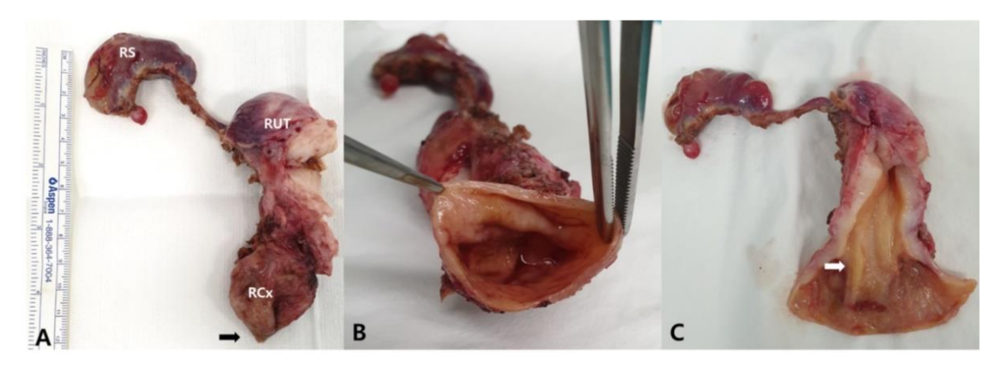
2. Case
3. Discussion
4. Conclusions
Author Contributions
Funding
Institutional Review Board Statement
Informed Consent Statement
Data Availability Statement
Conflicts of Interest
References
- Humphries, P.D.; Simpson, J.C.; Creighton, S.M.; Hall-Craggs, M.A. MRI in the assessment of congenital vaginal anomalies. Clin. Radiol. 2008, 63, 442–448. [Google Scholar] [CrossRef] [PubMed]
- Smith, N.A.; Laufer, M.R. Obstructed hemivagina and ipsilateral renal anomaly (OHVIRA) syndrome: Management and follow-up. Fertil. Steril. 2007, 87, 918–922. [Google Scholar] [CrossRef] [PubMed]
- Herlyn, U.; Werner, H. Das gemeinsame Vorkommen von effener Gartner-Gang-Zyste, gleichseitiger Nierenaplasie und Uterusdoppelmissbildung als typisches Missbildungssyndrom Simultaneous occurrence of an open Gartner-duct cyst, a homolateral aplasia of the kidney and a double uterus as a typical syndrome of abnormalities. Geburtshilfe Frauenheilkd. 1971, 31, 340–347. [Google Scholar] [PubMed]
- Zhang, H.; Ning, G.; Fu, C.; Bao, L.; Guo, Y. Herlyn-Werner-Wunderlich syndrome: Diverse presentations and diagnosis on MRI. Clin. Radiol. 2020, 75, 480.e17–480.e25. [Google Scholar] [CrossRef] [PubMed]
- Fedele, L.; Motta, F.; Frontino, G.; Restelli, E.; Bianchi, S. Double uterus with obstructed hemivagina and ipsilateral renal agenesis: Pelvic anatomic variants in 87 cases. Hum. Reprod. 2013, 28, 1580–1583. [Google Scholar] [CrossRef] [PubMed] [Green Version]
- Kiechl-Kohlendorfer, U.; Geley, T.; Maurer, K.; Gassner, I. Uterus didelphys with unilateral vaginal atresia: Multicystic dysplastic kidney is the precursor of “renal agenesis” and the key to early diagnosis of this genital anomaly. Pediatr. Radiol. 2011, 41, 1112–1116. [Google Scholar] [CrossRef] [PubMed]
- Grimbizis, G.F.; Tsalikis, T.; Mikos, T.; Papadopoulos, N.; Tarlatzis, B.C.; Bontis, J.N. Successful end-to-end cervico-cervical anastomosis in a patient with congenital cervical fragmentation: Case report. Hum. Reprod. 2004, 19, 1204–1210. [Google Scholar] [CrossRef] [PubMed] [Green Version]
- Acién, P.; Acién, M. The presentation and management of complex female genital malformations. Hum. Reprod. Update 2016, 22, 48–69. [Google Scholar] [CrossRef] [PubMed]
- Acién, P.; Susarte, F.; Romero, J.; Galán, J.; Mayol, M.J.; Quereda, F.J.; Sánchez-Ferrer, M. Complex genital malformation: Ectopic ureter ending in a supposed mesonephric duct in a woman with renal agenesis and ipsilateral blind hemivagina. Eur. J. Obs. Gynecol. Reprod. Biol. 2004, 117, 105–108. [Google Scholar] [CrossRef] [PubMed]
- Kriplani, A.; Kachhawa, G.; Awasthi, D.; Kulshrestha, V. Laparoscopic-assisted uterovaginal anastomosis in congenital atresia of uterine cervix: Follow-up study. J. Minim. Invasive Gynecol. 2012, 19, 477–484. [Google Scholar] [CrossRef] [PubMed]
- Berger, A.; Batzer, F.; Lev-Toaff, A.; Berry-Roberts, C. Diagnostic imaging modalities for Müllerian anomalies: The case for a new gold standard. J. Minim. Invasive Gynecol. 2014, 21, 335–345. [Google Scholar] [CrossRef] [PubMed]
- Ghi, T.; Casadio, P.; Kuleva, M.; Perrone, A.M.; Savelli, L.; Giunchi, S.; Meriggiola, M.C.; Gubbini, G.; Pilu, G.; Pelusi, C.; et al. Accuracy of three-dimensional ultrasound in diagnosis and classification of congenital uterine anomalies. Fertil. Steril. 2009, 92, 808–813. [Google Scholar] [CrossRef] [PubMed]
- Raga, F.; Bonilla-Musoles, F.; Blanes, J.; Osborne, N.G. Congenital Müllerian anomalies: Diagnostic accuracy of three-dimensional ultrasound. Fertil. Steril. 1996, 65, 523–528. [Google Scholar] [CrossRef]
- Kim, D.Y.; Nam, G.; Lee, S.R.; Kim, S.H.; Chae, H.D.; Kang, B.M. Congenital Obstructive Müllerian Anomaly: The Pitfalls of a Magnetic Resonance Imaging-Based Diagnosis and the Importance of Intraoperative Biopsy. J. Clin. Med. 2021, 10, 2414. [Google Scholar] [CrossRef] [PubMed]


Publisher’s Note: MDPI stays neutral with regard to jurisdictional claims in published maps and institutional affiliations. |
© 2021 by the authors. Licensee MDPI, Basel, Switzerland. This article is an open access article distributed under the terms and conditions of the Creative Commons Attribution (CC BY) license (https://creativecommons.org/licenses/by/4.0/).
Share and Cite
Yu, J.-H.; Lee, S.-R.; Choi, H.; Kim, K.-S.; Kang, B.-M. A New Case of Herlyn–Werner–Wunderlich Syndrome: Uterine Didelphys with Unilateral Cervical Dysgenesis, Vaginal Agenesis, Cervical Distal Ureteral Remnant Fistula, Ureterocele, and Renal Agenesis in a Patient with Contralateral Multicystic Dysplastic Kidney. Diagnostics 2022, 12, 83. https://0-doi-org.brum.beds.ac.uk/10.3390/diagnostics12010083
Yu J-H, Lee S-R, Choi H, Kim K-S, Kang B-M. A New Case of Herlyn–Werner–Wunderlich Syndrome: Uterine Didelphys with Unilateral Cervical Dysgenesis, Vaginal Agenesis, Cervical Distal Ureteral Remnant Fistula, Ureterocele, and Renal Agenesis in a Patient with Contralateral Multicystic Dysplastic Kidney. Diagnostics. 2022; 12(1):83. https://0-doi-org.brum.beds.ac.uk/10.3390/diagnostics12010083
Chicago/Turabian StyleYu, Jin-Hee, Sa-Ra Lee, Heayeon Choi, Kun-Suk Kim, and Byung-Moon Kang. 2022. "A New Case of Herlyn–Werner–Wunderlich Syndrome: Uterine Didelphys with Unilateral Cervical Dysgenesis, Vaginal Agenesis, Cervical Distal Ureteral Remnant Fistula, Ureterocele, and Renal Agenesis in a Patient with Contralateral Multicystic Dysplastic Kidney" Diagnostics 12, no. 1: 83. https://0-doi-org.brum.beds.ac.uk/10.3390/diagnostics12010083