
Synchronous Pancreatic Ductal Adenocarcinoma in the Head and Tail, a Double Trouble: A Case Report and Literature Review
 , , , , ,
Abstract
:1. Introduction
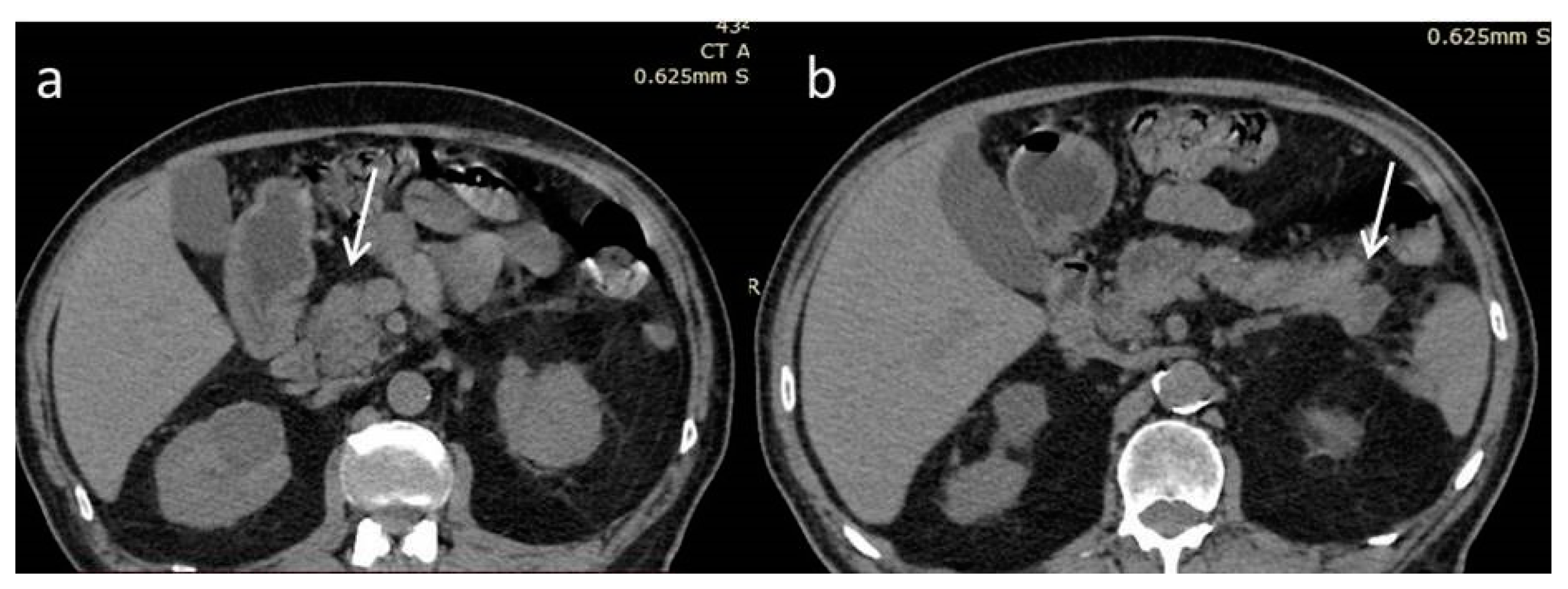
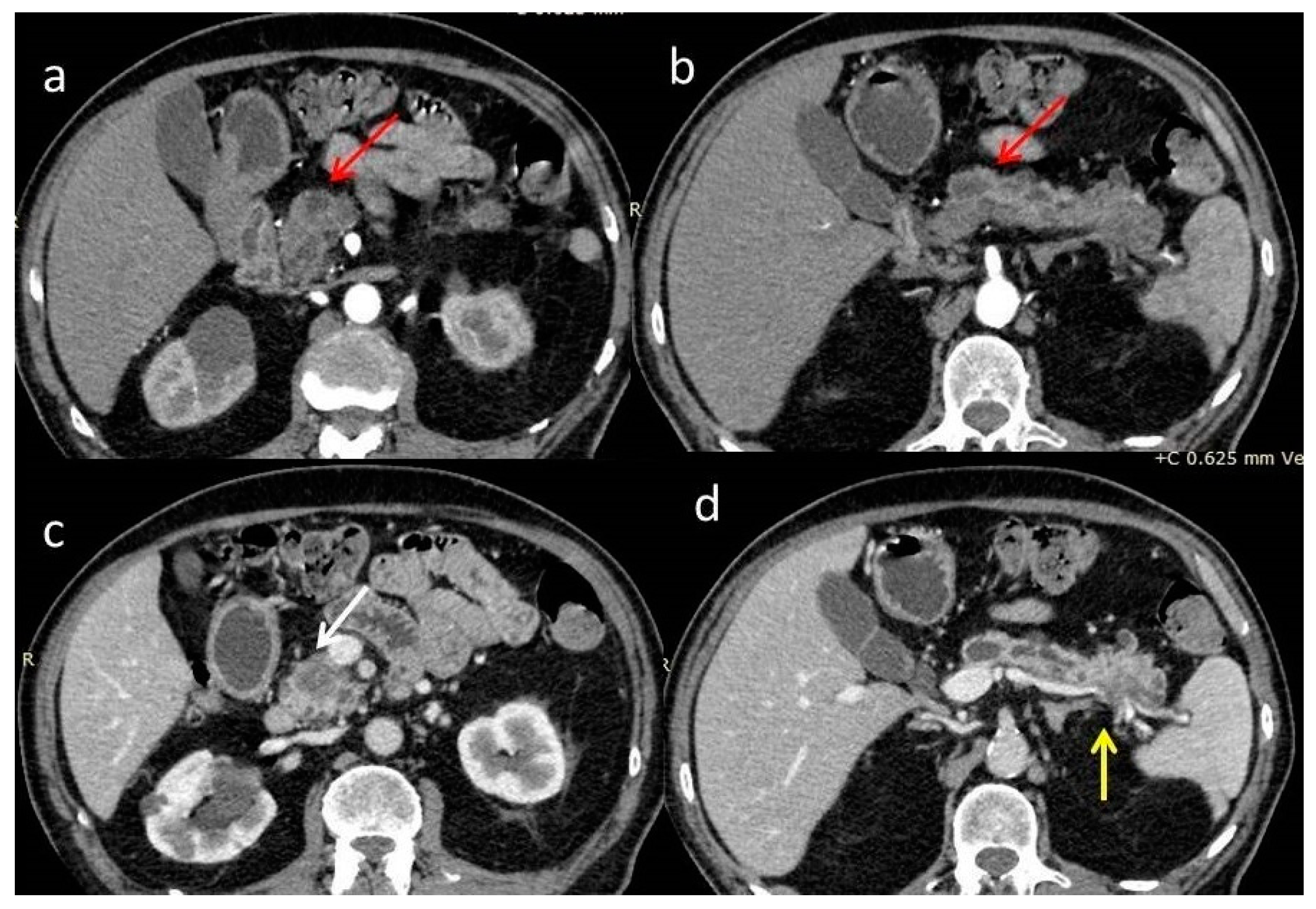
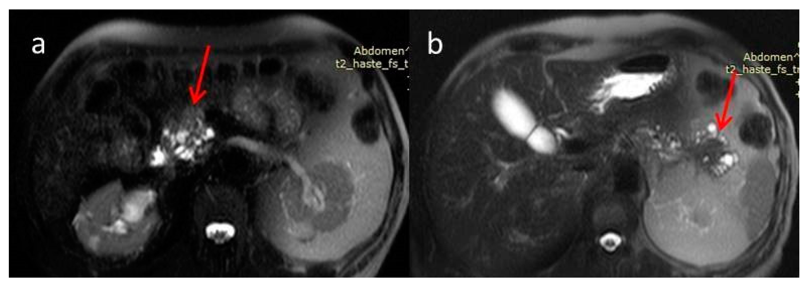
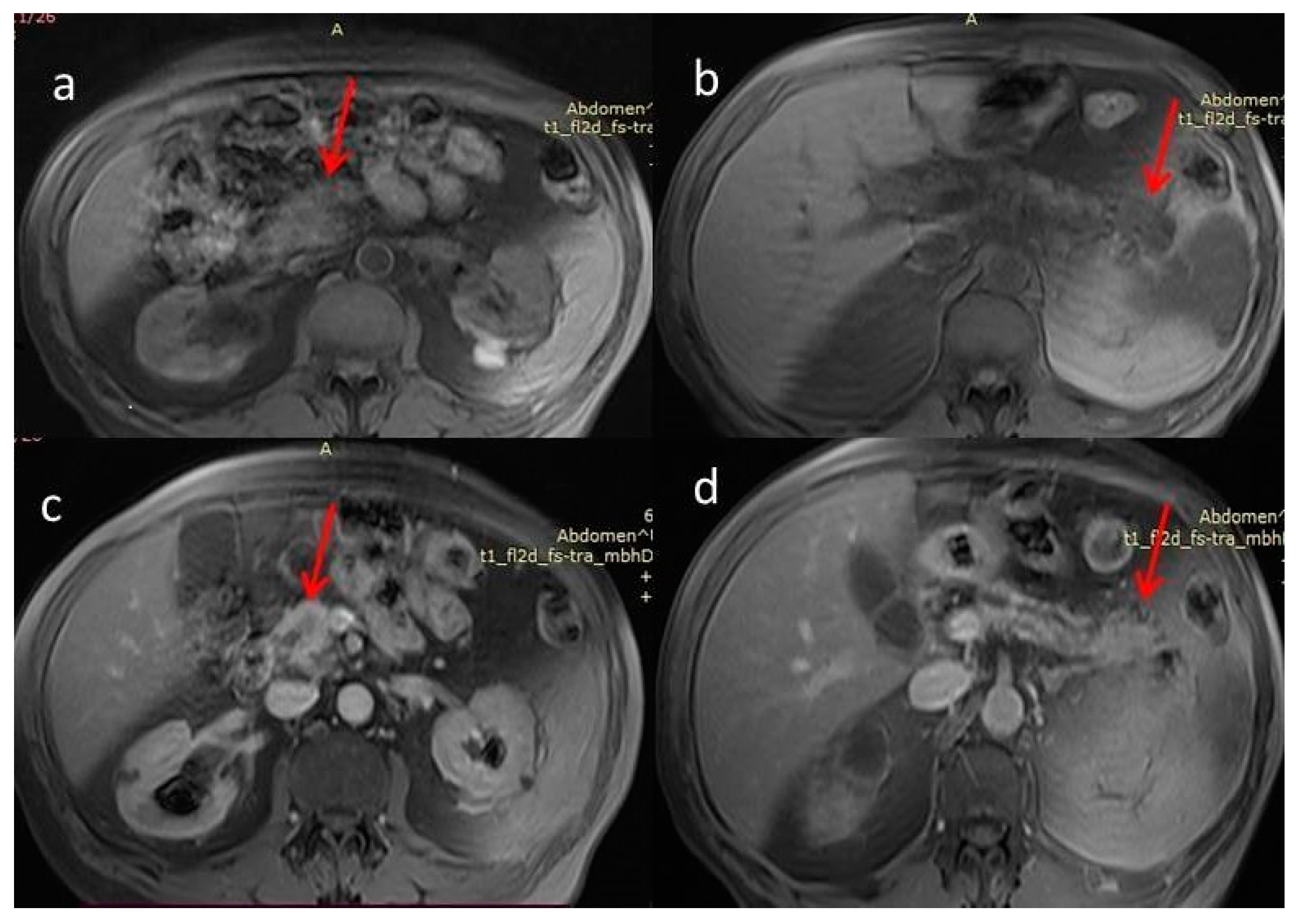
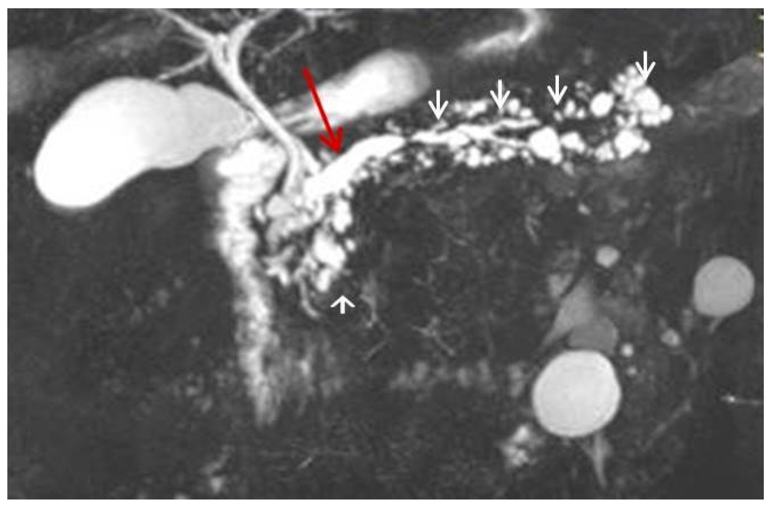
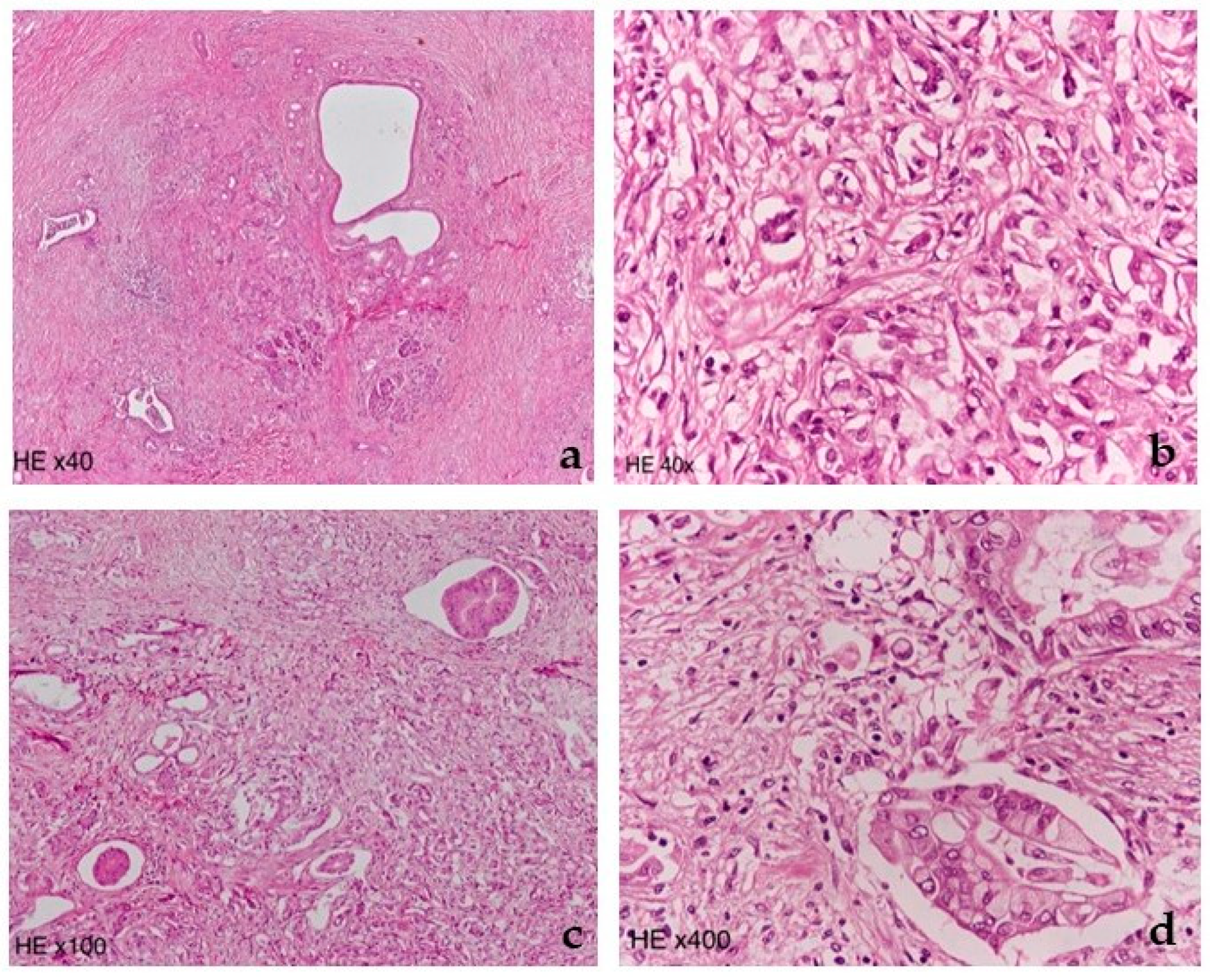
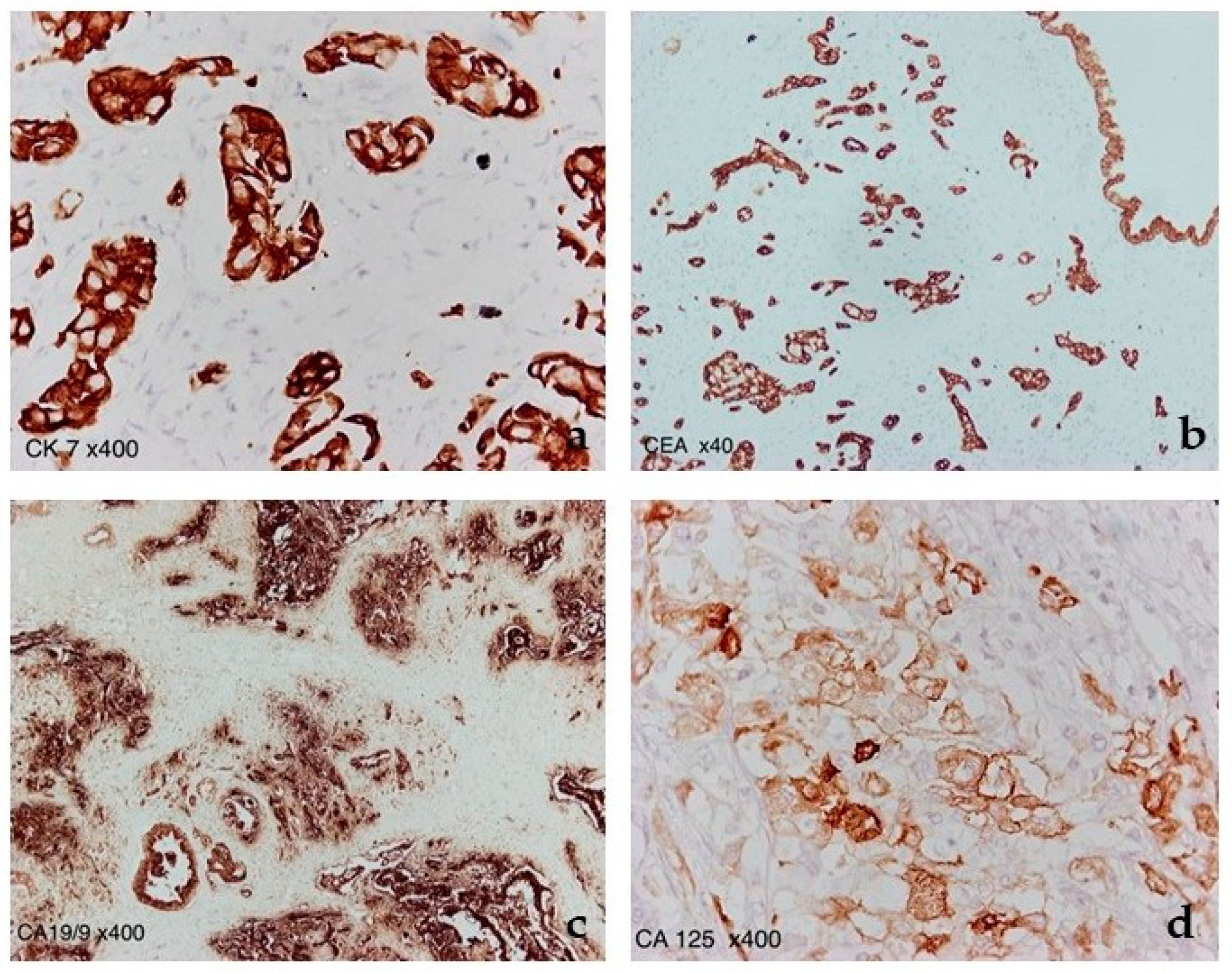
2. Case Report
3. Discussion
4. Conclusions
Author Contributions
Funding
Institutional Review Board Statement
Informed Consent Statement
Data Availability Statement
Conflicts of Interest
References
- National Cancer Institute Cancer Stat Facts: Pancreatic Cancer. Available online: https://seer.cancer.gov/statfacts/html/pancreas.html (accessed on 15 September 2022).
- Cancer Net Pancreatic Cancer: Statistics. Available online: https://www.cancer.net/cancer-types/pancreatic-cancer/statistics (accessed on 15 September 2022).
- Izumi, S.; Nakamura, S.; Mano, S.; Suzuka, I. Resection of four synchronous invasive ductal carcinomas in the pancreas head and body associated with pancreatic intraepithelial neoplasia: Report of a case. Surg. Today 2009, 39, 1091–1097. [Google Scholar] [CrossRef] [PubMed]
- Jiang, W.; Shen, Y.; Ding, Y.; Ye, C.; Zheng, Y.; Zhao, P.; Liu, L.; Tong, Z.; Zhou, L.; Sun, S.; et al. A naive Bayes algorithm for tissue origin diagnosis (TOD-Bayes) of synchronous multifocal tumors in the hepatobiliary and pancreatic system. Int. J. Cancer 2018, 142, 357–368. [Google Scholar] [CrossRef] [PubMed] [Green Version]
- Kim, H.J.; Park, M.H.; Shin, B. Double primary tumors of the pancreas: A case report. Medicine 2018, 97, e13616. [Google Scholar] [CrossRef]
- Rustagi, T.; Gleeson, F.C.; Chari, S.T.; Abu Dayyeh, B.K.; Farnell, M.B.; Iyer, P.G.; Kendrick, M.L.; Pearson, R.K.; Petersen, B.T.; Rajan, E.; et al. Endoscopic Ultrasound Fine-Needle Aspiration Diagnosis of Synchronous Primary Pancreatic Adenocarcinoma and Effects on Staging and Resectability. Clin. Gastroenterol. Hepatol. 2017, 15, 299–302. [Google Scholar] [CrossRef]
- Fujimori, N.; Nakamura, T.; Oono, T.; Igarashi, H.; Takahata, S.; Nakamura, M.; Tanaka, M.; Hayashi, A.; Aishima, S.; Ishigami, K.; et al. Adenocarcinoma involving the whole pancreas with multiple pancreatic masses. Intern. Med. 2010, 49, 1527–1532. [Google Scholar] [CrossRef] [PubMed] [Green Version]
- Ohike, N.; Norose, T.; Takano, Y.; Niiya, F.; Nagahama, M.; Matsuo, K.; Tanaka, K.; Furukawa, T. Resection of multiple invasive pancreatic ductal adenocarcinomas: A diagnostic dilemma distinguishing multicentric carcinogenesis from intrapancreatic metastasis. Pathol. Int. 2020, 70, 588–590. [Google Scholar] [CrossRef] [PubMed]
- Fujita, Y.; Matsuda, S.; Sasaki, Y.; Masugi, Y.; Kitago, M.; Yagi, H.; Abe, Y.; Shinoda, M.; Tokino, T.; Sakamoto, M.; et al. Pathogenesis of multiple pancreatic cancers involves multicentric carcinogenesis and intrapancreatic metastasis. Cancer Sci. 2020, 111, 739–748. [Google Scholar] [CrossRef] [Green Version]
- Imai, K.; Karasaki, H.; Ono, Y.; Sasajima, J.; Chiba, S.; Funakoshi, H.; Muraki, M.; Hanaoka, H.; Furukawa, T.; Furukawa, H.; et al. Metachronous pancreatic cancer originating from disseminated founder pancreatic intraductal neoplasias (PanINs). J. Pathol. Clin. Res. 2015, 1, 76–82. [Google Scholar] [CrossRef]
- WHO Classification of Tumours Editorial Board. Digestive System Tumours, 5th ed.; International Agency for Research on Cancer: Lyon, France, 2019; pp. 322–332. ISBN 978-928-324-499-8.
- Siegel, R.L.; Miller, K.D.; Jemal, A. Cancer statistics, 2018. CA Cancer J. Clin. 2018, 68, 7–30. [Google Scholar] [CrossRef]
- Tempero, M.A.; Malafa, M.P.; Al-Hawary, M.; Behrman, S.W.; Benson, A.B.; Cardin, D.B.; Chiorean, E.G.; Chung, V.; Czito, B.; Del Chiaro, M.; et al. Pancreatic Adenocarcinoma, Version 2.2021, NCCN Clinical Practice Guidelines in Oncology. J. Natl. Compr. Cancer Netw. JNCCN 2021, 19, 439–457. [Google Scholar] [CrossRef]
- Zakaria, A.; Al-Share, B.; Klapman, J.B.; Dam, A. The Role of Endoscopic Ultrasonography in the Diagnosis and Staging of Pancreatic Cancer. Cancers 2022, 14, 1373. [Google Scholar] [CrossRef] [PubMed]
- Vincent, A.; Herman, J.; Schulick, R.; Hruban, R.H.; Goggins, M. Pancreatic cancer. Lancet 2011, 378, 607–620. [Google Scholar] [CrossRef]
- Ren, B.; Liu, X.; Suriawinata, A.A. Pancreatic Ductal Adenocarcinoma and Its Precursor Lesions: Histopathology, Cytopathology, and Molecular Pathology. Am. J. Pathol. 2019, 189, 9–21. [Google Scholar] [CrossRef] [PubMed] [Green Version]
- Goong, H.J.; Moon, J.H.; Choi, H.J.; Lee, Y.N.; Choi, M.H.; Kim, H.K.; Lee, T.H.; Cha, S.W. Synchronous Pancreatic Ductal Adenocarcinomas Diagnosed by Endoscopic Ultrasound-Guided Fine Needle Biopsy. Gut Liver 2015, 9, 685–688. [Google Scholar] [CrossRef] [PubMed] [Green Version]
- van Erning, F.N.; Mackay, T.M.; van der Geest, L.G.M.; Groot Koerkamp, B.; van Laarhoven, H.W.M.; Bonsing, B.A.; Wilmink, J.W.; van Santvoort, H.C.; de Vos-Geelen, J.; van Eijck, C.H.J.; et al. Association of the location of pancreatic ductal adenocarcinoma (head, body, tail) with tumor stage, treatment, and survival: A population-based analysis. Acta Oncol. 2018, 57, 1655–1662. [Google Scholar] [CrossRef] [PubMed]
- Ling, Q.; Xu, X.; Zheng, S.S.; Kalthoff, H. The diversity between pancreatic head and body/tail cancers: Clinical parameters and in vitro models. Hepatobiliary Pancreat. Dis. Int. 2013, 12, 480–487. [Google Scholar] [CrossRef]
- Elsherif, S.B.; Virarkar, M.; Javadi, S.; Ibarra-Rovira, J.J.; Tamm, E.P.; Bhosale, P.R. Pancreatitis and PDAC: Association and differentiation. Abdom. Radiol. 2020, 45, 1324–1337. [Google Scholar] [CrossRef]
- Birnbaum, D.J.; Bertucci, F.; Finetti, P.; Birnbaum, D.; Mamessier, E. Head and Body/Tail Pancreatic Carcinomas Are Not the Same Tumors. Cancers 2019, 11, 497. [Google Scholar] [CrossRef] [PubMed] [Green Version]
- Dreyer, S.B.; Jamieson, N.B.; Upstill-Goddard, R.; Bailey, P.J.; McKay, C.J.; Australian Pancreatic Cancer Genome Initiative; Biankin, A.V.; Chang, D.K. Defining the molecular pathology of pancreatic body and tail adenocarcinoma. J. Br. Surg. 2018, 105, e183–e191. [Google Scholar] [CrossRef] [Green Version]
- Winer, L.K.; Dhar, V.K.; Wima, K.; Morris, M.C.; Lee, T.C.; Shah, S.A.; Ahmad, S.A.; Patel, S.H. The Impact of Tumor Location on Resection and Survival for Pancreatic Ductal Adenocarcinoma. J. Surg. Res. 2019, 239, 60–66. [Google Scholar] [CrossRef] [PubMed]
- Malleo, G.; Maggino, L.; Ferrone, C.R.; Marchegiani, G.; Luchini, C.; Mino-Kenudson, M.; Paiella, S.; Qadan, M.; Scarpa, A.; Lillemoe, K.D.; et al. Does Site Matter? Impact of Tumor Location on Pathologic Characteristics, Recurrence, and Survival of Resected Pancreatic Ductal Adenocarcinoma. Ann. Surg. Oncol. 2020, 27, 3898–3912. [Google Scholar] [CrossRef] [PubMed]
- Sung, M.K.; Park, Y.; Kwak, B.J.; Jun, E.; Lee, W.; Song, K.B.; Lee, J.H.; Hwang, D.W.; Kim, S.C. Comparison of Characteristics and Survival Rates of Resectable Pancreatic Ductal Adenocarcinoma according to Tumor Location. Biomedicines 2021, 9, 1706. [Google Scholar] [CrossRef] [PubMed]
- Pompella, L.; Tirino, G.; Pappalardo, A.; Caterino, M.; Ventriglia, A.; Nacca, V.; Orditura, M.; Ciardiello, F.; De Vita, F. Pancreatic Cancer Molecular Classifications: From Bulk Genomics to Single Cell Analysis. Int. J. Mol. Sci. 2020, 21, 2814. [Google Scholar] [CrossRef] [PubMed] [Green Version]
- Birnbaum, D.J.; Finetti, P.; Birnbaum, D.; Mamessier, E.; Bertucci, F. Validation and comparison of the molecular classifications of pancreatic carcinomas. Mol. Cancer 2017, 16, 168. [Google Scholar] [CrossRef] [Green Version]
- Scarà, S.; Bottoni, P.; Scatena, R. CA 19-9: Biochemical and Clinical Aspects. Adv. Exp. Med. Biol. 2015, 867, 247–260. [Google Scholar] [CrossRef] [PubMed]
- Winter, K.; Talar-Wojnarowska, R.; Dąbrowski, A.; Degowska, M.; Durlik, M.; Gąsiorowska, A.; Głuszek, S.; Jurkowska, G.; Kaczka, A.; Lampe, P.; et al. Diagnostic and therapeutic recommendations in pancreatic ductal adenocarcinoma. Recommendations of the Working Group of the Polish Pancreatic Club. Prz. Gastroenterol. 2019, 14, 1–18. [Google Scholar] [CrossRef]
- Pietryga, J.A.; Morgan, D.E. Imaging preoperatively for pancreatic adenocarcinoma. J. Gastrointest. Oncol. 2015, 6, 343–357. [Google Scholar] [CrossRef] [PubMed]
- Choi, S.Y.; Kim, Y.K.; Min, J.H.; Cha, D.I.; Jeong, W.K.; Lee, W.J. The value of gadoxetic acid-enhanced MRI for differentiation between hepatic microabscesses and metastases in patients with periampullary cancer. Eur. Radiol. 2017, 27, 4383–4393. [Google Scholar] [CrossRef] [PubMed]
- Motosugi, U.; Ichikawa, T.; Morisaka, H.; Sou, H.; Muhi, A.; Kimura, K.; Sano, K.; Araki, T. Detection of pancreatic carcinoma and liver metastases with gadoxetic acid-enhanced MR imaging: Comparison with contrast-enhanced multi-detector row CT. Radiology 2011, 260, 446–453. [Google Scholar] [CrossRef]
- Lami, G.; Biagini, M.R.; Galli, A. Endoscopic ultrasonography for surveillance of individuals at high risk for pancreatic cancer. World J. Gastrointest. Endosc. 2014, 6, 272–285. [Google Scholar] [CrossRef]
- Kojima, H.; Kitago, M.; Iwasaki, E.; Masugi, Y.; Matsusaka, Y.; Yagi, H.; Abe, Y.; Hasegawa, Y.; Hori, S.; Tanaka, M.; et al. Peritoneal dissemination of pancreatic cancer caused by endoscopic ultrasound-guided fine needle aspiration: A case report and literature review. World J. Gastroenterol. 2021, 27, 294–304. [Google Scholar] [CrossRef] [PubMed]
- Aloraini, A.M.; Helmi, H.A.; Aljomah, N.A.; Zubaidi, A.M. Multiple primary gastrointestinal tumors of gastric, pancreatic and rectal origin; a case report. Int. J. Surg. Case Rep. 2021, 89, 106610. [Google Scholar] [CrossRef] [PubMed]
- Shin, S.J.; Park, H.; Sung, Y.N.; Yoo, C.; Hwang, D.W.; Park, J.H.; Kim, K.P.; Lee, S.S.; Ryoo, B.Y.; Seo, D.W.; et al. Prognosis of Pancreatic Cancer Patients with Synchronous or Metachronous Malignancies from Other Organs Is Better than Those with Pancreatic Cancer Only. Cancer Res. Treat. 2018, 50, 1175–1185. [Google Scholar] [CrossRef] [PubMed] [Green Version]
- Brune, K.; Abe, T.; Canto, M.; O’Malley, L.; Klein, A.P.; Maitra, A.; Volkan Adsay, N.; Fishman, E.K.; Cameron, J.L.; Yeo, C.J.; et al. Multifocal neoplastic precursor lesions associated with lobular atrophy of the pancreas in patients having a strong family history of pancreatic cancer. Am. J. Surg. Pathol. 2006, 30, 1067–1076. [Google Scholar]
- Ikegawa, T.; Masuda, A.; Sakai, A.; Toyama, H.; Zen, Y.; Sofue, K.; Nakagawa, T.; Shiomi, H.; Takenaka, M.; Kobayashi, T.; et al. Multifocal cysts and incidence of pancreatic cancer concomitant with intraductal papillary mucinous neoplasm. Pancreatol. Off. J. Int. Assoc. Pancreatol. 2018, 18, 399–406. [Google Scholar] [CrossRef]
- Rotzinger, R.; Bläker, H.; Bahra, M.; Denecke, T.; Grieser, C. CT and MRI Findings of Autoimmune Polymorph Bifocal Pancreatitis Mimicking Pancreatic Adenocarcinoma: A Case Report and Review of the Literature. J. Investig. Med. High Impact Case Rep. 2015, 3, 2324709615576988. [Google Scholar] [CrossRef] [Green Version]
- Suzumura, K.; Hatano, E.; Uyama, N.; Okada, T.; Asano, Y.; Hai, S.; Nakasho, K.; Fujimoto, J. Multifocal Mass Lesions in Autoimmune Pancreatitis. Case Rep. Gastroenterol. 2017, 11, 678–685. [Google Scholar] [CrossRef] [Green Version]
- Chari, S.T.; Leibson, C.L.; Rabe, K.G.; Timmons, L.J.; Ransom, J.; de Andrade, M.; Petersen, G.M. Pancreatic cancer-associated diabetes mellitus: Prevalence and temporal association with diagnosis of cancer. Gastroenterology 2008, 134, 95–101. [Google Scholar] [CrossRef] [Green Version]
- Hart, P.A.; Bellin, M.D.; Andersen, D.K.; Bradley, D.; Cruz-Monserrate, Z.; Forsmark, C.E.; Goodarzi, M.O.; Habtezion, A.; Korc, M.; Kudva, Y.C.; et al. Type 3c (pancreatogenic) diabetes mellitus secondary to chronic pancreatitis and pancreatic cancer. Lancet Gastroenterol. Hepatol. 2016, 1, 226–237. [Google Scholar] [CrossRef] [Green Version]
- Scholten, L.; Mungroop, T.H.; Haijtink, S.; Issa, Y.; van Rijssen, L.B.; Koerkamp, B.G.; van Eijck, C.H.; Busch, O.R.; DeVries, J.H.; Besselink, M.G. New-onset diabetes after pancreatoduodenectomy: A systematic review and meta-analysis. Surgery 2018, 164, 6–16. [Google Scholar] [CrossRef]
- Lv, X.; Qiao, W.; Leng, Y.; Wu, L.; Zhou, Y. Impact of diabetes mellitus on clinical outcomes of pancreatic cancer after surgical resection: A systematic review and meta-analysis. PLoS ONE 2017, 12, e0171370. [Google Scholar] [CrossRef] [PubMed]
- Shingyoji, A.; Mikata, R.; Ogasawara, S.; Kusakabe, Y.; Yasui, S.; Sugiyama, H.; Ohno, I.; Kato, J.; Takano, S.; Yoshitomi, H.; et al. Diverse transitions in diabetes status during the clinical course of patients with resectable pancreatic cancer. Jpn. J. Clin. Oncol. 2020, 50, 1403–1411. [Google Scholar] [CrossRef] [PubMed]
- Roy, A.; Sahoo, J.; Kamalanathan, S.; Naik, D.; Mohan, P.; Kalayarasan, R. Diabetes and pancreatic cancer: Exploring the two-way traffic. World J. Gastroenterol. 2021, 27, 4939–4962. [Google Scholar] [CrossRef]
- Andersen, D.K.; Korc, M.; Petersen, G.M.; Eibl, G.; Li, D.; Rickels, M.R.; Chari, S.T.; Abbruzzese, J.L. Diabetes, Pancreatogenic Diabetes, and Pancreatic Cancer. Diabetes 2017, 66, 1103–1110. [Google Scholar] [CrossRef] [PubMed] [Green Version]
- Badowska-Kozakiewicz, A.; Fudalej, M.; Kwaśniewska, D.; Durlik, M.; Nasierowska-Guttmejer, A.; Mormul, A.; Włoszek, E.; Czerw, A.; Banaś, T.; Deptała, A. Diabetes Mellitus and Pancreatic Ductal Adenocarcinoma-Prevalence, Clinicopathological Variables, and Clinical Outcomes. Cancers 2022, 14, 2840. [Google Scholar] [CrossRef]
- Lu, R.; Yang, J.; Wei, R.; Ke, J.; Tian, Q.; Yu, F.; Liu, J.; Zhang, J.; Hong, T. Synergistic anti-tumor effects of liraglutide with metformin on pancreatic cancer cells. PLoS ONE 2018, 13, e0198938. [Google Scholar] [CrossRef]
- Chen, K.; Qian, W.; Jiang, Z.; Cheng, L.; Li, J.; Sun, L.; Zhou, C.; Gao, L.; Lei, M.; Yan, B.; et al. Metformin suppresses cancer initiation and progression in genetic mouse models of pancreatic cancer. Mol. Cancer 2017, 16, 131. [Google Scholar] [CrossRef] [Green Version]
- Pircher, A.; Zieher, M.; Eigentler, A.; Pichler, R.; Schäfer, G.; Fritz, J.; Puhr, M.; Steiner, E.; Horninger, W.; Klocker, H.; et al. Antidiabetic drugs influence molecular mechanisms in prostate cancer. Cancer Biol. Ther. 2018, 19, 1153–1161. [Google Scholar] [CrossRef]
- Izumo, W.; Higuchi, R.; Furukawa, T.; Yazawa, T.; Uemura, S.; Shiihara, M.; Yamamoto, M. Evaluation of preoperative prognostic factors in patients with resectable pancreatic ductal adenocarcinoma. Scand. J. Gastroenterol. 2019, 54, 780–786. [Google Scholar] [CrossRef]
- Mackay, T.M.; van Erning, F.N.; van der Geest, L.; de Groot, J.; Haj Mohammad, N.; Lemmens, V.E.; van Laarhoven, H.W.; Besselink, M.G.; Wilmink, J.W.; Dutch Pancreatic Cancer Group. Association between primary origin (head, body and tail) of metastasised pancreatic ductal adenocarcinoma and oncologic outcome: A population-based analysis. Eur. J. Cancer 2019, 106, 99–105. [Google Scholar] [CrossRef]
- Sakin, A.; Sahin, S.; Sakin, A.; Atci, M.M.; Arici, S.; Yasar, N.; Demir, C.; Geredeli, C.; Cihan, S. Factors affecting survival in operated pancreatic cancer: Does tumor localization have a significant effect on treatment outcomes? N. Clin. Istanb. 2020, 7, 487–493. [Google Scholar] [CrossRef] [PubMed]
- Yun, H.S.; Min, Y.W.; Lee, M.J.; Chang, W.I.; Lee, K.H.; Lee, K.T.; Lee, J.K.; Kim, Y.K.; Lim, J.H. Clinicoradiologic characteristics and outcomes of metastatic cancer to the pancreas and double primary pancreatic cancer. Clin. Res. Hepatol. Gastroenterol. 2013, 37, 182–188. [Google Scholar] [CrossRef] [PubMed]
- American Cancer Society. Surgery for Pancreatic Cancer. Available online: https://www.cancer.org/cancer/pancreatic-cancer/treating/surgery.html (accessed on 11 September 2022).
- Andrén-Sandberg, Å.; Ansorge, C.; Yadav, T.D. Are There Indications for Total Pancreatectomy in 2016? Dig. Surg. 2016, 33, 329–334. [Google Scholar] [CrossRef] [PubMed]
- Soufi, M.; Yip-Schneider, M.T.; Carr, R.A.; Roch, A.M.; Wu, H.H.; Schmidt, C.M. Multifocal High-Grade Pancreatic Precursor Lesions: A Case Series and Management Recommendations. J. Pancreat. Cancer 2019, 5, 8–11. [Google Scholar] [CrossRef] [PubMed] [Green Version]
- Casadei, R.; Ricci, C.; Ingaldi, C.; Alberici, L.; Minni, F. Contemporary indications for upfront total pancreatectomy. Updates Surg. 2021, 73, 1205–1217. [Google Scholar] [CrossRef]
- Nitta, N.; Yamamoto, Y.; Sugiura, T.; Okamura, Y.; Ito, T.; Ashida, R.; Ohgi, K.; Otsuka, S.; Sasaki, K.; Uesaka, K. Middle segment-preserving pancreatectomy for multifocal pancreatic ductal adenocarcinoma located in the head and tail of the pancreas: A case report. J. Surg. Case Rep. 2020, 2020, rjaa383. [Google Scholar] [CrossRef]
- Cheng, K.; Shen, B.Y.; Peng, C.H.; Na, L.M.; Cheng, D.F. Middle-preserving pancreatectomy: Report of two cases and review of the literature. World J. Surg. Oncol. 2013, 11, 106. [Google Scholar] [CrossRef] [Green Version]
- Kim, S.H.; Park, B.S.; Kim, H.S.; Kim, J.H. Synchronous quintuple primary gastrointestinal tract malignancies: Case report. World J. Gastroenterol. 2017, 23, 173–177. [Google Scholar] [CrossRef]
- Stoop, T.F.; Ateeb, Z.; Ghorbani, P.; Scholten, L.; Arnelo, U.; Besselink, M.G.; Del Chiaro, M. Impact of Endocrine and Exocrine Insufficiency on Quality of Life After Total Pancreatectomy. Ann. Surg. Oncol. 2020, 27, 587–596. [Google Scholar] [CrossRef]
- Barbier, L.; Jamal, W.; Dokmak, S.; Aussilhou, B.; Corcos, O.; Ruszniewski, P.; Belghiti, J.; Sauvanet, A. Impact of total pancreatectomy: Short- and long-term assessment. HPB Off. J. Int. Hepato Pancreato Biliary Assoc. 2013, 15, 882–892. [Google Scholar] [CrossRef] [Green Version]
- Parsaik, A.K.; Murad, M.H.; Sathananthan, A.; Moorthy, V.; Erwin, P.J.; Chari, S.; Carter, R.E.; Farnell, M.B.; Vege, S.S.; Sarr, M.G. Metabolic and target organ outcomes after total pancreatectomy: Mayo Clinic experience and meta-analysis of the literature. Clin. Endocrinol. 2010, 73, 723–731. [Google Scholar] [CrossRef] [PubMed]
- Serrano, P.E.; Cleary, S.P.; Dhani, N.; Kim, P.T.; Greig, P.D.; Leung, K.; Moulton, C.A.; Gallinger, S.; Wei, A.C. Improved long-term outcomes after resection of pancreatic adenocarcinoma: A comparison between two time periods. Ann. Surg. Oncol. 2015, 22, 1160–1167. [Google Scholar] [CrossRef] [PubMed]
- Latenstein, A.; Mackay, T.M.; Beane, J.D.; Busch, O.R.; van Dieren, S.; Gleeson, E.M.; Koerkamp, B.G.; van Santvoort, H.C.; Wellner, U.F.; Williamsson, C.; et al. The use and clinical outcome of total pancreatectomy in the United States, Germany, the Netherlands, and Sweden. Surgery 2021, 170, 563–570. [Google Scholar] [CrossRef] [PubMed]
- Li, H.J.; Chen, Y.T.; Yuan, S.Q. Proposal of a modified American Joint Committee on Cancer staging scheme for resectable pancreatic ductal adenocarcinoma with a lymph node ratio-based N classification: A retrospective cohort study. Medicine 2018, 97, e12094. [Google Scholar] [CrossRef]
- Song, Y.; Chen, Z.; Chen, L.; He, C.; Huang, X.; Duan, F.; Wang, J.; Lao, X.; Li, S. A Refined Staging Model for Resectable Pancreatic Ductal Adenocarcinoma Incorporating Examined Lymph Nodes, Location of Tumor and Positive Lymph Nodes Ratio. J. Cancer 2018, 9, 3507–3514. [Google Scholar] [CrossRef]
- Adamska, A.; Domenichini, A.; Falasca, M. Pancreatic Ductal Adenocarcinoma: Current and Evolving Therapies. Int. J. Mol. Sci. 2017, 18, 1338. [Google Scholar] [CrossRef] [PubMed]
- Müller, P.C.; Frey, M.C.; Ruzza, C.M.; Nickel, F.; Jost, C.; Gwerder, C.; Hackert, T.; Z’graggen, K.; Kessler, U. Neoadjuvant Chemotherapy in Pancreatic Cancer: An Appraisal of the Current High-Level Evidence. Pharmacology 2021, 106, 143–153. [Google Scholar] [CrossRef]
- Qian, Y.; Gong, Y.; Fan, Z.; Luo, G.; Huang, Q.; Deng, S.; Cheng, H.; Jin, K.; Ni, Q.; Yu, X. Molecular alterations and targeted therapy in pancreatic ductal adenocarcinoma. J. Hematol. Oncol. 2020, 13, 130. [Google Scholar] [CrossRef]
- Siassi, M.; Klein, P.; Hohenberger, W. Organ-preserving surgery for multicentric carcinoma of the pancreas. Eur. J. Surg. Oncol. 1999, 25, 548–550. [Google Scholar] [CrossRef]
- Koizumi, K.; Fujii, T.; Matsumoto, A.; Sugiyama, R.; Suzuki, S.; Sukegawa, R.; Ozawa, K.; Orii, F.; Taruishi, M.; Saitoh, Y. Synchronous double invasive ductal carcinomas of the pancreas with multifocal branch duct intraductal papillary mucinous neoplasms of the pancreas. Jpn. J. Gastro-Enterol. 2009, 106, 98–105. [Google Scholar] [PubMed]
- Mori, Y.; Ohtsuka, T.; Tsutsumi, K.; Yasui, T.; Sadakari, Y.; Ueda, J.; Takahata, S.; Nakamura, M.; Tanaka, M. Multifocal pancreatic ductal adenocarcinomas concomitant with intraductal papillary mucinous neoplasms of the pancreas detected by intraoperative pancreatic juice cytology. A case report. JOP J. Pancreas 2010, 11, 389–392. [Google Scholar] [PubMed]
- Kyokane, T.; Watanabe, K.; Morofuji, N.; Nakamura, H.; Kuze, S.; Baba, S. A case of synchronous multi-centric invasive ductal carcinomas of the pancreas. Jpn. J. Gastroenterol. Surg. 2011, 44, 729–737. [Google Scholar] [CrossRef]
- McGregor, A.; Kleiner, D. Use of an Insulin Pump in the Elderly Surgical Patient: Tolerance of Total Pancreatectomy After Neoadjuvant Chemotherapy for Multifocal Pancreatic Cancer. J. Pancreat. Cancer 2018, 4, 72–74. [Google Scholar] [CrossRef] [PubMed] [Green Version]
- Sugiura, R.; Kuwatani, M.; Hirata, K.; Kato, S.; Kawamoto, Y.; Kawakubo, K.; Mitsuhashi, T.; Asano, T.; Hirano, S.; Sakamoto, N. Synchronous multiple pancreatic cancers developed long after severe postendoscopic retrograde cholangiopancreatography pancreatitis. Endosc. Ultrasound 2019, 8, 213–214. [Google Scholar] [CrossRef]


| Author/Year | Sex/Age | Location of PDAC | Number of Lesions | Initial Blood Glucose Levels/Diabetes Mellitus | Clinical Presentation | |
|---|---|---|---|---|---|---|
| 1. | Siassi et al./1999 [73] | F/62 | body, tail | 2 | normal/new-onset due to pancreatic resection | jaundice |
| 2. | Izumi et al./2009 [3] | F/75 | head, head, body, body | 4 | NA | back pains |
| 3. | Koizumi et al./2009 [74] | M/52 | head, tail | 2 | NA | jaundice |
| 4. | Fujimori et al./2010 [7] | M/77 | head, body, tail | 3 | 166 mg/dL fasting glucose/type 2 | (-) |
| 5. | Mori et al./2010 [75] | M/57 | head, tail | 2 | NA/type 2 | NA |
| 6. | Kyokane et al./2011 [76] | F/71 | body, tail | 2 | NA | back pains |
| 7. | Goong et al./2015 [17] | F/61 | head, tail | 2 | NA | abdominal discomfort, jaundice |
| 8. | McGregor et al./2018 [77] | M/72 | head, tail | 2 | normal/new-onset due to pancreatic resection | NA |
| 9. | Sugiura et al./2019 [78] | F/69 | body, tail | 2 | NA | NA |
| 10. | Fujita et al./2020 [9] | NA | body, tail | 2 | NA | NA |
| 11. | Fujita et al./2020 [9] | NA | body, tail | 2 | NA | NA |
| 12. | Fujita et al./2020 [9] | NA | body, tail | 2 | NA | NA |
| 13. | Fujita et al./2020 [9] | NA | body, body | 2 | NA | NA |
| 14. | Fujita et al./2020 [9] | NA | body, tail | 2 | NA | NA |
| 15. | Fujita et al./2020 [9] | NA | body, tail | 2 | NA | NA |
| 16. | Fujita et al./2020 [9] | NA | body, tail | 2 | NA | NA |
| 17. | Nitta et al./2020 [60] | F/77 | head, tail | 2 | new-onset due to resection | NA |
| 18. | Ohike et al./2020 [8] | F/70 | body, tail | 2 | NA | NA |
| Tumor Markers in Serum | Endoscopic Findings | Biopsy | Surgery | Precursor Lesions | ||
| 1. | CEA, CA 19-9 normal | MPD splitting in the tail, MPD obstruction near the ampulla of Vater | negative for malignancy (CT-guided FNAB) | distal pancreatectomy, pylorus-preserving partial PD | NA | |
| 2. | CA 19-9, SPAN-1 elevated | MPD stenosis in the body and tail | NA | pylorus-preserving subtotal PD | PanIN | |
| 3. | CEA, CA 19-9 elevated | hypoechoic masses | NA | TP | IPMN | |
| 4. | CA 19-9, sIL-2R elevated | MPD dilatation and hypoechoic lesions | NA | TP | (-) | |
| 5. | CA 19-9 elevated | dilatation of the branch duct in the tail, stenotic lesion in the MPD in the body | ADC (ERCP cytology) | TP | IPMN | |
| 6. | CEA, CA 19-9 normal | NA | NA | distal pancreatectomy | PanIN | |
| 7. | CA 19-9 elevated | hypoechoic masses | PDAC (EUS-FNB) | (-) | (-) | |
| 8. | NA | NA | PDAC (EUS-FNB) | TP | NA | |
| 9. | NA | hypoechoic masses and atrophic pancreatic parenchyma between them | ADC (EUS-FNA) | (-) | pancreatitis | |
| 10. | NA | NA | NA | pancreatectomy | PanIN | |
| 11. | NA | NA | NA | pancreatectomy | PanIN | |
| 12. | NA | NA | NA | pancreatectomy | PanIN | |
| 13. | NA | NA | NA | pancreatectomy | (-) | |
| 14. | NA | NA | NA | pancreatectomy | (-) | |
| 15. | NA | NA | NA | pancreatectomy | PanIN | |
| 16. | NA | NA | NA | pancreatectomy | (-) | |
| 17. | normal | NA | ADC (EUS-FNA) | middle segment-preserving pancreatectomy | NA | |
| 18. | CA 19-9 elevated | NA | ADC (EUS-FNA) | distal pancreatectomy | PanIN | |
| Maximum Diameter of Tumor (mm) | Metastatic Lymph Nodes | Stage | Glycemic Control | Chemotherapy | Recurrence/Outcome (Months) | |
| 1. | 10 (body), 50 (tail) | (-) | I | insulin, glibenclamide | NA | (-)/Survival (12) |
| 2. | 25 (head), 20 (head), 10 (body), 10 (body) | NA | NA | NA | S-1 (adjuvant) | (+)/Survival (6) |
| 3. | 25 (head), 35 (tail) | (+) | IVb, III | NA | NA | (-)/Survival (11) |
| 4. | 20 (head), 35 (body), 15 (tail) | 1 | IIB | insulin | gemcitabine (adjuvant) | (-)/Survival (12) |
| 5. | 12 (head), 3 (tail) | 1 | NA | NA | gemcitabine (adjuvant) | (-)/Survival (6) |
| 6. | 35 (body), 20 (tail) | (+) | IVa | NA | gemcitabine, TS-1 (adjuvant) | NA/Survival (18) |
| 7. | 49 (head), 24 (tail) | (+) | IIB | NA | chemoradiotherapy | NA |
| 8. | 13 (head), 14 (tail) | (-) | NA | insulin pump | FOLFIRINOX (neoadjuvant) | (-)/Survival (39) |
| 9. | 35 (body), 23 (tail) | NA | IV | NA | (palliative) | NA |
| 10. | 16 (body), 29 (tail) | NA | IIB | NA | NA | (-)/Survival (53) |
| 11. | 20 (body), 30 (tail) | NA | IIB | NA | NA | (+)/Survival (48) |
| 12. | 8 (body), 24 (tail) | NA | IIB | NA | NA | (-)/Death (50) |
| 13. | 12 (body), 1 (body) | NA | IIB | NA | NA | (+)/Death (44) |
| 14. | 35 (body), 1 (tail) | NA | III | NA | NA | (+)/Death (27) |
| 15. | 32 (body), 1 (tail) | NA | III | NA | NA | (+)/Death (35) |
| 16. | 7 (body), 30 (tail) | NA | III | NA | NA | (+)/Death (15) |
| 17. | 18 (head), 32 (tail) | (+) | IIB/ IB | dipeptidyl peptidase-4 inhibitor | S-1 (adjuvant) | (-)/Survival (9) |
| 18. | 19 (body), 45 (tail) | (-) | IIA/ IA | NA | NA | (+)/Death (65) |
Publisher’s Note: MDPI stays neutral with regard to jurisdictional claims in published maps and institutional affiliations. |
© 2022 by the authors. Licensee MDPI, Basel, Switzerland. This article is an open access article distributed under the terms and conditions of the Creative Commons Attribution (CC BY) license (https://creativecommons.org/licenses/by/4.0/).
Share and Cite
Paramythiotis, D.; Fotiadou, G.; Karlafti, E.; Abba Deka, I.; Petrakis, G.; Psoma, E.; Mavropoulou, X.; Kyriakidis, F.; Netta, S.; Apostolidis, S. Synchronous Pancreatic Ductal Adenocarcinoma in the Head and Tail, a Double Trouble: A Case Report and Literature Review. Diagnostics 2022, 12, 2709. https://0-doi-org.brum.beds.ac.uk/10.3390/diagnostics12112709
Paramythiotis D, Fotiadou G, Karlafti E, Abba Deka I, Petrakis G, Psoma E, Mavropoulou X, Kyriakidis F, Netta S, Apostolidis S. Synchronous Pancreatic Ductal Adenocarcinoma in the Head and Tail, a Double Trouble: A Case Report and Literature Review. Diagnostics. 2022; 12(11):2709. https://0-doi-org.brum.beds.ac.uk/10.3390/diagnostics12112709
Chicago/Turabian StyleParamythiotis, Daniel, Georgia Fotiadou, Eleni Karlafti, Ioanna Abba Deka, Georgios Petrakis, Elisavet Psoma, Xanthippi Mavropoulou, Filippos Kyriakidis, Smaro Netta, and Stylianos Apostolidis. 2022. "Synchronous Pancreatic Ductal Adenocarcinoma in the Head and Tail, a Double Trouble: A Case Report and Literature Review" Diagnostics 12, no. 11: 2709. https://0-doi-org.brum.beds.ac.uk/10.3390/diagnostics12112709