Improved In Vitro and In Vivo Corrosion Resistance of Mg and Mg Alloys by Plasma Ion Implantation and Deposition Techniques—A Mini-Review


1
Department of Physics and Materials Science, City University of Hong Kong, Tat Chee Avenue, Kowloon, Hong Kong, China
2
Applied and Plasma Physics, School of Physics (A28), University of Sydney, Sydney, NSW 2006, Australia
3
CSIR-Central Electrochemical Research Institute, Chennai Unit, CSIR Madras Complex, Taramani, Chennai 600113, Tamil Nadu, India
4
Department of Chemistry, Division of Science and Humanities, VSB College of Engineering Technical Campus, Coimbatore 642109, Tamil Nadu, India
Lubricants 2022, 10(10), 255; https://0-doi-org.brum.beds.ac.uk/10.3390/lubricants10100255
Submission received: 13 September 2022
/
Revised: 2 October 2022
/
Accepted: 9 October 2022
/
Published: 13 October 2022
(This article belongs to the Special Issue Corrosion and Tribocorrosion Behavior of Metals and Alloys)
Abstract
:Enhanced in vitro corrosion resistance, cytocompatibility, in vitro antibacterial activities, in vivo antibacterial activities, in vivo corrosion resistance and in vivo stimulation of bone formation on plasma-modified biodegradable Mg and its alloys are reviewed, where the plasma modification includes plasma ion implantation (PII), plasma immersion ion implantation (PIII), or plasma immersion ion implantation and deposition (PIII&D) techniques. PII, PIII, and PIII&D are useful surface modification techniques, which can alter the surface properties of the biomaterials while preventing the bulk properties, which is much desirable factor especially for Mg based biomaterials. At first, this paper reviews the improved corrosion resistance by the formation of protective passive surface layer containing Zr-O, Zr-N, N, Si, Al-O, Zn-Al, Cr-O, Ti-O, Ti-N, Fe, Y, Sr, P, Pr, Ce, Nd, Hf, Ta, or C on Mg or its alloys using PII, PIII, or PIII&D techniques. Then, this paper reviews the improved biological properties such as cytocompatibility, in vitro antibacterial activities, and in vivo antibacterial activities on plasma-modified Mg or its alloys. Finally, this paper reviews the improved in vivo corrosion resistance and in vivo stimulation of bone formation on plasma modified Mg alloys. This review suggests that PII, PIII, and PIII&D techniques are effective techniques to improve the in vitro and in vivo corrosion resistance of Mg and its alloys for the development of degradable bio-implants.
1. Introduction
Mg and its alloys are considered for the development of biodegradable metallic implants for cardiovascular stents [1,2,3,4,5], wound closing rivet for stomach trauma [6], and orthopedic implants [7]. Biodegradable metallic implants are considered much attention because they can eliminate the need for secondary surgeries after sufficiently healing the tissues. Mg and its alloys are given much attention as a biodegradable metallic implant because Mg alloys have specific densities almost similar to that of human bone, and they have a Young’s modulus almost closer to that of human bone that can reduce the stress at the bone/implant interface [7]. Moreover, Mg-based materials are considered as a promising biodegradable biomaterial [8,9,10,11,12,13,14,15,16,17,18,19,20,21,22,23,24]. Mg is a vital element in the human body. It stimulates new bone growth. It takes part in human metabolic activities. It is a cofactor in many enzymes. It stabilizes the structure of deoxyribonucleic acid (DNA) and ribonucleic acid (RNA) [25]. Thus, Mg and its alloys have several kinds of advantages. However, the major obstacle of Mg and its alloys as biodegradable implants are their low corrosion resistance because of their high chemical activity in physiological environment. Mg corrodes too fast in physiological environment containing high chloride content, consequently the interfacial pH rises high and producing huge amount of hydrogen gas, resulting in loss of mechanical integrity prior to the healing of the tissues [7,26,27,28,29].
Various approaches including alloying, aging, and surface modification have been used to improve the corrosion resistance of Mg and its alloys. In vitro and/or in vivo degradation of various kinds of Mg alloys such as Mg-Zn [30], Mg-Ca [31], Mg-Sr [32], Mg-Cu [33], Mg-Al [33], Mg-Zn-Mn [34,35], Mg-RE [28], Mg-Al-Zn [28], Mg-Bi-Ca [36], Mg-Zn-Sr [37], and Mg-Nd-Zn-Zr[38] have been investigated. Sankara Narayanan’s group [34] has compared the corrosion behavior of pure Mg and Mg-Zn-Mn alloy at different immersion time intervals for 92 h in Ringer’s solution. They observed that Mg-Zn-Mn alloy exhibits lesser corrosion resistance than that of pure Mg in the earlier immersion time, while Mg-Zn-Mn alloy exhibits better corrosion resistance after 40 h of immersion, and then Mg-Zn-Mn alloy exhibits significant improvement in corrosion resistance after 92 h of immersion due to the formation of protective corrosion product on it. They further observed that the Mg-Zn-Mn alloy exhibits better corrosion resistance than pure Mg after 92 h of immersion because of the presence of Zn and Mn as alloying elements in Mg-Zn-Mn alloy. In addition, Jamesh et al. [26] have compared the corrosion behavior of ZK60 and WE43 Mg alloys at different immersion time intervals for 96 h in Ringer’s solution and simulated body fluid (SBF). They observed that the corrosion resistance of WE43 Mg alloy increases with increase in immersion time interval and protective corrosion product is formed on a WE43 Mg alloy containing calcium phosphate bio-mineral, while the formation of pitting corrosion and localized damage are observed on a ZK60 Mg alloy. They further observed that the WE43 Mg alloy exhibits better corrosion resistance than ZK60 Mg alloy after immersion for 96 h in Ringer’s solution and SBF, because WE43 Mg alloy contains Y and Nd as alloying elements while ZK60 Mg alloy contains Zn and Zr as alloying elements. Hagihara et al. [33] have compared the corrosion behavior of pure Mg, Mg-Al alloy, and Mg-Cu alloy with different crystal orientations in Hanks’ balanced salt solution (HBSS). They observed that the Mg-Al alloy exhibits better corrosion resistance than pure Mg while the Mg-Cu alloy exhibits lesser corrosion resistance than pure Mg and the influence of crystal orientation on the corrosion resistance of pure Mg, Mg-Al alloy, and Mg-Cu follows the same trend, which is in the following order: (0001) > (110) > (100) > (113) > (102). In addition, Wu et al. [39] observed the precipitation of β-phases (Mg17Al12) from the supersaturated α-phases and the β-phases distributed along grains or grain boundaries using aging treatment (heated at 440 °C for 24 h, quenched, heated again at 220 °C for 48 h, and quenched) on Mg-Al-Zn-Mn alloy and the aged Mg-Al-Zn-Mn alloy exhibits lesser corrosion resistance than untreated Mg-Al-Zn-Mn alloy in the initial stages of immersion time intervals but the aged Mg-Al-Zn-Mn alloy exhibits better corrosion resistance than the untreated Mg-Al-Zn-Mn alloy in the lateral stage of immersion time intervals (8 h to 30 h of immersion) in cell culture medium.
Surface modification is a useful approach to tailor the surface properties of the materials [40,41,42,43,44]. Sankara Narayanan’s group [45] has fabricated dicalcium phosphate dihydrate (brushite, CaHPO4·2H2O) coating on pure Mg using electrodeposition method and subsequently the brushite coating is converted into calcium phosphate (hydroxyapatite (HA), Ca10(PO4)6(OH)2) after immersion in NaOH. The HA coated Mg exhibits better corrosion resistance than untreated Mg in SBF. Cordoba et al. [46] have fabricated silane/TiO2 coating on AZ31 and ZE41 Mg alloys using sol-gel coating method and the silane/TiO2 coated Mg alloys (AZ31 and ZE41) exhibit better corrosion resistance than the untreated Mg alloys (AZ31 and ZE41) in SBF. Sankara Narayanan’s group [47] has prepared MgO-ZrO2 composite coating on Mg using anodizing method, micro-arc oxidation (MAO) coating on Mg, anodizing followed by MAO coating on Mg, and MAO coating followed by anodizing on Mg. The corrosion resistances of coated and uncoated Mg are in the following order in HBSS: anodizing followed by MAO coating on Mg > MAO coating followed by anodizing on Mg > MAO coating on Mg > MgO-ZrO2 composite (using anodizing) coating on Mg > uncoated Mg. Wong et al. [48] have fabricated porous polymeric membrane coating made up of polycaprolactone (PCL) and dichloromethane on AZ91 Mg alloy. The porous polymeric coated AZ91 Mg alloy exhibits better corrosion resistance (both in vitro and in vivo) than untreated AZ91 Mg alloy. Zhao et al. [49] observed enhanced corrosion resistance on Zr&O plasma-treated (contains oxides of Zr after Zr plasma ion implantation followed by O plasma immersion ion implantation) Mg alloys (Mg-Ca and Mg-Sr) than that of untreated Mg alloys in SBF, tryptic soy broth (TSB), and cell culture medium. Wu et al. [17] observed enhanced corrosion resistance on C2H2 plasma-treated Mg-Nd-Zn-Zr alloy than that of untreated Mg-Nd-Zn-Zr alloy in 0.9 wt% NaCl solution, where about 200 nm thick diamond-like carbon (DLC) film was formed on the surface of the Mg alloy after C2H2 plasma immersion ion implantation and deposition (PIII&D).
Among various surface modifications, plasma surface modification techniques are useful (for various biomaterials) to achieve strong adhesion between the modified layer and coating [50,51] even in complicated cases, and also useful to enhance corrosion resistance, cytocompatibility, antimicrobial properties, covalent immobilization of protein molecules, and in vivo stimulation of bone formation [49,52,53,54,55,56,57,58,59,60]. In particular, plasma ion implantation (PII), plasma immersion ion implantation (PIII), and plasma immersion ion implantation and deposition (PIII&D) are useful surface modification techniques, where the techniques have the following advantages: (a) using this techniques, the surface properties (usually in nanometer) of the biomaterials can be altered while the preservation of bulk properties and especially in Mg based biomaterials is vitally important; (b) the surface modification using these techniques are solution free route, where chemical wastes will not be generated after plasma treatment. More recently, significant progress has been achieved by improving the corrosion resistance [38,49,52,54,61,62,63,64,65,66,67,68,69,70,71,72,73,74,75,76,77], cytocompatibility [49,54,63,70,76,77], antibacterial activities [49,63], and in vivo stimulation of bone formation [54,63] of Mg and its alloys using plasma modification techniques (PII/PIII/PIII&D). However, the reviews on enhanced corrosion resistance and biological properties of Mg and its alloys by plasma modification techniques (PII/PIII/PIII&D) have been scarcely reported. This paper reviews the improved corrosion resistance, cytocompatibility, antibacterial activities, and in vivo stimulation of bone formation of Mg and/or its alloys using plasma modification techniques (PII/PIII/PIII&D).
2. Plasma Ion Implantation (PII), Plasma Immersion Ion Implantation (PIII), and Plasma Immersion Ion Implantation and Deposition (PIII&D) Processes
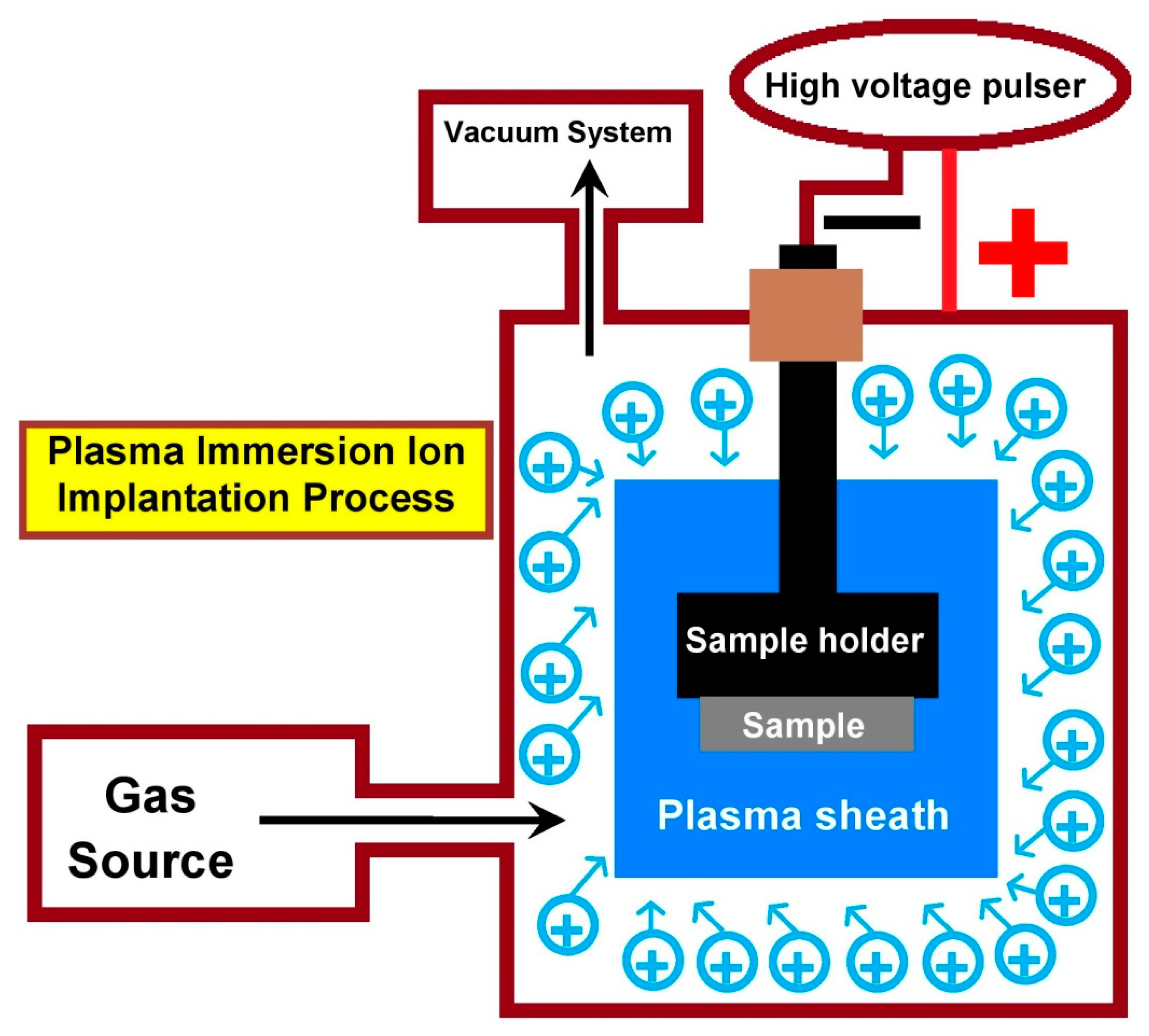
Plasma ion implantation and deposition process have been elaborately described earlier [41,78]. Accordingly, conventional beam-line ion implantation also indicated as plasma ion implantation (PII) is a line-of sight process where energetic ions are bombarded into the surface layer of a substrate. As shown in Figure 1, the plasma immersion ion implantation (PIII) eliminates the line-of sight process and the PIII process involves the immersion of substrate in plasma and a high negative potential is applied for biasing the substrate. As a result, the substrate is surrounded by plasma sheath and acceleration of ions across the plasma sheath occurs normal to the surface of the substrate. Plasma immersion ion implantation and deposition (PIII&D) process is a hybrid process which combines plasma ion implantation and deposition. PIII&D involves the use of metal plasmas in the presence or absence of gas plasmas. High efficiency surface modification can be achieved by bombarding energetic ions generated from a cathodic arc plasma source and the plasma process can be tailored to obtain only implantation or only deposition or combined implantation and deposition.
3. Corrosion Resistance of Plasma Modified (PII, PIII, or PIII&D) Mg and/or Its Alloys
3.1. Zr Based or N Based Plasma Modification
Preparing thin Zr oxide surface layer can improve the corrosion resistance of Mg alloy. Chu’s group [52] has observed enhanced corrosion resistance on zirconium (Zr) plasma-treated, and Zr&O plasma-treated ZK60 Mg alloy than that of untreated ZK60 Mg alloy in SBF, where the former contains 69 nm in thick Zr rich surface layer after Zr PII, and the later contains 80 nm in thick Zr rich surface layer (with O rich surface layer) after Zr PII followed by O PIII. Zr plasma-treated and Zr&O plasma-treated ZK60 Mg alloy exhibit lower icorr (Table 1), higher resistance, and lower capacitance than that of untreated ZK60 Mg alloy, where the corrosion resistances are in the following order: Zr&O plasma treated >Zr plasma treated > untreated ZK60 Mg alloy. The Zr or Zr&O plasma-treated Mg alloy contains Zr oxide surface layer with few nm in thick, where this thin surface passive layer can retard the charge transfer process for corrosion, and that can improve the corrosion resistance of ZK60 Mg alloy. Immersion tests disclose smaller surface damage after Zr or Zr&O plasma treatment initially and after 30 h immersion, while protective corrosion products including Mg(OH)2, zirconium phosphate, Ca10(PO4)6(OH)2, and Ca3(PO4)2·3H2O are formed (Figure 2 and Figure 3). Moreover, Zhao et al. [49] have observed enhanced corrosion resistance on Zr plasma-treated (Zr PII) Mg alloys (Mg-Ca and Mg-Sr) and Zr&O plasma-treated (Zr PII followed by O PIII) Mg alloys (Mg-Ca and Mg-Sr) than that of untreated Mg alloys in SBF, tryptic soy broth (TSB), and cell culture medium. In addition, Liu et al. [61] have observed improved corrosion resistance on Zr plasma-treated (Zr PIII&D) AZ91 Mg alloy than that of untreated AZ91 in Hank’s simulated body fluid. Moreover, Ba et al. [79] have observed enhanced corrosion resistance on Zr plasma-treated Mg than that of pure Mg in SBF. In addition, Jia et al. [80] have observed improved corrosion resistance on Zr plasma-treated ZK60 Mg alloy than that of untreated ZK60 Mg alloy in SBF. Moreover, Liang et al. [81] have observed enhanced corrosion resistance on Zr&O plasma-treated (Zr&O PIII) ZK60 Mg alloy than that of untreated ZK60 Mg alloy in SBF and TSB.
Preparing thin Zr nitride along with Zr oxide surface layer can enhance the corrosion resistance of Mg alloys. Chu’s group [62] has observed enhanced corrosion resistance on Zr&N plasma-treated (Zr PII followed by N PIII) WE43 Mg alloy than that of untreated WE43 Mg alloy in SBF and cell culture medium, where the plasma-treated Mg alloy contains 53 nm in thick Zr rich surface layer and 66 nm in thick N rich surface layer. Zr&N plasma-treated WE43 Mg alloy exhibits lower icorr, higher resistance, and lower capacitance than that of untreated WE43 Mg alloy in both solution, where the corrosion resistance is more enhanced in cell culture medium than in SBF. Calcium phosphate (Ca10(PO4)6(OH)2 and/or Ca3(PO4)2·3H2O) is formed in the corrosion product of Zr&N plasma-treated and untreated WE43 Mg alloy after corrosion studies in SBF and cell culture medium. The formation of oxides and nitrides of Zr along with oxides of Y on the surface layer of Zr&N plasma-treated WE43 Mg alloy are observed, where this thin surface passive layer can diminish the charge transfer process for corrosion, and that can enhance the corrosion resistance of WE43 Mg alloy. Moreover, Cheng et al. [63] have observed enhanced corrosion resistance on Zr-N plasma-treated (contains about 80 nm of modified layer after Zr-N PIII) AZ91 Mg alloy (icorr: 1.16 × 10⁻⁶ A/cm2) than that of untreated AZ91 (icorr: 4.26 × 10⁻⁶ A/cm2) in 0.9% NaCl solution. Preparing nitrogen rich surface layer can enhance the corrosion resistance of Mg alloy. Li et al. [82] have observed that N+ plasma-treated AZ31 Mg alloy exhibits higher corrosion resistance than that of untreated AZ31 Mg alloy in Hank’s solution. On the other hand, Wei et al. [83] have observed that NH2+ plasma-treated AZ31 Mg alloy exhibits higher corrosion resistance than that of untreated AZ31 Mg alloy in Hank’s solution, where the enhanced corrosion resistance on plasma-treated Mg alloy is ascribed to the formation protective surface layer comprising of oxides and amino group.

Table 1.
Ecorr andicorr of the untreated and Zr based/N based plasma-treated Mg/Mg alloys.
| Plasma Treatment | Substrate | Corrosion Medium | icorr (μA cm−2) | Ecorr (V vs. SCE) | References |
|---|---|---|---|---|---|
| Untreated | ZK60 | SBF | 409 | −1654 | [52] |
| Zr PII | ZK60 | SBF | 70 | −1569 | [52] |
| Zr PII + O PIII | ZK60 | SBF | 11 | −1571 | [52] |
| Untreated | Mg-Ca | SBF | 230 | −1.92 | [49] |
| Zr PII | Mg-Ca | SBF | 120 | −1.6 | [49] |
| Zr PII + O PIII | Mg-Ca | SBF | 26 | −1.78 | [49] |
| Untreated | Mg-Sr | SBF | 1000 | −1.79 | [49] |
| Zr PII | Mg-Sr | SBF | 250 | −1.6 | [49] |
| Zr PII + O PIII | Mg-Sr | SBF | 170 | −1.68 | [49] |
| Untreated | Mg-Ca | Tryptic Soy Broth | 280 | −1.91 | [49] |
| Zr PII | Mg-Ca | Tryptic Soy Broth | 49 | −1.51 | [49] |
| Zr PII + O PIII | Mg-Ca | Tryptic Soy Broth | 41 | −1.66 | [49] |
| Untreated | Mg-Sr | Tryptic Soy Broth | 430 | −1.81 | [49] |
| Zr PII | Mg-Sr | Tryptic Soy Broth | 42 | −1.45 | [49] |
| Zr PII + O PIII | Mg-Sr | Tryptic Soy Broth | 40 | −1.63 | [49] |
| Untreated | Mg-Ca | Cell culture medium | 15 | −1.76 | [49] |
| Zr PII | Mg-Ca | Cell culture medium | 6.4 | −1.48 | [49] |
| Zr PII + O PIII | Mg-Ca | Cell culture medium | 4.6 | −1.63 | [49] |
| Untreated | Mg-Sr | Cell culture medium | 23 | −1.56 | [49] |
| Zr PII | Mg-Sr | Cell culture medium | 5 | −1.44 | [49] |
| Zr PII + O PIII | Mg-Sr | Cell culture medium | 4 | −1.45 | [49] |
| Untreated | Mg | SBF | 52.6 | −1.7 | [79] |
| Zr PII | Mg | SBF | 19.9 | −1.43 | [79] |
| Untreated | ZK60 | SBF with glucose | 22.68 | −1.577 | [80] |
| Zr PII | ZK60 | SBF with glucose | 4.416 | –1.629 | [80] |
| Untreated | ZK60 | SBF | 740 | ~−1.7 | [81] |
| Zr PIII + O PIII | ZK60 | SBF | 102 | ~−1.62 | [81] |
| Untreated | ZK60 | Tryptic Soy Broth | 142 | ~−1.49 | [81] |
| Zr PIII + O PIII | ZK60 | Tryptic Soy Broth | 19.3 | ~−1.51 | [81] |
| Untreated | WE43 | SBF | 368 | −1.997 | [62] |
| Zr PII + N PIII | WE43 | SBF | 29.8 | −1.82 | [62] |
| Untreated | WE43 | Cell culture medium | 36.6 | −1.78 | [62] |
| Zr PII + N PIII | WE43 | Cell culture medium | 0.51 | −1.517 | [62] |
| Untreated | AZ91 | 0.9% NaCl | 4.26 | −1.49 | [63] |
| Zr-N PIII | AZ91 | 0.9% NaCl | 1.16 | −1.3 | [63] |
| Untreated | AZ91 | DMEM | 10.8 | −1.57 | [63] |
| Zr-N PIII | AZ91 | DMEM | 0.392 | −1.32 | [63] |
| Untreated | AZ31 | Hank’s solution | 869.72 | –1.03 | [83] |
| NH2+ II | AZ31 | Hank’s solution | 125.23 | –1.01 | [83] |
SCE: Saturated Calomel Electrode; PII: Plasma Ion Implantation; PIII: Plasma immersion ion implantation; SBF: Simulated Body Fluid; DMEM: Dulbecco’s modified Eagle’s medium; II: Ion Implantation.
3.2. Si, Al Based, Zn Based, Zn&Al, or Cr Based Plasma Modification
Preparing thin Si oxide surface layer can enhance the corrosion resistance of Mg alloy. Chu’s group [64] has observed enhanced corrosion resistance on Si plasma-treated (contains Si rich surface layer after Si PII) WE43 Mg alloy than that of untreated WE43 Mg alloy in SBF. Si plasma-treated WE43 Mg alloy exhibits lower icorr (Table 2), higher resistance, and lower capacitance than untreated WE43 Mg alloy, and the Si plasma-treated WE43 Mg alloy exhibits higher phase angle maximum (−60° at 180 Hz) than that of untreated WE43 Mg alloy (−26° at 202 Hz) indicating that Si plasma-treated WE43 Mg alloy exhibits better corrosion resistance than that of untreated WE43 Mg alloy in SBF. The Si plasma-treated Mg alloy contains Si oxide surface layer with few nm in thick, where this thin surface passive layer can hinder the charge transfer process for corrosion, and that can enhance the corrosion resistance of WE43 Mg alloy.
Preparing thin Al oxide surface layer can enhance the corrosion resistance of Pure Mg and Mg alloy. Wu et al. [67] have observed enhanced corrosion resistance (exhibits lower icorr) on Al plasma-treated (Al PII) pure Mg, AZ31 Mg alloy, and AZ91 Mg alloy than that of untreated pure Mg, AZ31 Mg alloy, and AZ91 Mg alloy in SBF, respectively. The Al plasma-treated pure Mg and Mg alloy contains Al oxide surface layer with few nm in thick, where this thin surface passive layer can retard the charge transfer process for corrosion, and that can enhance the corrosion resistance of pure Mg and Mg alloy. Moreover, Zhao et al. [65] have observed enhanced corrosion resistance on Al&O plasma-treated (Al PII followed by O PIII) WE43 Mg alloy than that of untreated WE43 Mg alloy in SBF, where the plasma-treated WE43 Mg alloy exhibits lower icorr, higher resistance, and lower capacitance than that of untreated WE43 Mg alloy. In addition, Wu et al. [66] have observed enhanced corrosion resistance on Al&O plasma-treated (Al PII followed by O PIII) Mg-Nd-Zn-Zr alloy than that of untreated Mg-Nd-Zn-Zr alloy in SBF. In addition, Liu et al. [61] have observed enhanced corrosion resistance on Al plasma-treated (contains Al rich surface layer after Al PIII&D) AZ91 Mg alloy than that of untreated AZ91 in Hank’s simulated body fluid. Moreover, Wong et al. [54] have observed enhanced corrosion resistance on Al-O plasma-treated AZ91 Mg alloy (contains Al and O rich surface layer for about 250 nm after Al-O PIII&D) than that of untreated AZ91 in SBF.
Preparing thin Zn surface layer can deteriorate the corrosion resistance of Pure Mg possibly due to galvanic effect. Wu et al. [84] have observed diminish in corrosion resistance on Zn plasma-treated (contains Zn rich surface layer after Zn PII) pure Mg than that of untreated Mg in SBF possibly due to galvanic effect between metallic Zn and Mg. On the other hand, preparing Zn deposition layer on Zn ion implanted layer can diminish the galvanic effect. Liu et al. [85] have observed a comparable corrosion current density on Zn plasma-treated Mg-1Ca alloy (Zn ion implantation and deposition; icorr: 321.8 μA cm−2) than that of untreated Mg-1Ca alloy (icorr: 248.1 μA cm−2) in SBF, while the plasma-treated Mg alloy (Ecorr: −1.58 V) exhibits higher corrosion potential than that of untreated Mg alloy (Ecorr: −1.92 V), where the positive shift in the potential can be attributed to the formation of ZnO as a passive layer. However, preparing thin Al surface layer over the Zn layer can enhance the corrosion resistance of Pure Mg. Xu et al. [68] have observed enhanced corrosion resistance on Zn&Al plasma-treated (Zn PII followed by Al PII) pure Mg than that of untreated Mg in SBF. The enhance corrosion resistance of the Zn&Al plasma-treated pure Mg is attributed to the formation of more compact hybrid oxide film along with the formation of the β-Mg17Al12 phase, where the hybrid oxide film is composed of three-layer surface film containing an inner metallic magnesium rich layer, a middle layer alloyed with metallic Zn, Al and Mg, and an outer layer of Mg oxide/Al oxide. The thin surface passive layer on Zn&Al plasma-treated pure Mg can diminish the charge transfer process for corrosion, and that can improve the corrosion resistance of pure Mg.
Preparing thin Cr oxide surface layer can enhance the corrosion resistance of Pure Mg and Mg alloy. Xu et al. [69] have observed diminish in corrosion resistance on Cr plasma-treated (Cr PII) pure Mg than that of untreated Mg in SBF, possibly due to the galvanic effect between metallic Cr and Mg. On the other hand, enhanced corrosion resistance is observed on Cr&O plasma-treated (Cr PII followed by O PIII) pure Mg than that of untreated Mg in SBF. The Cr&O plasma-treated pure Mg contains Cr and O rich surface layer with few nm in thick, where this thin surface passive layer can hinder the charge transfer process for corrosion, and that can enhance the corrosion resistance of pure Mg. Moreover, Cr&O plasma-treated (Cr PII followed by O PIII) pure Mg at different voltages (Cr PII at 15, 20, and 40 kV) exhibits higher corrosion resistance than that of untreated pure Mg in SBF, where the corrosion resistance are in the following order: 15 kV > 20 kV > 40 kV [70]. In addition, Cr&O plasma-treated (Cr PII followed by O PIII) Mg-Nd-Zn-Zr alloy exhibits higher corrosion resistance than that of untreated Mg-Nd-Zn-Zr alloy in SBF [66].
3.3. Ti Based, Ni, Ti&Ni, Fe, Mn, Ag, Y, Sr, Ca, P, or Pr Plasma Modification
Preparing thin Ti oxide surface layer can enhance the corrosion resistance of the Mg alloy. Zhao et al. [71] have observed enhanced corrosion resistance on Ti plasma-treated (Ti PII) and Ti&O plasma-treated (Ti PII followed by O PIII) WE43 Mg alloys than that of untreated WE43 Mg alloy in SBF, where the former contains 64 nm in thick Ti rich surface layer, and the later contains 104 nm in thick Ti rich surface layer (with O rich surface layer), while the corrosion resistances are in the following order: Ti&O plasma treated >Ti plasma treated > untreated WE43 Mg alloy. The Ti plasma-treated or Ti&O plasma-treated Mg alloy contains Ti oxide surface layer with few nm in thick, where this thin surface passive layer can retard the charge transfer process for corrosion, and that can improve the corrosion resistance of Mg alloy. Moreover, Chen et al. [72] have observed enhanced corrosion resistance on Ti plasma-treated (Ti PII) AZ31 Mg alloy than that of untreated AZ31 in 3.5% NaCl solution. In addition, Liu et al. [61] have observed enhanced corrosion resistance on Ti plasma-treated (Ti PIII&D) AZ91 Mg alloy than that of untreated AZ91 in Hank’s simulated body fluid.
Preparing thin Ti nitride along with Ti oxide surface layer can enhance the corrosion resistance of Mg alloy. Liu et al. [73] have observed enhanced corrosion resistance on Ti-N plasma-treated (Ti PII followed by N PIII) AZ31 Mg alloy (icorr: 1.5 ± 0.5 × 10⁻³ A/cm2; Table 3) than that of untreated AZ31 (icorr: 112 ± 0.5 × 10⁻³ A/cm2) in 0.56 M NaCl solution, where the plasma-treated modified layer contains MgO, TiO2 and TiN along with Ti and Mg. The formation of oxides and nitrides of Ti on the surface layer of Ti&N plasma-treated AZ31 Mg alloy is observed, where this thin surface passive layer can diminish the charge transfer process for corrosion, and that can enhance the corrosion resistance of AZ31 Mg alloy. In addition, Dai et al. [86] have observed diminish in the corrosion resistance on Ni plasma-treated as well as on Ti&Ni plasma-treated AM60 Mg alloys than that of untreated AM60 Mg alloy in 3.5% NaCl solution, possibly due to the galvanic effect.
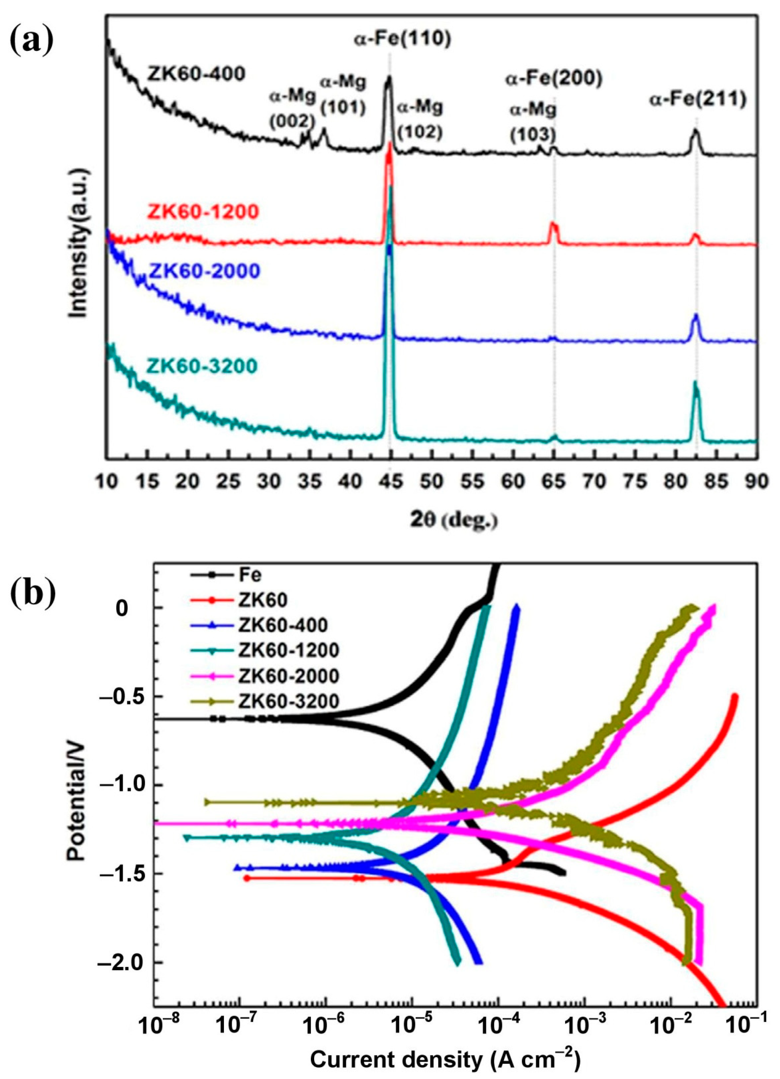
Preparing Fe rich surface layer by implantation can diminish the corrosion resistance of Mg alloy possibly due to galvanic effect. Liu et al. [87] have observed accelerated corrosion on the Fe plasma-treated (Fe PII) Mg-1Ca alloy than that of the untreated Mg-1Ca alloy in Hank’s solution possibly due to the galvanic effect between Fe and Mg. On the other hand, preparing Fe deposition layer on Fe ion implanted layer can enhance the corrosion resistance. Zheng et al. [88] have observed enhanced corrosion resistance on Fe plasma-treated (after Fe ion implantation and deposition) ZK60 Mg alloy than that of untreated ZK60 in Hank’s solution. They have observed the formation of α-Fe thin film on surface of the Fe plasma-treated ZK60 Mg alloy (Figure 4a), where this surface passive layer can retard the charge transfer process for corrosion, and that can enhance the corrosion resistance of ZK60 Mg alloy (Figure 4b). Moreover, Zheng et al. [89] have observed enhanced corrosion resistance on theFe&O plasma-treated ZK60 Mg alloy (Fe&O ion implantation and deposition) than that of the untreated ZK60 Mg alloy in Hank’s solution, where the improvement in corrosion resistance on plasma-treated Mg alloy is attributed to the formation of the protective passive layer comprising FeO/Fe-rich oxide (in the outer deposition zone) along with anFe&Mg mixture (in the inner implanted zone).
Preparing thin Mn rich surface layer can alter the corrosion behavior of Mg. Dong et al. [90] have observed diminish in corrosion resistance on Mn plasma-treated Mg than that of untreated Mg in the initial stage (after 20 minutes) possibly due to the galvanic effect between Mn and Mg, whereas the Mn plasma-treated Mg exhibits higher corrosion resistance than that of untreated Mg after 72 h in Hank’s solution possibly due to the formation of protective corrosion products including calcium phosphate along with a small amount of Mn oxide.
Table 3.
Ecorr and icorr of the untreated and Ti based/Fe based/Ag/Y/Sr/Ca/P/Pr plasma-treated Mg/Mg alloys.
Table 3.
Ecorr and icorr of the untreated and Ti based/Fe based/Ag/Y/Sr/Ca/P/Pr plasma-treated Mg/Mg alloys.
| Plasma Treatment | Substrate | Corrosion Medium | icorr (μA cm−2) | Ecorr (V vs. SCE) | References |
|---|---|---|---|---|---|
| Untreated | WE43 | SBF | 327 | ~−1.98 | [71] |
| Ti PII + O PIII | WE43 | SBF | 14.4 | ~−1.8 | [71] |
| Untreated | AZ31 | 0.56 M NaCl | 0.112 | −1460 | [73] |
| TiN | AZ31 | 0.56 M NaCl | 0.0015 | −850 | [73] |
| Untreated | ZK60 | Hank’s solution | 105.8 | −1.493 | [89] |
| Fe&O II&D | ZK60 | Hank’s solution | 1.25 | −1.066 | [89] |
| Untreated | ZK60 | Hank’s solution | 119 | −1.528 | [88] |
| Fe II&D | ZK60 | Hank’s solution | 1.363 | −1.295 | [88] |
| Untreated | Mg-Ca | Hank’s solution | 92.58 | −1.81 | [87] |
| Fe PII | Mg-Ca | Hank’s solution | 235.23 | −1.56 | [87] |
| Ag PII | Mg-Ca | Hank’s solution | 524.93 | −1.55 | [87] |
| Y PII | Mg-Ca | Hank’s solution | 30.65 | −1.60 | [87] |
| Untreated | Mg | SBF with glucose | 52.6 | −1.694 | [91] |
| Sr PII | Mg | SBF with glucose | 25.1 | −1.582 | [91] |
| Untreated | Mg | SBF | 161.66 | −1.820 | [92] |
| Ca PII | Mg | SBF | 100 | −1.825 | [92] |
| Untreated | Mg | SBF with glucose | 67.601 | −1.657 | [93] |
| P PII | Mg | SBF with glucose | 54.955 | −1.598 | [93] |
| Untreated | Mg | artificial hand sweat | 132.2 | −1.985 | [74] |
| Pr PII | Mg | artificial hand sweat | 79.62 | −1.881 | [74] |
| Untreated | AZ80 | artificial hand sweat | 354.1 | −1.653 | [74] |
| Pr PII | AZ80 | artificial hand sweat | 11.12 | −1.488 | [74] |
SCE: saturated calomel electrode; PII: Plasma Ion Implantation; PIII: Plasma immersion ion implantation; SBF: Simulated Body Fluid; II&D: Ion Implantation and Deposition.
Preparing an Ag rich surface layer by implantation can diminish the corrosion resistance of the Mg alloy possibly due to the galvanic effect. Liu et al. [87] have observed accelerated corrosion on an Ag plasma-treated (contains Ag rich surface layer after Ag PII) Mg-1Ca alloy than that of an untreated Mg-1Ca alloy in Hank’s solution, possibly due to the galvanic effect between Ag and Mg (Type I in Figure 5).
Preparing thin Y oxide surface layer can enhance the corrosion resistance of Mg alloy. Liu et al. [87] have observed enhanced corrosion resistance on Y plasma-treated (contains Y rich surface layer after Y PII) Mg-1Ca alloys (0.69 ± 0.18 mm/year) than that of untreated Mg-1Ca alloys (2.10 ± 0.21 mm/year) in Hank’s solution. The Y plasma-treated Mg alloy contains a Y oxide surface layer with a few nm in thickness, where this thin surface passive layer can diminish the charge transfer process for corrosion, and that can improve the corrosion resistance of Mg alloy. They proposed the schematic illustration to demonstrate the corrosion mechanism of plasma ion-implanted Mg alloys, where the formation of a compact passive protective oxide layer in Y plasma-treated Mg alloy improves its corrosion resistance (Type II in Figure 5), whereas the formation of non-protective layer (mainly metallic) in Ag or Fe plasma-treated Mg alloys accelerates its corrosion resistance (Type I in Figure 5). In addition, Jia et al. [91] have observed enhanced corrosion resistance on Sr plasma-treated Mg than that of pure Mg in SBF. The Sr plasma-treated Mg contains Sr oxide surface layer with about 30 nm in thick, where this thin surface passive layer can retard the charge transfer process for corrosion, and that can enhance the corrosion resistance of Mg. On the other hand, Somasundaram et al. [92] have observed only a slight improvement in corrosion resistance on Ca ion implanted Mg than that of pure Mg in SBF. In addition, Asdi et al. [93] have observed improvement in corrosion resistance on P ion implanted Mg than that of pure Mg in SBF, where the improvement in corrosion resistance on the ion implanted Mg is attributed to the formation of about 120 nm in thick passive layer comprising of PO along with MgO.
Preparing a thin Pr oxide surface layer can enhance the corrosion resistance of pure Mg and Mg alloy. Wang et al. [74] have observed enhanced corrosion resistance on Pr plasma-treated (Pr PII) pure Mg and AZ80 Mg alloy than that of untreated samples (pure Mg and AZ80 Mg alloy) in artificial hand sweat solution. The Pr plasma-treated pure Mg and Mg alloy contain Pr oxide surface layer with few nm in thick, where this thin surface passive layer can retard the charge transfer process for corrosion, and that can improve the corrosion resistance of pure Mg and Mg alloy.
3.4. Ce, Nd, Hf, Ta, H2O or Hydroxyl Plasma Modification
Preparing thin Ce oxide surface layer can enhance the corrosion resistance of pure Mg and Mg alloy. Wang et al. [94] have observed enhanced corrosion resistance on Ce plasma-treated (Ce PII) AZ31 Mg alloy than that of untreated AZ31 in 3.5 wt.% NaCl solution saturated with Mg(OH)2. Moreover, Wu et al. [75] have observed enhanced corrosion resistance on Ce plasma-treated (Ce PII) pure Mg than that of untreated pure Mg in Ringer’s solution, artificial hand sweat solution, and complete cell culture medium. The Ce plasma-treated pure Mg and Mg alloy contain Ce oxide surface layer with few nm in thick, where this thin surface passive layer can diminish the charge transfer process for corrosion, and that can enhance the corrosion resistance of pure Mg and Mg alloy.
Preparing thin Nd oxide surface layer can enhance the corrosion resistance of Mg alloy. Jin et al. [76] have observed enhanced corrosion resistance on Nd plasma-treated (Nd PII) WE43 Mg alloy than that of untreated WE43 Mg alloy in SBF and cell culture medium. The Nd plasma-treated Mg alloy contains Nd oxide surface layer with few nm in thick, where this thin surface passive layer can hinder the charge transfer process for corrosion, and that can improve the corrosion resistance of Mg alloy.
Preparing thin Hf oxide surface layer can enhance the corrosion resistance of Mg alloy. Jin et al. [77] have observed enhanced corrosion resistance (exhibits lower icorr (Table 4), and higher resistance, Figure 6) on Hf plasma-treated (Hf PII) WE43 Mg alloy than that of untreated WE43 Mg alloy in SBF. The Hf plasma-treated Mg alloy contains Hf oxide surface layer with a few nm in thickness, where this thin surface passive layer can retard the charge transfer process for corrosion, and that can enhance the corrosion resistance of Mg alloy.
Preparing thin Ta oxide surface layer can enhance the corrosion resistance of Mg alloy. Wang et al. [95] have observed enhanced corrosion resistance on Ta plasma-treated (Ta PII) AZ31 Mg alloy than that of untreated AZ31 in 3.5 wt.% NaCl solution saturated with Mg(OH)2. The Ta plasma-treated Mg alloy contains Ta oxide surface layer with few nm in thick, where this thin surface passive layer can retard the charge transfer process for corrosion, and that can enhance the corrosion resistance of Mg alloy.
Increasing the surface oxide layer can enhance the corrosion resistance of Mg alloy. Tian et al. [96] have observed enhanced corrosion resistance on H2O plasma-treated (H2O PIII and oxidation) AZ31B Mg alloy than that of untreated AZ31B in 3 wt.% NaCl solution. The plasma-treated Mg alloy contains increased surface oxide layer with a few nm in thickness, where this thin surface passive layer can diminish the charge transfer process for corrosion, and that can enhance the corrosion resistance of Mg alloy. Moreover, Wei et al. [97] have observed enhanced corrosion resistance on OH− plasma-treated ZK60 Mg alloy than that of untreated ZK60 Mg alloy in Hank’s solution, where the improvement in the corrosion resistance on the plasma-treated Mg alloy is attributed to the formation of passive surface layer comprising of (Mg(OH)2/MgO).
Table 4.
Ecorr andicorr of the untreated and Ce/Nd/Hf/Ta/hydroxyl plasma-treated Mg/Mg alloys.
| Plasma Treatment | Substrate | Corrosion Medium | icorr (μA cm−2) | Ecorr (V vs. SCE) | References |
|---|---|---|---|---|---|
| Untreated | AZ31 | 3.5% NaCl saturated with Mg(OH)2 | 29.1 | −1.45 | [94] |
| Ce PII | AZ31 | 3.5% NaCl saturated with Mg(OH)2 | 2.81 | −1.37 | [94] |
| Untreated | Mg | artificial hand sweat | 106.1 | −1.878 | [75] |
| Ce PII | Mg | artificial hand sweat | 39.9 | −1.789 | [75] |
| Untreated | Mg | Ringer’s solution | 41.8 | −1.748 | [75] |
| Ce PII | Mg | Ringer’s solution | 26.8 | −1.514 | [75] |
| Untreated | Mg | Cell culture medium | 20.4 | −1.731 | [75] |
| Ce PII | Mg | Cell culture medium | 5.2 | −1.552 | [75] |
| Untreated | WE43 | SBF | 555.7 | −1.992 | [76] |
| Nd PII | WE43 | SBF | 11.9 | −1.740 | [76] |
| Untreated | WE43 | Cell culture medium | 2.55 | −1.705 | [76] |
| Nd PII | WE43 | Cell culture medium | 0.5 | −1.560 | [76] |
| Untreated | WE43 | SBF | 625.7 | ~−1.9 | [77] |
| Hf PII | WE43 | SBF | 40 | ~−1.75 | [77] |
| Untreated | AZ31 | 3.5% NaCl saturated with Mg(OH)2 | 15 | −1.43 | [95] |
| Ta PII | AZ31 | 3.5% NaCl saturated with Mg(OH)2 | 3.9 | −1.25 | [95] |
| Untreated | ZK60 | Hank’s solution | 312.26 | −1.57967 | [97] |
| OH− II | ZK60 | Hank’s solution | 70.79 | −1.60067 | [97] |
SCE: saturated calomel electrode; PII: Plasma Ion Implantation; SBF: Simulated Body Fluid; II: Ion Implantation.
3.5. C Based Plasma Modification
Preparing amorphous thin C containing surface layer can enhance the corrosion resistance of pure Mg. Xu et al. [98] have observed enhanced corrosion resistance on C plasma-treated (C PII) pure Mg than that of untreated pure Mg in SBF and cell culture medium, where the C plasma-treated Mg (26.3 μA cm−2; Table 5) exhibits lower corrosion current density than that of untreated pure Mg (258 μA cm−2) in SBF, while the C plasma-treated Mg (1.88 μA cm−2) exhibits lower corrosion current density than that of untreated pure Mg (65.2 μA cm−2) in cell culture medium. The C plasma-treated pure Mg contains amorphous graphite surface layer with 58 nm in thick, where this thin surface passive layer can hinder the charge transfer process for corrosion, and that can enhance the corrosion resistance of pure Mg. On the other hand, Yu et al. [99] have observed enhanced corrosion resistance on the C plasma-treated AM60 Mg alloy than that of the untreated AM60 Mg alloy in 3.5% NaCl, where the improvement in corrosion resistance of the plasma-treated Mg alloy is attributed to the formation of about a 250 nm thick C rich passive surface layer comprising an Mg2C3 phase.
Preparing thin C containing surface layer with increased surface oxide layer can enhance the corrosion resistance of Mg alloy. Xu et al. [100] have observed enhanced corrosion resistance on CO2 plasma-treated (CO2 PIII) AZ31 Mg alloy than that of untreated AZ31 in SBF and cell culture medium. The CO2 plasma-treated AZ31 Mg alloy contains graphite surface layer along with increase in surface oxide layer (Mg and Al oxide), where this thin surface passive layer can diminish the charge transfer process for corrosion, and that can improve the corrosion resistance of AZ31 Mg alloy.
Preparing thin diamond-like carbon (DLC) film on the surface can enhance the corrosion resistance of Mg alloy. Wu et al. [38] have observed enhanced corrosion resistance on the C2H2 plasma-treated (C2H2 PIII&D) Mg-Nd-Zn-Zr alloy than that of the untreated Mg-Nd-Zn-Zr alloy in 0.9 wt% NaCl solution. The C2H2 plasma-treated Mg alloy contains about a 200 nm thick DLC film on the surface, where this thin surface passive layer can hinder the charge transfer process for corrosion, and that can improve the corrosion resistance of the Mg alloy. The C2H2 plasma-treated Mg-Nd-Zn-Zr alloy exhibits lesser surface damage than that of untreated Mg-Nd-Zn-Zr alloy after 24 h immersion in 0.9 wt% NaCl solution. Moreover the cross sectional images observed at different locations of untreated Mg-Nd-Zn-Zr alloys (Figure 7a,b) and C2H2 plasma-treated Mg-Nd-Zn-Zr alloys (Figure 7c,d) after 24 h immersion in 0.9 wt% NaCl solution revealed that the C2H2 plasma-treated Mg-Nd-Zn-Zr alloy exhibits lesser corrosion with the existence of a carbon film (top inset of Figure 7c) and small corrosion pits (indicated using arrows in Figure 7d) after 24 h of corrosion, while the untreated Mg-Nd-Zn-Zr alloy exhibits higher corrosion with the existence of a thicker corrosion product with larger pits (Figure 7b) indicating that the C2H2 plasma-treated Mg-Nd-Zn-Zr alloy exhibits better corrosion resistance than untreated Mg-Nd-Zn-Zr alloys. Moreover, Feng et al. [101] have observed enhanced corrosion resistance on C2H2 plasma-treated (C2H2 PIII&D) AZ31 Mg alloys than that of untreated AZ31 Mg alloys in a Lysogeny broth medium, where the plasma-treated surface contains about a 200 nm thick DLC film. In addition, Yekehtaz et al. [102] have observed enhanced corrosion resistance on CH4&C2H2 plasma-treated Mg (contains DLC film on the surface after CH4 PIII followed by C2H2 PIII&D) than that of CH4 plasma-treated Mg and untreated Mg in a slightly acidic acetate buffer solution, where the corrosion resistances are in the following order: CH4&C2H2 plasma-treated Mg > CH4 plasma-treated Mg > untreated Mg. On the other hand, Wei et al. [103] have observed that the COOH+ plasma-treated ZK60 Mg alloy exhibits higher corrosion resistance than that of the untreated ZK60 Mg alloy in Hank’s solution, where the improvement is ascribed to the smooth passive surface layer comprising carbonate, and oxide/hydroxide.
Table 5.
Ecorr and icorr of the untreated and C based plasma-treated Mg/Mg alloys.
| Plasma Treatment | Substrate | Corrosion Medium | icorr (μA cm−2) | Ecorr (V vs. SCE) | References |
|---|---|---|---|---|---|
| Untreated | Mg | SBF | 258 | −1.98 | [98] |
| C PII | Mg | SBF | 26.3 | −1.94 | [98] |
| Untreated | Mg | DMEM | 65.2 | −1.98 | [98] |
| C PII | Mg | DMEM | 1.88 | −1.71 | [98] |
| Untreated | AM60 | 3.5% NaCl | 50.6 | −1.48 | [99] |
| C PII | AM60 | 3.5% NaCl | 20.1 | −1.35 | [99] |
| Untreated | AZ31 | SBF | 561 | ~−1.77 | [100] |
| CO2 PIII | AZ31 | SBF | 260 | ~−1.73 | [100] |
| Untreated | AZ31 | DMEM | 241 | −1.72 | [100] |
| CO2 PIII | AZ31 | DMEM | 3.66 | −1.62 | [100] |
| Untreated | AZ31 | Lysogeny broth medium | 317 | ~−1.49 | [101] |
| C2H2 PIII&D | AZ31 | Lysogeny broth medium | 65.3 | ~−1.65 | [101] |
| Untreated | ZK60 | Hank’s solution | 291.94 | −1.585 | [103] |
| COOH+ II | ZK60 | Hank’s solution | 33.245 | −1.601 | [103] |
SCE: saturated calomel electrode; PII: Plasma Ion Implantation; PIII: Plasma immersion ion implantation; PIII&D: Plasma Immersion Ion Implantation and Deposition; SBF: Simulated Body Fluid; DMEM: Dulbecco’s modified Eagle’s medium; II: Ion Implantation.
Figure 7.
Cross sectional images (Inset: its corresponding EDS map) observed at different locations of (a,b) untreated Mg-Nd-Zn-Zr alloy and (c,d) C2H2 plasma-treated Mg-Nd-Zn-Zr alloy after 24 h immersion in 0.9 wt% NaCl solution. Top inset of the figure (c) depicts the existence of carbon film after 24 h corrosion in NaCl solution. The existence of small corrosion pits is indicated using arrows in figure (d) (Reproduced with permission from Ref. [17]; Copyright 2013, Elsevier Ltd.).

4. Biological Properties of Plasma Modified (PII, PIII, or PIII&D) Mg and/or Mg Alloys
4.1. In Vitro Cytocompatibility of Plasma Modified Mg and Mg Alloys
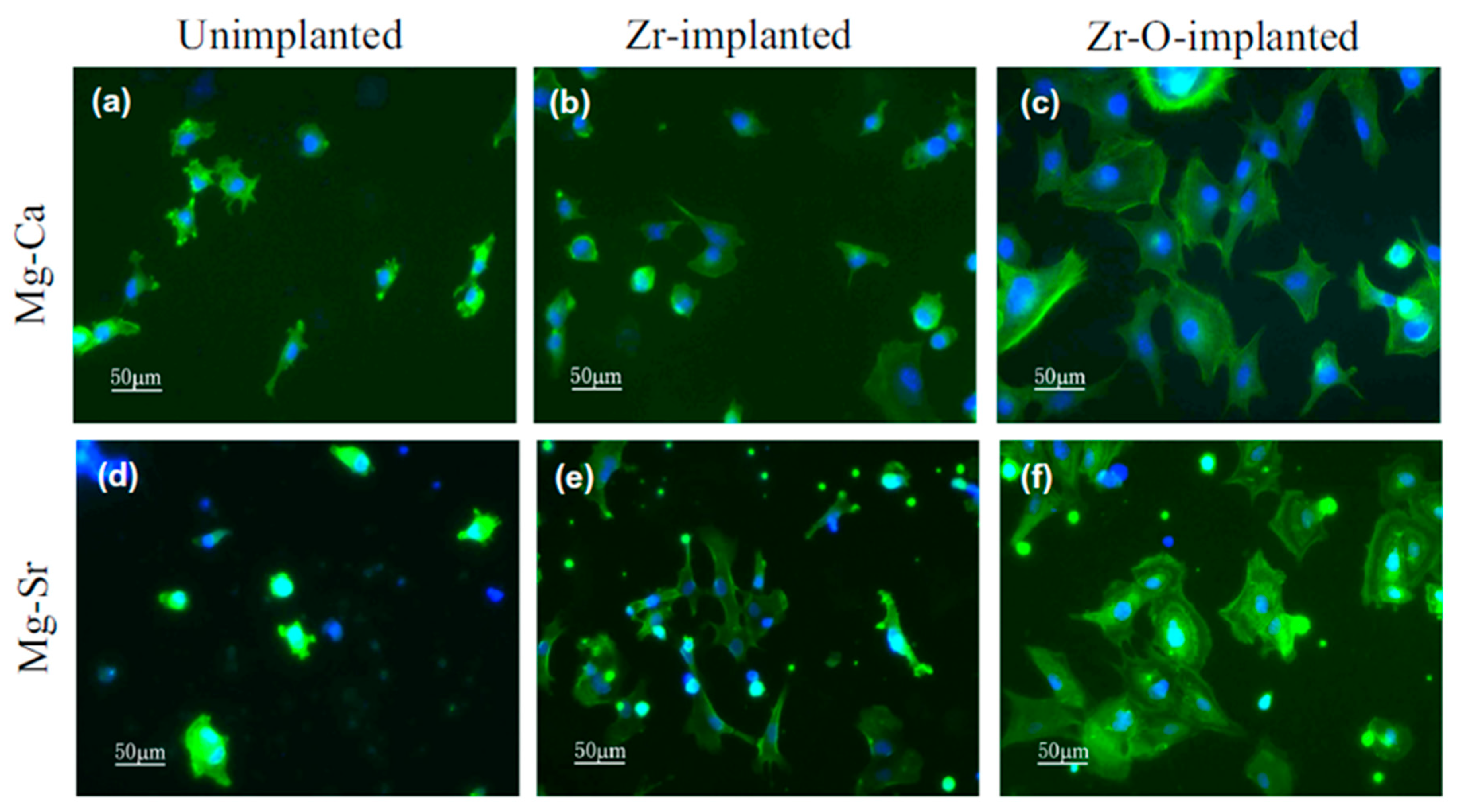
Preparing thin Zr oxide surface layer can enhance the cytocompatibility of Mg alloy. Zhao et al. [49] observed enhanced cell viability (after 72 h incubation), cell proliferation (using BrdU incorporation after 1 day and 3 days), and cell adhesion (MC3T3-E1 pre-osteoblasts after cultured for 5 h) on Zr plasma-treated and Zr&O plasma-treated Mg alloys (Mg-Ca and Mg-Sr) than that of untreated corresponding Mg alloys. The Zr plasma-treated and Zr&O plasma-treated Mg alloys (Mg-Ca and Mg-Sr) exhibit good MC3T3-E1 pre-osteoblasts attachment and cell spreading and the attached cells on Zr&O plasma-treated Mg alloys (Mg-Ca and Mg-Sr) exhibit apparent filopodia, flattened membranes, and ordered F-actin, whereas the attached pre-osteoblasts are not able to spread well on untreated Mg alloys (Mg-Ca and Mg-Sr) as shown in Figure 8. Zr plasma-treated and Zr&O plasma-treated Mg alloys (Mg-Ca and Mg-Sr) contains Zr oxides surface layer that possibly enhances their corrosion resistance and that possibly enhances their cell viability, cell proliferation, and cell adhesion. These indicate that Zr plasma-treated and Zr&O plasma-treated Mg alloys (Mg-Ca and Mg-Sr) exhibit better cytocompatibility than that of untreated corresponding Mg alloys. Moreover, Liang et al. [81] have observed good cytocompatibility on Zr&O plasma-treated (Zr&O PIII) ZK60 Mg alloys. Preparing thin Zr nitride along with Zr oxide surface layer can enhance the cytocompatibility of Mg alloy. Cheng et al. [63] observed enhanced cell viability (after incubated for 1, 3, and 7 days), cell adhesion (using MG63 osteoblasts after incubated for 1 and 3 days), and alkaline phosphatase (ALP) activity (using MG63 osteoblasts after incubated for 7 and 14 days) on Zr-N plasma-treated AZ91 Mg alloy than that of untreated AZ91 Mg alloy. Zr-N plasma-treated AZ91 Mg alloy contains surface layer with Zr oxides and nitrides that possibly enhance its corrosion resistance and that possibly enhance its cell viability, cell adhesion, and ALP activity. These indicate that Zr-N plasma-treated AZ91 Mg alloy exhibits better cytocompatibility than that of untreated AZ91 Mg alloy. Preparing nitrogen rich surface layer can maintain the cytocompatibility of Mg alloys. Li et al. [82] have performed the cytotoxicity test using NIH-3T3 mouse fibroblasts after cultured for 2 days, where the N+ plasma-treated AZ31 Mg alloy almost retains the cytocompatibility of AZ31 Mg alloy. On the other hand, Wei et al. [83] have performed the cytotoxicity test using MC3T3-E1 cells after cultured for 3 days, where the NH2+ plasma-treated AZ31 Mg alloy exhibits higher cytocompatibility than that of untreated AZ31 Mg alloy. Preparing thin Al oxide surface layer can enhance the cytocompatibility of Mg alloy. Wong et al. [54] observed enhanced cell adhesion (after cultured for 1 day and 3 days) on Al-O plasma-treated AZ91 Mg alloy than that of untreated AZ91 Mg alloy. Al-O plasma-treated AZ91 Mg alloy contains surface layer with Al oxides that possibly enhances its corrosion resistance and that possibly enhances its cell adhesion. These indicate that Al-O plasma-treated AZ91 Mg alloy exhibits better cytocompatibility than that of untreated AZ91 Mg alloy. Preparing Zn deposition layer on Zn ion implanted layer can enhance the cytocompatibility of Mg alloy. Liu et al. [85] have observed improved cytocompatibility of MC3T3-E1 cells on the Zn plasma-treated Mg-1Ca alloy (Zn ion implantation and deposition) than that of the untreated Mg alloy possibly due to the formation of ZnO layer on the surface. Preparing thin Cr oxide surface layer can enhance the cytocompatibility of Mg. Xu et al. [70] observed enhanced cell viability (after incubated for 1, 3, and 7 days), and cell adhesion (using MC3T3-E1 pre-osteoblasts after cultured for 1 day and 3 days) on Cr&O plasma-treated Mg than that of untreated pure Mg. Cr&O plasma-treated Mg contains surface layer with Cr oxides that possibly enhance its corrosion resistance and that possibly enhance its cell viability, and cell adhesion. These indicate that Cr&O plasma-treated Mg exhibits better cytocompatibility than that of untreated pure Mg. Preparing thin Nd oxide surface layer can enhance the cytocompatibility of Mg alloy. Jin et al. [76] observed enhanced cell viability (at higher concentration after incubated for 1 day and 3 days), and cell adhesion (using MC3T3-E1 pre-osteoblasts after cultured for 5 h) on Nd plasma-treated WE43 Mg alloy than that of untreated WE43 Mg alloy. Nd plasma-treated WE43 Mg alloy contains surface layer with Nd oxides that possibly enhance its corrosion resistance and that possibly enhance its cell viability, and cell adhesion. These indicate that Nd plasma-treated WE43 Mg alloy exhibits better cytocompatibility than that of untreated WE43 Mg alloy. Preparing thin Hf oxide surface layer can enhance the cytocompatibility of Mg alloy. Jin et al. [77] observed enhanced cell viability (after incubated for 1 day and 3 days), and cell adhesion (using MC3T3-E1 pre-osteoblasts after cultured for 5 h) on Hf plasma-treated WE43 Mg alloy than that of untreated WE43 Mg alloy. Hf plasma-treated WE43 Mg alloy contains surface layer with Hf oxides that possibly enhance its corrosion resistance and that possibly enhance its cell viability, and cell adhesion. These indicate that Hf plasma-treated WE43 Mg alloy exhibits better cytocompatibility than that of untreated WE43 Mg alloy. Preparing thin oxide/hydroxide surface layer can enhance the cytocompatibility of Mg alloy. Wei et al. [97] have performed the cytotoxicity test using MC3T3-E1 cells after cultured for 3 days, where the OH− plasma-treated ZK60 Mg alloy exhibits higher cytocompatibility than that of untreated ZK60 Mg alloy. Preparing thin carbonate surface layer can enhance the cytocompatibility of Mg alloy. Wei et al. [103] have performed the cytotoxicity test using MC3T3-E1 cells after cultured for 3 days, where the COOH+ plasma-treated ZK60 Mg alloy exhibits higher cytocompatibility than that of untreated ZK60 Mg alloy.
4.2. In Vitro and In Vivo Antibacterial Activities of Plasma Modified Mg Alloys
Preparing thin Zr oxide surface layer can enhance the antibacterial activities of Mg alloy. Zhao et al. [49] observed significantly lesser numbers of live bacteria (Staphylococcus aureus) on Zr plasma-treated and Zr&O plasma-treated Mg alloys (Mg-Ca and Mg-Sr) than that of untreated corresponding Mg alloys, as shown in Figure 9. Zr plasma-treated and Zr&O plasma-treated Mg alloys (Mg-Ca and Mg-Sr) contain Zr oxides surface layer that possibly enhance their antibacterial activities. This indicates that Zr plasma-treated and Zr&O plasma-treated Mg alloys (Mg-Ca and Mg-Sr) exhibit better antibacterial activities than that of untreated corresponding Mg alloys. Moreover, Liang et al. [81] have conducted in vitro and in vivo antibacterial tests, where Zr&O plasma-treated (Zr&O PIII) ZK60 Mg alloy exhibits higher antibacterial activities than that of untreated ZK60 Mg alloy. The improved antibacterial activities on Zr&O plasma-treated ZK60 Mg alloy are ascribed to the improved surface hydrophobicity, diminished surface roughness, and large concentration of oxygen vacancies in the ZrO2-containing surface layer, where the smooth and hydrophobic surface retard the bacterial adhesion. Preparing thin Zr nitride along with Zr oxide surface layer can enhance the antibacterial activities of Mg alloy. Cheng et al. [63] observed lesser numbers of live bacteria (Staphylococcus aureus) on Zr-N plasma-treated AZ91 Mg alloy than that of untreated AZ91 Mg alloy. Zr-N plasma-treated AZ91 Mg alloy contains surface layer with Zr oxides and nitrides that possibly enhance its antibacterial activities. This indicates that Zr-N plasma-treated AZ91 Mg alloy exhibits better antibacterial activities than that of untreated AZ91 Mg alloy.
4.3. In Vivo Stimulation of Bone Formation of Plasma Modified Mg Alloys
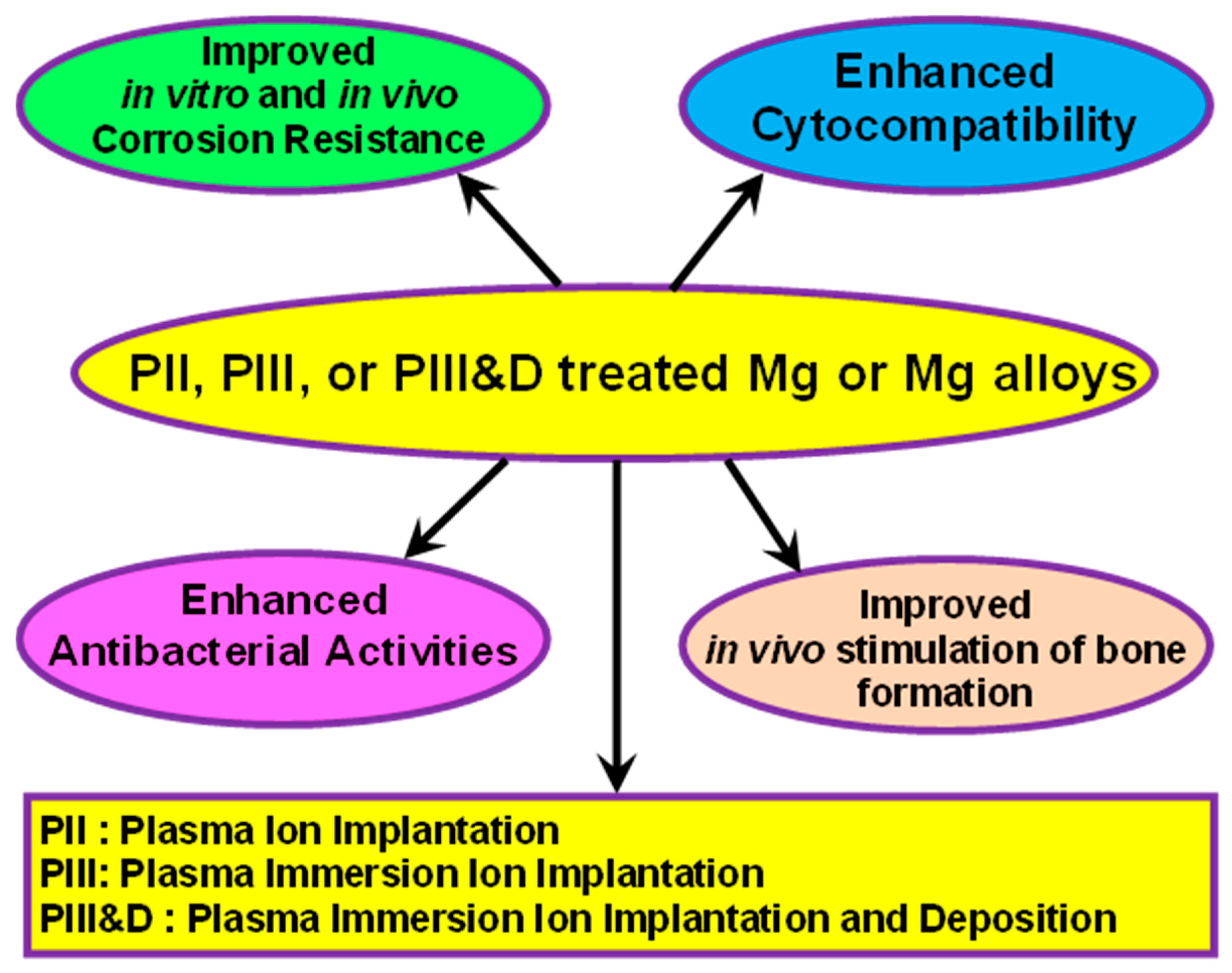
Preparing thin Al oxide surface layer can stimulate the new bone formation and improve the in vivo corrosion resistance of Mg alloy. Wong et al. [54] observed better in vivo stimulation of bone formation and in vivo corrosion resistance on Al-O plasma-treated AZ91 Mg alloy than that of untreated AZ91 Mg alloy after in vivo animal studies for a period of 2 months. To monitor the in vivo corrosion behavior, they have performed micro-computed tomography (CT), which shows the newly generated bone around the implants and the corrosion morphology of the implants. The micro-computed tomography 3D reconstruction models evaluation after 2 months post-operation revealed that the newly formed bone around the implant on Al-O plasma-treated AZ91 Mg alloy (7.11 mm3) is higher than that of untreated AZ91 Mg alloy (1.06 mm3). The Al-O plasma-treated AZ91 Mg alloy exhibits about 46% increase in bone volume while untreated AZ91 Mg alloy exhibits more than 54% bone resorption after 1 week of post-operation. In addition, the Al-O plasma-treated AZ91 Mg alloy exhibits about 138% increase in bone volume than that of untreated AZ91 Mg alloy after 2 months of post-operation (Figure 10). Moreover, the Al-O plasma-treated AZ91 Mg alloy (99%) exhibits lesser implant volume reduction than that of untreated AZ91 Mg alloy (97%) after 4 weeks of post-operation. In addition, the Al-O plasma-treated AZ91 Mg alloy (98%) exhibits further lesser implant volume reduction than that of untreated AZ91 Mg alloy (93%) after 2 months of post-operation; Al-O plasma-treated AZ91 Mg alloy contains surface layer with Al oxides that possibly enhance its in vivo corrosion resistance suggesting slower degradation rate than that of untreated, and that possibly exhibits higher new bone formation, much higher percentage changes in the bone volume, and lesser percentage changes in the implant volume reduction after 2 month of in vivo animal studies on Al-O plasma-treated AZ91 Mg alloy than that of untreated AZ91. These indicate that Al-Oplasma-treated AZ91 Mg alloy exhibits better in vivo stimulation of bone formation and in vivo corrosion resistance than that of untreated AZ91 Mg alloy. Preparing thin Zr nitride along with Zr oxide surface layer can stimulate the new bone formation and improve the in vivo corrosion resistance of Mg alloy. Cheng et al. [63] have conducted micro-computed tomography until 12 weeks to monitor the in vivo corrosion resistance on the implants and new bone formation around the implants. They have observed higher new bone formation, higher percentage changes in the bone volume, and lesser percentage changes in the implant volume reduction after 12 weeks in vivo animal studies on Zr-N plasma-treated AZ91 Mg alloy than that of untreated AZ91 (Figure 11). These indicate that Zr-N plasma-treated AZ91 Mg alloy exhibits better in vivo stimulation of bone formation than that of untreated AZ91 Mg alloy. The Zr-N plasma-treated AZ91 Mg alloy contains surface layer with Zr oxides and nitrides that possibly enhance its corrosion resistance and that possibly enhance its in vivo stimulation of bone formation and in vivo corrosion resistance. Thus, enhanced in vitro/in vivo corrosion resistance, cytocompatibility, antibacterial activities, and in vivo stimulation of bone formation are observed on plasma-treated Mg or Mg alloys (Figure 12), where the plasma treatment includes plasma ion implantation (PII), plasma immersion ion implantation (PIII), or plasma immersion ion implantation and deposition (PIII&D) techniques.
5. Summary and Outlook
Degradable metallic implants are potential candidates for bio-implants. Hence, Mg and its alloys are much attracted but their rapid degradation prevents their use. The improvement of in vitro corrosion resistance, in vivo corrosion resistance, cytocompatibility, in vitro antibacterial activities, in vivo antibacterial activities, and in vivo stimulation of bone formation on Mg or its alloys by plasma techniques have been reviewed, where the plasma techniques include plasma ion implantation (PII), plasma immersion ion implantation (PIII), and plasma immersion ion implantation and deposition (PIII&D) techniques.
The following conclusions have been drawn:
- (a)
- The in vitro corrosion resistance is improved on the plasma-treated Mg or its alloys possibly due to the formation of passive protective layer comprising of Zr-O [81], Zr-N [62], N [82], Si [64], Al-O [54], Zn-Al [68], Cr-O [69], Ti-O [71], Ti-N [73], Fe [88], Y [87], Sr [91], P [93], Pr [74], Ce [75], Nd [76], Hf [77], Ta [95], or C [98].
- (b)
- The in vivo corrosion resistance and in vivo stimulation of bone formation are improved on the plasma-treated Mg alloys possibly due to the formation of suitable surface layer comprising of Al-O [54], or Zr-N [63], where the surface layer not only control the release of Mg but also stimulate the bone formation.
- (c)
- (d)
- The cytocompatibility is improved on the plasma-treated Mg or Mg alloys possibly due to the formation of suitable surface layer comprising of Zr-O [49], Zr-N [63], N [83], Al-O [54], Zn [85], Cr-O [70], Nd [76], or Hf [77], where the surface layer not only control the release of Mg but also enhance the cell growth and adhesion.
The essential factors governing the performance of Mg or Mg alloys by using plasma treatment should be considered in future research:
- The in vivo corrosion resistance, in vivo stimulation of bone formation, and in vivo antibacterial activities are improved on the plasmatreated Mg alloys. However, the studies on these aspects are very limited [54,63,81]. Therefore, additional progresses are obviously needed by using suitable plasma treatment on Mg and Mg alloys.
- Surface modification of Mg and Mg alloys using other surface modification techniques followed by plasma treatment can tune the corrosion and biological properties. Nevertheless, the studies on these aspects are very limited [104,105,106,107,108,109,110]. Hence, additional efforts are obviously needed by combining other suitable surface modification with plasma treatment.
Funding
This research received no external funding, while the APC for this article is completely waived by the Editorial office of the “Lubricants”.
Acknowledgments
The author thanks the Science and Engineering Research Board (SERB), Department of Science and Technology, Government of India (Reference no.PDF/2017/000015).
Conflicts of Interest
The authors declare no conflict of interest.
References
- Grogan, J.A.; O’Brien, B.J.; Leen, S.B.; McHugh, P.E. A corrosion model for bioabsorbable metallic stents. Acta Biomater. 2011, 7, 3523–3533. [Google Scholar] [CrossRef] [PubMed]
- Singh, N.; Batra, U.; Kumar, K.; Ahuja, N.; Mahapatro, A. Progress in bioactive surface coatings on biodegradable Mg alloys: A critical review towards clinical translation. Bioact. Mater. 2023, 19, 717–757. [Google Scholar] [CrossRef] [PubMed]
- Zhang, Z.-Q.; Tong, P.-D.; Wang, L.; Qiu, Z.-H.; Li, J.-A.; Li, H.; Guan, S.-K.; Lin, C.-G.; Wang, H.-Y. One-step fabrication of self-healing poly(thioctic acid) coatings on ZE21B Mg alloys for enhancing corrosion resistance, anti-bacterial/oxidation, hemocompatibility and promoting re-endothelialization. Chem. Eng. J. 2023, 451, 139096. [Google Scholar] [CrossRef]
- Li, M.; Jiang, M.; Gao, Y.; Zheng, Y.; Liu, Z.; Zhou, C.; Huang, T.; Gu, X.; Li, A.; Fang, J.; et al. Current status and outlook of biodegradable metals in neuroscience and their potential applications as cerebral vascular stent materials. Bioact. Mater. 2022, 11, 140–153. [Google Scholar] [CrossRef] [PubMed]
- Fu, J.; Su, Y.; Qin, Y.-X.; Zheng, Y.; Wang, Y.; Zhu, D. Evolution of metallic cardiovascular stent materials: A comparative study among stainless steel, magnesium and zinc. Biomaterials 2020, 230, 119641. [Google Scholar] [CrossRef] [PubMed]
- Hänzi, A.C.; Metlar, A.; Schinhammer, M.; Aguib, H.; Lüth, T.C.; Löffler, J.F.; Uggowitzer, P.J. Biodegradable wound-closing devices for gastrointestinal interventions: Degradation performance of the magnesium tip. Mater. Sci. Eng. C 2011, 31, 1098–1103. [Google Scholar] [CrossRef]
- Staiger, M.P.; Pietak, A.M.; Huadmai, J.; Dias, G. Magnesium and its alloys as orthopedic biomaterials: A review. Biomaterials 2006, 27, 1728–1734. [Google Scholar] [CrossRef]
- Abazari, S.; Shamsipur, A.; Bakhsheshi-Rad, H.R.; Ismail, A.F.; Sharif, S.; Razzaghi, M.; Ramakrishna, S.; Berto, F. Carbon Nanotubes (CNTs)-Reinforced Magnesium-Based Matrix Composites: A Comprehensive Review. Materials 2020, 13, 4421. [Google Scholar] [CrossRef]
- Saberi, A.; Bakhsheshi-Rad, H.R.; Abazari, S.; Ismail, A.F.; Sharif, S.; Ramakrishna, S.; Daroonparvar, M.; Berto, F. A Comprehensive Review on Surface Modifications of Biodegradable Magnesium-Based Implant Alloy: Polymer Coatings Opportunities and Challenges. Coatings 2021, 11, 747. [Google Scholar] [CrossRef]
- Khorashadizade, F.; Abazari, S.; Rajabi, M.; Bakhsheshi-Rad, H.R.; Ismail, A.F.; Sharif, S.; Ramakrishna, S.; Berto, F. Overview of magnesium-ceramic composites: Mechanical, corrosion and biological properties. J. Mater. Res. Technol. 2021, 15, 6034–6066. [Google Scholar] [CrossRef]
- Abazari, S.; Shamsipur, A.; Bakhsheshi-Rad, H.R.; Ramakrishna, S.; Berto, F. Graphene Family Nanomaterial Reinforced Magnesium-Based Matrix Composites for Biomedical Application: A Comprehensive Review. Metals 2020, 10, 1002. [Google Scholar] [CrossRef]
- Bonithon, R.; Lupton, C.; Roldo, M.; Dunlop, J.N.; Blunn, G.W.; Witte, F.; Tozzi, G. Open-porous magnesium-based scaffolds withstand in vitro corrosion under cyclic loading: A mechanistic study. Bioact. Mater. 2023, 19, 406–417. [Google Scholar] [CrossRef] [PubMed]
- Zhang, Z.-Y.; An, Y.-L.; Wang, X.-S.; Cui, L.-Y.; Li, S.-Q.; Liu, C.-B.; Zou, Y.-H.; Zhang, F.; Zeng, R.-C. In vitro degradation, photo-dynamic and thermal antibacterial activities of Cu-bearing chlorophyllin-induced Ca-P coating on magnesium alloy AZ31. Bioact. Mater. 2022, 18, 284–299. [Google Scholar] [CrossRef] [PubMed]
- Zhao, Z.; Zong, L.; Liu, C.; Li, X.; Wang, C.; Liu, W.; Cheng, X.; Wang, J.; Jian, X. A novel Mg(OH)2/MgFx(OH)1−x composite coating on biodegradable magnesium alloy for coronary stent application. Corros. Sci. 2022, 208, 110627. [Google Scholar] [CrossRef]
- Zhang, D.; Cheng, S.; Tan, J.; Xie, J.; Zhang, Y.; Chen, S.; Du, H.; Qian, S.; Qiao, Y.; Peng, F.; et al. Black Mn-containing layered double hydroxide coated magnesium alloy for osteosarcoma therapy, bacteria killing, and bone regeneration. Bioact. Mater. 2022, 17, 394–405. [Google Scholar] [CrossRef]
- Cho, D.H.; Avey, T.; Nam, K.H.; Dean, D.; Luo, A.A. In vitro and in vivo assessment of squeeze-cast Mg-Zn-Ca-Mn alloys for biomedical applications. Acta Biomater. 2022, 150, 442–455. [Google Scholar] [CrossRef]
- Ye, L.; Xu, J.; Mi, J.; He, X.; Pan, Q.; Zheng, L.; Zu, H.; Chen, Z.; Dai, B.; Li, X.; et al. Biodegradable magnesium combined with distraction osteogenesis synergistically stimulates bone tissue regeneration via CGRP-FAK-VEGF signaling axis. Biomaterials 2021, 275, 120984. [Google Scholar] [CrossRef]
- Wang, Z.; Wang, X.; Pei, J.; Tian, Y.; Zhang, J.; Jiang, C.; Huang, J.; Pang, Z.; Cao, Y.; Wang, X.; et al. Degradation and osteogenic induction of a SrHPO4-coated Mg-Nd-Zn-Zr alloy intramedullary nail in a rat femoral shaft fracture model. Biomaterials 2020, 247, 119962. [Google Scholar] [CrossRef]
- van Gaalen, K.; Quinn, C.; Benn, F.; McHugh, P.E.; Kopp, A.; Vaughan, T.J. Linking the effect of localised pitting corrosion with mechanical integrity of a rare earth magnesium alloy for implant use. Bioact. Mater. 2023, 21, 32–43. [Google Scholar] [CrossRef]
- Zhang, Y.; Li, N.; Ling, N.; Zhang, J.; Wang, L. Enhanced long-term corrosion resistance of Mg alloys by superhydrophobic and self-healing composite coating. Chem. Eng. J. 2022, 449, 137778. [Google Scholar] [CrossRef]
- Yuan, B.; Chen, H.; Zhao, R.; Deng, X.; Chen, G.; Yang, X.; Xiao, Z.; Aurora, A.; Iulia, B.A.; Zhang, K.; et al. Construction of a magnesium hydroxide/graphene oxide/hydroxyapatite composite coating on Mg-Ca-Zn-Ag alloy to inhibit bacterial infection and promote bone regeneration. Bioact. Mater. 2022, 18, 354–367. [Google Scholar] [CrossRef] [PubMed]
- Liu, J.; Liu, B.; Min, S.; Yin, B.; Peng, B.; Yu, Z.; Wang, C.; Ma, X.; Wen, P.; Tian, Y.; et al. Biodegradable magnesium alloy WE43 porous scaffolds fabricated by laser powder bed fusion for orthopedic applications: Process optimization, in vitro and in vivo investigation. Bioact. Mater. 2022, 16, 301–319. [Google Scholar] [CrossRef] [PubMed]
- Kačarević, Ž.P.; Rider, P.; Elad, A.; Tadic, D.; Rothamel, D.; Sauer, G.; Bornert, F.; Windisch, P.; Hangyási, D.B.; Molnar, B.; et al. Biodegradable magnesium fixation screw for barrier membranes used in guided bone regeneration. Bioact. Mater. 2022, 14, 15–30. [Google Scholar] [CrossRef] [PubMed]
- Rider, P.; Kačarević, Ž.P.; Elad, A.; Tadic, D.; Rothamel, D.; Sauer, G.; Bornert, F.; Windisch, P.; Hangyási, D.B.; Molnar, B.; et al. Biodegradable magnesium barrier membrane used for guided bone regeneration in dental surgery. Bioact. Mater. 2022, 14, 152–168. [Google Scholar] [CrossRef]
- Hartwig, A. Role of magnesium in genomic stability. Mutat. Res. Fundam. Mol. Mech. Mutagen. 2001, 475, 113–121. [Google Scholar] [CrossRef]
- Jamesh, M.I.; Wu, G.; Zhao, Y.; McKenzie, D.R.; Bilek, M.M.M.; Chu, P.K. Electrochemical corrosion behavior of biodegradable Mg-Y-RE and Mg-Zn-Zr alloys in Ringer’s solution and simulated body fluid. Corros. Sci. 2015, 91, 160–184. [Google Scholar] [CrossRef]
- Xin, Y.; Hu, T.; Chu, P.K. In vitro studies of biomedical magnesium alloys in a simulated physiological environment: A review. Acta Biomater. 2011, 7, 1452–1459. [Google Scholar] [CrossRef]
- Witte, F.; Kaese, V.; Haferkamp, H.; Switzer, E.; Meyer-Lindenberg, A.; Wirth, C.; Windhagen, H. In vivo corrosion of four magnesium alloys and the associated bone response. Biomaterials 2005, 26, 3557–3563. [Google Scholar] [CrossRef]
- Song, G. Control of biodegradation of biocompatable magnesium alloys. Corros. Sci. 2007, 49, 1696–1701. [Google Scholar] [CrossRef]
- Zhang, S.; Zhang, X.; Zhao, C.; Li, J.; Song, Y.; Xie, C.; Tao, H.; Zhang, Y.; He, Y.; Jiang, Y.; et al. Research on an Mg-Zn alloy as a degradable biomaterial. Acta Biomater. 2010, 6, 626–640. [Google Scholar] [CrossRef]
- Li, Z.; Gu, X.; Lou, S.; Zheng, Y. The development of binary Mg-Ca alloys for use as biodegradable materials within bone. Biomaterials 2008, 29, 1329–1344. [Google Scholar] [CrossRef] [PubMed]
- Gu, X.N.; Xie, X.H.; Li, N.; Zheng, Y.F.; Qin, L. In vitro and in vivo studies on a Mg-Sr binary alloy system developed as a new kind of biodegradable metal. Acta Biomater. 2012, 8, 2360–2374. [Google Scholar] [CrossRef] [PubMed]
- Hagihara, K.; Okubo, M.; Yamasaki, M.; Nakano, T. Crystal-orientation-dependent corrosion behaviour of single crystals of a pure Mg and Mg-Al and Mg-Cu solid solutions. Corros. Sci. 2016, 109, 68–85. [Google Scholar] [CrossRef] [Green Version]
- Jamesh, M.; Kumar, S.; Sankara Narayanan, T.S.N. Corrosion behavior of commercially pure Mg and ZM21 Mg alloy in Ringer’s solution—Long term evaluation by EIS. Corros. Sci. 2011, 53, 645–654. [Google Scholar] [CrossRef]
- Xu, L.; Yu, G.; Zhang, E.; Pan, F.; Yang, K. In vivo corrosion behavior of Mg-Mn-Zn alloy for bone implant application. J. Biomed. Mater. Res. Part A 2007, 83A, 703–711. [Google Scholar] [CrossRef] [PubMed]
- Remennik, S.; Bartsch, I.; Willbold, E.; Witte, F.; Shechtman, D. New, fast corroding high ductility Mg-Bi-Ca and Mg-Bi-Si alloys, with no clinically observable gas formation in bone implants. Mater. Sci. Eng. B 2011, 176, 1653–1659. [Google Scholar] [CrossRef]
- Brar, H.S.; Wong, J.; Manuel, M.V. Investigation of the mechanical and degradation properties of Mg-Sr and Mg-Zn-Sr alloys for use as potential biodegradable implant materials. J. Mech. Behav. Biomed. Mater. 2012, 7, 87–95. [Google Scholar] [CrossRef]
- Wu, G.; Zhang, X.; Zhao, Y.; Ibrahim, J.M.; Yuan, G.; Chu, P.K. Plasma modified Mg-Nd-Zn-Zr alloy with enhanced surface corrosion resistance. Corros. Sci. 2014, 78, 121–129. [Google Scholar] [CrossRef]
- Wu, G.; Zhao, Y.; Zhang, X.; Ibrahim, J.M.; Chu, P.K. Self-protection against corrosion of aged magnesium alloy in simulated physiological environment. Corros. Sci. 2013, 68, 279–285. [Google Scholar] [CrossRef]
- Jamesh, M.I. Recent progress on earth abundant hydrogen evolution reaction and oxygen evolution reaction bifunctional electrocatalyst for overall water splitting in alkaline media. J. Power Sources 2016, 333, 213–236. [Google Scholar] [CrossRef]
- Liu, X.; Chu, P.K.; Ding, C. Surface modification of titanium, titanium alloys, and related materials for biomedical applications. Mater. Sci. Eng. R: Rep. 2004, 47, 49–121. [Google Scholar] [CrossRef] [Green Version]
- Wu, G.; Ibrahim, J.M.; Chu, P.K. Surface design of biodegradable magnesium alloys—A review. Surf. Coat. Technol. 2013, 233, 2–12. [Google Scholar] [CrossRef]
- Zhang, T.; Wang, W.; Liu, J.; Wang, L.; Tang, Y.; Wang, K. A review on magnesium alloys for biomedical applications. Front. Bioeng. Biotechnol. 2022, 10, 953344. [Google Scholar] [CrossRef] [PubMed]
- Han, B.; Ju, D.; Sato, S.; Zhao, H. Plasma preparation method and tribological properties of diamond-like carbon coating on magnesium alloy AZ31 substrate. Sci. China Technol. Sci. 2019, 62, 1939–1947. [Google Scholar] [CrossRef]
- Jamesh, M.; Kumar, S.; Sankara Narayanan, T.S.N. Electrodeposition of hydroxyapatite coating on magnesium for biomedical applications. J. Coat. Technol. Res. 2011, 9, 495–502. [Google Scholar] [CrossRef]
- Córdoba, L.C.; Montemor, M.F.; Coradin, T. Silane/TiO2 coating to control the corrosion rate of magnesium alloys in simulated body fluid. Corros. Sci. 2016, 104, 152–161. [Google Scholar] [CrossRef] [Green Version]
- Sankara Narayanan, T.S.N.; Lee, M.H. Incorporation of ZrO 2 particles in the oxide layer formed on Mg by anodizing: Influence of electrolyte concentration and current modes. J. Colloid Interface Sci. 2016, 464, 36–47. [Google Scholar] [CrossRef]
- Wong, H.M.; Yeung, K.W.K.; Lam, K.O.; Tam, V.; Chu, P.K.; Luk, K.D.K.; Cheung, K.M.C. A biodegradable polymer-based coating to control the performance of magnesium alloy orthopaedic implants. Biomaterials 2010, 31, 2084–2096. [Google Scholar] [CrossRef] [Green Version]
- Zhao, Y.; Jamesh, M.I.; Li, W.K.; Wu, G.; Wang, C.; Zheng, Y.; Yeung, K.W.K.; Chu, P.K. Enhanced antimicrobial properties, cytocompatibility, and corrosion resistance of plasma-modified biodegradable magnesium alloys. Acta Biomater. 2014, 10, 544–556. [Google Scholar] [CrossRef]
- Jamesh, M.I.; Boxman, R.L.; Nosworthy, N.J.; Falconer, I.S.; Chu, P.K.; Bilek, M.M.M.; Kondyurin, A.; Ganesan, R.; McKenzie, D.R. Graded metal carbon protein binding films prepared by hybrid cathodic arc—Glow discharge plasma assisted chemical vapor deposition. Surf. Coat. Technol. 2015, 265, 222–234. [Google Scholar] [CrossRef]
- Jamesh, M.I.; Boxman, R.L.; Bilek, M.M.M.; Kocer, C.; Hu, T.; Zhang, X.; McKenzie, D.R.; Chu, P.K. Effects of pulse voltage and deposition time on the adhesion strength of graded metal/carbon films deposited on bendable stainless steel foils by hybrid cathodic arc—Glow discharge plasma assisted chemical vapor deposition. Appl. Surf. Sci. 2016, 366, 535–544. [Google Scholar] [CrossRef]
- Jamesh, M.I.; Wu, G.; Zhao, Y.; McKenzie, D.R.; Bilek, M.M.M.; Chu, P.K. Effects of zirconium and oxygen plasma ion implantation on the corrosion behavior of ZK60 Mg alloy in simulated body fluids. Corros. Sci. 2014, 82, 7–26. [Google Scholar] [CrossRef]
- Jamesh, M.I.; Li, P.; Bilek, M.M.M.; Boxman, R.L.; McKenzie, D.R.; Chu, P.K. Evaluation of corrosion resistance and cytocompatibility of graded metal carbon film on Ti and NiTi prepared by hybrid cathodic arc/glow discharge plasma-assisted chemical vapor deposition. Corros. Sci. 2015, 97, 126–138. [Google Scholar] [CrossRef]
- Wong, H.M.; Zhao, Y.; Tam, V.; Wu, S.; Chu, P.K.; Zheng, Y.; To, M.K.T.; Leung, F.K.L.; Luk, K.D.K.; Cheung, K.M.C.; et al. In vivo stimulation of bone formation by aluminum and oxygen plasma surface-modified magnesium implants. Biomaterials 2013, 34, 9863–9876. [Google Scholar] [CrossRef]
- Nikoomanzari, E.; Karbasi, M.; Melo, W.C.M.A.; Moris, H.; Babaei, K.; Giannakis, S.; Fattah-alhosseini, A. Impressive strides in antibacterial performance amelioration of Ti-based implants via plasma electrolytic oxidation (PEO): A review of the recent advancements. Chem. Eng. J. 2022, 441, 136003. [Google Scholar] [CrossRef]
- Akdoğan, E.; Şirin, H.T. Plasma surface modification strategies for the preparation of antibacterial biomaterials: A review of the recent literature. Mater. Sci. Eng. C 2021, 131, 112474. [Google Scholar] [CrossRef]
- Fattah-alhosseini, A.; Molaei, M.; Attarzadeh, N.; Babaei, K.; Attarzadeh, F. On the enhanced antibacterial activity of plasma electrolytic oxidation (PEO) coatings that incorporate particles: A review. Ceram. Int. 2020, 46, 20587–20607. [Google Scholar] [CrossRef]
- Bilek, M.M.M.; Bax, D.V.; Kondyurin, A.; Yin, Y.; Nosworthy, N.J.; Fisher, K.; Waterhouse, A.; Weiss, A.S.; dos Remedios, C.G.; McKenzie, D.R. Free radical functionalization of surfaces to prevent adverse responses to biomedical devices. Proc. Natl. Acad. Sci. USA 2011, 108, 14405–14410. [Google Scholar] [CrossRef] [Green Version]
- Waterhouse, A.; Yin, Y.; Wise, S.G.; Bax, D.V.; McKenzie, D.R.; Bilek, M.M.M.; Weiss, A.S.; Ng, M.K.C. The immobilization of recombinant human tropoelastin on metals using a plasma-activated coating to improve the biocompatibility of coronary stents. Biomaterials 2010, 31, 8332–8340. [Google Scholar] [CrossRef]
- Waterhouse, A.; Wise, S.G.; Yin, Y.; Wu, B.; James, B.; Zreiqat, H.; McKenzie, D.R.; Bao, S.; Weiss, A.S.; Ng, M.K.C.; et al. In vivo biocompatibility of a plasma-activated, coronary stent coating. Biomaterials 2012, 33, 7984–7992. [Google Scholar] [CrossRef]
- Liu, C.; Xin, Y.; Tian, X.; Chu, P.K. Corrosion behavior of AZ91 magnesium alloy treated by plasma immersion ion implantation and deposition in artificial physiological fluids. Thin Solid Film. 2007, 516, 422–427. [Google Scholar] [CrossRef] [Green Version]
- Jamesh, M.I.; Wu, G.; Zhao, Y.; Jin, W.; McKenzie, D.R.; Bilek, M.M.M.; Chu, P.K. Effects of zirconium and nitrogen plasma immersion ion implantation on the electrochemical corrosion behavior of Mg-Y-RE alloy in simulated body fluid and cell culture medium. Corros. Sci. 2014, 86, 239–251. [Google Scholar] [CrossRef]
- Cheng, M.; Qiao, Y.; Wang, Q.; Qin, H.; Zhang, X.; Liu, X. Dual ions implantation of zirconium and nitrogen into magnesium alloys for enhanced corrosion resistance, antimicrobial activity and biocompatibility. Colloids Surf. B 2016, 148, 200–210. [Google Scholar] [CrossRef]
- Jamesh, M.; Wu, G.; Zhao, Y.; Chu, P.K. Effects of silicon plasma ion implantation on electrochemical corrosion behavior of biodegradable Mg-Y-RE Alloy. Corros. Sci. 2013, 69, 158–163. [Google Scholar] [CrossRef]
- Zhao, Y.; Wu, G.; Pan, H.; Yeung, K.W.K.; Chu, P.K. Formation and electrochemical behavior of Al and O plasma-implanted biodegradable Mg-Y-RE alloy. Mater. Chem. Phys. 2012, 132, 187–191. [Google Scholar] [CrossRef]
- Wu, G.; Feng, K.; Shanaghi, A.; Zhao, Y.; Xu, R.; Yuan, G.; Chu, P.K. Effects of surface alloying on electrochemical corrosion behavior of oxygen-plasma-modified biomedical magnesium alloy. Surf. Coat. Technol. 2012, 206, 3186–3195. [Google Scholar] [CrossRef]
- Wu, G.; Xu, R.; Feng, K.; Wu, S.; Wu, Z.; Sun, G.; Zheng, G.; Li, G.; Chu, P.K. Retardation of surface corrosion of biodegradable magnesium-based materials by aluminum ion implantation. Appl. Surf. Sci. 2012, 258, 7651–7657. [Google Scholar] [CrossRef]
- Xu, R.; Yang, X.; Suen, K.W.; Wu, G.; Li, P.; Chu, P.K. Improved corrosion resistance on biodegradable magnesium by zinc and aluminum ion implantation. Appl. Surf. Sci. 2012, 263, 608–612. [Google Scholar] [CrossRef]
- Xu, R.; Wu, G.; Yang, X.; Hu, T.; Lu, Q.; Chu, P.K. Controllable degradation of biomedical magnesium by chromium and oxygen dual ion implantation. Mater. Lett. 2011, 65, 2171–2173. [Google Scholar] [CrossRef]
- Xu, R.; Yang, X.; Jiang, J.; Li, P.; Wu, G.; Chu, P.K. Effects of chromium ion implantation voltage on the corrosion resistance and cytocompatibility of dual chromium and oxygen plasma-ion-implanted biodegradable magnesium. Surf. Coat. Technol. 2013, 235, 875–880. [Google Scholar] [CrossRef]
- Zhao, Y.; Wu, G.; Lu, Q.; Wu, J.; Xu, R.; Yeung, K.W.K.; Chu, P.K. Improved surface corrosion resistance of WE43 magnesium alloy by dual titanium and oxygen ion implantation. Thin Solid Film. 2013, 529, 407–411. [Google Scholar] [CrossRef]
- Chen, F.; Zhou, H.; Cai, S.; Lv, F.; Li, C. Corrosion resistance properties of AZ31 magnesium alloy after Ti ion implantation. Rare Met. 2007, 26, 142–146. [Google Scholar] [CrossRef]
- Liu, H.; Xu, Q.; Jiang, Y.; Wang, C.; Zhang, X. Corrosion resistance and mechanical property of AZ31 magnesium alloy by N/Ti duplex ion implantation. Surf. Coat. Technol. 2013, 228 (Suppl. S1), S538–S543. [Google Scholar] [CrossRef]
- Wang, W.; Zhang, X.; Wu, G.; Wang, C.; Chu, P.K. Praseodymium-surface-modified magnesium alloy: Retardation of corrosion in artificial hand sweat. Mater. Lett. 2016, 163, 85–89. [Google Scholar] [CrossRef]
- Wu, H.; Wu, G.; Chu, P.K. Effects of cerium ion implantation on the corrosion behavior of magnesium in different biological media. Surf. Coat. Technol. 2016, 306 Pt A, 6–10. [Google Scholar] [CrossRef]
- Jin, W.; Wu, G.; Feng, H.; Wang, W.; Zhang, X.; Chu, P.K. Improvement of corrosion resistance and biocompatibility of rare-earth WE43 magnesium alloy by neodymium self-ion implantation. Corros. Sci. 2015, 94, 142–155. [Google Scholar] [CrossRef]
- Jin, W.; Wu, G.; Gao, A.; Feng, H.; Peng, X.; Chu, P.K. Hafnium-implanted WE43 magnesium alloy for enhanced corrosion protection and biocompatibility. Surf. Coat. Technol. 2016, 306 Pt A, 11–15. [Google Scholar] [CrossRef]
- Chu, P.K.; Chen, J.Y.; Wang, L.P.; Huang, N. Plasma-surface modification of biomaterials. Mater. Sci. Eng. R: Rep. 2002, 36, 143–206. [Google Scholar] [CrossRef] [Green Version]
- Ba, Z.; Jia, Y.; Dong, Q.; Li, Z.; Kuang, J. Effects of Zr Ion Implantation on Surface Mechanical Properties and Corrosion Resistance of Pure Magnesium. J. Mater. Eng. Perform. 2019, 28, 2543–2551. [Google Scholar] [CrossRef]
- Jia, Y.; Ba, Z.; Chen, X.; Zhou, B.; Zhou, W.; Liu, H.; Dong, Q. Controlled surface mechanical property and corrosion resistance of ZK60 magnesium alloy treated by zirconium ion implantation. Surf. Topogr. Metrol. Prop. 2020, 8, 025015. [Google Scholar] [CrossRef]
- Liang, T.; Zeng, L.; Shi, Y.; Pan, H.; Chu, P.K.; Yeung, K.W.K.; Zhao, Y. In vitro and in vivo antibacterial performance of Zr & O PIII magnesium alloys with high concentration of oxygen vacancies. Bioact. Mater. 2021, 6, 3049–3061. [Google Scholar] [CrossRef]
- Li, Z.; Shang, Z.; Wei, X.; Zhao, Q. Corrosion resistance and cytotoxicity of AZ31 magnesium alloy with N+ ion implantation. Mater. Technol. 2019, 34, 730–736. [Google Scholar] [CrossRef]
- Wei, X.; Li, Z.; Liu, P.; Li, S.; Peng, X.; Deng, R.; Zhao, Q. Improvement in corrosion resistance and biocompatibility of AZ31 magnesium alloy by NH2+ ions. J. Alloy. Compd. 2020, 824, 153832. [Google Scholar] [CrossRef] [Green Version]
- Wu, G.; Gong, L.; Feng, K.; Wu, S.; Zhao, Y.; Chu, P.K. Rapid degradation of biomedical magnesium induced by zinc ion implantation. Mater. Lett. 2011, 65, 661–663. [Google Scholar] [CrossRef]
- Liu, J.; Zheng, Y.; Bi, Y.; Li, Y.; Zheng, Y. Improved cytocompatibility of Mg-1Ca alloy modified by Zn ion implantation and deposition. Mater. Lett. 2017, 205, 87–89. [Google Scholar] [CrossRef]
- Dai, J.; Liu, Z.; Yu, B.; Ruan, Q.; Chu, P.K. Effects of Ti, Ni, and Dual Ti/Ni Plasma Immersion Ion Implantation on the Corrosion and Wear Properties of Magnesium Alloy. Coatings 2020, 10, 313. [Google Scholar] [CrossRef] [Green Version]
- Liu, Y.; Bian, D.; Wu, Y.; Li, N.; Qiu, K.; Zheng, Y.; Han, Y. Influence of biocompatible metal ions (Ag, Fe, Y) on the surface chemistry, corrosion behavior and cytocompatibility of Mg-1Ca alloy treated with MEVVA. Colloids Surf. B 2015, 133, 99–107. [Google Scholar] [CrossRef]
- Zheng, Y.; Li, Y.; Chen, J.; Zou, Z. Surface characteristics and corrosion resistance of biodegradable magnesium alloy ZK60 modified by Fe ion implantation and deposition. Prog. Nat. Sci. Mater. Int. 2014, 24, 547–553. [Google Scholar] [CrossRef] [Green Version]
- Zheng, Y.; Ma, Y.; Zang, L.; Chen, Y. Microstructure, corrosion behavior, and surface mechanical properties of Fe oxide coatings on biomedical ZK60 Mg alloy. Mater. Corros. 2019, 70, 2292–2302. [Google Scholar] [CrossRef]
- Dong, Q.; Jia, Y.; Ba, Z.; Tao, X.; Wang, Z.; Xue, F.; Bai, J. Exploring the corrosion behavior of Mn-implanted biomedical Mg. J. Alloy. Compd. 2021, 873, 159739. [Google Scholar] [CrossRef]
- Jia, Y.; Ba, Z.; Dong, Q.; Li, Z.; Kuang, J. Surface properties of magnesium improved by Sr ion implantation. Mater. Res. Express 2018, 5, 066546. [Google Scholar] [CrossRef]
- Somasundaram, S.; Ionescu, M.; Mathan, B.K. Ion Implantation of Calcium and Zinc in Magnesium for Biodegradable Implant Applications. Metals 2018, 8, 30. [Google Scholar] [CrossRef] [Green Version]
- Asdi, M.H.; Khan, M.U.A.; Hussain, J.; Arshad, M.; Karim, M.R.A.; Javed, K.; Rehman, A.; Shafique, A.; Bashir, S.; Shah, S.A. Effect of Phosphorous Ion Implantation on the Surface, Crystal Structure, Mechanical, and Electrochemical Properties of Bioresorbable Magnesium for Biomedical Applications. J. Mater. Eng. Perform. 2022, 1–20. [Google Scholar] [CrossRef]
- Wang, X.; Zeng, X.; Yao, S.; Wu, G.; Lai, Y. The corrosion behavior of Ce-implanted magnesium alloys. Mater. Charact. 2008, 59, 618–623. [Google Scholar] [CrossRef]
- Wang, X.; Zeng, X.; Wu, G.; Yao, S.; Lai, Y. Effects of tantalum ion implantation on the corrosion behavior of AZ31 magnesium alloys. J. Alloy. Compd. 2007, 437, 87–92. [Google Scholar] [CrossRef]
- Tian, X.B.; Wei, C.B.; Yang, S.Q.; Fu, R.K.Y.; Chu, P.K. Water plasma implantation/oxidation of magnesium alloys for corrosion resistance. Nucl. Instrum. Methods Phys. Res. Sect. B 2006, 242, 300–302. [Google Scholar] [CrossRef]
- Wei, X.; Ma, J.; Ma, S.; Liu, P.; Qing, H.; Zhao, Q. Enhanced anti-corrosion and biocompatibility of a functionalized layer formed on ZK60 Mg alloy via hydroxyl (OH-) ion implantation. Colloids Surf. B 2022, 216, 112533. [Google Scholar] [CrossRef]
- Xu, R.; Yang, X.; Li, P.; Suen, K.W.; Wu, G.; Chu, P.K. Eelectrochemical properties and corrosion resistance of carbon-ion-implanted magnesium. Corros. Sci. 2014, 82, 173–179. [Google Scholar] [CrossRef]
- Yu, B.; Dai, J.; Ruan, Q.; Liu, Z.; Chu, P.K. Corrosion Behavior and Mechanism of Carbon Ion-Implanted Magnesium Alloy. Coatings 2020, 10, 734. [Google Scholar] [CrossRef]
- Xu, R.; Yang, X.; Zhang, X.; Wang, M.; Li, P.; Zhao, Y.; Wu, G.; Chu, P.K. Effects of carbon dioxide plasma immersion ion implantation on the electrochemical properties of AZ31 magnesium alloy in physiological environment. Appl. Surf. Sci. 2013, 286, 257–260. [Google Scholar] [CrossRef]
- Feng, H.; Zhang, X.; Wu, G.; Jin, W.; Hao, Q.; Wang, G.; Huang, Y.; Chu, P.K. Unusual anti-bacterial behavior and corrosion resistance of magnesium alloy coated with diamond-like carbon. RSC Adv. 2016, 6, 14756–14762. [Google Scholar] [CrossRef]
- Yekehtaz, M.; Baba, K.; Hatada, R.; Flege, S.; Sittner, F.; Ensinger, W. Corrosion resistance of magnesium treated by hydrocarbon plasma immersion ion implantation. Nucl. Instrum. Methods Phys. Res. Sect. B 2009, 267, 1666–1669. [Google Scholar] [CrossRef]
- Wei, X.; Liu, P.; Ma, S.; Li, Z.; Peng, X.; Deng, R.; Zhao, Q. Improvement on corrosion resistance and biocompability of ZK60 magnesium alloy by carboxyl ion implantation. Corros. Sci. 2020, 173, 108729. [Google Scholar] [CrossRef]
- Cheon, K.-H.; Park, C.; Kang, M.-H.; Kang, I.-G.; Lee, M.-K.; Lee, H.; Kim, H.-E.; Jung, H.-D.; Jang, T.-S. Construction of tantalum/poly(ether imide) coatings on magnesium implants with both corrosion protection and osseointegration properties. Bioact. Mater. 2021, 6, 1189–1200. [Google Scholar] [CrossRef] [PubMed]
- Wu, H.; Xi, K.; Xiao, S.; Qasim, A.M.; Fu, R.K.Y.; Shi, K.; Ding, K.; Chen, G.; Wu, G.; Chu, P.K. Formation of self-layered hydrothermal coating on magnesium aided by titanium ion implantation: Synergistic control of corrosion resistance and cytocompatibility. Surf. Coat. Technol. 2020, 401, 126251. [Google Scholar] [CrossRef]
- Shanaghi, A.; Souri, A.R.; Mehrjou, B.; Chu, P.K. Corrosion resistance, nano-mechanical properties, and biocompatibility of Mg-plasma-implanted and plasma-etched Ta/TaN hierarchical multilayered coatings on the nitrided AZ91 Mg alloy. Biomed. Mater. 2021, 16, 045028. [Google Scholar] [CrossRef]
- Shanaghi, A.; Mehrjou, B.; Chu, P.K. Enhanced corrosion resistance and reduced cytotoxicity of the AZ91 Mg alloy by plasma nitriding and a hierarchical structure composed of ciprofloxacin-loaded polymeric multilayers and calcium phosphate coating. J. Biomed. Mater. Res. Part A 2021, 109, 2657–2672. [Google Scholar] [CrossRef]
- Cheon, K.-H.; Park, C.; Kang, M.-H.; Park, S.; Kim, J.; Jeong, S.-H.; Kim, H.-E.; Jung, H.-D.; Jang, T.-S. A combination strategy of functionalized polymer coating with Ta ion implantation for multifunctional and biodegradable vascular stents. J. Magnes. Alloy. 2021, 9, 2194–2206. [Google Scholar] [CrossRef]
- Zhang, G.; Xu, Y.; Zeng, Z.; Cao, B. Enhanced the corrosion resistance and biocompatibility of magnesium alloy by hydroxyapatite composite coating of AZ31/Ti/PDA/HA. Surf. Topogr. Metrol. Prop. 2021, 9, 025042. [Google Scholar] [CrossRef]
- Dou, Z.; Jiang, H.; Ao, R.; Luo, T.; Zhang, D. Improving the Surface Friction and Corrosion Resistance of Magnesium Alloy AZ31 by Ion Implantation and Ultrasonic Rolling. Coatings 2022, 12, 899. [Google Scholar] [CrossRef]
Figure 1.
Schematic diagram to illustrate the process of plasma immersion ion implantation (PIII).

Figure 2.
(a) XPS survey scan spectra and (b–f) High resolution XPS spectra (Mg 1s, Ca 2p, P 2p, Zn 2p, and Zr 3d) of the ZK60 Mg alloy after 30 h immersion in SBF (Reproduced with permission from Ref. [29]; Copyright 2014, Elsevier Ltd.).
Figure 2.
(a) XPS survey scan spectra and (b–f) High resolution XPS spectra (Mg 1s, Ca 2p, P 2p, Zn 2p, and Zr 3d) of the ZK60 Mg alloy after 30 h immersion in SBF (Reproduced with permission from Ref. [29]; Copyright 2014, Elsevier Ltd.).

Figure 3.
Schematic illustration of different stages of corrosion mechanism of untreated, Zr, Zr&O, and O plasma-treated ZK60 Mg alloy for 30 h immersion in SBF (Reproduced with permission from Ref. [29]; Copyright 2014, Elsevier Ltd.).
Figure 3.
Schematic illustration of different stages of corrosion mechanism of untreated, Zr, Zr&O, and O plasma-treated ZK60 Mg alloy for 30 h immersion in SBF (Reproduced with permission from Ref. [29]; Copyright 2014, Elsevier Ltd.).

Figure 4.
(a) Glancing Angle XRD patterns of Fe plasma-treated ZK60 Mg alloy, and (b) Potentiodynamic polarization curves of Fe plasma-treated and untreated ZK60 Mg alloy in Hank’s solution (Reproduced with permission from Ref. [52]; Copyright 2014, Chinese Materials Research Society and Elsevier B.V.).

Figure 5.
Schematic illustration of the degradation of plasma ion implanted Mg alloys in simulated physiological fluids (Reproduced with permission from Ref. [51]; Copyright 2015, Elsevier B.V.).
Figure 5.
Schematic illustration of the degradation of plasma ion implanted Mg alloys in simulated physiological fluids (Reproduced with permission from Ref. [51]; Copyright 2015, Elsevier B.V.).

Figure 6.
(a) Potentiodynamic polarization curves and (b) Nyquist plots with their corresponding equivalent circuits of Hf plasma-treated and untreated WE43 Mg alloy in SBF (Reproduced with permission from Ref. [48]; Copyright 2016, Elsevier B.V.).
Figure 6.
(a) Potentiodynamic polarization curves and (b) Nyquist plots with their corresponding equivalent circuits of Hf plasma-treated and untreated WE43 Mg alloy in SBF (Reproduced with permission from Ref. [48]; Copyright 2016, Elsevier B.V.).

Figure 8.
Fluorescent microscopic images of MC3T3-E1 pre-osteoblasts after culturing for 5 h on (a) untreated Mg-Ca, (b) Zr plasma-treated Mg-Ca, (c) Zr&O plasma-treated Mg-Ca, (d) untreated Mg-Sr, (e) Zr plasma-treated Mg-Sr, and (f) Zr&O plasma-treated Mg-Sr (Reproduced with permission from Ref. [26]; Copyright 2013, Acta Materialia Inc. and Elsevier Ltd.).

Figure 9.
Fluorescent microscopic images of plasma treated and untreated Mg alloys after 30 min of Staphylococcus aureus bacterial culture: (a) untreated Mg-Ca, (b) Zr plasma treated Mg-Ca, (c) Zr&O plasma treated Mg-Ca, (d) untreated Mg-Sr, (e) Zr plasma treated Mg-Sr, and (f) Zr&O plasma treated Mg-Sr (Reproduced with permission from Ref. [26]; Copyright 2013, Acta Materialia Inc. and Elsevier Ltd.).

Figure 10.
Micro-computed tomography reconstructed images of the lateral epicondyle having (a) untreated AZ91 Mg alloy, and (b) Al-O plasma treated AZ91 Mg alloy after immediate surgery and 1, 2, 3, 4, and 8 weeks of post operation. Yellow arrow indicates the progressive new bone formation through the time intervals. (c) The bone volume changes (in %) around the Al-O plasma treated AZ91 Mg alloy and untreated AZ91 Mg alloy after immediate surgery and 1, 2, 3, 4, and 8 weeks of post operation, where the Al-O plasma treated AZ91 Mg alloy exhibits about 46 % increase in bone volume while untreated AZ91 Mg alloy exhibits more than 54 % bone resorption after 1 week of post-operation (Reproduced with permission from Ref. [31]; Copyright 2013, Elsevier Ltd.).

Figure 11.
Micro-CT characterization of surrounding bones and implants volume. (a) 2D Micro-CT after surgery for 1, 4, 8, and 12 weeks, where the new bone formation is indicated by the red arrows, and 3D reconstruction models depicting the implants (in gray color) and bone (in pink color) response after surgery for 12 weeks [34]. (b) Bone volume changes around the implants after immediate surgery, and 4, 8, and 12 weeks of post-operation [34], and (c) Implant volume changes after immediate surgery, and 4, 8, and 12 weeks of post-operation; Error bars indicate mean ± SD for n = 4, where * represents a significant difference between un-treated and Zr-N- plasma treated AZ91 Mg alloy (p < 0.05) (Reproduced with permission from Ref. [34]; Copyright 2016, Elsevier B.V.).

Figure 12.
Schematic diagram illustrate the potential applications of plasma treated biodegradable Mg or Mg alloys, where the plasma treatment includes plasma ion implantation (PII), plasma immersion ion implantation (PIII), or plasma immersion ion implantation and deposition (PIII&D) techniques.
Figure 12.
Schematic diagram illustrate the potential applications of plasma treated biodegradable Mg or Mg alloys, where the plasma treatment includes plasma ion implantation (PII), plasma immersion ion implantation (PIII), or plasma immersion ion implantation and deposition (PIII&D) techniques.

Table 2.
Ecorr and icorr of the untreated, and Si/Al based/Zn based/Zn&Al/Cr based plasma-treated Mg/Mg alloys.
Table 2.
Ecorr and icorr of the untreated, and Si/Al based/Zn based/Zn&Al/Cr based plasma-treated Mg/Mg alloys.
| Plasma Treatment | Substrate | Corrosion Medium | icorr (μA cm−2) | Ecorr (V vs. SCE) | References |
|---|---|---|---|---|---|
| Untreated | WE43 | SBF | 642 | −1.972 | [64] |
| Si PII | WE43 | SBF | 27 | −1.895 | [64] |
| Untreated | Mg | SBF | 394.2 | ~−1.98 | [67] |
| Al PII | Mg | SBF | 33.66 | ~−1.76 | [67] |
| Untreated | AZ31 | SBF | 230.4 | ~−1.68 | [67] |
| Al PII | AZ31 | SBF | 28.27 | ~−1.55 | [67] |
| Untreated | AZ91 | SBF | 150.1 | ~−1.70 | [67] |
| Al PII | AZ91 | SBF | 58.24 | ~−1.47 | [67] |
| Untreated | WE43 | SBF | 602.5 | −1.972 | [65] |
| Al PII + O PIII | WE43 | SBF | 44.68 | −1.586 | [65] |
| Untreated | Mg-Nd-Zn-Zr | SBF | 434.1 | −1.987 | [66] |
| Cr PII + O PIII | Mg-Nd-Zn-Zr | SBF | 38.32 | −1.592 | [66] |
| Al PII + O PIII | Mg-Nd-Zn-Zr | SBF | 152.7 | −1.637 | [66] |
| Untreated | Mg | SBF | 173.8 | −1.998 | [84] |
| Zn PII | Mg | SBF | 1147 | −1.517 | [84] |
| Untreated | Mg-1Ca | SBF | 248.1 | −1.92 | [85] |
| Zn II&D | Mg-1Ca | SBF | 321.8 | −1.58 | [85]‘ |
| Untreated | Mg | SBF | 359 | −1.976 | [68] |
| Zn PII + Al PII | Mg | SBF | 22.7 | −1.8125 | [68] |
| Untreated | Mg | SBF | 168 | −1.98 | [69] |
| Cr PII + O PIII | Mg | SBF | 58.9 | −1.63 | [69] |
| Untreated | Mg | SBF | 395.4 | −2.00 | [70] |
| Cr PII + O PIII | Mg | SBF | 38.04 | −1.57 | [70] |
SCE: saturated calomel electrode; PII: Plasma Ion Implantation; PIII: Plasma immersion ion implantation; SBF: Simulated Body Fluid; II&D: Ion Implantation and Deposition.
Publisher’s Note: MDPI stays neutral with regard to jurisdictional claims in published maps and institutional affiliations. |
© 2022 by the author. Licensee MDPI, Basel, Switzerland. This article is an open access article distributed under the terms and conditions of the Creative Commons Attribution (CC BY) license (https://creativecommons.org/licenses/by/4.0/).
Share and Cite
MDPI and ACS Style
Jamesh, M.-I. Improved In Vitro and In Vivo Corrosion Resistance of Mg and Mg Alloys by Plasma Ion Implantation and Deposition Techniques—A Mini-Review. Lubricants 2022, 10, 255. https://0-doi-org.brum.beds.ac.uk/10.3390/lubricants10100255
AMA Style
Jamesh M-I. Improved In Vitro and In Vivo Corrosion Resistance of Mg and Mg Alloys by Plasma Ion Implantation and Deposition Techniques—A Mini-Review. Lubricants. 2022; 10(10):255. https://0-doi-org.brum.beds.ac.uk/10.3390/lubricants10100255
Chicago/Turabian StyleJamesh, MOHAMMED-IBRAHIM. 2022. "Improved In Vitro and In Vivo Corrosion Resistance of Mg and Mg Alloys by Plasma Ion Implantation and Deposition Techniques—A Mini-Review" Lubricants 10, no. 10: 255. https://0-doi-org.brum.beds.ac.uk/10.3390/lubricants10100255
Note that from the first issue of 2016, this journal uses article numbers instead of page numbers. See further details here.