Article

Detection of Babesia spp. in High Altitude Cattle in Ecuador, Possible Evidence of the Adaptation of Vectors and Diseases to New Climatic Conditions

by
María A. Chávez-Larrea
1,2,
Cristina Cholota-Iza
2,
Viviana Medina-Naranjo
2,
Michelle Yugcha-Díaz
2,
Jorge Ron-Román
3,
Sarah Martin-Solano
2,
Gelacio Gómez-Mendoza
4,
Claude Saegerman
1,* and
Armando Reyna-Bello
2,*
1
Research Unit of Epidemiology and Risk Analysis Applied to Veterinary Sciences (UREAR-ULiège), Fundamental and Applied Research for Animal and Health (FARAH) Center, Department of Infections and Parasitic Diseases, Faculty of Veterinary Medicine, University of Liège, 4000 Liège, Belgium
2
Grupo de Investigación en Sanidad Animal y Humana (GISAH), Carrera de Ingeniería en Biotecnología, Departamento de Ciencias de la Vida y la Agricultura, Universidad de las Fuerzas Armadas-ESPE, Sangolquí P.O. Box 171-5-231, Ecuador
3
Grupo de Investigación en Sanidad Animal y Humana (GISAH), Carrera de Ingeniería Agropecuaria, Departamento de Ciencias de la Vida y la Agricultura, Universidad de las Fuerzas Armadas-ESPE, Sangolquí P.O. Box 171-5-231, Ecuador
4
Carrera de Ingeniería Agropecuaria, Departamento de Ciencias de la Vida y la Agricultura Extensión Santo Domingo, Universidad de las Fuerzas Armadas-ESPE, Sangolquí P.O. Box 171-5-231, Ecuador
*
Authors to whom correspondence should be addressed.
Submission received: 3 November 2021 / Revised: 26 November 2021 / Accepted: 6 December 2021 / Published: 8 December 2021
(This article belongs to the Special Issue Ticks and Tick-Borne Diseases―Pathogens, Parasites and People)

Abstract

:
Background: Babesia species are intraerythrocytic protozoa, distributed in tropical and subtropical areas of the world, causing anemic diseases in many animals, including cattle. This disease, called babesisosis, is transmitted from one animal to another through ticks (Tick Borne-Disease or TBD). On the other hand, Ecuador has a tropical climate that allows the development of the vector Rhipicephalus microplus, and therefore favors the transmission of Babesia spp. in cattle. Methods and principal findings: We determined the presence of Babesia spp. by PCR using 18s ribosomal gene as target (18s PCR) in 20 farms in the area of El Carmen (zone below 300 m above sea level) and 1 farm in Quito (2469 m.a.s.l.). In addition, we analyzed parameters such as age, sex, and packed cell volume (PCV) as explanatory variable associated with the disease. Results: The 18s PCR test showed that 18.94% (14.77% Babesia bovis and 4.17% Babesia bigemina) and 20.28% (14.69% B. bovis and 5.59% B. bigemina) of the cattle were positive for Babesia spp in farms sampled in El Carmen and in Quito, respectively. Age influenced the presence of animals positive for Babesia spp., but sex and PCV did not. The phylogenetic analysis of sequences showed 4 isolates of B. bovis and 3 isolates of B. bigemina in the 2 study zones, with similarities between 99.73 and 100% with other sequences. One B. bovis isolate was similar in the zone of El Carmen and Quito. Conclusion and significance: This work is the first molecular characterization of B. bigemina and B. bovis in Ecuador, and it is also the first evidence of Babesia spp. in cattle in the area of Quito at an altitude of 2469 m.a.s.l., being the highest altitude reported for animals with babesiosis and for the tick R. microplus. Climatic factors as well as mobility of tick-carrying animals without any control allow the presence of Babesiosis outbreaks in new geographical areas.

1. Introduction

Babesiosis, a disease caused by intraerythrocytic protozoa of the genus Babesia spp., affects a wide variety of domestic and wild animals and even birds [1]. Two species, Babesia bovis and Babesia bigemina, are present in cattle in Central and South America [2]. The main clinical manifestations in cattle are fever, anemia, hemoglobinuria, and nervous signs associated with sequestration of infected erythrocytes in the microcapillaries of the brain [2,3].
Bovine babesiosis is found in Latin American countries such as Venezuela, Colombia, Peru, and Brazil [4,5,6,7]. In the state of Santa Catarina-Brazil, cattle presented high prevalences of B. bovis (29%) and B. bigemina (16%) [8]. Similarly, water buffaloes showed prevalences of 16.2% for B. bovis and 24% for B. bigemina in the county of Papaloapan, state of Veracruz, Mexico [9].
The presence of ticks and tick-associated diseases generate economic losses not only due to decreased production, mortality, and control [10], but also in animal trade [2]. Rhipcephalus microplus (R. microplus) is described as the main vector of Babesia spp. in South America [2,4,11]. For example, in Brazil, in 2011, R. microplus caused a decrease in milk and meat production, resulting in economic losses of US$3.24 million [12].
R. microplus ticks cohabit with cattle in tropical and subtropical areas, resulting in the presence of persistently infected cattle [3]. In Europe, climatic conditions such as humidity and temperature would limit the distribution of ticks such as I. ricinus [13]. In Central and South America, optimal environmental conditions such as climate, soil and cattle biotypes favor R. microplus populations as well as the infection rate of cattle and ticks [4].
Studies in Latin America have shown that climatic conditions and altitude influence indirectly the distribution and prevalence of Babesia spp. In Bolivia, cattle at altitudes of 300 to 1300 m above sea level have a higher risk of babesiosis than those at altitudes of 700 to 2000 m above the sea level (m.a.s.l) [14]. In Colombia, Babesia spp. infection values are higher in cattle, buffaloes, and ticks, during and at the end of the rainy season [7]. Recent findings suggest that “climate change” could be modifying the habitats of ticks, evidencing their presence in areas where they have not been traditionally reported, increasing the possibilities of babesiosis outbreaks [1].
Ecuador is located in the equatorial zone and has several bioclimatic levels ranging from over 2000 m.a.s.l (Andean Cordillera) to lowlands at sea level with tropical climates [15], which facilitate the presence of ticks, becoming a problem in cattle production systems. The presence of ticks was found in 88% of cattle farms in the province of Santo Domingo de los Tsáchilas, with a predominance of R. microplus (83%) and Amblyomma cajennense sensu lato (s.l.) (21%) [16]. In addition, molecular tests as Polymerase Chain Reaction (PCR) and quantitative polymerase chain reaction (qPCR) demonstrated the presence of B. bovis, B. bigemina, and mixed infections in ticks (R. microplus) [16,17,18]. On the contrary, the use of a PCR test using ribosomal 18s as a target (PCR) with primers described by Carret et al., 1999 [19], did not demonstrate the presence of Babesia spp. in cattle in the province of Pastaza, in the Amazon region of Ecuador, since this is a tick-free zone [20].
Since there is no national program for the control of ticks and tick-borne diseases in Ecuador, producers use different strategies, products, and doses for the treatment against ticks, which has generated problems of resistance to acaricides [16,21].
The aim of this study was to demonstrate the detection and molecular characterization of Babesia spp. in cattle from two study areas (coastal area in the province of Manabi; and mountain area in the province of Pinchincha—study of a clinical outbreak), as possible evidence of the adaptation of the vector and the disease to high altitude climates.

2. Results

In the El Carmen area, 18.94% (50/264) of the cattle were positive for Babesia spp. by PCR. Of the 264 cattle, 14.77% (39/264) were positive for B. bovis and 4.17% (11/264) for B. bigemina. Of the 143 cattle from the Quito farm, 20.28% (29/143) were positive to Babesia spp. by PCR, and 14.69% (21/143) were positive to B. bovis and 5.59% (8/143) to B. bigemina using restriction enzyme analysis (Table 1).
In El Carmen, 85.61% (226/264) of the total animals sampled were females. For the distribution of animals by age, information was only available for 183 animals. Of the animals positive for Babesia spp., 42.86% were found in the 10 to 18 months age group. Regarding the distribution of positive animals, there was no significant difference for sex (Fisher’s Exact test; p-value = 0.38) and age group (Fisher’s Exact test; p-value = 0.48). R. microplus ticks were present in 21.59% (57/264) of the cattle.
Of the total number of animals (n = 143) sampled from Cantón Quito, 95.10% (136/143) were females. As for the age distribution, there was a significant difference (Fisher’s Exact test; p-value = 0.004). There were 72.41% (21/70) of the Babesia spp. positive animals in the 19 to 36 months age group and no positive animals were found in the 0 to 9 months age group (Table 2). No significant difference was found in relation to sex (Fisher’s exact test; p-value = 0.35) since the number of males was low (5%), thus this variable was not an explanatory factor (Table 2).
In Quito, three cattle showed symptoms at the time of the visit to the farm, two of which were positive for B. bovis. The three animals were females and had been separated from the herd because they presented symptoms such as decay, prostration, and temperature. They received treatment based on Inmidocarp (Imicar®, Imidocarb dipropionate 12 g), using 2.5 mL/100 kg of weight. At the time of the inspection, the presence of R. microplus ticks was found in the three animals and clinical and zootechnical information was collected (Table 3).
In the examination of the rest of the animals (140), no cattle had placket cell volume (PCV) values less than or equal to 24%, but 2 animals had a body temperature of 39.3 °C and 39.9 °C and these animals were positive for B. bovis.
Both Quito and El Carmen were exclusively dairy farms. In El Carmen, there were animals with crossbreeds of Bos indicus (Brahman, Gyr, Nelore) and Bos taurus (Brown suis, Hosltein, Charolais). In Quito, the animals were Bos taurus and there were crossbred animals of Brown suis, Hosltein and Jersey breeds.

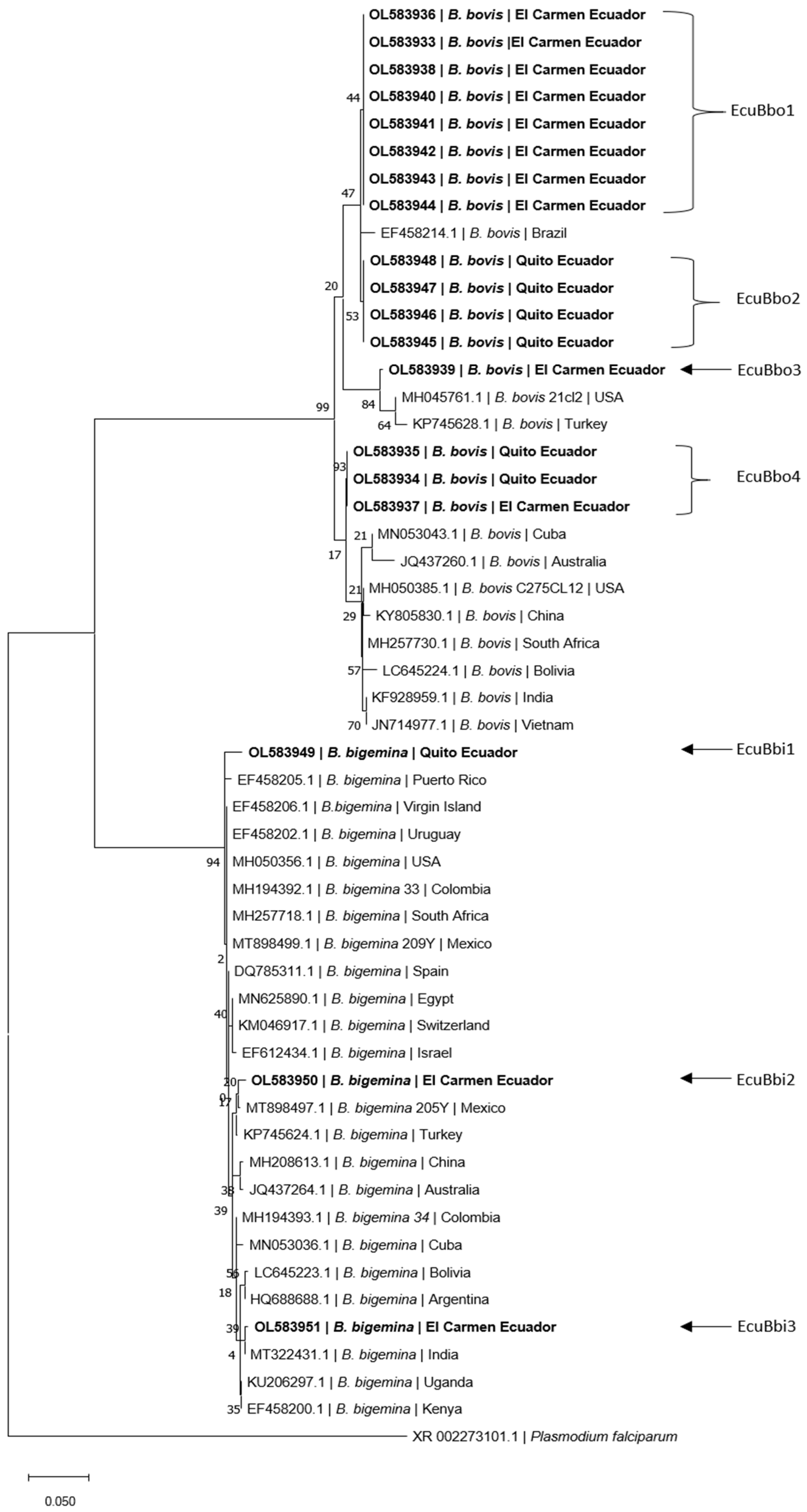
Phylogenetic Analysis

Of the consensus sequences (n = 19) evaluated in BLAST, 10 samples from El Carmen and 6 from Quito showed similarity to the 18s rRNA of B. bovis, while 2 samples from El Carmen and 1 from Quito had similarity to B. bigemina (Figure 1).
In El Carmen, of the 10 sequences identified as B. bovis, 8 were identical (EcuBbo 1), while the remaining 2 (EcuBbo 2 and EcuBbo 3) differed from each other, as well as from isolate EcuBbo 1. The two sequences of B. bigemina were also different from each other (EcuBbi 1 and EcuBbi 2). In Quito, of the six sequences identified as B. bovis, four sequences were identical forming the EcuBbo 4 isolate and the remaining two sequences were identical to the EcuBbo 2 isolate from El Carmen. The sequence of B. bigemina formed an isolate EcuBbi 3 that differs from El Carmen sequences (Table 4).

3. Discussion

In South America, R. microplus is widely distributed in the Mesoamerican corridor towards Venezuela and Colombia, and in southern Brazil and Argentina [23]. In Ecuador, data on tick ecology and distribution information are scarce [24]. The present study would be the first finding of Babesia spp. and R. microplus in a high altitude area, although the farm in Quito is not a preferred area for the tick due to its climatological and altitudinal characteristics [25]. Possibly, in this zone, the environmental temperature influences the adaptation process of the tick where the development phase can be extended (120 days) at temperatures of 18 °C [25]. Due to climate change in recent years, the abundance and distribution of ticks is increasing to new areas [26,27]. In India, ticks on domestic animals are absent at altitudes above 1900 m.a.s.l. However, in closer countries, such as Colombia, Amblyomma cajennense is recorded at a maximum altitude of 1771 m.a.s.l. [28]. For Ixodes spp., there are records of up to 2410 m.a.s.l. in Venezuela [29].
However, climate change alone should not be a determining factor in tick distribution. Other factors such as land use change [30], as well as the mobility of cattle, can also play an important role in the introduction of ticks to new areas and therefore the introduction of diseases such as Babesia spp. [26]. In the city of Quito, cattle arrive from various regions of the country to be butchered in slaughter centers, many of them are carriers of ectoparasites. This mobility without any control can introduce ticks to the pastures occasionally, which would be causing an enzootic instability for hemoparasites [25].
Previous studies in Ecuador demonstrated the presence of Babesia spp. in ticks, but not in cattle [16,18]. Our study shows for the first time B. bovis and B. bigemina in two geographical areas El Carmen and Quito, with a prevalence of 18.93% and 20.28%, respectively for Babesia spp. In both regions, B. bovis is more prevalent than B. bigemina. This is similar to those found in Brazil where a prevalence of 16% for B. bigemina and 29% for B. bovis was reported in cattle [8], but differs from that found in Colombia where a prevalence of 24.2% for B. bigemina and 14.4% for B. bovis was recorded [7]. Usually, tick infection rates are lower for B. bovis than for B. bigemina [2], which may allow tick survival and therefore favor transmission [31]. Moreover, B. bovis has a strong ability to survive against host immune pressure [32] and therefore, in the two study areas, B. bovis is more predominant. B. bovis and B. bigemina species can persist for several months and years with a low level of parasitemia in the host, which allows the long-term maintenance of the parasite in the environment and therefore the presence of persistently infected asymptomatic animals [31]. At the level of the bovine population, this condition originates a state called enzootic equilibrium, where no sick animals are observed [2], which is possibly what happened in the area of El Carmen. In the case of Quito, where clinically sick animals were observed, it is likely that there was a recent introduction of the parasite in the area, together with the presence of R. microplus, which resulted in the appearance of the outbreak, showing that this area is not in enzootic equilibrium. This imbalance in the host–parasite relationship would lead us to think that the presence of Babesiosis is relatively new in the area.
In South America, most of the outbreaks have been associated with B. bovis [4], which is consistent with what was found in the province of Pichincha, where symptomatic animals positive for B. bovis were found; this may be due to a delayed, inadequate, and insufficient immune response by the host [31], and to the pathogenicity of the parasite [4].
The age of cattle is correlated with the clinical stages of Babesiosis [2,5,31], so that young animals between 3 and 9 months are more resistant than adult animals [2]; in the Pichincha area the animals with symptomatology were older than 15 months of age. On the other hand, 42.18% of the asymptomatic animals in the El Carmen area belonged to the 10 to 18 months age group, and 28.57% were in the 0 to 9 months age group. Because, in El Carmen, the young animals are the ones that are mostly infected, these animals acquire early immunity, developing what is known as concomitant immunity [5,7] and for this reason, clinical cases are rare. In Quito, 30% belonged to the age group of 19 to 36 months that were positive for Babesia spp. These animals were of reproductive age and were inseminated for the first time. In Brazilian buffaloes, pregnant females had the highest infection rate, which was associated with hormonal and immunological changes [33]. The PCV analysis in all PCR positive animals in Quito was normal, excluding one with clinical finding. This is in agreement with what was found in Colombia, where the prevalence of B. bigemina and B. bovis was higher in animals with normal PCV values [34]. The breeds Bos taurus are associated with the presence of Babesiosis [2,31,35]. In the area of Manabí, the animals sampled were Bos taurus and Bos indicus crossbreeds, and in the area of Pichincha, they were Bos taurus, so that crossbreeding with Bos taurus in the study areas increases the probability of being infected by the R. microplus and therefore, of acquiring Babesiosis [2].
The phylogenetic analysis of B. bovis and B. bigemina in the two zones of Ecuador was grouped with sequences from other countries of South America, North America, Asia, Africa, and Oceania; this grouping has also been evidenced in isolates from the Brazilian Pantanal [35]. This suggests that most of the sequences found are not exclusive to Ecuador. This analysis also revealed four isolates of B. bovis: three isolates (EcuBbo 1, EcuBbo 2 and EcuBbo 3) in the area of El Carmen and two isolates (EcuBbo 2 and EcuBbo 4) in the area of Quito, showing that EcuBbo 2 is present in both areas, indicating that the parasites were transferred from the Ecuadorian coast to the temperate zone of the Sierra (i.e., Quito), possibly due to the mobility of animals. On the other hand, for B. bigemina in El Carmen, two isolates were determined (EcuBbi 1 and EcuBbi 2) and in the Quito area, only one isolate EcuBbi 3 was identified, none of which was similar between the two zones. Thirty percent of the genetic diversity of Babesia spp. is associated with genetic differences between isolates in different geographic regions [36], but this diversity could also be associated with other factors such as the introduction of strains from different geographic locations, diversity of biological and mechanical vectors [7].
When observing the phylogenetic study carried out from the sequences downloaded from BLAST and comparing them to the 19 samples analyzed in this work, two large clades are observed, in the upper branches are the B. bovis and in the lower B. bigemina. Of the 19 samples evaluated in this study, we identified 7 different genotypes called EcuBbo1 to EcuBbo4 for those similar to B. bovis and EcuBbi1 to EcuBbi3 for those similar to B. bigemina.
For B. bigemina, the three genotypes are slightly different from each other and, in turn, similar to other genotypes described in Latin America and the world; for example, EcuBbi1 migrates very close to isolates from Puerto Rico, Virgin Island, Uruguay, etc. However, EcuBbi3 is similar to isolates from Cuba, Bolivia, and Argentina, but is closer to an isolate from India (see Figure 1).
On the other hand, when considering the genotypes similar to B. bovis identified as EcuBbo in this work, it can be highlighted that the similarity between them is much lower, finding that under the EcuBbo1 genotype, there are eight specimens identical to each other and all of them are from Carmen and equally similar to a Brazilian isolate. Four individuals conformed to the genotype identified as EcuBbo2 and all of them came from Quito (2469 m.a.s.l.). The EcuBbo3 isolate forms a unique genogroup that together with the two previous groups constitutes a single clade where sequences from Brazil, USA, and Turkey are found.
Very curiously, the EcuBbo4 group is constituted by three individuals, one comes from Carmen and the other two from Quito. This demonstrates the genetic closeness of both isolates, making clear the probability that the outbreak occurred in the bovines of Quito had its origin in babesiosis from the coastal region, in Ecuador itself.

4. Materials and Methods

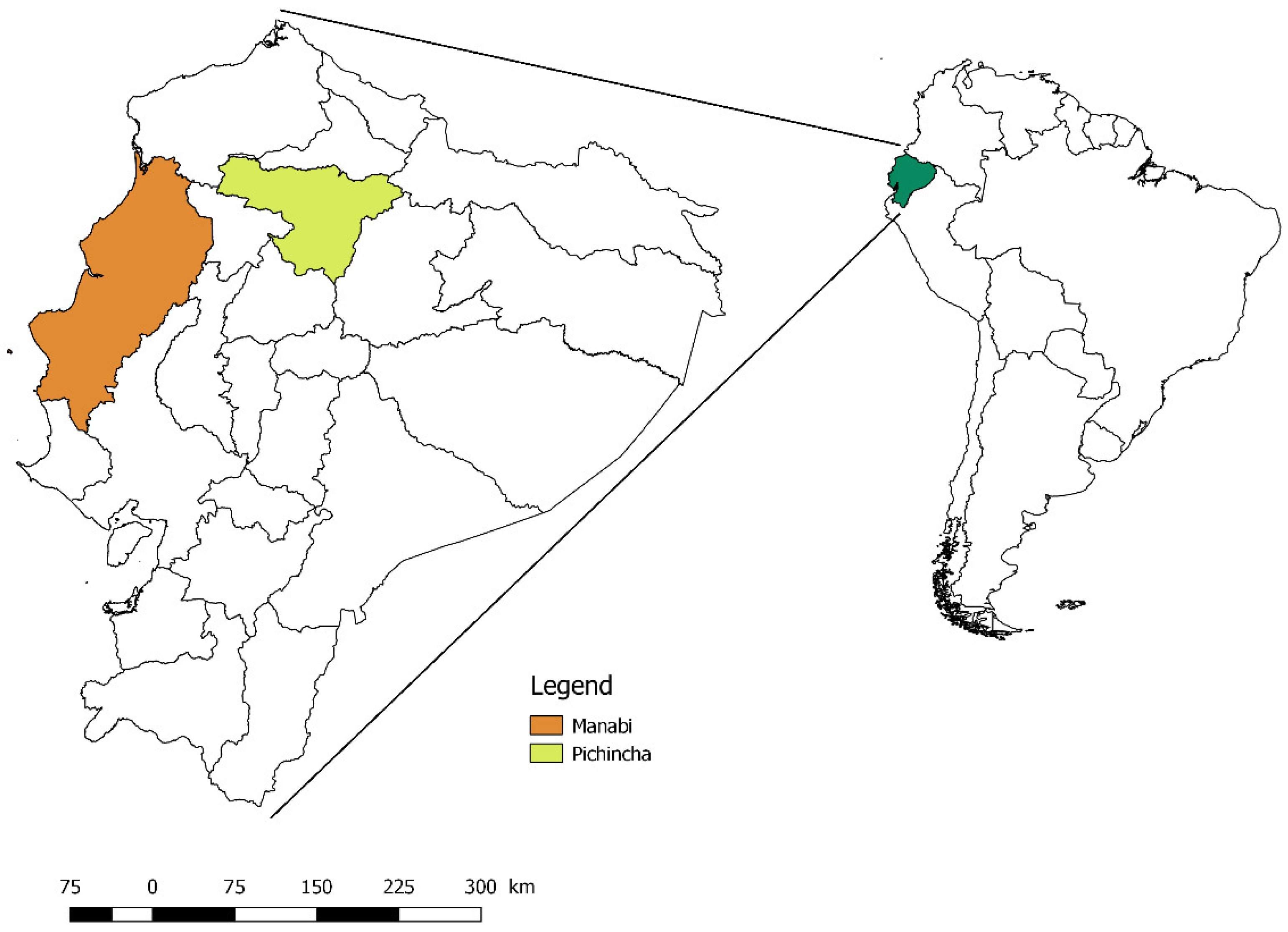
4.1. Study 1—Area of the Province of Manabí

Between March–April 2016, this study aimed to study bovine babesiosis in the parish of San Pedro de Suma the canton of El Carmen in the province of Manabí, in the coastal region of Ecuador, because this area concentrates 21.95% of the national bovine production [37] (Figure 2). The area belongs to the ecosystem: seasonal lowland evergreen forest of the Equatorial Chocó; with altitudes from 0 to 300 m.a.s.l., it has an infratropical thermotype and a humid bioclimate [15], with average temperatures of 27.2 °C (min 20.2 °C–max 34.4 °C), and average relative humidity of 79% (min 40%–max 92%) [38].
We collected blood samples from cattle (n = 264) on twenty farms (n = 20). Zootechnical information such as age and sex were collected from each animal. Age was distributed in four groups: 0 to 9 months, 10 to 18 months, 19 to 36 months, and older than 48 months.
For the sampling design, a database of the farms (n = 100) existing in the parish of San Pedro de Suma from El Carmen, was obtained by interviewing the President of the Producers’ Association. Depending on the number of existing cattle, the farms were categorized into: small (less than 20 cattle), medium (21 to 70 cattle), or large (more than 70 cattle). The sampled cattle came from small (n = 10), medium (n = 6), and large (n = 4) farms, which were randomly selected from the database. The blood samples were obtained from a random sampling in a percentage depending on the category of the farm: small (minimum 50% of bovines), medium (minimum 25%), and large (minimum 13%), without the existence of exclusion variables.

4.2. Study 2—Area of the Province of Pinchincha

In May 2020, an outbreak of Babesiosis was reported in cattle on a farm in the parish of Conocoto (Canton Quito), 18 km from Quito, in the province of Pichincha in the Sierra Region (Figure 2). The area belongs to the evergreen montane shrubland ecosystem of the northern Andes. It corresponds to the Montane bioclimatic floor, with altitudes of 2000–3000 m.a.s.l, and a mesotropical thermotype; with a bioclimate as humid with an average relative humidity of 74% (min 27%–max 94%) [15] (Ecosystem Classification Systems of Continental Ecuador, 2013) and characterized by temperatures min 14 °C–max 21 °C [39]. The altitude of the farm is 2469 m.a.s.l.
We collected blood samples from cattle (n = 143) of this outbreak, and zootechnical information such as age and sex were collected; furthermore, clinical parameters as PCV (packed cell volume) and rectal temperature were recorded in order to identify another possible sick animal.

4.3. Collection and Analysis of Blood Samples

In the two study areas, blood samples in tubes without (VANTUBO®) and with anticoagulant (EDTA-K2 0.75 mm × 25 mm VACUTECH®) were collected from each animal by puncture of the coccygeal vein.

4.4. Packed Cell Volume Determination

The packed cell volume value (≤24% is related to anemia) [40] was determined from each blood sample (EDTA tube); for this, a fraction of blood was transferred to a capillary with heparin (TECNAN®) and centrifuged (TG12M Madell Techinology Corporation, Riverside, CA, USA) at 10,000 rpm for 5 min and its reading was performed according to the usual procedures [40].

4.5. DNA Extraction

DNA extraction from blood was performed following the protocol described by Tana-Hernandez et al., (2017) [41]. It was quantified by UV spectrophotometry in NanoDrop 2000 equipment (Thermo Fisher Scientific, Waltham, MA, USA), obtaining an average concentration of 114.7 ng/mL, and DNA integrity was verified by electrophoresis in a 0.8% agarose gel.

4.6. RFLP-PCR for the Detection of the 18s Fragment for Babesia spp.

For the detection of Babesia, a conventional PCR was performed using as specific primers: PIRO A (5’-AATACCCAATCCTGACACACAGGG-3’) and PIRO B (5’-TTAAATACACGAATGCCCCCCCAAC-3’), which partially amplify the 18s rRNA gene of Babesia spp. [42]; subsequently, the amplicons were cut with restriction enzymes, following the protocol described by Figueroa (2014) [43], sing HpaII (Thermo Scientific, Waltham, MA, USA) to identify B. bovis and BoxI (Thermo Scientific, Waltham, MA, USA) for B. bigemina, according to the conditions of the commercial house.
PCR products from positive samples were purified using the Wizard® SV Gel and Clean-Up System (Promega, Wisconsin, Madison, WI, USA), and sent for sequencing to MACROGEN (Seoul, Korea).

4.7. Analysis of the Sequence Obtained

In order to obtain the sequences to carry out the present work, one sample for each farm (n = 20) (the most prominent amplicon on each farm) was sent three times to sequence, of which we only worked with those sequenced with higher quality indices of more than 90%. In the case of the Pichincha farm (outbreak), 17 samples were sequenced; all of them exceeded the required quality index.
These sequences were assembled using the Clustal W and BioEdit programs to obtain a consensus sequence for each of the samples. The consensus sequences were up-loaded to GenBank (Accession numbers: OL583933, OL583934, OL583935, OL583936, OL583937, OL583938, OL583939, OL583940, OL583941, OL583942, OL583943, OL583944, OL583945, OL583946, OL583947, OL583948, OL583949, OL583950, OL583951). Subsequently, the similarity and homology of the consensus sequences were evaluated with the database of the National Center for Biotechnology Information (NCBI) using the Basic Local Aligment Search Tool (BLAST). Evolutionary analyses were performed in MEGA X [44], using the maximum likelihood method and Tamura’s 3-parameter model [23]. For this analysis, 34 partial 18s rRNA sequences of B. bovis and B. bigemina from Genbank and one sequence of Plasmodium falciparum as an outgroup were used.

4.8. Statistical Analysis

Age and sex were the only two variables studied, and the significance of the difference in the distribution of the results was analyzed by the Fisher’s Exact test, due to the low number of observations.
The PCV and the breed of the bovines were not analyzed as a possible explanatory factor, because in study 1 (El Carmen), complete information on these variables was not obtained, whereas in study 2 (Quito), complete information on these variables was obtained and the cattle had crosses with the Holstein, Brown Swiss, and Jersey breeds.

5. Conclusions

There is little information on the abundance and distribution of R. microplus in Ecuador. The presence of bovine Babesiosis in the area of Quito suggests that this tick can reach increasingly higher altitudes; this may be due to climatic factors, but also to the mobility of animals carrying ticks without any restriction or control.
This study evidenced for the first time the presence of Babesia spp. in the area of El Carmen located in the coastal region and in the area of Quito located in the highlands region. In addition, we found two circulating species B. bovis and B. bigemina in cattle. Babesisa bovis was the most predominant in both study zones; this species is associated with the outbreak reported in the Quito farm where symptomatic animals were found.
The sex of the cattle in both the El Carmen and Quito areas was not an explanatory factor. Age was an explanatory factor associated with the presence of Babesiosis in cattle in Quito, in which symptomatology was evidenced. It is possible that Babesiosis is entering the area, while in El Carmen, it is likely that this parasite is in enzootic equilibrium.
This would be the first molecular characterization of B. bovis and B. bigemina in cattle in two geographic zones of Ecuador, finding similarities in both zones, which would indicate that the hemotropics of the Quito zone come from the coastal region, possibly introduced due to animal mobility.

Author Contributions

C.S. and J.R.-R., funding acquisition and administration; A.R.-B. and C.S., supervision; M.A.C.-L., J.R.-R., and A.R.-B., conception and design of the study; M.A.C.-L., J.R.-R., G.G.-M., C.C.-I., V.M.-N. and M.Y.-D., executor of field survey; C.C.-I., V.M.-N., M.Y.-D. and A.R.-B., conducted the laboratory analyses; M.A.C.-L. and G.G.-M., data analysis; M.A.C.-L., C.C.-I. and S.M.-S., drafted the manuscript; A.R.-B., J.R.-R. and C.S., critical review & comments on draft. All authors contributed equally to this work. All authors have read and agreed to the published version of the manuscript.

Funding

This work was funded by the Academy of Research and Higher Education (ARES) through the project: “Establishment of a Platform to support training and awareness-raising, diagnosis and development of a control strategy for Brucellosis and Trypanosomiasis in Ecuador” (Bru-Tryp Project), with the participation of the University of Liège in Belgium and the Universidad de las Fuerzas Armadas ESPE in Ecuador.

Institutional Review Board Statement

The president of the producers’ association of the San Pedro de Suma-Manabí parish, and the owner of the Quito farm authorized the collection of blood samples and application of diagnostic tests for bovine babesiosis.

Informed Consent Statement

The farmers were properly informed and gave their written consent prior to sampling their animals.

Data Availability Statement

The data that support the findings of this study are available from the corresponding author upon reasonable request.

Acknowledgments

The authors would like to thank all farmers who participated in the study.

Conflicts of Interest

The authors declare no conflict of interest. The funders had no role in the design of the study; in the collection, analyses, or interpretation of data; in the writing of the manuscript, or in the decision to publish the results.

References

  1. Martínez-García, G.; Santamaría-Espinosa, R.M.; Lira-Amaya, J.J.; Figueroa, J.V. Challenges in Tick-Borne Pathogen Detection: The Case for Babesia spp. Identification in the Tick Vector. Pathogens 2021, 10, 92. [Google Scholar] [CrossRef] [PubMed]
  2. Bock, R.; Jackson, L.; De Vos, A.; Jorgensen, W. Babesiosis of Cattle. Parasitology 2004, 129, S247–S269. [Google Scholar] [CrossRef]
  3. Alvarez, J.A.; Rojas, C.; Figueroa, J.V. Diagnostic Tools for the Identification of Babesia sp. in Persistently Infected Cattle. Pathogens 2019, 8, 143. [Google Scholar] [CrossRef] [Green Version]
  4. Guglielmone, A.A. Epidemiology of Babesiosis and Anaplasmosis in South and Central America. Vet. Parasitol. 1995, 57, 109–119. [Google Scholar] [CrossRef]
  5. Amorim, L.S.; Wenceslau, A.A.; Carvalho, F.S.; Carneiro, P.L.S.; Albuquerque, G.R. Bovine Babesiosis and Anaplasmosis Complex: Diagnosis and Evaluation of the Risk Factors from Bahia, Brazil. Rev. Bras. Parasitol. Vet. 2014, 23, 328–336. [Google Scholar] [CrossRef] [PubMed] [Green Version]
  6. De Assis Leite Souza, F.; de Siqueira Cajado Liarte, A.; de Carvalho Castro, K.N.; Beserra, E.E.A.; Bernardi, J.S.M.; de Sousa, G.V.; Costa-Júnior, L.M.; de Sousa Silva, S.M.M. Dynamics of Natural Infection by Babesia Bovis and Babesia Bigemina in Dairy Cattle from an Enzootic Instability Area in Northeastern Brazil. Rev. Bras. Parasitol. Vet. 2018, 27, 2–6. [Google Scholar] [CrossRef] [PubMed] [Green Version]
  7. Jaimes-Dueñez, J.; Triana-Chávez, O.; Holguín-Rocha, A.; Tobon-Castaño, A.; Mejía-Jaramillo, A.M. Molecular Surveillance and Phylogenetic Traits of Babesia bigemina and Babesia bovis in Cattle (Bos taurus) and Water Buffaloes (Bubalus bubalis) from Colombia. Parasites Vectors 2018, 11, 510. [Google Scholar] [CrossRef] [PubMed]
  8. Vieira, L.L.; Canever, M.F.; Cardozo, L.L.; Cardoso, C.P.; Herkenhoff, M.E.; Neto, A.T.; Vogel, C.I.G.; Miletti, L.C. Prevalence of Anaplasma marginale, Babesia bovis and Babesia bigemina in Cattle in the Campos de Lages Region, Santa Catarina State, Brazil, Estimated by Multiplex-PCR. Parasite Epidemiol. Control 2019, 6, e00114. [Google Scholar] [CrossRef] [PubMed]
  9. Romero-Salas, D.; Mira, A.; Mosqueda, J.; García-Vázquez, Z.; Hidalgo-Ruiz, M.; Vela, N.A.O.; de León, A.A.P.; Florin-Christensen, M.; Schnittger, L. Molecular and Serological Detection of Babesia bovis- and Babesia bigemina-Infection in Bovines and Water Buffaloes Raised Jointly in an Endemic Field. Vet. Parasitol. 2016, 217, 101–107. [Google Scholar] [CrossRef] [PubMed] [Green Version]
  10. Rashid, M.; Rashid, M.I.; Akbar, H.; Ahmad, L.; Hassan, M.A.; Ashraf, K.; Saeed, K.; Gharbi, M. A Systematic Review on Modelling Approaches for Economic Losses Studies Caused by Parasites and Their Associated Diseases in Cattle. Parasitology 2019, 146, 129–141. [Google Scholar] [CrossRef]
  11. Gray, J.S.; Estrada-Peña, A.; Zintl, A. Vectors of Babesiosis. Annu. Rev. Entomol. 2019, 64, 149–165. [Google Scholar] [CrossRef] [PubMed]
  12. Grisi, L.; Leite, R.C.; de Souza Martins, J.R.; de Barros, A.T.M.; Andreotti, R.; Cançado, P.H.D.; de León, A.A.P.; Pereira, J.B.; Villela, H.S. Reassessment of the Potential Economic Impact of Cattle Parasites in Brazil. Rev. Bras. Parasitol. Vet. 2014, 23, 150–156. [Google Scholar] [CrossRef] [PubMed] [Green Version]
  13. Semenza, J.C.; Suk, J.E. Vector-Borne Diseases and Climate Change: A European Perspective. FEMS Microbiol. Lett. 2018, 365, fnx244. [Google Scholar] [CrossRef] [PubMed]
  14. Carrique Mas, J.J.; Widdowson, M.-A.; Cuéllar, A.M.; Ribera, H.; Walker, A.R. Risk of Babesiosis and Anaplasmosis in Different Ecological Zones of Santa Cruz Department, Bolivia. Vet. Parasitol. 2000, 93, 29–38. [Google Scholar] [CrossRef]
  15. Ministerio del Ambiente del Ecuador. Sistema de Clasificacion de Ecosistemas del Ecuador Continental; Subsecretaría de Patrimonio Natural Quito: Quito, Ecuador, 2013; Volume 1. [Google Scholar]
  16. Maya-Delgado, A.; Madder, M.; Benitez-Ortiz, W.; Saegerman, C.; Berkvens, D.; Ron-Garrido, L. Molecular Screening of Cattle Ticks, Tick-Borne Pathogens and Amitraz Resistance in Ticks of Santo Domingo de Los Tsáchilas Province in Ecuador. Ticks Tick-Borne Dis. 2020, 11, 8. [Google Scholar] [CrossRef]
  17. Vasco Aguas, K.A.; Vasco, L. Estandarización de la Técnica de Análisis de Fusión de Alta Resolución Para la Detección de Babesia en Garrapatas Utilizando Polimorfismos de Nucleótidos. Tesis para la Obtencion del Titulo en Medicina Veterinaria, Facultad de Medicina Veterinaria y Zootecnia, Universidad Central del Ecuador, Quito, Ecuador, 2012. [Google Scholar]
  18. Gioia, G.V.; Vinueza, R.L.; Marsot, M.; Devillers, E.; Cruz, M.; Petit, E.; Boulouis, H.J.; Moutailler, S.; Monroy, F.; Coello, M.A.; et al. Bovine Anaplasmosis and Tick-Borne Pathogens in Cattle of the Galapagos Islands. Transbound Emerg. Dis. 2018, 65, 1262–1271. [Google Scholar] [CrossRef] [PubMed]
  19. Carret, C.; Walas, F.; Carcy, B. Babesia Canis Canis, Babesia Canis Voaeli, Babesia canis Rossi. Society 1999, 46, 298–303. [Google Scholar]
  20. Medina-Naranjo, V.L.; Reyna-Bello, A.; Tavares-Marques, L.M.; Campos, A.M.; Ron-Román, J.W.; Moyano, J.C.; Jarrín-Porras, E.C.; Sandoval-Morejón, E.D.; Chávez-Larrea, M.A. Diagnóstico de Los Hemotrópicos Anaplasma Marginale, Trypanosoma spp. y Babesia spp. Mediante Las Técnicas de Elisai y PCR En Tres Fincas Ganaderas de La Provincia de Pastaza, Ecuador. Rev. Cient. Fac. Cienc. Vet. Univ. Zulia 2017, 27, 162–171. [Google Scholar]
  21. Rodríguez-Hidalgo, R.; Pérez-Otáñez, X.; Garcés-Carrera, S.; Vanwambeke, S.O.; Madder, M.; Benítez-Ortiz, W. The Current Status of Resistance to Alpha-Cypermethrin, Ivermectin, and Amitraz of the Cattle Tick (Rhipicephalus microplus) in Ecuador. PLoS ONE 2017, 12, e0174652. [Google Scholar] [CrossRef]
  22. Tamura, K. Estimation of the Number of Nucleotide Substitutions When There Are Strong Transition-Transversion and G+C-Content Biases. Mol. Biol. Evol. 1992, 9, 678–687. [Google Scholar] [CrossRef] [Green Version]
  23. Estrada-Peña, A.; Bouattour, A.; Camicas, J.-L.; Guglielmone, A.; Horak, I.; Jongejan, F.; Latif, A.; Pegram, R.; Walker, A.R. The Known Distribution and Ecological Preferences of the Tick Subgenus Boophilus (Acari: Ixodidae) in Africa and Latin America. Exp. Appl. Acarol. 2006, 38, 219–235. [Google Scholar] [CrossRef]
  24. Enríquez, S.; Guerrero, R.; Arrivillaga-Henríquez, J.; Araujo, P.; Villacrés, E.; Enríquez, A.; Benítez-Ortíz, W. New Records of Ticks of Genus Amblyomma Koch, 1844 (Acari: Ixodidae) for Ecuador. Acta Parasit. 2020, 65, 430–440. [Google Scholar] [CrossRef] [PubMed]
  25. Benavides Ortiz, E. Las Garrapatas del Ganado Bovino y los Agentes de Enfermedad que Transmiten en Escenarios Epidemiológicos de Cambio Climático: Guia Para el Manejo de Garrapatas Y Adaptación al Cambio Climático; Instituto Interamericano de Cooperación para la Agricultura (IICA), Universidad de La Salle: San José, Costa Rica, 2016; ISBN 978-92-9248-655-6. [Google Scholar]
  26. Léger, E.; Vourc’h, G.; Vial, L.; Chevillon, C.; McCoy, K.D. Changing Distributions of Ticks: Causes and Consequences. Exp. Appl. Acarol. 2013, 59, 219–244. [Google Scholar] [CrossRef]
  27. Ogden, N.H.; Ben Beard, C.; Ginsberg, H.S.; Tsao, J.I. Possible Effects of Climate Change on Ixodid Ticks and the Pathogens They Transmit: Predictions and Observations. J. Med. Entomol. 2021, 58, 1536–1545. [Google Scholar] [CrossRef]
  28. Acevedo-Gutiérrez, L.Y.; Paternina, L.E.; Londoño, A.F.; Parra-Henao, G.; Rodas, J.D. Modelos potenciales de distribución geográfica y climática del complejo Amblyomma cajennense (Acari: Ixodidae), potencial vector de Rickettsia rickettsii en Colombia. Biomedica 2018, 38. [Google Scholar] [CrossRef]
  29. Durden, L.A.; Keirans, J.E. Description of the Larva, Diagnosis of the Nymph and Female Based on Scanning Electron Microscopy, Hosts, and Distribution of Ixodes (Ixodes) venezuelensis. Med. Vet. Entomol. 1994, 8, 310–316. [Google Scholar] [CrossRef] [PubMed]
  30. Yang, X.; Gao, Z.; Wang, L.; Xiao, L.; Dong, N.; Wu, H.; Li, S. Projecting the Potential Distribution of Ticks in China under Climate and Land Use Change. Int. J. Parasitol. 2021, 51, 749–759. [Google Scholar] [CrossRef] [PubMed]
  31. Chauvin, A.; Moreau, E.; Bonnet, S.; Plantard, O.; Malandrin, L. Babesia and Its Hosts: Adaptation to Long-Lasting Interactions as a Way to Achieve Efficient Transmission. Vet. Res. 2009, 40, 37. [Google Scholar] [CrossRef] [Green Version]
  32. Suarez, C.E.; Noh, S. Emerging Perspectives in the Research of Bovine Babesiosis and Anaplasmosis. Vet. Parasitol. 2011, 180, 109–125. [Google Scholar] [CrossRef]
  33. da Silva, J.B.; André, M.R.; da Fonseca, A.H.; de Albuquerque Lopes, C.T.; da Silva Lima, D.H.; de Andrade, S.J.T.; Oliveira, C.M.C.; Barbosa, J.D. Molecular and Serological Prevalence of Babesia bovis and Babesia bigemina in Water Buffaloes in the North Region of Brazil. Vet. Parasitol. 2013, 197, 678–681. [Google Scholar] [CrossRef] [PubMed]
  34. Jaimes-Dueñez, J.; Triana-Chávez, O.; Mejía-Jaramillo, A.M. Parasitological and Molecular Surveys Reveal High Rates of Infection with Vector-Borne Pathogens and Clinical Anemia Signs Associated with Infection in Cattle from Two Important Livestock Areas in Colombia. Ticks Tick-Borne Dis. 2017, 8, 290–299. [Google Scholar] [CrossRef] [PubMed]
  35. Mendes, N.S.; de Souza Ramos, I.A.; Herrera, H.M.; Campos, J.B.V.; de Almeida Alves, J.V.; de Macedo, G.C.; Machado, R.Z.; André, M.R. Genetic Diversity of Babesia bovis in Beef Cattle in a Large Wetland in Brazil. Parasitol. Res. 2019, 118, 2027–2040. [Google Scholar] [CrossRef] [PubMed]
  36. Flores, D.A.; Minichiello, Y.; Araujo, F.R.; Shkap, V.; Benítez, D.; Echaide, I.; Rolls, P.; Mosqueda, J.; Pacheco, G.M.; Petterson, M.; et al. Evidence for Extensive Genetic Diversity and Substructuring of the Babesia Bovis Metapopulation. Transbound. Emerg. Dis. 2013, 60, 131–136. [Google Scholar] [CrossRef] [PubMed] [Green Version]
  37. Instituto Nacional de Estadística y Censo. Agricultura, Silvicultura y Pesca; ES: Quito, Ecuador, 2020. [Google Scholar]
  38. Instituto Nacional de Metereología e Hidrología INAMHI. Boletin Climatológico Semestral 2016; ES: Quito, Ecuador, 2016; p. 22. [Google Scholar]
  39. Instituto Nacional de Meteorología e Hidrología INAMHI. Boletín Climático Mensual; ES: Quito, Ecuador, 2021; p. 6. [Google Scholar]
  40. Rockett, J.; Bosted, S. Veterinary Clinical Procedures in Large Animal Practice. Bosted, S., Ed.; Thompson-Delmar Learning: Boston, MA, USA, 2007; ISBN 978-1-4018-5787-5. [Google Scholar]
  41. Tana-Hernández, L.; Navarrete-Arroyo, K.; Ron-Román, J.; Reyna-Bello, A.; Chávez-Larrea, M.A. PCR-Diagnosis of Anaplasma marginale in Cattle Populations of Ecuador and Its Molecular Identification through Sequencing of Ribosomal 16S Fragments. BMC Vet. Res. 2017, 13, 392. [Google Scholar] [CrossRef] [PubMed] [Green Version]
  42. Carret, C.; Delbecq, S.; Labesse, G.; Carcy, B.; Precigout, E.; Moubri, K.; Schetters, T.P.M.; Gorenflot, A. Characterization and Molecular Cloning of an Adenosine Kinase from Babesia canis rossi: Molecular Cloning of a B. canis rossi Adenosine Kinase. Eur. J. Biochem. 2001, 265, 1015–1021. [Google Scholar] [CrossRef]
  43. Figueroa, J.V. Optimización de una prueba de pcr-rflp para detección y diferenciación de Babesia sp. en garrapatas Rhipicephalus microplus. Entomol. Vet. 2014, 1, 978–983. [Google Scholar]
  44. Kumar, S.; Stecher, G.; Li, M.; Knyaz, C.; Tamura, K. MEGA X: Molecular Evolutionary Genetics Analysis across Computing Platforms. Mol. Biol. Evol. 2018, 35, 1547–1549. [Google Scholar] [CrossRef] [PubMed]
Figure 1. Maximum likelihood, 3-parameter Tamura [22] phylogenetic tree of the 18s rRNA gene sequences of B. bovis and B. bigemina from Ecuador. It involved 54 nucleotide sequences and a total of 519 positions in the final data set. The following terminology was used to identify the isolates: Ecu = Ecuador, Bbo = B. bovis, Bbi = B. bigemina and the isolate number. Taxa names consist in GeneBank code/isolates of Babesia spp./geographical origin.
Pathogens 10 01593 g001
Figure 2. Location of the provinces of Manabí and Pichincha in Ecuador.
Pathogens 10 01593 g002
Table 1. Presence of Babesia spp., in the canton El Carmen province of Manabí and in the canton Quito province of Pichincha, Ecuador.
Farm CodeTotal Animals Present on the FarmSamplePositive Babesia spp.Positive B. bovisPositive B. bigemina
PCR 18sRestriction EnzymesRestriction Enzymes
No% by FarmNo%No%No%
El Carmen-Manabí Zone
F-188100112.5112.500
F-221523.8000000
F-316743.8000000
F-41943920.1000000
F-543614000000
F-61521711.2000000
F-76466.725025000
F-8602033.3735420315
F-91123632.1822.2616.725.6
F-1038821.1000000
F-111616100743.8637.516.3
F-12165281727.127.100
F-1317952.9000000
F-14272592.6624312312
F-158450000000
F-165510012000120
F-1722100000000
F-1817635.335035000
F-19271244.4975866.718.3
F-209777.8457.1457.100
All farms943264285018.933914.77114.17
Quito-Pichincha Zone
F-116414387, 202920, 282114, 6985, 59
Legend: No: number; %: percentage; PCR: Polymerase chain reaction. In bold are the totals in column identified with “No” and the average in column with %, respectively.
Table 2. Distribution of samples and analysis of risk factors: sex and age groups for bovine Babesia spp. in Ecuador.
VariablesSamplePositiveFisher’s
Exact Test
(p-Value)
Number%Number%
El Carmen-Manabí Zone (n = 264)
Sex 0.38
Male3814.39513.16
Female22685.614519.91
Age group 0.48
0 to 9217.95628.57
10 to 1872.65342.86
19 to 36186.82422.22
>36 months13751892921.17
ND8130.6889.88
Quito-Pichincha Zone (n = 143)
Sex 0.35
Male74.900
Female13695.12921.32
Age group 0.004
0 to 92618.1800
10 to 182718.88413.79
19 to 367048.952172.41
>36 months2013.99413.79
Legend: The % value corresponds to the total number of animals sampled in the area or in each farm; ND, not dertermined.
Table 3. Clinical information of three symptomatic bovines in the farm of Cantón Quito.
NoFarm CodeAgeWeightTPCVHeart RateRespiratory RateOther Clinical FindingsPositive B. bovis by PCR
14251223239.5198447Pale mucous membranes, swollen cervical glands
24331519939168564Salivation, pale mucous membranes, jaundice+
33815202402910057Swollen groin glands+
Legend: Age in months; Weight in kilograms; T°: temperature in Celsius degree; PCV: packed cell volume (values less than or equal to 24% is related to anemia); PCR: polymerase chain reaction; −: negative; +: positive.
Table 4. Percentage of identity of B. bovis and B. bigemina species found in El Carmen and Quito.
Study AreasEcuadorian CodeIsolatedSpeciesTotal
Score
Identity
Percentage
Query
Coverage
No. Access
Gen Bank
El Carmen (province of Manabi)M5EcuBbo 1B. bovis684100%98%OL583933
M92OL583936
M100OL583938
M149OL583940
M154OL583941
M187OL583942
M245OL583943
M279OL583944
M97EcuBbo 2B. bovis684100%98%OL583937
M103EcuBbo 3B. bovis67899.73%98%OL583939
M232EcuBbi 1B. bigemina71799.74%99%OL583950
M260EcuBbi 2B. bigemina72199.75%100%OL583949
Quito
(province of Pinchincha)
85EcuBbo 2B. bovis684100%98%OL583934
86OL583935
310EcuBbo 4B. bovis67899.73%98%OL583946
300OL583945
314OL583947
315OL583948
260EcuBbi 3B. bigemina70198.98%99%OL583951
Legend: The total score is the sum of alignment scores of all segments from the same database sequence that match the query sequence (calculated over all segments), the percent identity is a number that describes how similar the query sequence is to the target sequence, and the query cover is a number that describes how much of the query sequence is covered by the target sequence (taken from the National Center for Biotechnology Information, NCBI).
Publisher’s Note: MDPI stays neutral with regard to jurisdictional claims in published maps and institutional affiliations.

