Article

Antibiogram Profile of Acinetobacterbaumannii Recovered from Selected Freshwater Resources in the Eastern Cape Province, South Africa

by
Mary Ayobami Adewoyin
1,2,*,
Kingsley Ehi Ebomah
1,2 and
Anthony Ifeanyi Okoh
1,2,3
1
SAMRC Microbial Water Quality Monitoring Centre, University of Fort Hare, Alice 5700, South Africa
2
Applied and Environmental Microbiology Research Group, Department of Biochemistry and Microbiology, University of Fort Hare, Alice 5700, South Africa
3
Department of Environmental Health Sciences, College of Health Sciences, University of Sharjah, Sharjah P.O. Box 27272, United Arab Emirates
*
Author to whom correspondence should be addressed.
Submission received: 11 May 2021 / Revised: 4 July 2021 / Accepted: 16 July 2021 / Published: 31 August 2021
(This article belongs to the Section Bacterial Pathogens)

Abstract

:
Acinetobacter species have been found in a variety of environments, including soil, food, plants, hospital environments and water. Acinetobacter baumannii is an opportunistic and emerging waterborne pathogen. It has been implicated in several nosocomial infections that demonstrate resistance to commonly administered antibiotics. We investigated phenotypic antibiotic resistance (PAR) and relevant antibiotic resistance genes (ARGs) in A. baumannii isolated from three freshwater resources in the Eastern Cape Province, South Africa; A. baumannii (410) was confirmed by the recA and gyrB genes of 844 suspected Acinetobacter species in the water samples. The PAR of the confirmed isolates was assessed using a panel of 11 antibiotics by the disc diffusion method, while ARGs were investigated in isolates exhibiting PAR. The A. baumannii isolates were resistant to piperacillin-tazobactam (11.2%), ceftazidime (12%), cefotaxime (18.8%), cefepime (8.8%), imipenem (2.7%), meropenem (4.15%), amikacin (2.4%), gentamicin (8.8%), tetracycline (16.8%), ciprofloxacin (11%) and trimethoprim/sulfamethoxazole (20.5%). For multidrug resistance (MDR), two isolates were resistant to all antibiotics and 28 isolates were resistant to imipenem and meropenem. Moreover, β-lactamases blaTEM (64.4%) and blaOXA-51 (28.70%) as well as sulphonamides sul1 (37.1%) and sul2 (49.4%) were common ARGs. Overall, PAR and ARGs had positive correlations (r) in all rivers. Detection of MDR-A. baumannii in freshwater resources could be linked to possible wastewater discharge from the nearby animal farms, indicating potential implications for public health.

1. Introduction

Acinetobacter species are known lactose non-fermenters that were formerly regarded as colonisers of different natural habitats, with low ability to cause infection [1,2]. Recent reports have shown A. baumannii is the most virulent species in the genus Acinetobacter, causing severe infections among patients on prolonged admission in hospital intensive care units (ICUs) [3]. Similarly, individuals with diabetes and compromised immune systems are prone to A. baumannii infections [4,5,6]. Some of the reasons for the rise in Acinetobacter species infections are their ability to adapt to harsh environmental conditions (e.g., dry surfaces) and their resistance to different commonly prescribed antimicrobial agents [7,8].
Antibiotic resistance in bacterial strains is a global health problem of major concern. The cause for concern with regard to antimicrobial resistance is the failure of some front-line and last-line antimicrobial agents to combat the rising resistance in pathogenic microorganisms, including A. baumannii [7,8,9]. In addition, incidences of multidrug-resistant A. baumannii (MDRAB) infections are now happening on a global scale [10]. The mechanism of antibiotic resistance in pathogenic bacteria could be intrinsic or acquired through horizontal gene transfer (HGT) or genetic mutations [11]. A. baumannii possesses in its chromosome genes the code for enzymes and efflux pumps that is responsible for its high natural resistance [12,13]. The tendency for A. baumannii and other related species to acquire resistance-encoding genes [14] from other closely related species even of different genus increases their resistance profile. These genes are often the basis for the acquisition of new antimicrobial resistance genes (ARGs), as they themselves or their regulators are subject to mutations.
Therefore, ARGs are essential determinants for pathogenic A. baumannii to thrive in the host system. For example, resistance to β-lactams was associated with the expression of beta-lactamase genes such as blaTEM, blaSHV, blaOXA-51, blaCTX-M (GROUP 1) and blaCTX-M (GROUP 2) and a few other ARGs harboured by Acinetobacter spp. [15,16,17]. Similarly, the genes responsible for antibiotic resistance against aminoglycosides (aacC2, aphA1 and aphA2), fluoroquinolones (qnrA, qnrB, qnrD and qnrS), sulfonamides (sul1 and sul2) and tetracyclines (tet(A), tet(B), tet(O), tet(L) and tet(M)) were previously reported in A. baumannii [18].
This study investigated the antibiogram profile as well as antibiotic resistance genes (ARGs) of A. baumannii species recovered from Great Fish, Keiskamma and Tyhume Rivers in the Eastern Cape Province, South Africa as shown in Scheme 1. The isolation of pathogenic A. baumannii strains from freshwater sources have been reported [19,20,21,22,23]. The goal of this study was to evaluate the impact of the anthropogenic activities such as in-flow of wastewater treatment plant (WWTP) effluent, animal husbandry and indiscriminate disposal of waste to the freshwater resources in that area. These human activities have the tendency to expose existing bacterial species to antibiotics and influence the behaviour of the microbial communities in these rivers.

2. Results

2.1. Identification and Antibiotic Resistance Profile of Acinetobacter Species

Eight hundred and forty-four (844) presumptive isolates of Acinetobacter species were selected for molecular identification and confirmation. The results in Figures S1 and S2 show that 410 (48.58%) isolates were confirmed to be A. baumannii across the three rivers, from which 153, 102 and 155 isolates were recovered from Great Fish, Keiskamma and Tyhume Rivers, respectively.

2.2. Phenotypic Antibiotic Resistance (PAR) Profile of A. baumannii

While the majority of the confirmed A. baumannii isolates were susceptible to the test antibiotics (Table S1), a certain percentage of the isolates were resistant to the antimicrobial agents (Table 1). According to the values obtained in comparison to CLSI guidelines for antimicrobial susceptibility testing for A. baumannii, the isolates were classified as susceptible (S), intermediate resistant (I) or resistant (R). However, to better explain the outcome of the antibiotic susceptibility testing, a non-susceptible (R + I) column was created in this study, which gives a summarized view of the level of resistance to each of the antibiotics used. Although resistance to SXT 84 (20.5%) was higher than other antibiotics, there was no significant difference (p < 0.05) when compared to other antimicrobial agents. Nonetheless, non-susceptibility (R + I) or intermediate resistance (I) to CTX was significantly higher (p < 0.05) than other antibiotics, followed by CAZ, which also belongs to the same class of antimicrobial agent-cephems. Comparably, CTX and TET had a similar level of resistance. Notably, at least ten A. baumannii isolates were resistant to each of the antibiotics tested, while 11 (2.7%) and 17 (4.15%) of the A. baumannii isolates were resistant to imipenem and meropenem, respectively.

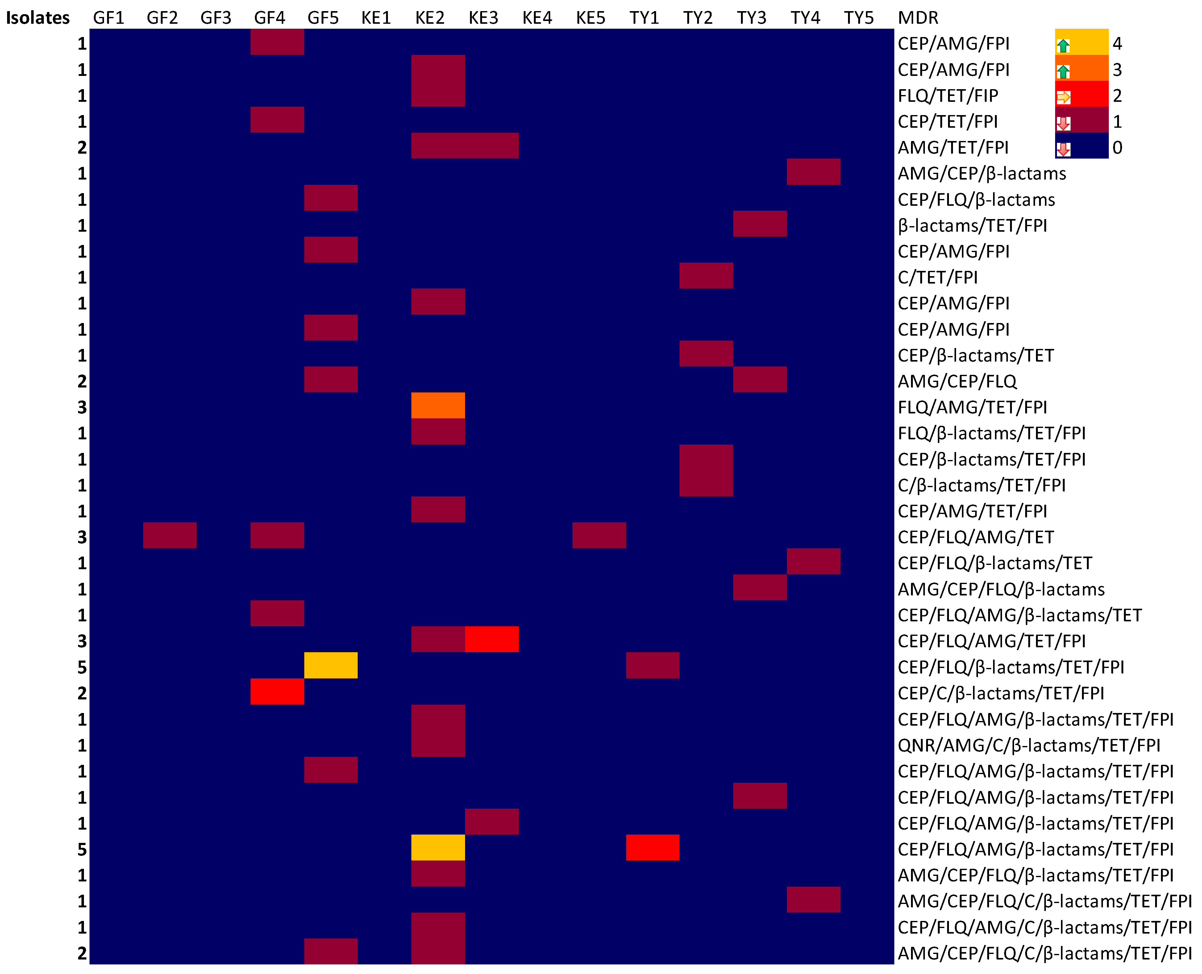
2.3. Multiple Drug Resistance (MDR) Phenotypes

The multiple drug resistance (MDR) phenotypes of A. baumannii isolated from the freshwater sources (Great Fish, Keiskamma and Tyhume Rivers) are highlighted in Figure 1 (heatmap). A. baumannii exhibited some degree of MDR phenotypes, ranging from three to seven of the antimicrobials used. However, out of 410 A. baumannii isolates, 54 (13.17%) exhibited multiple drug resistances. Figure 1 describes the patterns of MDR observed in this study according to source of isolation of the A. baumannii. Of the 54 MDR A. baumannii isolates, 16 (29.63%) were resistant to three classes of antibiotics, 12 (22.22%) were resistant to four, and 11 (20.37%) were resistant to five and six, while four (7.41%) were resistant to all (seven) of the classes of the antimicrobial agents. The highest MDR value was observed in sampling site KE2 (19; 35.19%) followed by site GF5 (10; 18.52%) and site GF4 (6; 11.11%). There were no MDR phenotypes observed in the remaining sampling sites.

2.4. Profile of ARGs among A. baumannii

Scheme 1 illustrates the collection of water sample from the three rivers, the antibiotic susceptibility testing and resistance genes in A. baumannii isolates. Table 2 summarizes the phenotypic antibiotic resistance (PAR) and ARGs of the A. baumannii recovered in this study. Both sul1 and sul2 genes were investigated in 83 sulfonamide-resistant A. baumannii. Figure S3 shows the gel picture of some A. baumannii isolates harbouring sul1 and sul2 resistance genes. Among the screened isolates, 32 (37.1%) possessed sul1, 43 (49.4%) harboured sul2 (Table 2), 18 (21.7%) had both genes, while 26 (31.3%) isolates did not harbour any of the genes. Figure 2 (heatmap below) outlines the distribution patterns of the ARGs among the isolates recovered from the river water samples, and this shows that the Great Fish River possessed the highest number of A. baumannii isolates that harboured ARGs. However, factorial analysis of variance (ANOVA) indicated no significant difference (p < 0.05) between sul1 and sul2 ARGs in the Great Fish and Tyhume Rivers, whereas the presence of sul2 in A. baumannii was significantly higher (p < 0.05) than sul1 in the Keiskamma River water samples.
Similarly, Figure S4 shows the gel picture of some A. baumannii isolates harbouring apHA1 and apHA2 resistance genes. Among 40 A. baumannii isolates evaluated for aminoglycoside-resistance genes (apHA1 and apHA2), 13 (25.5%) and 21 (41.2%) possessed apHA1 and apHA2 genes, respectively (Table 2), two isolates possessed all the genes, and 15 did not harbour any genes. From ANOVA, detection of apHA1 gene in the Great Fish River and apHA2 gene in the Keiskamma River showed higher significance (p < 0.05) when compared to other aminoglycoside resistance genes.
Five relevant fluoroquinolone resistance genes, qnrA, qnrB, qnrC, qnrD and qnrS, were investigated in the Acinetobacter spp. (Figure S5 and Table 2). Forty-four A. baumannii isolates were screened for the presence of the selected ARGs, and only qnrB and qnrD were detected in all the isolates. Out of 44 isolates, only seven (15.9%) harboured qnrB and eight (18.2%) harboured qnrD genes, three (6.8%) possessed both qnrB and qnrD genes, and 35 (79.5%) did not possess either of the two genes. Isolates from both the Great Fish and Tyhume River water samples harboured qnrD, while some isolates from all the water samples possessed qnrB (Figure 2). However, while no significant difference (p < 0.05) was seen among the A. baumannii isolates that harboured qnrB in the samples, those that possessed qnrD genes in the Great Fish (p < 0.05) and Tyhume (p < 0.05) Rivers were significantly higher (p < 0.05) than other fluoroquinolone resistance genes investigated in all the rivers.
Seventy isolates of A. baumannii that exhibited phenotypic resistance to tetracyclines were screened for six tetracycline resistance genes (Table S1 and Figure S6). Among the tet genes, tet(B) was more prevalent and was detected in 19 (27.1%) isolates, followed by tet(A) in eight (11.4%) isolates; other tetracycline resistance genes were in the following order: tet(L), three (4.3%) isolates; tet(C), 2 (2.9%) isolates; tet(M), two (2.9%) isolates (Table 2). None of the isolates harboured tet(O), while 46 (63%) isolates did not harbour any of the tet genes. Similarly, tet(B) was significantly higher (p < 0.05) than all other tetracycline resistance genes in both the Great Fish and Keiskamma Rivers, whereas a significant number of A. baumannii isolates bearing the gene were recovered from the Keiskamma River compared to the Great Fish and Tyhume Rivers.
The β-lactamase resistance genes were harboured by 101 A. baumannii isolates. These isolates demonstrated phenotypic resistance to β-lactam antibiotics, including carbapenems. β-lactamase resistance genes, namely blaTEM, blaSHV, blaCTX-M (blaCTX-M-1group, 2group, blaCTX-M-9group and blaCTX-M-8/-25group), blaVEB, blaGES, blaPER, blaOXA-48-like, blaOXA-51, blaVIM, blaIMP and blaKPC, were investigated, and the results are highlighted in Figure 2 and summarized in Table 2. Figure S7A–F show the gel pictures of some A. baumannii isolates harbouring β-lactamase resistance genes. All the ARGs except blaOXA-23, blaOXA-40, blaOXA-58, blaCTX-M-9group and blaIMP were detected in at least one isolate of A. baumannii recovered from the river water samples. However, blaTEM was prevalent (65; 64.4%) followed by blaOXA-51 (29; 28.71%) among the A. baumannii isolates, while 12 (11.9%), 13 (12.9%), 12 (11.9%), 12 (11.9%), two (2.0%), 10 (9.4%), nine (8.9%), five (4.9%), and three (2.9%) harboured blaCTX-M-8/-25, blaGES, blaSHV, blaCTX-M-1group, blaCTX-M-2group, blaOXA-48-like, blaKPC, blaVEB, and blaPER, respectively. While blaVEB, blaIMP, blaCTX-M-8/-25group, and blaOXA-48-like were predominantly recovered from the Tyhume River water samples, blaSHV, blaGES, blaKPC and blaCTX-M-1group were detected more in the Great Fish and Keiskamma River water samples (Figure 2).
β-lactamase resistance genes indicated that the recovery of the blaTEM gene was significantly higher (p < 0.05) than other β-lactamase genes investigated in this study, using Tukey HSD (preferred post hoc). Additionally, isolates harbouring blaTEM in the Great Fish River were significantly higher than those recovered from the Keiskamma (p < 0.05) or Tyhume (p < 0.05) Rivers. However, blaSHV, blaCTX-M-1group, blaCTX-M-8/-25group and blaGES harboured by the isolates from the Great Fish River were significantly higher (p < 0.05) than those from the Keiskamma or Tyhume Rivers. Similarly, there was no significant difference (p < 0.05) between A. baumannii that harboured blaOXA-51 in the Great Fish and Keiskamma Rivers, whereas isolates bearing blaOXA-51 in both rivers were significantly higher (p < 0.05) than isolates from the Tyhume River.
Eleven A. baumannii were resistant to imipenem, while 17 were resistant to meropenem, which is usually the last choice of antimicrobial agents to treat infections caused by Acinetobacter baumannii. Some of the isolates that were resistant to both meropenem and imipenem harboured blaTEM, blaSHV, blaCTX-M-1group, blaOXA-48-like and blaOXA-51 genes.

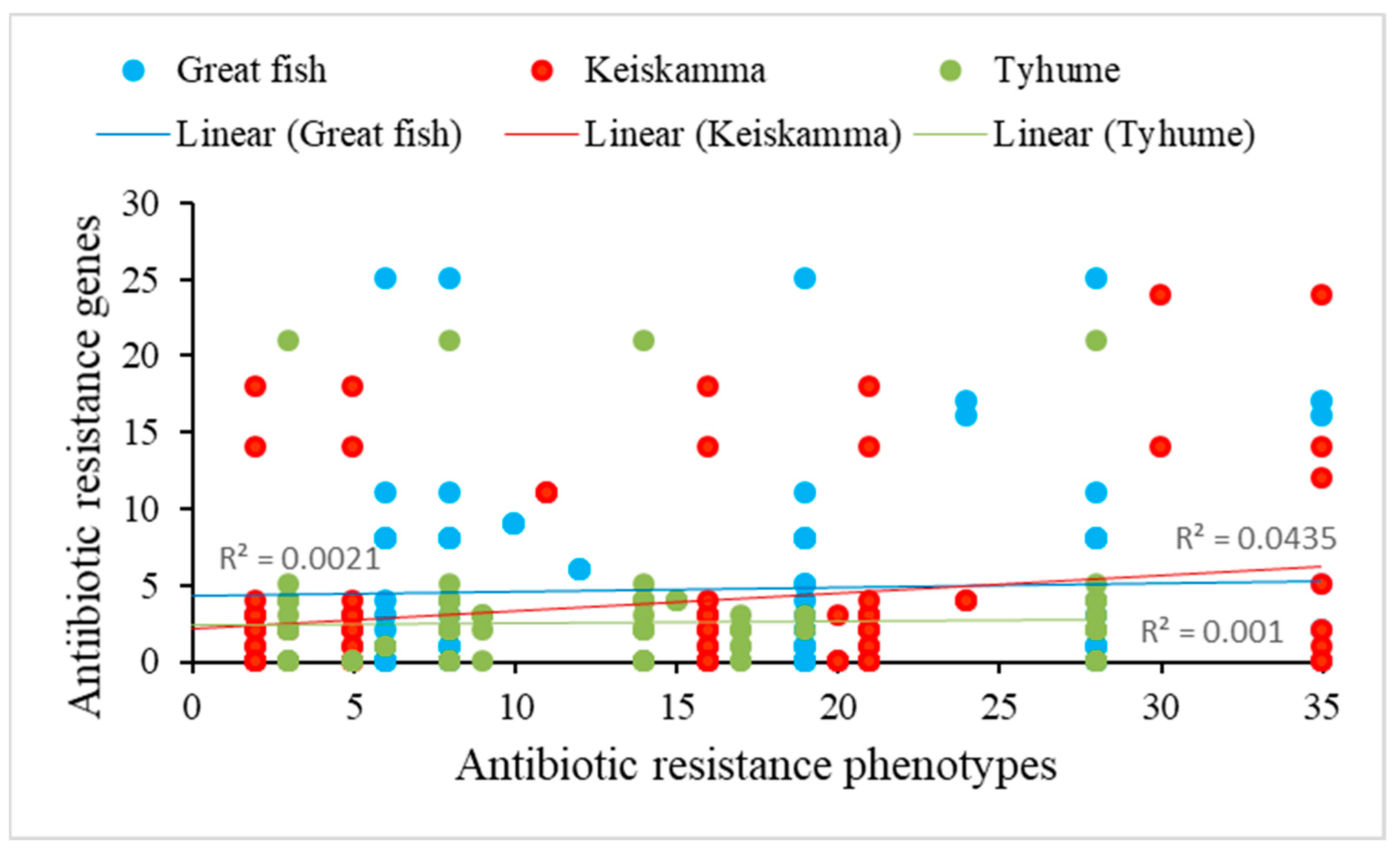
2.5. Correlation between PAR and ARGs

Correlation in this study was indicative of the strength of the relationship between the two variables—PAR and ARGs—in which ARGs were assumed to be the likely cause or contributed to the PAR observed in A. baumannii in each of the rivers. The summary of PAR and ARGs in A. baumannii recovered from specific sampling sites in the rivers studied is presented in Table 2, while Figure 3 describes the correlation between the PAR and ARGs of A. baumannii across the rivers studied. The linear relationship was determined using Pearson’s correlation coefficient (r). The coefficients of correlation (r) between the PAR and ARGs were generally positive in all the rivers, where 0.0456, 0.2131 and 0.0206 were determined for Great Fish, Keiskamma and Tyhume Rivers, respectively. However, the p-values of the correlation between PAR and ARGs in the Great Fish and Tyhume Rivers were 0.6643 and 0.8397, respectively, indicating weak correlations. The p-value in the Keiskamma River was 0.0403, which suggested a significant (p ≤ 0.05) relationship.

3. Discussion

The antibiogram profile of A. baumannii evaluated in this work revealed MDR isolates at several sites in three rivers in the Eastern Cape of South Africa (Scheme 1), which might be due to the anthropogenic impact in the study area. While A. baumannii in the freshwater resources were generally susceptible to antibiotics, few of the isolates exhibited MDR, and the correlation between the PAR and ARGs was positive in all the rivers. Fernando et al. [24] and Kittinger et al. [21] reported high susceptibility to the antimicrobial agents by A. baumannii from the natural environment, whereas Tsai et al. [25] emphasized resistance to antibiotics (SXT) belonging to the class of folate pathway inhibitors in Acinetobacter species recovered from water resources. This further validated the claim that non-clinical A. baumannii are more susceptible to antimicrobial agents than clinical isolates [26,27].
In addition, a few A. baumannii isolates demonstrated MDR, exhibiting resistance to meropenem and imipenem similar to a study conducted by [1]. In that study, some isolates (A. baumannii) were 100% resistant to all tested antibiotics, including meropenem and imipenem. As such, our findings, in accordance with their surveys, suggested an additional risk to the global therapeutic battle. Nonetheless, this may represent an index of secondary exposure of water sources to drugs; alternatively, these isolates originated from sources where the use of antibiotics is grossly abused [28,29]. Summarily, MDR phenotypes revealed that Keiskamma sampling point 2 (KE2) was a hotspot of antibiotic-resistant A. baumannii in this study.
For the ARGs studied, β-lactamases (blaTEM) were the most prevalent genes harboured by A. baumannii, as was previously shown by [30,31,32]. Apparently, some of the A. baumannii strains that were resistant to carbapenems (IMI and MEM) possessed blaTEM, blaSHV, blaVEB, blaSHV, blaCTX-M-1, blaOXA-48-like and blaOXA-51 (Table 2). As noted above, Maravic et al. showed that carbepenem-resistant strains of Acinetobacter species isolated from freshwater bore blaTEM [1]. Therefore, apart from other reasons, possession of these genes could induce resistance to carbapenems (IMI and MEM), β-lactams (PTZ), and cephems (CAZ, CTX and CPM) in A. baumannii because these isolates typically exhibited MDR to all the four Ambler classes (A, B, C and D) of β-lactamases [33,34].
Additionally, sul1 and sul2 were common in the A. baumannii. For instance, the resistance to SXT that carried the highest percentage [25,31] correlated to the prevalence of sulfonamide resistance genes, particularly in sites KE2, GF4 and GF5 (Table 2). Compared to this study, the prevalence of sulfonamide resistance was reported in A. baumannii [35,36] while [37] showed that A. baumannii uses plasmid as a medium of transfer/acquisition of sul2 resistance genes.
Likewise, the detection of tet(B) in A. baumannii isolates among other tetracycline resistance genes was prominent. Among the sites investigated, KE2 had the highest number of A. baumannii strains that possessed this gene. The efflux system or protection of the ribosomal system has been the channel of resistance to tetracycline by A. baumannii [25]. The gene coding for both tet(A) and tet(B) is involved in the tetracycline efflux system and has been detected previously in both aquatic and clinical A. baumannii isolates [38,39,40,41]. In addition to protection of the ribosome by tet(M) [17,36,42], the gene is also used to mediate resistance to tetracycline, but was found in only two isolates in this study.
In the same way, both qnrB and qnrD were detected in A. baumannii, though qnrD was more prevalent than other fluoroquinolone genes. Some of the A. baumannii isolates bearing the qnrB and qnrD genes also showed PAR to ciprofloxacin, especially isolates that were recovered from GF5 (Table 2). The ARGs could mediate resistance to CIP by inhibiting the activity of bacterial-DNA gyrase (encoded by gyrA and gyrB genes) and topoisomerase IV (encoded by parA and parC genes) [38,43,44,45].
Considering the presence of aminoglycoside resistance genes in A. baumannii, while apHA1 and apHA2 were recovered from GF4 at a lower amount compared to the amount at KE2 (Table 2), PAR to amikacin and gentamicin was the least among the antibiotics used (Table 1). All the same, a few A. baumannii isolates harboured the two aminoglycoside resistance genes, suggesting the possibility of HGT between different bacterial species. Nonetheless, the inactivation of the antibiotics by certain aminoglycoside-modifying enzymes such as phosphotransferases, acetyltransferases and adenyltransferases [38,45,46,47] have been linked to resistance. These modifying enzymes are produced by plasmids, integrons or transposons [35], which could be acquired via HGT [27,28].
Generally, blaTEM was the most prevalent ARG detected in Acinetobacter spp. in all the three rivers studied, with its highest recovery from sampling site GF5, followed by the sul2 gene, which had its highest detection at sampling site KE2. In addition to the MDR phenotypes and ARGs, there was a positive correlation between PAR and ARGs in A. baumannii across the rivers.
Overall, the differences observed in the recovery of isolates exhibiting phenotypic resistance as well as resistance genes in one river compared to another could be a function of individual exposure, intrinsic factors such as gene transfer, porin loss and upregulations of efflux pumps, or human interactions and accumulation of anthropogenic activities at a particular sampling site. Future studies on the rivers should be conducted to examine residual antibiotics in the freshwater resources.

4. Materials and Methods

4.1. Description of Sampling Areas

Water samples were collected from three rivers, namely the Great Fish, Keiskamma and Tyhume Rivers, located at Chris Hani and Amathole District Municipalities of the Eastern Cape Province, South Africa, between April 2017 and March 2018. Five sampling points along each river course were selected for the study, based on different visible activities such as in-flow of wastewater plant (WWTP) final effluents, irrigation farming, fishing and animal rearing in the communities.
Figure 4A shows the coordinates of the Great Fish River in the Chris Hani District Municipality of the Eastern Cape Province and the five sampling points. At GF1 there is irrigation farming and fishing, whereas GF2 is characterized by fishing, crop farming and animal rearing. Similarly, site GF3 is used for swimming, animal rearing and domestic purposes, while GF4 is a downstream of Craddock WWTP, where animal rearing and refuse dumping occurs. GF5 is known for fishing, recreational activities such as swimming, traditional ritual washing and dumping of refuse. The Great Fish River is major River that is used for irrigation and livestock farming in the area. The study sites are characterized by fish farming, irrigation, animal rearing, refuse dumping as well as effluents from WWTPs in Craddock. Figure 4B,C describe the flow paths of the Keiskamma and Tyhume Rivers. Both rivers are located in the Amathole District Municipality of the Eastern Cape Province and are exposed to different anthropogenic activities. For instance, crop farming and animal rearing are the normal activities at KE1, while KE2 is used for domestic activities and is also affected by wastewater pipe leakage and animal rearing. KE3 receives community runoff, wastewater pipe leakage, and domestic refuse. In addition, KE4 and KE5 are located downstream of Sandile Dam and a WWTP; animal husbandry and crop farming also take place at these locations. In the Tyhume River, sampling site TY1 is a recreational site where tourists visit and swim. Site TY2 is used for domestic activities, animal rearing and other farming activities. Both TY3 and TY4 are used for fishing, recreational activities and farming, whereas TY5 is located downstream of hospital waste discharge and waste discharge from the University of Fort Hare.
The sampling sites and the corresponding sampling points are highlighted in the maps in Figure 4A–C. Water samples were collected aseptically in sterile 1 L glass bottles from the different sampling points along the river courses by midstream dipping of sample bottles at 25–30 cm down the water column, with the mouth tilting against the flow of the river. All the samples were labelled appropriately and transported to the Applied and Environmental Microbiology Research Group (AEMREG) laboratory in an ice chest and processed within six hours of collection.

4.2. Isolation and Molecular Confirmation of A. baumannii

Water samples were collected from Great Fish, Keiskamma and Tyhume Rivers in Eastern Cape Province of South Africa between April 2017 and March 2018 (Figure 4A–C). Isolation of the presumptive Acinetobacter species was carried out according to American Public Health Association [48] usingselective medium (CHROMagar Acinetobacter base) containing selective supplements (CHROMagar, Paris, France) at 37 °C for 24 h. The selective medium was prepared as instructed by the manufacturer. After incubation, 20 distinct A. baumannii colonies per sampling site were subcultured on nutrient agar (Oxoid, Hampshire, UK) using a streak plate method and then incubated at 37 °C for 24 h. Fifty percent (50%) glycerol stocks of the pure culture were prepared and stored at −80 °C. DNA extraction from the bacterial isolates was carried out using the direct boiling method. Molecular identification of Genus Acinetobacter was performed by targeting Acinetobacter recA gene using a forward primer, P-rA1 (5′-CCTGAATCTTCTGGTAAAAC-3′), and a reverse primer, P-rA2 (5′-GTTTCTGGGCTGCCAAACATTAC-3′), while for the confirmation of A. baumannii, the primer sets P-Ab-ITS (5′-CATTATCACGGTAATTAGTG-3′–forward; 5′-AGAGCACTGTGCACTTAAG-3′-reverse) were used according to [49], while reference strain DSM-102929 was used as the positive control. The components of the PCR assay are as follows: total volume of 25 µL reaction mixture contained 12.5 µL Taq PCR Master Mix (Qiagen, Hilden, Germany), 1.0 µL each of the 100 µM primers (Inqaba Biotech., Pretoria, South Africa), 6.5 µL nuclease-free water and 5.0 µL DNA templates. The condition for the PCR amplification included an initial denaturation step (94 °C, 5 min), followed by 35 cycles (92 °C, 40 s), annealing (58 °C, 40 s), and the final extension step (72 °C, 10 min) was performed using a thermocycler (Bio-Rad Thermal cycler, Hercules, CA, USA). Five microlitres (5 µL) of the amplicon was subjected to gel (1.5% agarose) electrophoresis at 100 Volts for 45 min in Tris Boric EDTA buffer (pH 8.0) (0.089 M Tris, 0.089 M boric acid, and 0.002 M EDTA). Ethidium bromide (5 µL of 0.5 mg/mL) (Sigma-Aldrich, St. Louis, MO, USA) was used for gel staining, and DNA ladder (100 bp) (Thermo Scientific, Vilnius, Lithuania) was added into the gels as a standard. Finally, DNA bands were visualized under an ultraviolet transilluminator (Alliance 4.7, Cambridge, UK).

4.3. Antibiotics Susceptibility Testing (AST)

Susceptibility of A. baumannii isolates to the commonly prescribed antimicrobial agents (Table 1) was determined using the disc diffusion method [50]. After 18 h of culture on nutrient agar (NA) plates, a few distinct colonies were picked and transferred into a test tube containing 10 mL of sterile normal saline (0.85%) and then vortexed gently to allow a proportional assortment of the bacterial cells. The turbidity of the bacterial suspensions was adjusted to 0.5 McFarland standards. A sterile swab stick was dipped into bacterial suspension and used to inoculate the freshly prepared Mueller Hinton agar (MHA) plate by spreading evenly on the agar surface. After air-drying the inoculated plates, the antibiotic discs were impregnated onto them using a sterile disc dispenser and incubated at 37 °C for 20 to 24 h [50]. The diameter of the zone of antibiotic inhibition was rounded to the nearest millimetre, while the isolates were classified as susceptible (S), intermediate resistant (I) or resistant (R) according to the values obtained in comparison to CLSI guidelines for antimicrobial susceptibility testing for A. baumannii. A panel of eleven antibiotics was selected from different antimicrobial groups or classes (Table 3) as representative drugs for the treatment of A. baumannii infections. Multidrug resistance (MDR) phenotype is defined as the resistance of isolates to at least three classes of antimicrobial agents (Table 3).

4.4. Screening of A. baumannii for Antibiotic Resistance Genes (ARGs)

A. baumannii that exhibited phenotypic resistance to test antibiotics were screened for antibiotic resistance genes (ARGs) using multiplex or singleplex PCR assays. The list of primers used for the detection of different ARGs and PCR conditions are presented in Table S2. An aliquot of 25 μL reaction mixture containing a Master Mix (12.5 μL) (Thermo Scientific, Vilnius, Lithuania), oligonucleotide primer (0.5 μL each) (Inqaba Biotech, Pretoria, South Africa), nuclease-free water (6.5 μL) and template DNA (5 μL) was constituted for singleplex PCR assay. The multiplex PCR of 50 μL reaction volume was comprised of Master Mix (25 μL) (Thermo Scientific, (EU) Lithuania), each oligonucleotide primer (1 μL each) (Inqaba Biotech, Pretoria, South Africa), nuclease-free water (14 μL) and template DNA (5 μL). A thermocycler (Bio-Rad Thermal cycler, Hercules, CA, USA) was used for all PCR analysis. A reference strain of A. baumannii (DSM 102929) was used as positive control throughout the ARG screening. Ethidium bromide (5 µL of 0.5 mg/mL) (Sigma-Aldrich, St. Louis, MO, USA) was used for gel staining, and 5 µL of DNA ladder (100 bp) (Thermo Scientific, Vilnius, Lithuania) was added as a standard. Five microlitres (5 µL) of the amplicons were dispensed into the wells of the gel (1.5% agarose) and placed in Tris Boric EDTA buffer (pH 8.0) (0.089 M Tris, 0.089 M boric acid, and 0.002 M EDTA). The condition used for electrophoresis was 100 Volts for 45 min. Finally, DNA bands were viewed under an ultraviolet transilluminator (Alliance 4.7, Cambridge, UK).

4.5. Statistical Analysis

Factorial analysis of variance (ANOVA) was done to determine the variations in the ARGs harboured by Acinetobacter isolates across the three rivers studied in Statistica 13 software, using the Tukey HSD post hoc test (p < 0.05) to identify datasets that are significantly different from their counterparts. Correlations (Pearson) and test of significance were considered statistically significant when p values were ≥95% using Statistica v13 software. Pearson correlation coefficient was used to determine the strength of the linear association between the two variables—PAR and ARGs—in the rivers. The heatmaps were created in Microsoft Excel v16 to highlight the value of MDR phenotypes and ARGs.

5. Conclusions

The detection of ARGs in A. baumannii recovered from the aquatic environments in the study area might be an indication of the secondary impact of antibiotic mismanagement, which presents a significant challenge to public health. This could be the case, especially at sampling site GF4, which is characterized with discharge of wastewater effluent from the downstream Craddock WWTP, animal rearing and refuse dumping. The GF5 site is also exposed to community runoff and a dumpsite where domestic refuse is dumped at the river bank for biodegradation. KE2 is subject to domestic wastewater sludge, wastewater pipe leakage and animal rearing. Antibiotics such as tetracycline, penicillin, noritrine and the sulfonamide group are commonly administered to livestock in the area.
In this study, MDR was observed, and a positive correlation was detected between the PAR and ARGs of the A. baumannii strains recovered from the freshwater resources; in the case of the Keiskamma River, this was significant. The significant relationship between PAR and ARGs in the Keiskamma River could be an indication of selective pressure on antibiotics in that area. It was also evident that some of the isolates were resistant to ciprofloxacin, carbapenems and few other antibiotics. The prevalence of genes encoding for resistance to β-lactamase (blaTEM and blaOXA-51), sulphonamide (sul1 and sul2) and other antimicrobials might be responsible for the resistance as well as exposure to antibiotics due to anthropogenic activities, especially overuse of drugs in the livestock farming in the sampling areas. However, some of the resistant strains might be using other mechanisms to mediate resistance to antimicrobial agents in addition to harbouring the resistance genes. As such, the sampling sites where most of the ARG-bearing isolates were recovered could be categorised as hotspots for the pathogens. Thus, necessary measures should be taken against indiscriminate disposal of waste into the environment to prevent exposure of waterborne pathogens to unused or expired antibiotics and the propagation of resistant pathogens into the water sources.

Supplementary Materials

The following are available online at https://0-www-mdpi-com.brum.beds.ac.uk/article/10.3390/pathogens10091110/s1, Figure S1: PCR analyses resolved by gel electrophoresis showing confirmed Acinetobacter spp. targeting the recA gene at 425 bp., Figure S2: PCR analyses resolved by gel electrophoresis showing confirmed A. baumannii targeting the specific primer Ab-ITS gene at 208 bp., Figure S3: A gel electrophoresis showing sulfonamide resistance genes in A. baumannii, Figure S4: A gel electrophoresis showing aminoglycoside genes in A. baumannii, Figure S5: A gel electrophoresis showing Fluoroquinolones genes in A. baumannii, Figure S6: A gel electrophoresis showing Tetracycline genes A. baumannii, Figure S7A–F: Gel electrophoresis showing beta-lactamase genes in A. baumannii, Table S1: Phenotypic antibiotic resistance of A. baumannii recovered from each of the sites where water samples were collected along Great Fish, Keiskamma and Tyhume Rivers. S, R and I stand for susceptible, resistance and intermediate resistance, respectively, Table S2: Screening of A. baumannii that exhibited phenotypic resistance for antibiotic-resistant genes.

Author Contributions

Conceptualization, M.A.A. and A.I.O.; methodology, M.A.A.; software, M.A.A.; validation, M.A.A., K.E.E., and A.I.O.; formal analysis, M.A.A.; investigation, M.A.A.; resources, M.A.A. and A.I.O.; data curation, M.A.A.; writing—original draft preparation, M.A.A.; writing—review and editing, M.A.A. and K.E.E.; visualization, M.A.A., K.E.E., and A.I.O.; supervision, A.I.O.; project administration, A.I.O.; funding acquisition, M.A.A. and A.I.O. All authors have read and agreed to the published version of the manuscript.

Funding

We would like to express our gratitude to the National Research Foundation, South Africa, and The World Academy of Science, Italy (NRF/TWAS), with grant numbers 99767 and 116387, respectively, and the South African Medical Research Council (SAMRC/UFH/P790) for their financial support.

Institutional Review Board Statement

Not applicable.

Informed Consent Statement

Not applicable.

Data Availability Statement

The data presented in this study are available on request from the corresponding author. The data are not publicly available because the authors still need the data to further research in those areas and on Acinetobacter species recovered from the Rivers.

Conflicts of Interest

On behalf of all authors, the corresponding author states that there is no conflict of interest.

References

  1. Maravić, A.; Skočibušić, M.; Fredotović, Z.; Šamanić, I.; Cvjetan, S.; Knezović, M.; Puizina, J. Urban riverine environment is a source of multidrug-resistant and ESBL-producing clinically important Acinetobacter spp. Environ. Sci. Pollut. Res. 2016, 23, 3525–3535. [Google Scholar] [CrossRef]
  2. Doughari, H.J.; Ndakidemi, P.A.; Human, I.S.; Benade, S. The ecology, biology and pathogenesis of Acinetobacter spp.: An overview. Microbes Environ. 2011, 26, 101–112. [Google Scholar] [CrossRef] [PubMed] [Green Version]
  3. Basri, R.; Zueter, A.R.; Mohamed, Z.; Alam, M.K.; Norsa’adah, B.; Hasan, S.A.; Hasan, H.; Ahmad, F. Burden of bacterial meningitis: A retrospective review on laboratory parameters and factors associated with death in meningitis, Kelantan Malaysia. Nagoya J. Med. Sci. 2015, 77, 59. [Google Scholar] [PubMed]
  4. Chiang, T.; Pastagia, M.; Huang, D.B. Bacteremia Caused by Acinetobacter baumannii: Epidemiologic Features, Antimicrobial Susceptibility, and Outcomes. Adv. Infect. Dis. 2014, 4, 66–71. [Google Scholar] [CrossRef] [Green Version]
  5. Gundeslioglu, O.O.; Gokmen, T.G.; Horoz, O.O.; Aksaray, N.; Koksal, F.; Yaman, A.; Yıldızdas, R.D.; Alhan, E.; Kocabas, E.; Alabaz, D. Molecular epidemiology and antibiotic susceptibility pattern of Acinetobacter baumannii isolated from children in a Turkish University hospital. Turk. J. Pediatrics 2014, 56, 360–367. [Google Scholar]
  6. Guerrero, D.M.; Perez, F.; Conger, N.G.; Solomkin, J.S.; Adams, M.D.; Rather, P.N.; Bonomo, R.A. Acinetobacter baumannii-Associated Skin and Soft Tissue Infections: Recognizing a Broadening Spectrum of Disease. Surg. Infect. 2010, 11, 49–57. [Google Scholar] [CrossRef] [Green Version]
  7. Darvishi, M. Virulence factors profile and antimicrobial resistance of Acinetobacter baumannii strains isolated from various infections recovered from immunosuppressive patients. Biomed. Pharm. J. 2016, 9, 1057–1062. [Google Scholar] [CrossRef]
  8. European Centre for Disease Prevention and Control (ECDPC). Annual Epidemiological Report Food-and Waterborne Diseases and Zoonoses; European Centre for Disease Prevention and Control (ECDPC): Stockholm, Sweden, 2014. [Google Scholar]
  9. World Health Organization (WHO). Global Report on Surveillance of Antimicrobial Resistance; WHO: Geneva, Switzerland, 2014. [Google Scholar]
  10. Kanafani, A.Z.; Kanj, S.S. Ministry of Health, Kingdome of Saudi Arabia. 2014. Available online: http://www.uptodate.com/contents/acinetobacterinfection-treatment-and-prevention (accessed on 6 April 2020).
  11. Gallagher, L.A.; Ramage, E.; Weiss, E.J.; Radey, M.; Hayden, H.S.; Held, K.G.; Huse, H.K.; Zurawski, D.V.; Brittnacher, M.J.; Manoil, C. Resources for genetic and genomic analysis of emerging pathogen Acinetobacter baumannii. J. Bacteriol. 2015, 197, 2027–2035. [Google Scholar] [CrossRef] [Green Version]
  12. Abdi, S.N.; Ghotaslou, R.; Ganbarov, K.; Mobed, A.; Tanomand, A.; Yousefi, M.; Asgharzadeh, M.; Kafil, H.S. Acinetobacter baumannii Efflux Pumps and Antibiotic Resistance Infection and Drug Resistance. Infect. Drug Resist. 2020, 13, 423–434. [Google Scholar] [CrossRef] [Green Version]
  13. Rumbo, C.; Gato, E.; López, M.; de Alegría, C.R.; Fernández-Cuenca, F.; Martínez-Martínez, L.; Vila, J.; Pachón, J.; Cisneros, J.M.; Rodríguez-Baño, J.; et al. Contribution of efflux pumps, porins, and β-lactamases to multidrug resistance in clinical isolates of Acinetobacter baumannii. Antimicrob. Agents Chemother. 2013, 57, 5247–5257. [Google Scholar] [CrossRef] [Green Version]
  14. Gheorghe, I.; Novais, A.; Grosso, F.; Rodrigues, C.; Chifiriuc, M.C.; Lazar, V.; Peixe, L. Snapshot on carbapenemase-producing Pseudomonas aeruginosa and Acinetobacter baumannii in Bucharest hospitals reveals unusual clones and novel genetic surroundings for blaOXA-23. J. Antimicrob. Chemother. 2015, 70, 1016–1020. [Google Scholar] [CrossRef] [PubMed] [Green Version]
  15. Vázquez-López, R.; Solano-Gálvez, S.G.; Vignon-Whaley, J.J.J.; Vaamonde, J.A.A.; Alonzo, L.A.P.; Reséndiz, A.R.; Álvarez, M.M.; López, E.N.V.; Franyuti-Kelly, G.; Álvarez-Hernández, D.A.; et al. Acinetobacter baumannii Resistance: A Real Challenge for Clinicians. Antibiotics 2020, 9, 205. [Google Scholar] [CrossRef] [PubMed]
  16. Ardehal, S.H.; Azimi, T.; Fallah, F.; Owrang, M.; Aghamohammadi, N.; Azimi, L. Role of efflux pumps in reduced susceptibility to tigecycline in Acinetobacter baumannii. New Microbes New Infect. 2019, 30, 1–6. [Google Scholar] [CrossRef]
  17. Perez, F.; Hujer, A.M.; Hujer, K.M.; Decker, B.K.; Rather, P.N.; Bonomo, R.A. Global challenge of multidrug-resistant Acinetobacter baumannii. Antimicrob. Agents Chemother. 2007, 51, 3471–3484. [Google Scholar] [CrossRef] [PubMed] [Green Version]
  18. Falagas, M.E.; Vardakas, K.Z.; Kapaskelis, A.; Triarides, N.A.; Roussos, N.S. Tetracyclines for multidrug-resistant Acinetobacter baumannii infections. Int. J. Antimicrob. Agents 2015, 45, 455–460. [Google Scholar] [CrossRef] [PubMed]
  19. Hrenovic, J.; Durn, G.; Goic-Barisic, I.; Kovacic, A. Occurrence of an Environmental Acinetobacter baumannii Strain Similar to a Clinical Isolate in Paleosol from Croatia. Appl. Environ. Microbiol. 2014, 80, 2860–2866. [Google Scholar] [CrossRef] [Green Version]
  20. Goswami, R.; Mukherjee, S.; Rana, V.S.; Saha, D.R.; Raman, R.; Padhy, P.K.; Mazumder, S. Isolation and Characterization of Arsenic-Resistant Bacteria from Contaminated Water-Bodies in West Bengal, India. Geomicrobiol. J. 2015, 32, 17–26. [Google Scholar] [CrossRef]
  21. Kittinger, C.; Kirschner, A.; Lipp, M.; Baumert, R.; Mascher, F.; Farnleitner, A.H.; Zarfel, G.E. Antibiotic Resistance of Acinetobacter spp. isolates from the River Danube: Susceptibility Stays High. Int. J. Environ. Res. Public Health 2018, 15, 52. [Google Scholar] [CrossRef] [Green Version]
  22. Ashbolt, N.J. Microbial contamination of drinking water and human health from community water systems. Curr. Environ. Health Rpt. 2015, 2, 95–106. [Google Scholar] [CrossRef] [Green Version]
  23. Bitton, G. Microbiology of Drinking Water Production and Distribution, 1st ed.; John Wiley & Sons, Inc.: Hoboken, NJ, USA, 2014; p. 312. [Google Scholar]
  24. Fernando, D.M.; Tun, H.M.; Poole, J.; Patidar, R.; Li, R.; Mi, R.; Amarawansha, G.E.A.; Fernando, W.G.D.; Khafipour, E.; Farenhorst, A.; et al. Detection of antibiotic resistance genes in source and drinking water samples from a First Nations community in Canada. Appl. Environ. Microbiol. 2016, 82, 4767–4775. [Google Scholar] [CrossRef] [PubMed] [Green Version]
  25. Tsai, H.C.; Chou, M.Y.; Shih, Y.J.; Huang, T.Y.; Yang, P.Y.; Chiu, Y.C.; Chen, J.S.; Hsu, B.M. Distribution and genotyping of aquatic Acinetobacter baumannii strains isolated from the Puzi River and its tributaries near areas of livestock farming. Water 2018, 10, 1374. [Google Scholar] [CrossRef] [Green Version]
  26. Cikman, A.; Gulhan, B.; Aydin, M.; Ceylan, M.R.; Parlak, M.; Karakecili, F.; Karagoz, A. In vitro Activity of Colistin in Combination with Tigecycline against Carbapenem-Resistant Acinetobacter baumannii Strains Isolated from Patients with Ventilator-Associated Pneumonia. Int. J. Med. Sci. 2015, 12, 695–700. [Google Scholar] [CrossRef] [Green Version]
  27. Anh, N.T.; Thieu Nga, T.V.; Tuan, H.M.; Tuan, N.S.; Dao, Y.; Chau, N.V.V.; Baker, S.; Duong, H.H.T. Molecular epidemiology and antimicrobial resistance phenotypes of Acinetobacter baumannii isolated from patients in three hospitals in southern Vietnam. J. Med. Microbiol. 2017, 66, 46–53. [Google Scholar]
  28. Ekwealor, P.A.; Ugwu, M.C.; Ezeobi, I.; Amalukwe, G.; Ugwu, B.C.; Okezie, U.; Stanley, C.; Esimone, C. Antimicrobial Evaluation of Bacterial Isolates from Urine Specimen of Patients with Complaints of Urinary Tract Infections in Awka, Nigeria. Int. J. Microbiol. 2016, 2016, 9740273. [Google Scholar] [CrossRef] [Green Version]
  29. Al-Bahry, S.; Mahmoud, I.; Elshafie, A.; Al-Harthy, A.; Al-Ghafri, S.; Al-Amri, I.; Alkindi, A. Bacterial flora and antibiotic resistance from eggs of green turtles Chelonia myans: An indication of polluted effluents. Mar. Pollut. Bull. 2009, 58, 720–725. [Google Scholar] [CrossRef]
  30. Leungtongkam, U.; Thummeepak, R.; Tasanapak, K.; Sitthisak, S. Acquisition and transfer of antibiotic resistance genes in association with conjugative plasmid or class 1 integrons of Acinetobacter baumannii. PLoS ONE 2018, 13, e0208468. [Google Scholar] [CrossRef] [PubMed] [Green Version]
  31. Taitt, C.R.; Leski, T.A.; Stockelman, M.G.; Craft, D.W.; Zurawski, D.V.; Kirkup, B.C.; Vora, G.J. Antimicrobial resistance determinants in Acinetobacter baumannii isolates taken from Military Treatment Facilities. Antimicrob. Agents Chemother. 2013, 58, 767–781. [Google Scholar] [CrossRef] [Green Version]
  32. Hujer, K.M.; Hujer, A.M.; Hulten, E.A.; Bajaksouzian, S.; Adams, J.M.; Donskey, C.J.; Ecker, D.J.; Massire, C.; Eshoo, M.W.; Sampath, R.; et al. Analysis of antibiotic resistance genes in multidrug-resistant Acinetobacter sp. isolates from military and civilian patients treated at the Walter Reed Army Medical Center. Antimicrob. Agents Chemother. 2006, 50, 4114–4123. [Google Scholar] [CrossRef] [PubMed] [Green Version]
  33. Lin, M.F.; Lan, C.Y. Antimicrobial resistance in Acinetobacter baumannii: From bench to bedside. World J. Clin. Cases 2014, 2, 787–814. [Google Scholar] [CrossRef] [PubMed]
  34. Potron, A.; Poirel, L.; Nordmann, P. Emerging broad-spectrum resistance in Pseudomonas aeruginosa and Acinetobacter baumannii: Mechanisms and epidemiology. Int. J. Antimicrob. Agents 2015, 45, 568–585. [Google Scholar] [CrossRef] [Green Version]
  35. Poirel, L.; Corvec, S.; Rapoport, M.; Mugnier, P.; Petroni, A.; Pasteran, F.; Faccone, D.; Galas, M.; Drugeon, H.; Cattoir, V.; et al. Identification of the novel narrow-spectrum beta-lactamase SCO1 in Acinetobacter spp. from Argentina. Antimicrob. Agents Chemother. 2007, 51, 2179–2184. [Google Scholar] [CrossRef] [Green Version]
  36. Peleg, A.Y.; Seifert, H.; Paterson, D.L. Acinetobacter baumannii: Emergence of a successful pathogen. Clin. Microbiol. Rev. 2008, 21, 538–582. [Google Scholar] [CrossRef] [Green Version]
  37. Hamidian, M.; Ambrose, S.J.; Hall, R.M. A large conjugative Acinetobacter baumannii plasmid carrying the sul2 sulphonamide and strAB streptomycin resistance genes. Plasmid 2016, 87, 43–50. [Google Scholar] [CrossRef] [PubMed]
  38. Jamal, S.; Al Atrouni, A.; Rafei, R.; Dabboussi, F.; Hamze, M.; Osman, M. Molecular mechanisms of antimicrobial resistance in Acinetobacter baumannii, with a special focus on its epidemiology in Lebanon. J. Glob. Antimicrob. Resist. 2018, 15, 154–163. [Google Scholar] [CrossRef] [PubMed]
  39. Knight, D.B.; Rudin, S.D.; Bonomo, R.A.; Rather, P.N. Acinetobacter nosocomialis: Defining the Role of Efflux Pumps in Resistance to Antimicrobial Therapy, Surface Motility, and Biofilm Formation. Front. Microbiol. 2018, 9, 1902. [Google Scholar] [CrossRef] [PubMed]
  40. Roberts, M.C. Tetracycline resistance determinants: Mechanisms of action, regulation of expression, genetic mobility, and distribution. FEMS Microbiol. Rev. 1996, 19, 1–24. [Google Scholar] [CrossRef] [PubMed]
  41. Guardabassi, L.; Dijkshoorn, L.; Collard, J.M.; Olsen, J.E.; Dalsgaard, A. Distribution and in-vitro transfer of tetracycline resistance determinants in clinical and aquatic Acinetobacter strains. J. Med. Microbiol. 2000, 49, 929–936. [Google Scholar] [CrossRef] [PubMed] [Green Version]
  42. Ribera, A.; Ruiz, J.; Vila, J. Presence of the tetM determinant in a clinical isolate of Acinetobacter baumannii. Antimicrob. Agents Chemother. 2003, 47, 2310–2312. [Google Scholar] [CrossRef] [PubMed] [Green Version]
  43. Cattoir, V. Quinolones de l’antibiogramme aux phénotypes de résistance Quinolones: From antibiogram to resistance phenotypes. Revue Franco-Phone Lab. 2012, 445, 79–87. [Google Scholar] [CrossRef]
  44. Nordmann, P.; Poirel, L. Emergence of plasmid-mediated resistance to quinolones in Enterobacteriaceae. J. Antimicrob. Chemother. 2005, 56, 463–469. [Google Scholar] [CrossRef] [Green Version]
  45. Poirel, L.; Bonnin, R.A.; Nordmann, P. Genetic basis of antibiotic resistance in pathogenic Acinetobacter species. IUBMB Life 2011, 63, 1061–1067. [Google Scholar] [CrossRef] [PubMed]
  46. Towner, K.J. Antibiotic resistance in Acinetobacter spp. In Acinetobacter Molecular Biology; Gerischer, U., Ed.; Caister Academic Press: Norfolk, UK, 2008; pp. 331–343. [Google Scholar]
  47. Nemec, A.; Dolzani, L.; Brisse, S.; van den Broek, P.; Dijkshoorn, L. Diversity of aminoglycoside-resistance genes and their association with class 1 integrons among strains of pan-European Acinetobacter baumannii clones. J. Med. Microbiol. 2004, 53, 1233–1240. [Google Scholar] [CrossRef] [PubMed]
  48. American Public Health Association (APHA)/American Water Works Association (AWWA)/Water Environment Federation (WEF). Standard Methods for the Examination of Water and Wastewater; APHA/AWWA/WEF: Washington, DC, USA, 2005. [Google Scholar]
  49. Chen, T.L.; Lee, Y.T.; Kuo, S.C.; Yang, S.P.; Fung, C.P.; Lee, S.D. Rapid identification of Acinetobacter baumannii, Acinetobacter nosocomialis and Acinetobacter pittii with a multiplex PCR assay. J. Med. Microbiol. 2014, 63, 1154–1159. [Google Scholar] [CrossRef] [PubMed]
  50. Clinical and Laboratory Standards Institute (CLSI). Performance Standards for Antimicrobial Disk Susceptibility Tests, 28th ed.; CLSI Supplement M100: Wayne, PA, USA, 2018. [Google Scholar]
Scheme 1. Illustration of natural environment and water collection from river and antibiotic susceptibility testing for A. baumannii isolated from water samples. Although the majority of the A. baumannii strains were susceptible to the antibiotics tested, some strains were resistant to the drugs, perhaps due to antibiotic resistance genes they harbour.
Pathogens 10 01110 sch001
Figure 1. Heatmap showing the multiple antibiotic resistance phenotypes (MARPs) of A. baumannii, isolated from each of the sampling points along the Great Fish, Keiskamma, and Tyhume Rivers. Each resistant isolate demonstrated MARPs against different antibiotics, ranging from 3 to 11 antimicrobial agents.
Pathogens 10 01110 g001
Figure 2. Heatmap (colour legend: inset) showing the distribution of ARGs in A. baumannii in Great Fish, Keiskamma and Tyhume Rivers. PAR isolates were screened for the presence of ARGs belonging to the following classes: aminoglycoside (aaC2, aphA1 and aphA2), β-lactamase (blaTEM, blaSHV, blaOXA-1-like, blaCTX-M1group, blaCTX-M-2group, blaCTX-M9group, blaVEB, blaGES, blaPER, blaCTX-M-8/-25group, blaOXA-48-like, blaVIM, blaIMP and blaKPC), fluoroquinolone (qnrA, qnrB, qnrC, qnrD and qnrS), sulfonamide (sul1 and sul2), and tetracycline (tet(A), tet(B), tet(C), tet(M), tet(L) and tet(O)).
Pathogens 10 01110 g002
Figure 3. Correlation of PAR with ARGs of Acinetobacter spp. recovered from Great Fish, Keiskamma and Tyhume Rivers. Coefficient of correlation (r) is significant when p ≤ 0.05. Coefficient of correlation and p-values were determined using Pearson’s correlation coefficient.
Pathogens 10 01110 g003
Figure 4. (A) Map of Great Fish River showing the sampling points. (B) Map of Keiskamma River showing the sampling points. (C) Map of Tyhume River showing the sampling points.
Pathogens 10 01110 g004aPathogens 10 01110 g004b
Table 1. Antibiogram profiles of Acinetobacter baumannii in Great Fish, Keiskamma and Tyhume Rivers.
Antimicrobial AgentPercentage of Isolates Sensitive to Antibiotics
Susceptible (S)Resistance (R)Intermediate Resistance (I)Non-Susceptible
R + I
PTZ299 (72.9%)46 (11.2%)65 (15.8%)111 (27%)
CAZ289 (70.5%)49 (12%)72 (17.6%)121 (29.6%)
CTX69 (16.8%)77 (18.8%)264 (64.4%)341 (83.2%)
CPM363 (88.5%)36 (8.8%)11 (2.7%)47 (11.5%)
IMI394 (96.1%)11 (2.7%)5 (1.2%)16 (3.9%)
MEM381 (92.9%)17 (4.15%)12 (2.9%)29 (7.05%)
AK400 (97.6%)10 (2.4%)0 (0%)10 (2.4%)
GM369 (90.0%)36 (8.8%)5 (1.2%)41 (10%)
TET307 (74.9%)69 (16.8%)34 (8.3%)103 (25.1%)
CIP312 (76.1%)45 (11%)53 (12.9%)98 (23.9%)
SXT304 (74.1%)84 (20.5%)22 (5.4%)106 (25.9%)
Phenotypic antibiotic resistance (PAR) in A. baumannii from Great Fish, Keiskamma and Tyhume Rivers. Aminoglycosides (AK, GM), β-lactam/β-lactamase-inhibitor combinations (PTZ), cephems (CAZ, CTX, CPM), carbapenems (IMI, MEM), fluoroquinolones (CIP), folate pathway inhibitors (TS), tetracyclines (TET).
Table 2. Distribution of ARGs among A. baumannii recovered from specific sampling sites in the rivers studied.
Antibiotic ClassAntibiotic AgentResistance GenesNo. of Positive Isolates Per Sampling SiteTotal (%)
GF1GF2GF3GF4GF5KE1KE2KE3KE4KE5TY1TY2TY3TY4TY5
apHAI00054003010000013 (25.5)
apHA2000330112000011021 (41.2)
β-lactamases
(n = 101)
PTZ

CAZ

CTX

CPM

IMI

MEM
blaTEM1309140125002137865 (64.4)
blaSHV00053002000001112 (11.9)
blaCTX-M-100035002001000112 (11.9)
blaCTX-M-20000100000100002 (2.0)
blaCTX-M-90000000000000000
blaCTX-M-8/-2510106021001000212 (11.9)
blaPER1000100100000003 (2.9)
blaVEB0001001000000125 (4.9)
blaGES02015012000000213 (12.9)
blaOXA-230000000000000000
blaOXA-400000000000000000
blaOXA-480012003000200019 (8.9)
blaOXA-51002360113002100129 (28.7)
blaOXA-580000000000000000
blaVIM0000000000000000
blaIMP0000002000000406 (5.9)
blaKPC0001102200010018 (7.9)
Fluoroquilonone
(n = 44)
CIPqnrA0000000000000000
qnrB0000201200001107 (14.6)
qnrC0000000000000000
qnrD0001400000101108 (16.7)
qnrS0000000000000000
Sulfonamides
(n = 83)
SXTsul111167283001100132 (37.1)
sul2010790203001000243 (49.4)
Tetracyclines
(n = 70)
TETtet(A)0001102300010008 (11.4)
tet(B)000410120001000119 (27.1)
tet(C)0000001000100002 (2.9)
tet(L)0001100000100003 (4.3)
tet(M)0000002000000002 (2.9)
tet(O)0000000000000000
Table 3. List of antimicrobial agents for AST of Acinetobacter isolates.
Antimicrobial GroupAntimicrobial AgentDisc Content
β-lactam/β-lactamase-inhibitor combinationsPiperacillin-tazobactam (PTZ)100/10 μg
Cephems (CEP)Ceftazidime (CAZ)30 μg
Cefepime (CPM)30 μg
Cefotaxime (CTX)30 μg
Carbapenems (C)Imipenem (IMI)10 μg
Meropenem (MEM)10 μg
Aminoglycosides (AMG)Amikacin (AK)30 μg
Gentamicin (GM)10 μg
Tetracyclines (TET)Tetracycline (TET)30 μg
Fluoroquinolones (FLQ)Ciprofloxacin (CIP)5 μg
Folate pathway inhibitors (FPI)Trimethoprim/sulfamethoxazole (SXT)1.25/23.75 μg
Publisher’s Note: MDPI stays neutral with regard to jurisdictional claims in published maps and institutional affiliations.

