Article

Achromobacter spp. in a Cohort of Non-Selected Pre- and Post-Lung Transplant Recipients

by
Cornelia Geisler Crone
1,2,*,†,
Omid Rezahosseini
2,†,
Hans Henrik Lawaetz Schultz
3,
Tavs Qvist
2,
Helle Krogh Johansen
4,5,6,
Susanne Dam Nielsen
2,5 and
Michael Perch
3,5
1
Centre of Excellence for Health, Immunity and Infections (CHIP), Copenhagen University Hospital, Rigshospitalet, 2100 Copenhagen, Denmark
2
Department of Infectious Diseases, Copenhagen University Hospital, Rigshospitalet, 2100 Copenhagen, Denmark
3
Department of Cardiology, Section for Lung Transplantation, Copenhagen University Hospital, Rigshospitalet, 2100 Copenhagen, Denmark
4
Department of Clinical Microbiology, Copenhagen University Hospital, Rigshospitalet, 2100 Copenhagen, Denmark
5
Department of Clinical Medicine, Faculty of Health and Medical Sciences, University of Copenhagen, 2200 Copenhagen, Denmark
6
The Novo Nordisk Foundation Center for Biosustainability, Technical University of Denmark, 2800 Lyngby, Denmark
*
Author to whom correspondence should be addressed.
These authors contributed equally to this work.
Submission received: 23 December 2021 / Revised: 14 January 2022 / Accepted: 25 January 2022 / Published: 28 January 2022
(This article belongs to the Special Issue Achromobacter xylosoxidans, an Emergent Pathogen in Lung Infections)

Abstract

:
Achromobacter is an opportunistic pathogen that mainly causes chronic lung infections in cystic fibrosis (CF) patients and is associated with increased mortality. Little is known about Achromobacter spp. in the lung transplant recipient (LTXr) population. We aimed at describing rates of Achromobacter spp. infection in LTXr prior to, in relation to, and after transplantation, as well as all-cause mortality proportion in infected and uninfected LTXr. We included 288 adult LTXr who underwent lung transplantation (LTX) between 1 January 2010 and 31 December 2019 in Denmark. Bronchoalveolar lavage was performed at regular intervals starting two weeks after transplantation. Positive cultures of Achromobacter spp. were identified in nationwide microbiology registries, and infections were categorized as persistent or transient, according to the proportion of positive cultures. A total of 11 of the 288 LTXr had transient (n = 7) or persistent (n = 4) Achromobacter spp. infection after LTX; CF was the underlying disease in 9 out of 11 LTXr. Three out of the four patients, with persistent infection after LTX, also had persistent infection before LTX. The cumulative incidence of the first episode of infection one year after LTX was 3.8% (95% CI: 1.6–6.0). The incidence rates of transient and persistent infection in the first year after LTX were 27 (12–53) and 15 (5–37) per 1000 person-years of follow-up, respectively. The all-cause mortality proportion one year after LTX was 27% in the Achromobacter spp. infected patients and 12% in the uninfected patients (p = 0.114). Achromobacter spp. mainly affected LTXr with CF as the underlying disease and was rare in non-CF LTXr. Larger studies are needed to assess long-term outcomes of Achromobacter spp. in LTXr.

1. Introduction

Achromobacter is a genus of non-fermentative aerobic Gram-negative rods, mainly found in the environment in water and soil. Opportunistic disease in humans is well described and typically manifests as chronic respiratory infections in persons with chronic lung disease, such as cystic fibrosis (CF) or in immunocompromised patients [1,2,3,4]. Elderly patients with comorbidities and patients with malignancies are also known to be susceptible. Extra-pulmonary manifestations such as sepsis, bacteremia, urine-, and catheter or device infections are seen [1,3,5,6].
Infections with Achromobacter spp. are difficult to treat because of intrinsic and acquired antibiotic resistance. In addition, there is no standard treatment protocol [7], although it has been suggested that early treatment with inhaled and oral antibiotics postpones time to the next culture of Achromobacter spp. in CF patients [8].
In addition, Achromobacter form biofilm in the respiratory and conductive zones of the lung, the latter of which is poorly vascularized, leads to lower antibiotic availability when treatment is attempted either by oral intake or intravenously [8,9]. Due to this, infections in the respiratory tract often become persistent. These lead to continuous inflammation and are associated with poor long-term outcomes in infected CF patients [10,11], where an excess decline in lung function of −1.6% annually (95% CI −2.2 to −0.90) has been reported. This leads to premature death or lung transplantation 16.8 years after initial colonization [12].
In elderly patients with hospital-acquired Achromobacter pneumonia, 30-day mortality has been reported as high as 33%, despite antibiotic therapy [4]. A. xylosoxidans has been found as the causative agent in outbreaks of hospital-acquired infection, possibly associated with the bacilli’s capability of forming biofilms, creating reservoirs in water systems or even contaminating solutions [4,7,13,14].
Lung transplantation (LTX) is a last resort treatment for patients with end-stage lung disease, such as CF, emphysema, or pulmonary fibrosis. All lung transplant recipients (LTXr) are treated with immunosuppressives following transplantation to avoid graft rejection. This iatrogenic immunosuppression leads to increased risk of infections [15]. In most transplant centers, LTXr are frequently hospitalized as part of the follow-up program, where routine examinations include bronchoscopy with bronchoalveolar lavage (BAL).
Achromobacter in CF patients undergoing LTX have previously been described and early re-infection has been reported [16,17,18]. However, these studies also found that Achromobacter re-infection did not reduce survival in CF patients after LTX [16,17,18].
The general LTXr population has several risk factors for developing Achromobacter spp. infection including prior severe lung disease, immunosuppression, and frequent contacts to the healthcare system with invasive procedures. However, information about Achromobacter spp. infections in the general LTXr population is lacking [19]. We aimed to determine the incidence of Achromobacter spp. prior to, in relation to, and after LTX in LTXr, regardless of underlying disease. We also aimed to describe manifestation of infection, time to infection/re-infection, persistency of infection, and all-cause mortality proportion in Achromobacter infected LTXr.

2. Results

We identified 298 patients who had received a LTX during the inclusion period. We excluded three patients due to age < 18 years and seven patients, where the LTX in the inclusion period was not the first LTX.

2.1. Baseline Characteristics

Eleven out of the 288 LTXr who were included had transient or persistent Achromobacter spp. infection after transplantation. The median age at transplantation was significantly lower in LTXr with Achromobacter spp. infection compared to the uninfected group (33 vs. 53 years, p = 0.006). This was due to the high proportion of CF patients in the Achromobacter spp. group having 82% with CF as the underlying disease, compared to 12% in the uninfected group (Table 1). Most patients underwent double lung transplantation in both groups.

2.2. Achromobacter spp. Pre-Transplantation

Within the year prior to transplantation, 155 of the 288 included LTXr had at least one microbial culture performed, and 9 out of the 155 had at least one culture-positive for Achromobacter spp. Six out of the nine fulfilled the criteria of having persistent infection within the year prior to LTX. The remaining three had transient infection in the year leading up to LTX and had never had persistent infection prior to LTX.
All nine patients had CF as the underlying disease and were positive in respiratory tract specimens. The median (IQR) time difference from LTX to the last positive culture prior to transplantation was −6 (−19 to −4) days.

2.3. Achromobacter spp. in Relation to Transplantation

All 288 LTXr had at least one culture performed in relation to the time of transplantation, and five had at least one culture-positive for Achromobacter spp. All these patients had persistent infections in the year leading up to LTX. The Achromobacter spp. were cultured in blood in all of the five patients (two peripheral blood, two central venous catheters, and one port-a-cath). Four out of the five patients had simultaneous positive cultures from airway secretions taken related to LTX.

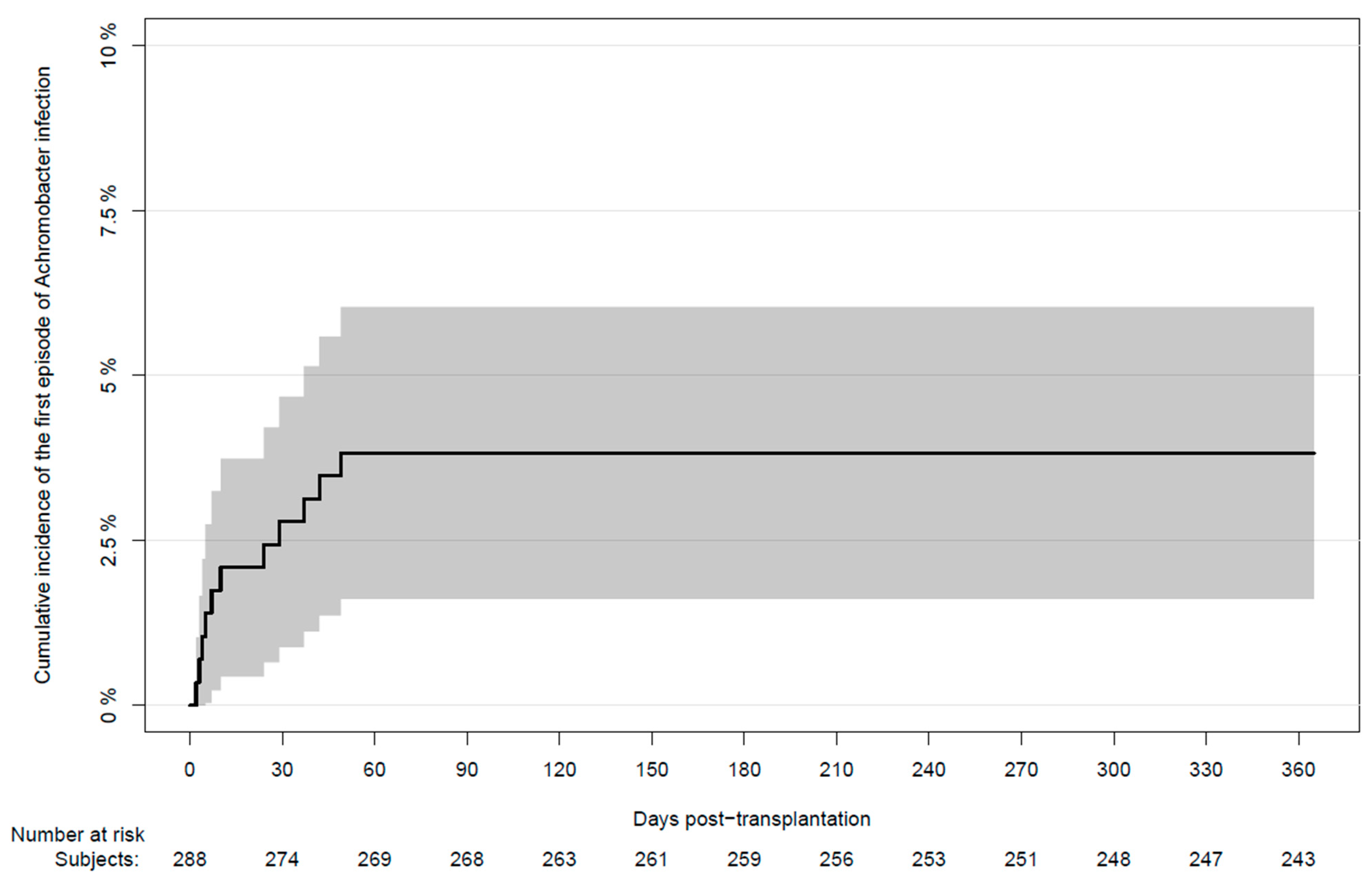
2.4. Achromobacter spp. after Transplantation

During the first year after transplantation, 11 LTXr had one or more cultures positive for Achromobacter spp. The cumulative incidence of the first episode of infection at day 365 post-LTX was 3.8% (95% CI: 1.6–6.0) (Figure 1).
Of the 11 infected patients, 9 (82%) were CF patients and 2 (22%) were non-CF patients (Table 2). The median time to the first Achromobacter spp. positive culture after transplantation was 10 days (IQR 5–33). In 7 out of 11 (64%) A. xylosoxidans was cultured after LTX. Three patients (27%) had positive cultures with A. rhulandii. One patient had positive cultures with Achromobacter species without further identification (Table 2). Whole genome sequencing was applied to pre- and post-LTX isolates in seven patients (patient no. 1, 3, 5–9, Table 2). Patients 5 and 6 had different species identified post-LTX compared to the one pre-LTX. Patients 1, 3, 7–9 had identical strains of Achromobacter xylosoxidans identified pre- and post-LTX, where isolates that differed <5000 SNVs in the core genome were considered identical [20]. Achromobacter rhulandii was found in patients 2, 4, and 6. The A. rhulandii is also called the Danish epidemic strain as described by Ridderberg et al. in 2011 and 2012 [21,22].
With 259 person-years of follow up, the incidence rate of persistent and transient infection in the first year post-LTX was 15 (5–37) and 27 (12–53) per 1000 person-years of follow up, respectively.

2.5. Characteristics of Infected Non-CF Patients

Two non-CF patients were transiently positive early after transplantation and were not positive prior to LTX (patients 10 and 11, Table 2). Patient 10, with emphysema as an underlying disease, had one positive culture of A. xylosoxidans in a sputum sample at day 37. The patient did not have clinical signs of respiratory infection and no antibiotic treatment was prescribed. The patient was alive at end of the study.
Patient 11, with pulmonary fibrosis as an underlying disease, had a positive Achromobacter species culture of BAL at day 42. The patient did not have respiratory symptoms and was not treated with antibiotics at that point. Achromobacter species was found again in a BAL culture at day 90. Due to no signs of clinical infection, antibiotic treatment was not initiated. A total of 23 respiratory specimens were cultured during the first year after LTX and only the two were positive. At end of follow-up, the patient was alive and had had no further positive cultures of Achromobacter spp.

2.6. Characteristics of Infected CF Patients

Nine CF patients were infected in the first year after LTX whereas eight were also positive prior to LTX (Table 2). The one patient, who was not infected prior to transplantation, had transient infection after LTXr and died on day 12 (patient 4, details in Appendix A.1). Six (67%) had persistent infections and three (33%) had transient infections during the first year after LTX. Of the five transiently infected patients, two had only one positive respiratory specimen during the first year after LTX (patients 2 and 9). Of the nine patients infected prior to LTX, one patient cleared the infection related to LTX (details in Appendix A.2).

2.7. Mortality

The crude all-cause mortality proportion within the first-year post-LTX was 3 (27%) out of 11 in LTXr with Achromobacter spp. compared to 33 (12%) out of 277 uninfected (p = 0.114). In the CF population during the first year, the crude all-cause mortality proportion was 6 (15%) out of all 41 CF patients and 3 (33%) out of the 9 Achromobacter spp. infected CF patients compared to 3 (9.5%) out of the 32 uninfected CF patients (p = 0.108).
The cause of death in the four patients surviving >1-year post-LTX was chronic graft rejection (N = 3) and bacterial infection (N = 1, patient 3) (Table 2). Patient 3 was chronically infected with Achromobacter spp. at time of death and died due to respiratory failure from superinfection with Staphylococcal pneumonia. The cause of death of patient 7, who died on day 179 post-LTX was “Infection,” which referred to cytomegalovirus infection

3. Discussions

In our cohort of 288 LTXr, Achromobacter spp. was mainly found in patients with CF and was rare in non-CF patients. Achromobacter spp. was transiently found in two non-CF patients who had no clinical signs of infection.
Previous studies on multi-drug resistant Gram-negative bacilli in solid organ transplant recipients (SOT) have been performed but only limited data are available on Achromobacter spp. infection in these patients. In a recent study of 143 SOT, A. xylosoxidans was reported in one patient, resulting in a prevalence of 0.4%. However, no information of organ transplant type or underlying disease was reported [23]. In our study, Achromobacter spp. was cultured in two non-CF patients with emphysema and pulmonary fibrosis as the underlying diseases. One patient was positive in a single sputum sample during hospital admission and had no symptoms of respiratory infection. This positive culture could be caused by a swap of samples from patients, leading to incorrect patient identification, or due to contamination while handling the specimen. The other patient, also positive during hospital admission, had two positive cultures from the lower respiratory tract, reducing the likelihood of contamination. Despite immunosuppression due to recent transplantation, this patient did not seem susceptible to infection with Achromobacter spp. The patients had no signs of respiratory infection and no need for antibiotic treatment. In a review of 20 case studies of Achromobacter respiratory infection, the most common chronic conditions, after CF, was malignancies (N = 15) and non-CF bronchiectasis (N = 3) [3]. The review did not report any cases of emphysema, pulmonary fibrosis patients, or LTXr. Achromobacter pneumonia has recently been reported in patients with similar conditions to our non-CF patients in a study of 15 cases of hospital-acquired Achromobacter pneumonia from China, including patients with chronic obstructive pulmonary disease and chronic bronchitis [4]. However, the two non-CF patients did not seem susceptible for Achromobacter spp. infection in our study.
In the CF patients infected prior to LTX, we found re-infection occurred early after transplantation, which has also been reported previously in our center in a study on CF patients following LTX [16] with an overlap in patients included in our study (see Appendix B). The study reported a median of 23 days to re-colonization of four common Gram-negative pathogens including Achromobacter [16].
In our study, most CF patients, with persistent infection in the year leading up to LTX, were also positive at the time of transplantation, often in both respiratory specimens and blood cultures from catheters. Whether these findings represent contamination or actual bacteremia is uncertain. High mortality of Achromobacter bacteremia has been reported with 23% all-cause 30-day mortality [5]. The patients with positive cultures related to the transplant surgery did not have high early mortality rates in our study. It is possible that these positive findings represent contamination of skin or catheters during admission to the surgical ward. We found 9 CF patients infected with Achromobacter prior to LTX and 8 (89%) of them were re-infected after transplantation. Previous studies have reported lower proportions of recurrence. A study by Nolley et al. included 26 CF patients infected prior to LTX and isolated Achromobacter after transplantation in 12 (46%) patients [18]. Within the first year after LTX, Lobo et al. reported recurrence in 9 out of 9 (100%) CF patients with pan-resistant Achromobacter and 5 out of 15 (33%) with multi-drug resistant Achromobacter [17]. Our relatively high rate of recurrence may relate to antibiotic resistance patterns of the Achromobacter spp. involved or to insufficient eradication treatment in the window of opportunity after LTX. Bacterial seeding from the paranasal sinus could also affect the high rate of re-infection since the sinuses often serve as a reservoir for these chronic infective pathogens [24,25]. Eradication of sinus infection, with sinus surgery related to LTX, is also practiced in some patients in our center, but results regarding the effect of this invasive procedure are ambiguous [26,27,28,29].
When evaluating the all-cause mortality during the first year after LTX, we found a not statistically significant, higher proportion in the infected group compared to the uninfected. The small numbers limit the validity of this finding and must be interpreted thereafter. Previous larger studies have not been able to detect an effect on mortality in Achromobacter infected patients. In a study of 186 LTXr with CF, Lobo et al. identified 24 patients infected with Achromobacter and found no difference in the 5 year-survival rates when comparing infected with uninfected [17]. Another study of 89 LTXr with CF, identified 26 patients infected with Achromobacter prior to LTX and found no significant difference in the one year (0.84% vs. 0.94%) and three year survival rates (0.68% vs. 0.84%) [18]. In LTXr, infections are believed to be a possible trigger of chronic rejection in the form of chronic lung allograft dysfunction (CLAD) [30]. It is likely that Achromobacter infection also serves as such a trigger, affecting the long-term outcomes in timelines beyond the existing literature. We found that three out of four infected patients, surviving more than one year after LTX, had chronic graft rejection as the cause of death. Larger studies with a focus on long-term outcomes, including pulmonary exacerbations, lung function decline, CLAD, and late mortality are warranted.
To our knowledge, this is the first study describing Achromobacter in a cohort of LTXr. Our study is strengthened by the access to national registries of all microbiological results collected in the clinical practice and linked to each patient through the Danish Civil Registration System. Our study also has several limitations including its retrospective design and a small number of Achromobacter spp. infected patients. Patients with even a single positive sputum culture after LTX were categorized as transient infection, although the positive culture could have unclear clinical significance. Furthermore, a degree of sampling or detection bias could confound our results. Sampling in CF patients could be more frequent compared to sampling in non-CF patients. However, all LTXr were followed with routine microbiological cultures during the first year after LTX including bronchoscopies. Lack of sampling prior to the first two-week routine BAL could affect the time-to-infection reported and should be kept in mind. In addition, we did not have information about Achromobacter spp. colonization in the upper airways, which could be an important source for reinfection. We also did not have information about other co-existing microorganisms with poor outcomes such as Burkholderia cepacia complex and non-tuberculous mycobacteria. Due to overlap between the data sources, some of the CF patients that we included were also included in previous reports (Appendix B).

4. Materials and Methods

We included all adult first-time LTXr, who underwent lung transplantation between 1 January 2010 and 31 December 2019 at the Danish National Lung Transplantation Centre, Rigshospitalet, University of Copenhagen. We excluded patients under age 18 and patients where the LTX performed in the inclusion period was not their first LTX.

4.1. Data Sources

Data regarding transplantation and patient characteristics were retrieved from the national lung transplantation database. Data regarding microbiological cultures and death were collected from nationwide registries through the Centre of Excellence for Personalized Medicine of Infectious Complications in Immune Deficiency (PERSIMUNE) data warehouse [31]. In PERSIMUNE, data were prospectively collected and merged as part of the routine care of the transplant recipients. Data from several national registries and clinical databases, such as the national Danish Microbiology Database (MiBa), are merged in the PERSIMUNE data repository. MiBa contains all microbiological data from patients who were examined in both general practices or in Denmark hospitals since 2010 [32].

4.2. Definitions

4.2.1. Achromobacter spp. Infections

Achromobacter spp. infections were categorized according to the type of specimen and the proportion of positive cultures. Respiratory tract infection was defined as a positive culture of Achromobacter spp. in BAL fluid, sputum, or tracheal/endo-laryngeal specimens.
Persistent infection was defined as a patient with ≥50% of all respiratory tract specimens positive for Achromobacter spp. (with ≥4 specimens collected), during a 365-day period leading up to or after LTX, according to modified Leeds criteria [33]. Patients had to be positive in specimens taken over a minimum period of three months to qualify as having persistent infection. Transient infection was defined as having ≥1 positive specimen of any type and not meeting the definition for persistent infection [34]. Infection related to transplantation was defined as any specimens positive for Achromobacter spp. sampled at day −1, 0, or 1 after transplantation.

4.2.2. Cultures

Microbiological specimens were cultured according to the standard of care, e.g., Gram-stain and microscopy. Culture was performed on selective and diagnostic agar including antibiotic susceptibility testing. Before 2011 the isolate of Achromobacter spp. was identified using analytical profile index with 20 miniature biochemical tests for identification of Gram-negative non-Enterobacteriaceae (API 20NE) (bioMérieux,Marcy-l’SEtoile, France). After 2011 a matrix-assisted laser desorption ionization-time of flight (MALDI-TOF) (Bruker, Billerica, MA, USA) technique was used for identification [20]. Since 2020 Whole genome sequencing (WGS) for typing and identification has been done on all first Achromobacter spp. since MALDI-TOF typing is not accurate for Achromobacter spp. level typing [8,20].

4.2.3. Immunosuppression and Antimicrobial Prophylaxis

All LTXr received induction therapy consisting of methylprednisolone and thymoglobuline followed by maintenance therapy with a calcineurin-inhibitor, prednisolone, and an antiproliferative agent (azathioprine or Mycophenolate Mofetil). Two antibacterial agents were administered systemically for the first couple of weeks after transplantation, with meropenem and ciprofloxacin being the standard choice. In some patients with known bacterial infections, treatment was targeted according to susceptibility. During the first three months after transplantation, antiviral prophylaxis consisted of valganciclovir. Antifungal prophylaxis consisted of universal voriconazole (2010–2016) or targeted posaconazole and amphotericin B inhalations (2016–2020). All patients received lifelong sulfamethoxazol and trimethoprim as Pneumocystis jirovecii (PCP) prophylaxis.

4.2.4. Microbiological Surveillance

All patients were followed with a routine surveillance program consisting of bronchoscopy with BAL sampling after transplantation at weeks 2, 4, 6, and 12 and at months 6, 12, 18, and 24 after transplantation. All BAL were examined with microscopy, culturing, antibiotic susceptibility testing, and typing. Additional bronchoscopy with BAL or other respiratory tract sampling was performed on clinical indication.

4.2.5. Incidence of Achromobacter spp. Infections after Transplantation

Included LTXr were followed from day two after transplantation to a positive Achromobacter spp. culture, re-transplantation, death, end of the first-year post-transplantation, or 28 February 2021, whichever came first. The last date of inclusion was 31 December 2019 to allow a minimum of one-year follow-up time for all included LTXr.

4.2.6. Cause of Death

The cause of death was obtained by the Classification of Death Causes after Transplantation (CLASS) method, where all deceased LTXr were evaluated by clinicians in a standardized procedure, as previously described [35].

4.3. Statistics

Proportions and continuous data were reported as percentages and medians with interquartile ranges (IQR). Fisher’s exact test was used to test the frequency distributions. Mann–Whitney U test was used to compare the differences in medians. The 95% confidence intervals (CI) of incidence rate were calculated using Byar’s approximation to the Poisson distribution. Estimates of the cumulative incidence of the first episode of infection were calculated using the Aalen–Johansen estimator. Statistical analyses were performed using R software version 4.1 (RStudio Version 1.2.5001) and p-values ≤ 0.05 were considered statistically significant.

5. Conclusions

We found that Achromobacter spp. infection was not common in our LTXr cohort and was mainly found in CF patients. Achromobacter spp. was cultured in two non-CF patients, who did not show significant clinical symptoms of Achromobacter spp. infection. A high proportion of CF patients were re-infected after transplantation. Re-infection occurred early, but not all CF patients became persistently infected. No statistically significant difference in all-cause mortality proportions the first year after LTX was found between infected and uninfected patients. Larger studies are needed to assess long-term outcomes of Achromobacter spp. infection in LTXr.

Author Contributions

Conceptualization and methodology, C.G.C., O.R., S.D.N. and M.P.; data curation, C.G.C.; formal analysis, O.R.; investigation, H.K.J.; resources, C.G.C., M.P. and H.K.J.; writing—original draft preparation, C.G.C. and O.R.; writing—review and editing, H.H.L.S., T.Q., H.K.J., S.D.N. and M.P.; supervision, S.D.N. and M.P.; project administration, C.G.C. All authors have read and agreed to the published version of the manuscript.

Funding

This work was supported by the Danish National Research Foundation, grant number DNRF126, and the Research Foundation of Rigshospitalet.

Institutional Review Board Statement

We conducted the study in accordance with the declaration of Helsinki. All relevant approval for this project was obtained from the Danish Health and Medicine Authorities by the Danish National Board of Health (R-21018786) and the Data Protection Agency (04433, RH-2016-47).

Informed Consent Statement

According to national legislation, informed consent for this type of study is not required. The Danish National Board of Health of Denmark waived the need for informed consent. All data was retrieved anonymously from the PERSIMUNE data warehouse.

Data Availability Statement

Data used in this study are available on request from the corresponding author. The data cannot be made public due to Danish legislation.

Conflicts of Interest

O.R. received a grant from The Research Foundation of Rigshospitalet related to this work. Outside of this current work, M.P. received a grant from Roche, non-financial support from Boehringer Ingelheim, personal fees from Mallinckrodt, Novartis, GSK, and Astra-Zeneca not related to this work. Outside of this current work, S.D.N. received unrestricted research grants from Novo Nordisk Foundation and Independent Research Fund (FSS). Outside of this current work, T.Q. has received grants and personal fees from Chiesi Pharma, Rigshospitalets Research Council, The Danish CF Foundation, the University of Queensland, the Belgian CF Foundation, AstraZenica A/S, Zambon Netherlands, and Vertex Pharmaceuticals. C.G.C., H.H.L.S. and H.K.J. declare no conflicts of interest.

Appendix A

Appendix A.1. Cystic Fibrosis Patient without Infection Prior to Lung Transplantation

Patient 4 was cultured positive after transplantation on day 5 in a sputum sample where Achromobacter rhulandii was detected. The patient had 3 out of 5 positive respiratory tract specimens in total. Transplantation was complicated by massive bleeding and multiorgan failure leading to death on day 12.

Appendix A.2. Patient with Achromobacter spp. Clearance after Lung Transplantation

One CF patient with persistent infection prior to transplantation was not positive after transplantation. This patient had A. xylosoxidans cultured in sputum samples during the 6 months leading up to transplantation with the latest positive culture 16 days prior to LTX. The patient was positive in relation to transplantation in one blood culture drawn from a permanent catheter (port-a-cath) with A. xylosoxidans susceptible for imipenem, piperacillin, and tazobactam and intermediate susceptible for sulfamethoxazole and trimethoprim. The catheter was removed, and the patient received intravenous treatment with imipenem (500 mg 4 times a day) for 19 days, intravenous colistin (6 MIE once daily) for 7 days, and inhalation treatment with ceftazidime for 7 days. Sulfamethoxazole and trimethoprim were continually given in prophylactic dosage. Piperacillin and tazobactam was not given due to intolerance. The patient did not have any positive cultures after transplantation until day 2462, where one solitary sputum sample was culture positive for Achromobacter species. The patient was still alive at the end of the study.

Appendix B

Patients Included in Previous Publications

Patients 3–6 and 9 were included in the study by Gabrielaite et al. [20]. The aim of the study was (1) to compare traditional bacterial identification of Achromobacter spp. with whole genome sequencing, (2) to detect possible transmission among CF patients, and (3) to look into antibiotic resistance.
Patients 1–6 and 8 were included in the study by Holm et al. [16]. The aim of the study was (1) to calculate time to re-colonization in CF patients after LTX with Pseudomonas aeruginosa, Stenotrophomonas maltophilia, Achromobacter xylosoxidans, and Burkholderia cepacia complex, (2) to compare survival in patients colonized after LTX and those not colonized.

References

  1. Isler, B.; Kidd, T.J.; Stewart, A.G.; Harris, P.; Paterson, D.L. Achromobacter Infections and Treatment Options. Antimicrob. Agents Chemother. 2020, 64, e01025-20. [Google Scholar] [CrossRef] [PubMed]
  2. Spilker, T.; Vandamme, P.; Lipuma, J.J. Identification and Distribution of Achromobacter Species in Cystic Fibrosis. J. Cyst. Fibros. 2013, 12, 298–301. [Google Scholar] [CrossRef] [PubMed] [Green Version]
  3. Swenson, C.E.; Sadikot, R.T. Achromobacter Respiratory Infections. Ann. Am. Thorac. Soc. 2015, 12, 252–258. [Google Scholar] [CrossRef] [PubMed]
  4. Liu, C.; Pan, F.; Guo, J.; Yan, W.; Jin, Y.; Liu, C.; Qin, L.; Fang, X. Hospital Acquired Pneumonia Due to Achromobacter Spp. in a Geriatric Ward in China: Clinical Characteristic, Genome Variability, Biofilm Production, Antibiotic Resistance and Integron in Isolated Strains. Front. Microbiol. 2016, 7, 621. [Google Scholar] [CrossRef] [PubMed]
  5. Pérez Barragán, E.; Sandino Pérez, J.; Corbella, L.; Orellana, M.A.; Fernández-Ruiz, M. Achromobacter Xylosoxidans Bacteremia: Clinical and Microbiological Features in a 10-Year Case Series. Rev. Esp. Quimioter. 2018, 31, 268–273. [Google Scholar] [PubMed]
  6. Marion-Sanchez, K.; Pailla, K.; Olive, C.; Le Coutour, X.; Derancourt, C. Achromobacter Spp. Healthcare Associated Infections in the French West Indies: A Longitudinal Study from 2006 to 2016. BMC Infect. Dis. 2019, 19, 795. [Google Scholar] [CrossRef] [PubMed]
  7. Steinberg, J.P.; Lutgring, J.D.; Burd, E.M. Other Gram-Negative and Gram-Variable Bacilli. In Mandell, Douglas, and Bennett’s Principles and Practice of Infectious Diseases; Elsevier Health Sciences: Amsterdam, The Netherlands, 2020; pp. 2847–2864.e5. [Google Scholar]
  8. Wang, M.; Ridderberg, W.; Hansen, C.R.; Høiby, N.; Jensen-Fangel, S.; Olesen, H.V.; Skov, M.; Lemming, L.E.; Pressler, T.; Johansen, H.K.; et al. Early Treatment with Inhaled Antibiotics Postpones next Occurrence of Achromobacter in Cystic Fibrosis. J. Cyst. Fibros. 2013, 12, 638–643. [Google Scholar] [CrossRef] [Green Version]
  9. Nielsen, S.M.; Penstoft, L.N.; Nørskov-Lauritsen, N. Motility, Biofilm Formation and Antimicrobial Efflux of Sessile and Planktonic Cells of Achromobacter Xylosoxidans. Pathogens 2019, 8, 14. [Google Scholar] [CrossRef] [Green Version]
  10. Somayaji, R.; Stanojevic, S.; Tullis, D.E.; Stephenson, A.L.; Ratjen, F.; Waters, V. Clinical Outcomes Associated with Achromobacter Species Infection in Patients with Cystic Fibrosis. Ann. Am. Thorac. Soc. 2017, 14, 1412–1418. [Google Scholar] [CrossRef]
  11. Hansen, C.R.; Pressler, T.; Nielsen, K.G.; Jensen, P.Ø.; Bjarnsholt, T.; Høiby, N. Inflammation in Achromobacter Xylosoxidans Infected Cystic Fibrosis Patients. J. Cyst. Fibros. 2010, 9, 51–58. [Google Scholar] [CrossRef] [Green Version]
  12. Qvist, T.; Taylor-Robinson, D.; Waldmann, E.; Olesen, H.V.; Hansen, C.R.; Mathiesen, I.H.; Høiby, N.; Katzenstein, T.L.; Smyth, R.L.; Diggle, P.J.; et al. Comparing the Harmful Effects of Nontuberculous Mycobacteria and Gram Negative Bacteria on Lung Function in Patients with Cystic Fibrosis. J. Cyst. Fibros. 2016, 15, 380–385. [Google Scholar] [CrossRef] [PubMed] [Green Version]
  13. Schoch, P.E.; Cunha, B.A. Nosocomial Achromobacter Xylosoxidans Infections. Infect. Control Hosp. Epidemiol. 1988, 9, 84–87. [Google Scholar] [CrossRef] [PubMed]
  14. Marion-Sanchez, K.; Olive, C.; Platon, M.-G.; Cesarine, M.; Derancourt, C.; Pailla, K. Achromobacter Xylosoxidans in Hospital Environments: Still Waters Run Deep. Trans. R. Soc. Trop. Med. Hyg. 2020, 114, 470–472. [Google Scholar] [CrossRef] [PubMed]
  15. Costa, J.; Benvenuto, L.J.; Sonett, J.R. Long-Term Outcomes and Management of Lung Transplant Recipients. Best Pract. Res. Clin. Anaesthesiol. 2017, 31, 285–297. [Google Scholar] [CrossRef]
  16. Holm, A.E.; Schultz, H.H.L.; Johansen, H.K.; Pressler, T.; Lund, T.K.; Iversen, M.; Perch, M. Bacterial Re-Colonization Occurs Early after Lung Transplantation in Cystic Fibrosis Patients. J. Clin. Med. 2021, 10, 1275. [Google Scholar] [CrossRef]
  17. Lobo, L.J.; Tulu, Z.; Aris, R.M.; Noone, P.G. Pan-Resistant Achromobacter Xylosoxidans and Stenotrophomonas Maltophilia Infection in Cystic Fibrosis Does Not Reduce Survival After Lung Transplantation. Transplantation 2015, 99, 2196–2202. [Google Scholar] [CrossRef]
  18. Nolley, E.; Robinson, K.; Pilewski, J.; Sanchez, P.; D’Cunha, J.; Morrell, M. Lung Transplantation for Patients with Cystic Fibrosis and Achromobacter Xylosoxidans in the Lung Allocation Score Era. J. Heart Lung Transpl. 2019, 38, S315–S316. [Google Scholar] [CrossRef]
  19. Pouch, S.M.; Patel, G. Practice, the A.I.D.C. of Multidrug-Resistant Gram-Negative Bacterial Infections in Solid Organ Transplant Recipients—Guidelines from the American Society of Transplantation Infectious Diseases Community of Practice. Clin. Transpl. 2019, 33, e13594. [Google Scholar] [CrossRef]
  20. Gabrielaite, M.; Bartell, J.A.; Nørskov-Lauritsen, N.; Pressler, T.; Nielsen, F.C.; Johansen, H.K.; Marvig, R.L. Transmission and Antibiotic Resistance of Achromobacter in Cystic Fibrosis. J. Clin. Microbiol. 2021, 59, e02911-20. [Google Scholar] [CrossRef]
  21. Ridderberg, W.; Bendstrup, K.E.M.; Olesen, H.V.; Jensen-Fangel, S.; Nørskov-Lauritsen, N. Marked Increase in Incidence of Achromobacter Xylosoxidans Infections Caused by Sporadic Acquisition from the Environment. J. Cyst. Fibros. 2011, 10, 466–469. [Google Scholar] [CrossRef] [Green Version]
  22. Ridderberg, W.; Wang, M.; Nørskov-Lauritsen, N. Multilocus Sequence Analysis of Isolates of Achromobacter from Patients with Cystic Fibrosis Reveals Infecting Species Other than Achromobacter Xylosoxidans. J. Clin. Microbiol. 2012, 50, 2688–2694. [Google Scholar] [CrossRef] [PubMed] [Green Version]
  23. Heldman, M.R.; Guo, K.; Nelson, B.; Babu, T.; Ison, M.G. Treatment of Multidrug-Resistant Gram-Negative Bacilli after Solid Organ Transplant: Outcomes and Complications. Transpl. Infect. Dis. 2021, 23, e13474. [Google Scholar] [CrossRef] [PubMed]
  24. Choi, K.J.; Cheng, T.Z.; Honeybrook, A.L.; Gray, A.L.; Snyder, L.D.; Palmer, S.M.; Abi Hachem, R.; Jang, D.W. Correlation between Sinus and Lung Cultures in Lung Transplant Patients with Cystic Fibrosis. Int. Forum. Allergy Rhinol. 2018, 8, 389–393. [Google Scholar] [CrossRef] [PubMed]
  25. Johansen, H.K.; Aanaes, K.; Pressler, T.; Nielsen, K.G.; Fisker, J.; Skov, M.; Høiby, N.; von Buchwald, C. Colonisation and Infection of the Paranasal Sinuses in Cystic Fibrosis Patients Is Accompanied by a Reduced PMN Response. J. Cyst. Fibros. 2012, 11, 525–531. [Google Scholar] [CrossRef] [PubMed] [Green Version]
  26. Leung, M.-K.; Rachakonda, L.; Weill, D.; Hwang, P.H. Effects of Sinus Surgery on Lung Transplantation Outcomes in Cystic Fibrosis. Am. J. Rhinol. 2008, 22, 192–196. [Google Scholar] [CrossRef]
  27. Holzmann, D.; Speich, R.; Kaufmann, T.; Laube, I.; Russi, E.W.; Simmen, D.; Weder, W.; Boehler, A. Effects of Sinus Surgery in Patients with Cystic Fibrosis after Lung Transplantation: A 10-Year Experience. Transplantation 2004, 77, 134–136. [Google Scholar] [CrossRef]
  28. Aanaes, K.; von Buchwald, C.; Hjuler, T.; Skov, M.; Alanin, M.; Johansen, H.K. The Effect of Sinus Surgery with Intensive Follow-up on Pathogenic Sinus Bacteria in Patients with Cystic Fibrosis. Am. J. Rhinol. Allergy 2013, 27, e1–e4. [Google Scholar] [CrossRef]
  29. Aanaes, K.; Johansen, H.K.; Skov, M.; Buchvald, F.F.; Hjuler, T.; Pressler, T.; Hoiby, N.; Nielsen, K.G.; von Buchwald, C. Clinical Effects of Sinus Surgery and Adjuvant Therapy in Cystic Fibrosis Patients—Can Chronic Lung Infections Be Postponed? Rhinology 2013, 51, 222–230. [Google Scholar] [CrossRef]
  30. Verleden, G.M.; Glanville, A.R.; Lease, E.D.; Fisher, A.J.; Calabrese, F.; Corris, P.A.; Ensor, C.R.; Gottlieb, J.; Hachem, R.R.; Lama, V.; et al. Chronic Lung Allograft Dysfunction: Definition, Diagnostic Criteria, and Approaches to Treatment-A Consensus Report from the Pulmonary Council of the ISHLT. J. Heart Lung Transpl. 2019, 38, 493–503. [Google Scholar] [CrossRef] [Green Version]
  31. Centre of Excellence for Personalized Medicine of Infectious Complications in Immune Deficiency (PERSIMUNE). Available online: http://www.Persimune.Dk/ (accessed on 1 September 2020).
  32. Voldstedlund, M.; Haarh, M.; Mølbak, K. The Danish Microbiology Database (MIBA) 2010 to 2013. Eurosurveillance 2014, 19, 20667. [Google Scholar] [CrossRef] [Green Version]
  33. Proesmans, M.; Balinska-Miskiewicz, W.; Dupont, L.; Bossuyt, X.; Verhaegen, J.; Høiby, N.; de Boeck, K. Evaluating the “Leeds Criteria” for Pseudomonas Aeruginosa Infection in a Cystic Fibrosis Centre. Eur. Respir. J. 2006, 27, 937–943. [Google Scholar] [CrossRef] [PubMed] [Green Version]
  34. Edwards, B.D.; Greysson-Wong, J.; Somayaji, R.; Waddell, B.; Whelan, F.J.; Storey, D.G.; Rabin, H.R.; Surette, M.G.; Parkins, M.D. Prevalence and Outcomes of Achromobacter Species Infections in Adults with Cystic Fibrosis: A North American Cohort Study. J. Clin. Microbiol. 2017, 55, 2074–2085. [Google Scholar] [CrossRef] [PubMed] [Green Version]
  35. Wareham, N.E.; Da Cunha-Bang, C.; Borges, Á.H.; Ekenberg, C.; Gerstoft, J.; Gustafsson, F.; Hansen, D.; Heilmann, C.; Helleberg, M.; Hillingsø, J.; et al. Classification of Death Causes after Transplantation (CLASS): Evaluation of Methodology and Initial Results. Medicine 2018, 97, e11564. [Google Scholar] [CrossRef] [PubMed]
Figure 1. Cumulative incidence of the first positive Achromobacter spp. culture in lung transplant recipients during the first year after transplantation. Bronchoalveolar lavage was performed regularly from week two after transplantation and onwards. Additional respiratory sampling was performed on the indication.
Pathogens 11 00181 g001
Table 1. Baseline demographics of lung transplant recipients grouped according to Achromobacter spp. infection status after transplantation.
Non-Achromobacter (n = 277)Achromobacter spp. (n = 11)Total (n = 288)p-Value
Age at transplantation,
median (IQR)
53 (46–59)33 (29–45)53 (44–58)0.006
Male gender, n (%)144 (52)6 (55)150 (52)1.0
Double lung transplantation, n (%)249 (90)11 (100)260 (90)0.540
Underlying disease, n (%)
Cystic fibrosis32 (12)9 (82)41 (14)
Emphysema136 (49)1 (9.1)137 (48)
Pulmonary fibrosis100 (36)1 (9.1)101 (35)
Pulmonary hypertension9 (3.2)0 (0.0)9 (3.1)<0.001
IQR = interquartile range, n = number of patients.
Table 2. Characteristics of lung transplant recipients with positive Achromobacter spp. culture after transplantation.
Patient NoUnderlying DiseaseAchromobacter spp. Pre-LTXPersistent Infection Pre-LTXAchromobacter spp. in Relation to LTXDays to Positive Culture SpeciesPersistent InfectionDeadDays to DeathCause of Death
1CFYesYesYes2A.X 1,2NoYes1560Rejection 3
2CFYesYesYes7A.R 2NoYes3284Rejection 3
3CFYesNoNo49A.X 1,2NoYes1950 Bacterial infection
4CFNoNoNo5A.R 2NoYes12Primary graft failure 4
5CFYesNoNo24A.S 1, A.X 2YesNo
6CFYesYesYes4A.X 1, A.R 2YesYes2096Rejection 3
7CFYesYesNo3A.X 1,2YesYes179Infection
8CFYesYesYes10A.X 1,2YesYes349Cardiac or vascular failure 5
9CFYesNoNo29A.X 1,2NoNo
10EmphysemaNoNoNo37A.XNoNo
11FibrosisNoNoNo42A.SNoNo
CF = Cystic fibrosis, Fibrosis = Pulmonary Fibrosis, pre-LTX = prior to lung transplantation, Achromobacter spp. in relation to LTX = positive culture at day −1, 0, or 1 from transplantation, and Days to positive culture = days to first positive Achromobacter culture from time of transplantation. Dead = dead status by end of follow-up, A.X= Achromobacter xylosoxidans, A.S = Achromobacter species unknown, and A.R = Achromobacter rhulandii. (1) Identified pre-LTX by whole genome sequencing, (2) Identified after LTX by whole genome sequencing, (3) Graft rejection, chronic, (4) Graft failure, primary non-function, (5) Organ failure or dysfunction (not due to graft rejection, graft failure, GvHD, or infection), Cardiac or vascular.
Publisher’s Note: MDPI stays neutral with regard to jurisdictional claims in published maps and institutional affiliations.

