Article

The Role of Serratomolide-like Amino Lipids Produced by Bacteria of Genus Serratia in Nematicidal Activity

by
Catarina Marques-Pereira
,
Diogo Neves Proença
* and
Paula V. Morais
Department of Life Sciences, Centre for Mechanical Engineering, Materials and Processes, University of Coimbra, Calçada Martim de Freitas, 3000-456 Coimbra, Portugal
*
Author to whom correspondence should be addressed.
Current address: Center for Neuroscience and Cell Biology, Center for Innovative Biomedicine and Biotechnology, University of Coimbra, 3004-504 Coimbra, Portugal.
Submission received: 30 September 2021 / Revised: 13 January 2022 / Accepted: 29 January 2022 / Published: 1 February 2022
(This article belongs to the Special Issue Biological Control of Phytopathogens: Mechanisms and Applications)

Abstract

:
Bursaphelenchus xylophilus, also known as pinewood nematode (PWN), is the pathogenic agent of pine wilt disease (PWD), which affects pine trees around the world. Infection spread globally through international wood commerce and locally by vector beetles, threatening the wood world economy. As climate changes, more countries are becoming susceptible to PWD and, to prevent disease spread and limit economic and ecological losses, better knowledge about this pathogenic agent is needed. Serratia strains, present in the endophytic community of pine trees and carried by PWN, may play an important role in PWD. This work aimed to better understand the interaction between Serratia strains and B. xylophilus and to assess the nematicidal potential of serratomolide-like molecules produced by Serratia strains. Serrawettin gene presence was evaluated in selected Serratia strains. Mortality tests were performed with bacteria supernatants, and extracted amino lipids, against Caenorhabditis elegans (model organism) and B. xylophilus to determine their nematicidal potential. Attraction tests were performed with C. elegans. Concentrated supernatants of Serratia strains with serratamolide-like lipopeptides were able to kill more than 77% of B. xylophilus after 72 h. Eight specific amino lipids showed a high nematicidal activity against B. xylophilus. We conclude that, for some Serratia strains, their supernatants and specific amino lipids showed nematicidal activity against B. xylophilus.

1. Introduction

Pine forests face a global threat, pine wilt disease (PWD), caused by pinewood nematode (PWN) Bursaphelenchus xylophilus which is carried from tree to tree by Monochamus galloprovincialis vector beetles [1]. B. xylophilus is native to North America and was first reported in Japan, where it rapidly caused a major pine forestry catastrophe [2]. Subsequently, it was spread to other Asian countries as China, Korea and Taiwan, causing severe economic damages [3]. PWN was later reported in Nigeria and Mexico [4,5]. Portugal was the first European country to detect PWD; PWN was detected in 1999 [6] and its vector M. galloprovincialis was confirmed in 2001 [7]. The B. xylophilus is listed as a European and Mediterranean Plant Protection Organization (EPPO) A2 quarantine pest in the European Union. In Europe, until this date, PWN was only reported in Portugal, currently affecting all of the mainland [8] and Madeira Island [9]; a few detections in Spain were soon after considered eradicated [10]. De la Fuente and Saura (2021) modelled the spatiotemporal patterns of future PWN natural spread in the Iberian Peninsula and found that, in the absence of effective containment measures, the PWN will spread naturally to the entire Iberian Peninsula, including the Pyrenees, where it would find a gateway for spread into France [11].
Phylum Nematoda includes five major taxonomic groups, involving Caenorhabditis elegans and B. xylophilus [12]. C. elegans was the first multicellular organism with a complete genome sequence available and continues to be considered an excellent model animal [13,14]. Plant pathogens such as PWN possess the ability to produce enzymes, such as cellulases, to degrade cell walls and penetrate and migrate over plant tissues [15]. The life cycle of PWN and PWD development have been addressed by several reviews [3,16,17]. Briefly, PWN is carried by insect vector Monochamus spp. inside their tracheal system and on their body surfaces. The insect vector is attracted by healthy young pine shoots and releases the nematodes during maturation feeding. PWN carried by vectors enter through feeding wounds and multiply in resin canals, attacking epithelial plant cells. The insect vector is later attracted by dead or dying trees for oviposition; during larvae development, B. xylophilus feeds on blue stain fungi. Before the adult insect vector emerges from the pupal chamber, the nematodes are attracted, enter the callow adult tracheal system and restart the disease cycle. After only three weeks of infection, trees start to show disease visual symptoms and weaker defense mechanisms [18]. There is no current way to control PWN after infection; damaged trees must be removed and destroyed. Insecticides against vectors are still in used in South Korea [19], but no longer in Japan, and were not used in Portugal. By infecting pine trees, PWN interacts with their endophytic microbial community [20], carrying its own bacteria [16]. In Portugal, bacteria associated with PWN belong mostly to the genera Pseudomonas, Burkholderia, Enterobacteria, Serratia, Ewingella, Pantoea and Erwinia [21]. Bacteria associated with PWD may be pathogenic and carried by PWN or endophytic and activated by B. xylophilus presence through a mutualistic effect [22,23]. Endophytic bacteria, in order to provide protection against plant pathogens and to promote plant growth, produce secondary metabolites such as siderophores (iron chelators and plant growth promoters), and enzymes as lipases [16,21,24,25]. Previous studies concluded that bacteria associated with PWN and the comprising endophytic community showed nematicidal activity and may contribute to the defense of host trees [26]. Strains from genus Serratia, present in the endophytic community and carried by PWN, were already reported as pathogenic to the PWD vector, Monochamus [27], and revealed an active nematicidal effect towards PWN [26]. These findings suggest that Serratia strains may help in future PWD management.
In order to colonize different surfaces, Serratia strains produce serratomolide biosurfactants, extracellular lipopeptides able to reduce surface tension and enhance flagellum spread growth [28]. Three different serratomolide lipopeptides were described with complex structures and synthesized by non-ribosomal enzymatic processes: serrawettin W1, a cyclodepsipeptide [29], serrawettin W2, a surface-active exolipid [30] and serrawetin W3 [31]. Biosynthesis of serrawettin W1 is thought to occur by condensation of two molecules of serratamic acid (D-3-hydroxydecanoyl)-L-serine [30], which is dependent on PPTase (4′-phosphopantetheinyl transferase). The serrawettin W1 gene (swrW) possesses 4476 bp, presents a large open reading frame (ORF) and has condensation, adenylation, thiolation and thioesterase domains in functional order [32]. Serrawettin W2 is a cyclic peptide containing a single fatty acid (3-hydroxydecanoic acid) and five amino acids (Leucine-Serine-Threonine-Phenylalanine-Isoleucine). This lipopeptide contributes specifically to surface bacterial translocation and has shown antimicrobial activity against many bacteria and fungi, as well as antitumor activity against Hela cells [30]. The serrawettin W2 biosynthetic gene (swrA) showed an architecture composed of five modules, each with a condensation, adenylation, and thiolation domain. Module 5 possesses an additional thioesterase (TE) domain. Biosynthesis is dependent on PPTase [33] and a hybrid polyketide synthase (PKS-NRPS) gene cluster was found to be involved in this process in S. surfactantfaciens YD25 [34]. The genome analysis of 84 publicly available genomes of the genus Serratia showed four genes common to all serrawettin gene clusters encoding a CTP (cytidine triphosphate) synthase, glyoxalase/bleomycin resistance protein/dioxygenase, LrgA family protein, and LrgB family protein, highlighting their essential potential in the serrawettins biosynthetic process [35].
Here, we aim to understand better the role-played in PWD by some Serratia strains and by its serratomolide-like lipopeptides in order to assess their nematicidal potential towards PWN. The presence of serrawettin genes was determined in nineteen selected Serratia strains from the University of Coimbra Bacteria Culture collection (UCCCB). Bacteria-nematode interactions were assessed through attraction and mortality tests with bacterial strains, respective supernatants, and amino lipids extracted from Serratia strains able to produce serratomolides. Attraction tests analyzed the olfactory ability of nematodes to discriminate between odors and to modify their behavior through olfactory learning. Nematicidal activity of supernatants, concentrated supernatants, and extracted amino lipids were performed.

2. Results

2.1. Amplification, Purification and Sequencing of swrW and swrA Genes

Nineteen bacterial strains were screened by PCR for the presence of swrW and swrA genes since these genes are the key genes to all serrawettin biosynthetic gene clusters; however, each Serratia strain showed only one type when present [35]. The PCR products corresponding to the expected gene-fragments sizes were sequenced and compared with known serrawettin genes. BLASTn analysis revealed that Serratia strains Arv-22-2.5c, Arv-22-2.6, Arv-29-3.9, NBRC 102599T and AS13 carried the swrW gene and that Serratia strains A88copa7 and A88copa13 carried the swrA gene (Table 1). Both genes were not detected on the remaining Serratia strains after PCR amplification and gene sequencing.

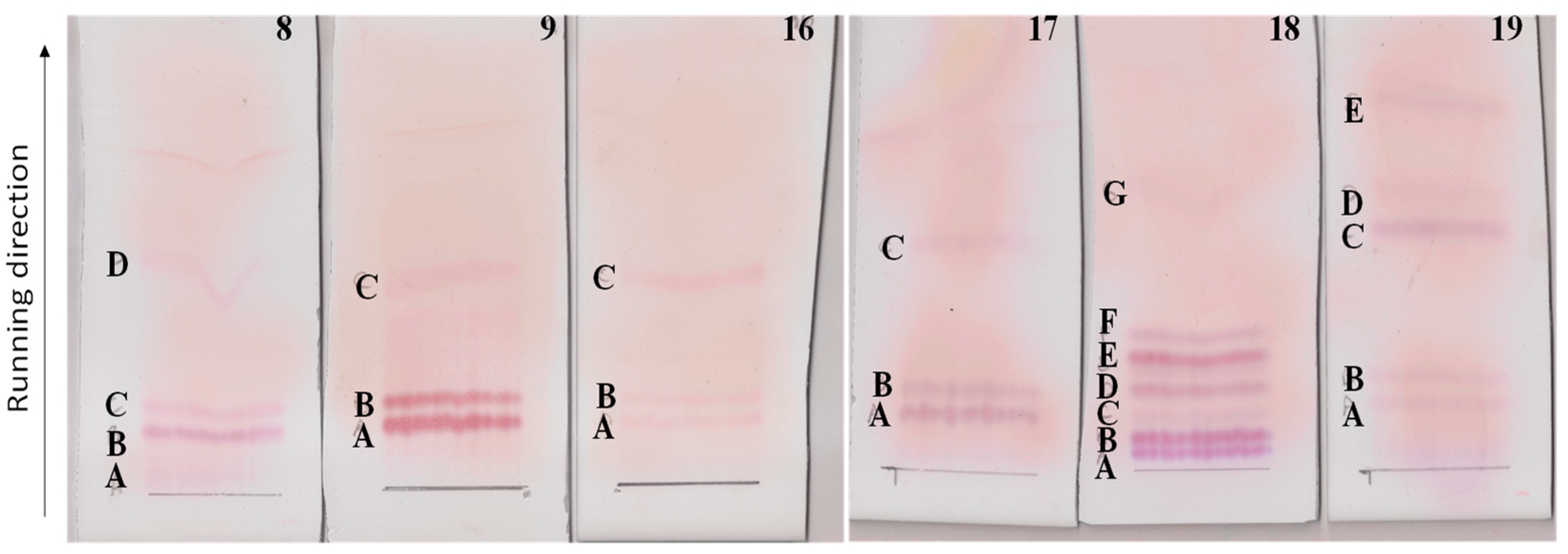
2.2. Lipopeptides Extraction and Purification

After a 24 h growth in CAA medium to promote secundary metabolits production [26], Serratia strains Arv-22-2.5c, Arv-22-2.6, A88copa7, A88copa13, NBRC 102599T and AS13 with serratomolides genes, the OD600 were observed (Table S3) followed by lipopeptides extraction and purification. Strain Arv-29-3.11b was not further analyzed since it was not able to recover after plate streaking.Thin layer chromatography (TLC) of polar lipids from each Serratia strain growth revealed four amino lipid spots on Serratia strain Arv-22-2.5c (8A, 8B, 8C, 8D); three on Serratia stains Arv-22-2.6 (9A, 9B, 9C), A88copa7 (16A, 16B, 16C) and A88copa13 (17A, 17B, 17C); seven on Serratia strain NBRC 102599T (18A, 18B, 18C, 18D, 18E, 18F, 18G) and five on Serratia strain AS13 (19A, 19B, 19C, 19D, 19E) (Figure 1).
The TLC profiles of the different strains share band staining as amino lipids. Moreover, the amino lipids profiles were not directly related with the serrawitin genes detected (swrW or swrA).

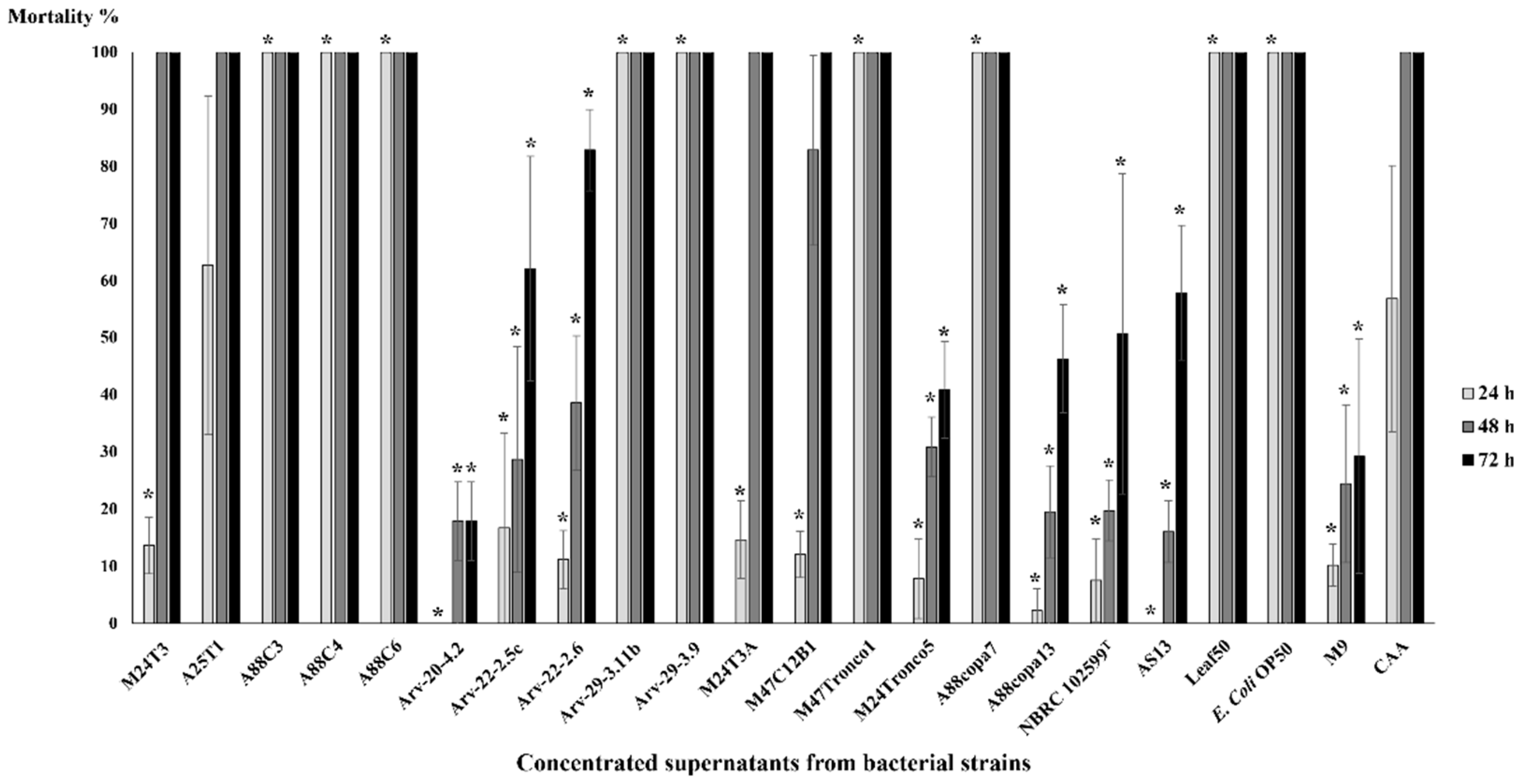
2.3. Mortality Tests in C. elegans

Mortality tests were performed to assess the nematicidal properties of bacterial supernatants. Ten C. elegans were used in each test in order to evaluate which strains have nematicidal properties against them. Mortality tests were performed in triplicate with concentrated supernatants (Figure 2) and extracted amino lipids (Figure 3).
Only concentrated supernatants from Serratia strains A88C3, A88C4, A88C6, Arv-29-3.11b, Arv-29-3.9, A88copa7, Leaf50, Pseudomonas strain M47Tronco1 and E. coli OP50 were able to kill 100% of C. elegans after 24 h, with statistical significance (Figure 2, Table S4). Concentrated supernatants from Serratia strains M24T3, A25T1, M24T3A and M47C12B1 showed 100% dead C. elegans. This was also visible for control CAA at the same time.
Mortality tests in C. elegans with extracted amino lipids revealed no statistical difference when compared to the 0.1 M NaCl solution (the solution used to resuspend TLC fractions); overall mortality rates were less than 15% (Figure 3). The Serratia strain A88copa13 supernatant that was previously identified as nematicidal for B. xylophilus [26] because it produced a serine protease to the supernatant was considered as a positive control for the experiment. Nevertheless, the strain supernatant was unable to have a nematicidal effect against C. elegans. E. coli OP50 with an OD600 = 0.4 was able to kill all nematodes in 24 h.

2.4. Mortality Tests in B. xylophilus

Mortality tests were performed to assess the nematicidal properties towards PWN of bacterial supernatants. Ten B. xylophilus in each test were used in order to evaluate which strains have nematicidal properties against this nematode (Figure 4, Table S5).
B. xylophilus’ mortality tests with concentrated supernatant revealed that this nematode is susceptible to Serratia strains. Almost every concentrated supernatant, when compared with negative control, showed statistical differences after 24 h, namely concentrated supernatants from Serratia strains M24T3, A25T1, A88C3, A88C4, A88C6, Arv-20-4.2, Arv-22-2.5c, Arv-22-2.6, Arv-29-3.11b, Arv-29-3.9, M24Tronco5, A88copa7, A88copa13, NBRC 102599T, AS13 and Leaf50, Pseudomonas strain M47Tronco1 and E. coli OP50. In these mortality tests, strains Arv-22-2.5c, Arv-22-2.6, Arv-29-3.9, A88copa7 and A88copa13, which have serrawettin genes, were able to kill more than 60% of B. xylophilus. After 48 h and 72 h, concentrated supernatants from all strains showed statistical differences when compared to control (CAA medium); all strains had a nematicidal activity against PWN. Serratia strains A25T1 and A88C6 were the only ones able to kill all B. xylophilus at 24 h (Figure 4).
Mortality tests performed with extracted amino lipids in B. xylophilus (Figure 5) revealed higher mortality rates when compared to C. elegans (Figure 3). When compared with the 0.1 M NaCl solution, amino lipids 8D from Serratia strain Arv-22-2.5c, 9C from Serratia strain Arv-22-2.6, 17C from Serratia strain A88copa13, 18F and 18G from Serratia strain NBRC 102599T, 19D and 19E from Serratia strain AS13 showed statistical significance, killing more than 55% of B. xylophilus after 24 h (Figure 5, Table S6). Amino lipids 8C and 8D from Serratia strain Arv-22-2.5c, 9C from Serratia strain Arv-22-2.6, 17A, 17B and 17C from Serratia strain A88copa13, 18E, 18F and 18G from Serratia strain NBRC 102599T, 19D and 19E from Serratia strain AS13 were able to kill B. xylophilus after 48 h, with statistical significance. Negative controls 0.1 M NaCl solution and H2O were unable to kill more than 15% of B. xylophilus. The supernatant of Serratia strain A88copa13, a strain that produces a serine protease and that was used as a nematicidal positive control, was unable to kill more than 60% of nematodes.

2.5. Attraction Tests in C. elegans

Attraction tests are designed to demonstrate if young, starved C. elegans are more attracted to bacteria or supernatant of selected strains or to control E. coli OP50.
Serratia strains M24T3, A25T1, A88C3, A88copa7 and Leaf50 were able to attract more C. elegans than control after 2 h and maintained their attraction effect after 24 h, with statistical significance (Figure 6a, Table S7). Pseudomonas strain M47Tronco1 and Serratia strains A88copa13 and AS13 were able to attract more C. elegans than control after 2 h but lost attraction effect after 24 h, with statistical significance. Serratia strains A88C4 and A88C6 were not able to attract C. elegans more than control after 2 h, but were able to do it after 24 h, with statistical significance. Serratia strains Arv-22-2.5c, Arv-29-3.9, M24T3A and M47C12B1 were not able to attract C. elegans; in these cases, nematodes were more attracted to E. coli OP50 both after 2 h and after 24 h (Figure 6a).
Supernatants of Serratia strains A25T1, A88C3 and A88C6 were able to attract more C. elegans than the E. coli OP50 control after 2 h and after 24 h, with statistical significance. Serratia strains Arv-22-2.5c and A88copa7 attracted nematodes after 2 h with statistical significance but lost their ability to attract after 24 h. Nematodes were more attracted to E. coli OP50 control than to Serratia strains M24T3, Arv-20-4.2, Arv-22-2.6, Arv-29-3.9, M24T3A, M47C12B1, M24Tronco5, A88copa13, AS13 and Pseudomonas strain M47Tronco1 after 2 h and after 24 h (Figure 6b, Table S8).
All bacteria supernatants were able to attract more C. elegans than E. coli OP50 supernatant control at 2 h with two exceptions—strain M24T3A, for which nematodes were more attracted to E. coli OP50 supernatant, and strain M47C12B1, which was able to attract nematodes after 2 h but lost this ability after 24 h (Figure 6c, Table S9).
Attraction tests with extracted amino lipids against 0.1 M NaCl solution revealed a very low percentage of attraction (Figure 7a, Table S10). After 2 h, only amino lipids 9C from Serratia strain Arv-22-2.6, 16C from Serratia strain A88copa7, 17A and 17C from Serratia strain A88copa13, 18E and 18G from Serratia strain NBRC 102599T and 19D from Serratia strain AS13 were able to attract more nematodes than control, with statistical significance. In attraction tests with amino lipids 8A and 8B from Serratia strain Arv-22-2.5c, 18B from Serratia strain NBRC 102599T and 19B from Serratia strain AS13, nematodes preferred the NaCl solution. After 24 h, amino lipids 8A and 8B from Serratia strain Arv-22-2.5c and 18C from Serratia strain NBRC 102599T were able to attract more nematodes than 0.1 M NaCl solution, with statistical significance. In attraction tests performed with amino lipids 18B and 18G from Serratia strain NBRC 102599T and 19A from Serratia strain AS13, nematodes preferred the control solution.
Attraction tests performed with extracted amino lipids against supernatants revealed a poor attraction by amino lipids (Figure 7b, Table S11). After 2 h, only amino lipids 8B from Serratia strain Arv-22-2.5c and 18C from Serratia strain NBRC 102599T were able to attract more nematodes than respective supernatants, with statistical significance. In attraction tests with amino lipids 8A from Serratia strain Arv-22-2.5c, 16B from Serratia strain A88copa7,17B and 17C from Serratia strain A88copa13, 18B and 18F from Serratia strain NBRC 102599T, nematodes preferred supernatants. After 48 h, almost all nematodes were attracted to supernatants, except for attraction tests with amino lipids 8C from Serratia strain Arv-22-2.5c, 18A and 18C from Serratia strain NBRC 102599T, and 19B from Serratia strain AS13, where most nematodes were dispersed over the NGM plate.
Attraction tests performed with extracted amino lipids against E. coli OP50 revealed that starved C. elegans preferred the food source over the amino lipids (Figure 7c, Table S12). After 2 h, only amino lipid 17A from Serratia strain A88copa13 was able to attract more nematodes than E. coli OP50, with statistical significance. After 24 h, nematodes were attracted by the food source in all tests.

2.6. HPLC Analysis of Selected TLC Bands

Eight extracted amino lipids were chosen for HPLC analysis since they showed better nematicidal activity against B. xylophilus when compared to other amino lipids. Amino lipids 8D from Serratia strain Arv-22-2.5c, 9C from Serratia strain Arv-22-2.6, 16C from Serratia strain A88copa7, 17C from Serratia strain A88copa13, 18F and 18G from Serratia strain NBRC 102599T and 19D and 19E from Serratia strain AS13.
HPLC analysis showed similar peaks in all amino lipids (Figure 8). All samples showed a peak between 3.48 and 3.86 min of retention time. Control with methanol reagent had no relevant peaks (Figure 8). High nematicidal activity of these extracted amino lipids may be associated with higher peaks.

3. Discussion

PWN, the known pathogenic agent of PWD, threatens pine and conifer forests all over the world. As global climate changes, a new scenario of PWN distribution evolves [36]. In order to prevent disease, spread, interactions between PWN and host trees, insect vectors and bacterial community must be understood. Serratia strains, previously described as part of the pine tree endophytic community [16] and associated with PWN [21,37], were chosen to study their nematicidal potential. This work intends to better understand the role of serratomolides, produced by Serratia strains, in PWD and, especially, to assess if they are effective against PWN. Serrawettin genes have already been found in several Serratia strains [30,32,33,34,35,38,39,40,41] and were present in seven of nineteen selected strains. The swrW gene was present in five Serratia strains and swrA gene was present in two Serratia strains. Serrawettin genes were also found in several Serratia strains resorting to computational approaches through AntiSMASH software [42]. Previously, several Serratia strains have been shown to produce high quantities of serrawettins, up to 17% of their mass dried weight [30]; some strains were already described as nematicidal [26,43]. The presence of other secondary metabolites described with nematicidal activity cannot be overruled [44].
Overall, the nematicidal effect of supernatants from selected strains was higher against B. xylophilus than against C. elegans. All supernatants were able to kill more than 30% of B. xylophilus after 48 h and more than 50% after 72 h, suggesting that this nematode is more susceptible to Serratia strains when compared to C. elegans. Our results confirm previous studies that identified some Serratia strains with nematicidal activity against C. elegans [33,45,46] and confirmed the nematicidal activity of Serratia strains against B. xylophilus, previously described [26]. It also expanded our knowledge about nematicidal activity of Serratia strains collected in different sampling sites, carried by PWN from USA [37] and from Portugal [21], and endophytic isolates from Portugal [16].
Concentrated supernatants presented better nematicidal results than non-concentrated supernatants and were able to kill more than 57% of B. xylophilus after 72 h. These results show that nematicidal activity increased as expected when supernatant components were more concentrated, which suggests that some extracellular components in Serratia supernatants possess strong nematicidal activity against nematodes. Concentrated supernatants from endophytic bacterial strains (Serratia strains A25T1, A88C4, A88C6), bacteria carried by PWN from Portugal (Serratia strain M24T3) and bacteria carried by PWN from USA (Serratia strains Arv-20-4.2, Arv-22-2.5c, Arv-22-2.6) were able to kill all B. xylophilus after 72 h. These results suggest that nematicidal activity is a characteristic that can be found in Serratia strains independently of the sample site. Mortality tests with concentrated supernatants and C. elegans revealed that Serratia strains A88C3, A88C4, A88C6, Arv-29-3.11b, Arv-29-3.9, A88copa7 and Leaf50, Pseudomonas strain M47Tronco1 and E. coli OP50 were able to kill all nematodes in 24 h. Even strains without swrW and swrA genes were able to produce high mortality rates, suggesting that nematicidal potential is not exclusively dependent on serratomolide presence, as observed for strains M24T3, A88C6 and A25T1. A previous study [26] had already concluded that some extracellular proteases, such as serine protease and serralysin, produced by Serratia strains, possess nematicidal activity against B. xylophilus.
Regarding attraction tests, 2 h results were considered more informative, as they had likely better reflected immediate attraction, whereas results after 24 h were probably more influenced by random nematode movements. Serratia strains M24T3, A25T1, A88C3, A88copa7 and Leaf50 were able to attract more nematodes than E. coli OP50 after 2 h and 24 h. Since C. elegans were starved, the preference for other bacteria than their usual food source reveals a strong attraction ability. Our findings confirm that Serratia strains have the ability to attract nematodes, as previously described [47] and that C. elegans is more attracted to some Serratia strains than to E. coli OP50, as previously described [46]. Supernatants of Serratia strains A25T1, A88C3, A88C4 and A88C6 were able to attract more nematodes than E. coli OP50 after 2 h and 24 h. None of these strains presented serratomolides genes, suggesting that another component in their supernatants is responsible for attraction. These results are in line with previous studies that showed that serrawettins produced by Serratia were able to act as repellent towards C. elegans [33] and showed nematostatic activity [48]. Some metabolites present in Serratia supernatants, such as amino acids, odors or autoinducers, may induce a food seeking behavior [33,49]. Almost all Serratia supernatants were able to attract more C. elegans than E. coli OP50 supernatant. This suggested that C. elegans may be attracted by some other E. coli OP50 cellular structure or component, that is absent or in low concentration in its respective supernatant, and/or that C. elegans preferred Serratia supernatant odors over E. coli OP50 odors, as previously described [50].
A total of 25 amino lipids from Serratia strains with swrW and swrA genes were extracted and purified from supernatants. Extracted amino lipids’ overall mortality rates against C. elegans were very low and identical to control. Mortality rates showed that B. xylophilus was more susceptible to these amino lipids than C. elegans. These results suggest that B. xylophilus may have more target proteins [51] susceptible to lipopeptides than C. elegans. Attraction tests with C. elegans revealed a poor attraction response to amino lipids and, overall, nematodes were more attracted to Serratia supernatants and food source E. coli OP50. As C. elegans were starved, these results suggest that amino lipids are not recognized as a potential food source. These results are consistent with the amino lipids’ absence in the nutritional requirements of C. elegans [52]. Amino lipids 8D, 9C, 16C, 17C, 18F, 18G, 19D and 19E had higher nematicidal activity against B. xylophilus than other extracted amino lipids from respective supernatants. These results seemed to indicate that, considering the mixture of amino lipids produced to the supernatant by the Serratia strains, the more polar amino lipids had more nematicidal activity against B. xylophylus and the less polar to C. elegans.
Subsequent HPLC analysis of these amino lipids extracted from Serratia strains with swrW and with swrA genes showed similar retention times on higher peaks, from 3.48–3.86 min. The results from this work indicated that amino lipids, serratomolides or other biosurfactants may be involved in Serratia nematicidal activity.

4. Materials and Methods

4.1. Bacterial Strains, Media and Growth Conditions

Twenty bacterial strains, 19 Serratia strains (seven strains isolated from Portuguese Pinus pinaster trees endophytic community [53], four PWN-carried strains from Portugal [21], five PWN-carried strains from USA [37] and three belonging to S. plymuthica) and one Pseudomonas strain carried by PWN from Portugal [21], used as control, were studied for lipopeptides serrawettin W1 and serrawettin W2 presence. Strains were grown on R2A agar medium for 24 h at 30 °C for DNA extraction. For mortality and attraction tests, bacterial strains were grown in Erlenmeyer’s flasks containing 50 mL of casamino acids (CAA) broth medium at 30 °C for 24 h, at 180 rpm [26]. Four mL of each bacterial growth were stored to be used on attraction tests. Bacterial growths were centrifuged at 12,100 rpm, at 4 °C, for 10 min, and 4 mL of each supernatant was used in mortality and attraction tests. Remainder supernatant was collected and concentrated with vacuum at −40 °C to be used on mortality tests.

4.2. DNA Extraction and Quantification

Genomic DNA extraction from all strains was performed using an NZY Microbial gDNA Isolation kit, according to manufacturer’s instructions. DNA samples were quantified using NanoDrop technology (Thermo ScientificTM, Waltham, MA, USA).

4.3. Primers Design, Polymerase Chain Reaction and DNA Sequence Analysis

To partially amplify swrW and swrA genes using PCR, new specific primers were designed (summarized in Tables S1 and S2). Eighty-five Serratia genomes available on NCBI were individually queried in the AntiSMASH database [42] to identify all secondary metabolite gene clusters (results published by authors [35]). Some of those secondary metabolite gene clusters presented DNA sequences similar to serrawettins’ genes: seventeen genomes with the swrW gene cluster and eleven with the swrA gene cluster [35]. Serratomolides genes were aligned and specific primers for swrW and swrA genes were designed, complementary to genetically conserved regions. Two primers for swrW gene were designed: swrW_1F, and swrW_7R. For the swrA gene, primers swrAA_1F and swrAA_10R were designed (Table S1).
In order to screen the presence of swrW and swrA genes in Serratia strains, PCRs were performed with selected primers. For each PCR, a mix was prepared containing, for each sample, 6.25 µL of MasterMix (NZYTaq II 2× Green Master Mix 0.2 U/μL), 0.2 µM of forward and reverse primers and 16.25 µL of ddH2O. One µL of DNA samples (Table S2) were added to a PCR tube containing the correspondent mix. For every mix, a negative control was made without DNA. PCR primers and gene length are summarized in Table S1. According to gene size and G+C mol% content of primers, temperature of annealing was adjusted. Electrophoreses were performed on 1% agarose gels at 90 V to separate and extract swrW and swrA genes. For PCR products that showed a band in the correspondent molecular size, DNA bands were removed and purified, using Omega’s Gel DNA Extraction Kit, according to the manufacturer’s instructions. DNA samples after gel extraction were sequenced by Stab Vida and the resulting sequences were compared with sequences available in EMBL/GenBank database using BLASTn network services [54].

4.4. Lipopeptides Extraction and Purification

Serratia strains Arv-22-2.5c, Arv-22-2.6, A88copa7, A88copa13, NBRC 102599T and AS13, containing the swrW gene, were grown in 100 mL of R2A broth medium at 120 rpm and 30 °C with an initial OD600 of 0.07. After 24 h, OD600 was measured; cultures were centrifuged at 12,100 rpm for 30 min at 4 °C. Supernatants were transferred to clean frosted Erlenmeyers and were adjusted to pH 2 with a 12 M HCl solution. One hundred mL of a chloroform–methanol (2:1, v/v) solution was added to each supernatant and these were shook for 2 h. Organic phases were transferred, after phase separation, to clean frosted flasks [55]. Organic phases were concentrated using a rotary evaporator at 60 °C and residual liquid was concentrated with nitrogen. Crude extracts with lipids were resuspended with 2 mL of chloroform–methanol (2:1, v/v) solution. Polar lipids were separated from other lipids trough a column with 1 g of activated silica-gel 60. Columns were previously washed with 4 mL of methanol and 4 mL of a chloroform–methanol (9:1, v/v) solution before 2 mL of eluted crudes were passed through the silica column. Particles unable to bind to silica-gel were rescued on a flask. Apolar lipids were eluted with 7 mL of a chloroform–methanol (9:1, v/v) solution and polar lipids were eluted with 7 mL of methanol [55]. Flasks with polar lipid fractions were concentrated with nitrogen and resuspended in 250 µL of methanol. Purified polar lipids were examined with 1D TLC. TLCs were prepared in triplicate for each sample and were performed on silica 10 × 10 cm plates with 15 µL of each sample along 3 cm. Chloroform–acetic acid–methanol–water (80:15:12:4, v/v) solvent was used to run TLCs. When the run was concluded, TLC plates were dried, and ninhydrin reagent was sprayed on one of the three plates and stayed at 120 °C for 5 min to reveal amino lipids [56]. Positive spots with amino lipids were scraped from the other two plates without dye, and were extracted with 3 mL of chloroform–methanol (2:1, v/v) solution. After vortex and centrifugation at 13,300 rpm for 1 min in order to remove silica gel, chloroform–methanol (2:1, v/v) solutions with individualized polar lipids were transferred to clean flasks and evaporated with nitrogen. Extractions from one TLC plate were used for attraction and mortality tests on nematodes and were resuspended with 750 µL of 0.1 M NaCl solution. Assays regarding mortality tests in C. elegans and B. xylophilus and attraction tests in C. elegans were performed. The extracts obtained from a second TLC plate were used for HPLC analysis and were resuspended with 100 µL of methanol. HPLC was performed using an eluent gradient as previously described in [34], with some modifications. Eluent A was composed by a 0.05% trifluoracetic acid solution in purified water and eluent B was methanol. The eluent B gradient was 87–92% for 15 min. Flow rate was 1 mL/min and UV detection was measured at 215 nm. Methanol was used as control.

4.5. Maintenance of C. elegans

Nematodes were synchronized on nematode growth medium (NGM) agar plates with E. coli OP50 (source of food) twice a week in order to renew the medium and maintain young and adult nematodes. To synchronize C. elegans, NGM agar plates with nematodes in different stages were washed up and down with 2 mL of ddH2O. A fresh NaOH–bleach solution with 0.5 mL of NaOH (2 M) and 1 mL of 3% bleach was added to C. elegans suspension. After 10 min with vigorous vortex every 2 min, nematode suspension with dead nematodes and viable eggs was centrifuged at 13,000 rpm for 1 min, supernatant was discarded and pellet was washed with 1 mL of pure water, twice. Then, 150 µL of the final resuspended solution was distributed into new NGM plates with E. coli OP50. For attraction tests, C. elegans synchronization took place 24 h before the experiment in order to use stage L3 larvae in all tests. Attraction tests were performed with starved synchronized C. elegans: eggs hatched on NGM plates without E. coli OP50. For mortality tests, fed C. elegans were synchronized 48 h before tests [57]. For all experiments, Petri dishes with synchronized C. elegans were washed with 2 mL of ddH2O and the volume was adjusted in order to have a nematode suspension with 20–30 C. elegans on 10 µL for attraction tests and with 10 C. elegans on 10 µL for mortality tests.

4.6. Attraction Tests in C. elegans

Attraction tests were performed on NGM plates divided in three parts and equidistant spots. For each bacteria strain, three different attraction tests were made: (i) bacterial suspension on one side and E. coli OP50 on the other side as a control; (ii) supernatant of bacteria on one side and E. coli OP50 on the other side; (iii) and supernatant of bacteria on one side and supernatant of E. coli OP50 on the other side. Three attraction tests were performed with C. elegans and amino lipids: (i) with amino lipids versus 0.1 M NaCl solution; (ii) with amino lipids versus respective supernatant; (iii) and with amino lipids versus E. coli OP50. With attraction tests, we assessed the percentage of nematodes drawn to bacteria, supernatants and extracted amino lipids. For bacterial suspension, 10 µL with OD600 = 1.0 were placed on the correspondent side of NGM agar plates. 10 µL of supernatants obtained by bacterial growth centrifugation and 10 µL of eluted amino lipids were used. After 30 min, for drops to dry, 10 µL of a nematode suspension, with approximately 20–30 C. elegans, was added to the third side of the NGM plate. The initial number of C. elegans in every plate was counted. Each attraction test was performed in triplicate. All NGM plates were sealed with Parafilm and were put at 19 °C. After 2 h and 24 h, nematodes on each drop were assessed to see if bacteria or supernatants were able to attract more nematodes than control [47,58].

4.7. Mortality Tests in C. elegans and B. xylophilus

Mortality tests were performed in 24 multi-well plates for both C. elegans and B. xylophilus. For C. elegans’ mortality tests, 10 µL of a nematode suspension with approximately 10 C. elegans was inserted in the middle of the well. The initial number of C. elegans in each well was assessed. For B. xylophilus’ mortality tests, 50 µL of ddH2O was put on the middle of the well and, for each well, exactly 10 young B. xylophilus were added using a disinfected eyelash. Every test was performed in triplicate with 500 µL of concentrated bacterial supernatants and once with supernatants. After 24 h, 48 h and 72 h, dead nematodes were assessed [26]. CAA medium (bacterial growth medium) and M9 medium (liquid medium used on C. elegans maintenance) were used as negative controls. To assess which amino lipid had a greater nematicidal effect, dead nematodes percentage was determined after a period of 24 h and 48 h with 500 µL of amino lipid suspension in NaCl solution [26].

4.8. Statistical Analysis

In order to find statistical differences among mortality test results, a one-way ANOVA was performed, separately, with data of mortality tests obtained from B. xylophilus and C. elegans, for each time point using the exposure treatment (strain or amino lipid) as a factor for comparison. Subsequently, when significant differences were detected by one-way ANOVA, a pairwise comparison was performed using the Tukey’s test. Attraction test results were analyzed using a Chi-Square statistical test to detect significant differences in the nematodes’ proportions between (i) bacterial suspensions and E. coli OP50 suspension as control; (ii) bacterial supernatants and E. coli OP50 suspension as control; (iii) bacterial supernatants and E. coli OP50 supernatant as control; (iv) amino lipids eluted in 0.1 M NaCl and 0.1 M NaCl solution as control; (v) amino lipids eluted in 0.1 M NaCl and corresponding bacterial supernatants; and (vi) amino lipids eluted in 0.1 M NaCl and E. coli OP50 suspension. All statistical analyses were performed using R packages stats (v4.1.2) and dplyr (v1.0.7) in R software (v4.1.2, https://www.r-project.org/, accessed on 15 December 2021) [59] and results were considered statistically different when p < 0.05.

4.9. Nucleotide Sequence Accession Numbers

The swrW and swrA gene sequences of the bacterial isolates reported in this study were deposited at the Genbank database, under the accession numbers OK500210-OK500216.

Supplementary Materials

The following supporting information can be downloaded at: https://0-www-mdpi-com.brum.beds.ac.uk/article/10.3390/pathogens11020198/s1, Table S1: Primers information to test the presence of swrW gene (PCR A) and swrA gene (PCR C); Table S2: Bacterial DNA concentrations of 19 Serratia strains M24T3, A25T1, A52T1, A88C3, A88C4, A88C6, Arv-20-4.2, Arv-22-2.5c, Arv-22-2.6, Arv-29-3.11b, Arv-29-3.9, M24T3A, M47C12B1, M24Tronco5, A88copa7, A88copa13, NBRC 102599T, AS13 and Leaf50 and Pseudomonas strain M47Tronco1; Table S3: Optical density of 24 h growth of Serratia strains Arv-22-2.5c, Arv-22-2.6, A88copa7, A88copa13, NBRC 102599T and AS13; Table S4: Mean and standard deviation of C. elegans’ mortality tests (%) with bacterial strains and controls M9 and CAA media. Tukey’s test p values for each strain versus control CAA medium are given for each time point 24, 48 and 72 h; Table S5: Mean and standard deviation of Bursaphenchus xylophilus’ mortality tests (%) with concentrated supernatants of bacterial strains and controls M9 and CAA media. Tukey’s test p values for each strain versus control CAA medium are given for each time point 24, 48 and 72 h; Table S6: Mean and standard deviation of B. xylophilus’ mortality tests (%) with amino lipids eluted in 0.1 M NaCl solution and controls H2O and 0.1 M NaCl solution. Tukey’s test p values for each amino lipid versus control 0.1 M NaCl solution are given for each time point 24 and 48 h; Table S7: Statistical analysis of attraction tests of C. elegans by bacterial suspensions versus E. coli OP50 suspension as control, using the mean proportion of nematodes as the dependent variable. Chi-square test value and the associated p value are given for C. elegans’ attraction tests at 2 and 24 h; Table S8: Statistical analysis of attraction tests of C. elegans by bacterial supernatants versus E. coli OP50 suspension as control, using the mean proportion of nematodes as dependent variable. Chi-square test value and the associated p value are given for C. elegans’ attraction tests at 2 and 24 h; Table S9: Statistical analysis of attraction tests of C. elegans by bacterial supernatants versus E. coli OP50 supernatant as control, using the mean proportion of nematodes as dependent variable. Chi-square test value and the associated p value are given for C. elegans’ attraction tests at 2 and 24 h; Table S10: Statistical analysis of attraction tests of C. elegans by amino lipids eluted in 0.1 M NaCl versus 0.1 M NaCl solution as control, using the mean proportion of nematodes as dependent variable. Chi-square test value and the associated p value are given for C. elegans’ attraction tests at 2 and 24 h; Table S11: Statistical analysis of attraction tests of C. elegans by amino lipids eluted in 0.1 M NaCl versus corresponding bacterial supernatants, using the mean proportion of nematodes as dependent variable. Chi-square test value and the associated p value are given for C. elegans’ attraction tests at 2 and 24 h; Table S12: Statistical analysis of attraction tests of C. elegans by amino lipids eluted in 0.1 M NaCl versus E. coli OP50 suspension, using the mean proportion of nematodes as dependent variable. Chi-square test value and the associated p value are given for C. elegans’ attraction tests at 2 and 24 h.

Author Contributions

Conceptualization, D.N.P. and P.V.M.; methodology, C.M.-P. and D.N.P.; formal analysis, C.M.-P. and D.N.P.; investigation, C.M.-P. and D.N.P.; resources, P.V.M.; data curation, D.N.P. and P.V.M.; writing—original draft preparation, C.M.-P., D.N.P., P.V.M.; writing—review and editing, D.N.P. and P.V.M.; supervision, D.N.P. and P.V.M.; project administration, P.V.M.; funding acquisition, P.V.M. All authors have read and agreed to the published version of the manuscript.

Funding

This research was funded by FEDER funds through the program COMPETE—Programa Operacional Factores de Competitividade and by national funds through FCT—Fundação para a Ciência e a Tecnologia, under the projects UID/EMS/00285/2020, PTWPTDC/AAG-REC/3839/2014; and by BIORECOVER Horizon2020 under grant agreement No 821096.

Institutional Review Board Statement

Not applicable.

Informed Consent Statement

Not applicable.

Data Availability Statement

The data presented in this study are available within this article and in supplementary material.

Acknowledgments

We would like to thank Isabel Abrantes and Luís Fonseca for providing B. xylophilus. We would like to thank Luís Cunha for his help on statistical analysis.

Conflicts of Interest

The authors declare no conflict of interest.

References

  1. Zhao, B.G.; Futai, K.; Sutherland, J.R.; Takeuchi, Y. (Eds.) Pine Wilt Disease; Springer: Tokyo, Japan, 2008; ISBN 978-4-431-75654-5. [Google Scholar]
  2. Jones, J.T.; Moens, M.; Mota, M.; Li, H.; Kikuchi, T. Bursaphelenchus xylophilus: Opportunities in comparative genomics and molecular host-parasite interactions. Mol. Plant Pathol. 2008, 9, 357–368. [Google Scholar] [CrossRef] [PubMed]
  3. Futai, K. Pine wilt in Japan: From First Incidence to the Present. 2008, pp. 5–12. Available online: https://0-link-springer-com.brum.beds.ac.uk/chapter/10.1007/978-4-431-75655-2_2 (accessed on 15 December 2021).
  4. Khan, F.A.; Gbadegesin, R.A. On the occurrence of nematode-induced pine wilt disease in Nigeria. Pakistan J. Nematol. 1991, 9, 57–58. [Google Scholar]
  5. Dwinnel, L.D. First report of pinewood nematode (Bursaphelenchus xylophilus) in Mexico. Plant Dis. 1993, 77, 846. [Google Scholar] [CrossRef]
  6. Mota, M.; Braasch, H.; Bravo, M.A.; Penas, A.C.; Burgermeister, W.; Metge, K.; Sousa, E. First report of Bursaphelenchus xylophilus in Portugal and in Europe. Nematology 1999, 1, 727–734. [Google Scholar] [CrossRef]
  7. Sousa, E.; Bonifácio, L.; Pires, J.; Penas, A.C.; Mota, M.; Bravo, M.A.; Naves, P. Bursaphelenchus xylophilus (Nematoda; Aphelenchoididae) associated with Monochamus galloprovincialis (Coleoptera; Cerambycidae) in Portugal. Nematology 2001, 3, 89–91. [Google Scholar] [CrossRef]
  8. Rodrigues, J.M.; Sousa, E.; Abrantes, I. Pine Wilt Disease historical overview. In Pine Wilt Disease in Europe: Biological Interactions and Integrated Management; Sousa, E., Vale, F., Abrantes, I., Eds.; FNAPF—Federação Nacional das Associações de Proprietários Florestais: Lisbon, Portugal, 2015; pp. 12–32. [Google Scholar]
  9. Fonseca, L.; Cardoso, J.M.S.; Lopes, A.; Pestana, M.; Abreu, F.; Nunes, N.; Mota, M.; Abrantes, I. The pinewood nematode, Bursaphelenchus xylophilus, in Madeira island. Helminthologia 2012, 49, 96–103. [Google Scholar] [CrossRef] [Green Version]
  10. Robertson, L.; Cobacho Arcos, S.; Escuer, M.; Santiago Merino, R.; Esparrago, G.; Abelleira, A.; Navas, A. Incidence of the pinewood nematode Bursaphelenchus xylophlius Steiner & Buhrer, 1934 (Nickle, 1970) in Spain. Nematology 2011, 13, 755–757. [Google Scholar] [CrossRef]
  11. de la Fuente, B.; Saura, S. Long-term projections of the natural expansion of the pine wood nematode in the Iberian Peninsula. Forests 2021, 12, 849. [Google Scholar] [CrossRef]
  12. Kikuchi, T.; Cotton, J.A.; Dalzell, J.J.; Hasegawa, K.; Kanzaki, N.; McVeigh, P.; Takanashi, T.; Tsai, I.J.; Assefa, S.A.; Cock, P.J.A.; et al. Genomic insights into the origin of parasitism in the emerging plant pathogen Bursaphelenchus xylophilus. PLoS Pathog. 2011, 7, e1002219. [Google Scholar] [CrossRef] [Green Version]
  13. Blaxter, M.L.; De Ley, P.; Garey, J.R.; Liu, L.X.; Scheldeman, P.; Vierstraete, A.; Vanfleteren, J.R.; Mackey, L.Y.; Dorris, M.; Frisse, L.M.; et al. A molecular evolutionary framework for the phylum Nematoda. Nature 1998, 392, 71–75. [Google Scholar] [CrossRef]
  14. Brenner, S. The genetics of Caenorhabditis elegans. Genetics 1974, 77, 71–94. [Google Scholar] [CrossRef] [PubMed]
  15. Kikuchi, T.; Jones, J.T.; Aikawa, T.; Kosaka, H.; Ogura, N. A family of glycosyl hydrolase family 45 cellulases from the pine wood nematode Bursaphelenchus xylophilus. FEBS Lett. 2004, 572, 201–205. [Google Scholar] [CrossRef] [Green Version]
  16. Proença, D.N.; Grass, G.; Morais, P.V. Understanding pine wilt disease: Roles of the pine endophytic bacteria and of the bacteria carried by the disease-causing pinewood nematode. Microbiologyopen 2017, 6, e00415. [Google Scholar] [CrossRef] [PubMed]
  17. Zhao, L.; Mota, M.; Vieira, P.; Butcher, R.A.; Sun, J. Interspecific communication between pinewood nematode, its insect vector, and associated microbes. Trends Parasitol. 2014, 30, 299–308. [Google Scholar] [CrossRef] [PubMed] [Green Version]
  18. EPPO/OEPP Diagnostic protocols for regulated pests: Bursaphelenchus xylophilus. Bull. OEPP/EPPO 2009, 39, 344–353.
  19. Ikeda, T. Control program of Pine Wilt Disease for landscape conservation–The case of Amanohashidate, Kyoto, Japan. In Pine Wilt Disease: A Worldwide Threat to Forest Ecosystems; Springer: Dordrecht, The Netherlands, 2008; pp. 397–404. [Google Scholar]
  20. Petrini, O. Fungal endophytes of tree leaves. In Microbial Ecology of Leaves; Andrews, J., Hirano, S., Eds.; Brock/Springer series in contemporary bioscience; Springer: New York, NY, USA, 1991; pp. 179–197. ISBN 978-1-4612-3168-4. [Google Scholar]
  21. Proença, D.N.; Francisco, R.; Santos, C.V.; Lopes, A.; Fonseca, L.; Abrantes, I.M.O.; Morais, P.V. Diversity of bacteria associated with Bursaphelenchus xylophilus and other nematodes isolated from Pinus pinaster trees with pine wilt disease. PLoS ONE 2010, 5, e15191. [Google Scholar] [CrossRef] [Green Version]
  22. Alves, M.; Pereira, A.; Vicente, C.; Matos, P.; Henriques, J.; Lopes, H.; Nascimento, F.; Mota, M.; Correia, A.; Henriques, I. The role of bacteria in pine wilt disease: Insights from microbiome analysis. FEMS Microbiol. Ecol. 2018, 94, fiy077. [Google Scholar] [CrossRef]
  23. Vicente, C.S.L.; Nascimento, F.X.; Espada, M.; Barbosa, P.; Hasegawa, K.; Mota, M.; Oliveira, S. Characterization of bacterial communities associated with the pine sawyer beetle Monochamus galloprovincialis, the insect vector of the pinewood nematode Bursaphelenchus xylophilus. FEMS Microbiol. Lett. 2013, 347, 130–139. [Google Scholar] [CrossRef] [Green Version]
  24. Proença, D.N.; Schwab, S.; Vidal, M.S.; Baldani, J.I.; Xavier, G.R.; Morais, P.V. The nematicide Serratia plymuthica M24T3 colonizes Arabidopsis thaliana, stimulates plant growth, and presents plant beneficial potential. Brazilian J. Microbiol. 2019, 50, 777–789. [Google Scholar] [CrossRef]
  25. Proença, D.N.; Heine, T.; Senges, C.H.R.; Bandow, J.E.; Morais, P.V.; Tischler, D. Bacterial metabolites produced under iron limitation kill pinewood nematode and attract Caenorhabditis elegans. Front. Microbiol. 2019, 10, 2166. [Google Scholar] [CrossRef]
  26. Paiva, G.; Proença, D.N.; Francisco, R.; Verissimo, P.; Santos, S.S.; Fonseca, L.; Abrantes, I.M.O.; Morais, P.V. Nematicidal bacteria associated to pinewood nematode produce extracellular proteases. PLoS ONE 2013, 8, e79705. [Google Scholar] [CrossRef] [PubMed] [Green Version]
  27. Shimazu, M. Use of microbes for control of Monochamus alternatus, vector of the invasive pinewood nematode. In Use of Microbes for Control and Eradication of Invasive Arthropods; Hajek, A.E., Glare, T.R., O’Callaghan, M., Eds.; Springer: Dordrecht, The Netherlands, 2009; Volume 6, pp. 141–157. ISBN 978-1-4020-8559-8. [Google Scholar]
  28. Matsuyama, T.; Fujita, M.; Yano, I. Wetting agent produced by Serratia marcescens. FEMS Microbiol. Lett. 1985, 28, 125–129. [Google Scholar] [CrossRef]
  29. Wasserman, H.H.; Keggi, J.J.; McKeon, J.E. The structure of Serratamolide1-3. J. Am. Chem. Soc. 1962, 84, 2978–2982. [Google Scholar] [CrossRef]
  30. Matsuyama, T.; Kaneda, K.; Nakagawa, Y.; Isa, K.; Hara-Hotta, H.; Yano, I. A novel extracellular cyclic lipopeptide which promotes flagellum-dependent and -independent spreading growth of Serratia marcescens. J. Bacteriol. 1992, 174, 1769–1776. [Google Scholar] [CrossRef] [PubMed] [Green Version]
  31. Matsuyama, T.; Murakami, T.; Fujita, M.; Fujita, S.; Yano, I. Extracellular vesicle formation and biosurfactant production by Serratia marcescens. Microbiology 1986, 132, 865–875. [Google Scholar] [CrossRef] [Green Version]
  32. Li, H.; Tanikawa, T.; Sato, Y.; Nakagawa, Y.; Matsuyama, T. Serratia marcescens gene required for surfactant serrawettin W1 production encodes putative aminolipid synthetase belonging to nonribosomal peptide synthetase family. Microbiol. Immunol. 2005, 49, 303–310. [Google Scholar] [CrossRef] [PubMed]
  33. Pradel, E.; Zhang, Y.; Pujol, N.; Matsuyama, T.; Bargmann, C.I.; Ewbank, J.J. Detection and avoidance of a natural product from the pathogenic bacterium Serratia marcescens by Caenorhabditis elegans. Proc. Natl. Acad. Sci. USA 2007, 104, 2295–2300. [Google Scholar] [CrossRef] [Green Version]
  34. Su, C.; Xiang, Z.; Liu, Y.; Zhao, X.; Sun, Y.; Li, Z.; Li, L.; Chang, F.; Chen, T.; Wen, X.; et al. Analysis of the genomic sequences and metabolites of Serratia surfactantfaciens sp. nov. YD25T that simultaneously produces prodigiosin and serrawettin W2. BMC Genom. 2016, 17, 865. [Google Scholar] [CrossRef] [Green Version]
  35. Marques-Pereira, C.; Proença, D.N.; Morais, P.V. Genome sequences of Serratia strains revealed common genes in both serratomolides gene clusters. Biology. 2020, 9, 482. [Google Scholar] [CrossRef]
  36. Hirata, A.; Nakamura, K.; Nakao, K.; Kominami, Y.; Tanaka, N.; Ohashi, H.; Takano, K.T.; Takeuchi, W.; Matsui, T. Potential distribution of pine wilt disease under future climate change scenarios. PLoS ONE 2017, 12, e0182837. [Google Scholar] [CrossRef] [Green Version]
  37. Proença, D.N.; Fonseca, L.; Powers, T.O.; Abrantes, I.M.O.; Morais, P.V. Diversity of bacteria carried by pinewood nematode in USA and phylogenetic comparison with isolates from other countries. PLoS ONE 2014, 9, e105190. [Google Scholar] [CrossRef]
  38. Matsuyama, T.; Nakagawa, Y. Bacterial wetting agents working in colonization of bacteria on surface environments. Colloids Surf. B Biointerfaces 1996, 7, 207–214. [Google Scholar] [CrossRef]
  39. Nascimento, F.; Vicente, C.; Cock, P.; Tavares, M.; Rossi, M.; Hasegawa, K.; Mota, M. From plants to nematodes: Serratia grimesii BXF1 genome reveals an adaptation to the modulation of multi-species interactions. Microb. Genom. 2018, 4, e000178. [Google Scholar] [CrossRef] [PubMed]
  40. Roberts, D.P.; McKenna, L.F.; Lakshman, D.K.; Meyer, S.L.F.; Kong, H.; de Souza, J.T.; Lydon, J.; Baker, C.J.; Buyer, J.S.; Chung, S. Suppression of damping-off of cucumber caused by Pythium ultimum with live cells and extracts of Serratia marcescens N4-5. Soil Biol. Biochem. 2007, 39, 2275–2288. [Google Scholar] [CrossRef]
  41. Zhang, L.; Sun, J.; Hao, Y.; Zhu, J.; Chu, J.; Wei, D.; Shen, Y. Microbial production of 2,3-butanediol by a surfactant (serrawettin)-deficient mutant of Serratia marcescens H30. J. Ind. Microbiol. Biotechnol. 2010, 37, 857–862. [Google Scholar] [CrossRef] [PubMed]
  42. Weber, T.; Blin, K.; Duddela, S.; Krug, D.; Kim, H.U.; Bruccoleri, R.; Lee, S.Y.; Fischbach, M.A.; Müller, R.; Wohlleben, W.; et al. antiSMASH 3.0—A comprehensive resource for the genome mining of biosynthetic gene clusters. Nucleic Acids Res. 2015, 43, W237–W243. [Google Scholar] [CrossRef] [PubMed] [Green Version]
  43. Rahul, S.; Chandrashekhar, P.; Hemant, B.; Chandrakant, N.; Laxmikant, S.; Satish, P. Nematicidal activity of microbial pigment from Serratia marcescens. Nat. Prod. Res. 2014, 28, 1399–1404. [Google Scholar] [CrossRef]
  44. Hellberg, J.E.E.U.; Matilla, M.A.; George, P.; Salmond, C. The broad-spectrum antibiotic, zeamine, kills the nematode worm Caenorhabditis elegans. Front. Microbiol. 2015, 6, 137. [Google Scholar] [CrossRef] [Green Version]
  45. Glater, E.E.; Rockman, M.V.; Bargmann, C.I. Multigenic natural variation underlies Caenorhabditis elegans olfactory preference for the bacterial pathogen Serratia marcescens. G3 (Bethesda) 2014, 4, 265–276. [Google Scholar] [CrossRef] [Green Version]
  46. Gibson, A.K.; Morran, L.T. A Model for Evolutionary Ecology of Disease: The case for Caenorhabditis nematodes and their natural parasites. J. Nematol. 2017, 49, 357–372. [Google Scholar] [CrossRef] [Green Version]
  47. Pujol, N.; Link, E.M.; Liu, L.X.; Kurz, C.L.; Alloing, G.; Tan, M.W.; Ray, K.P.; Solari, R.; Johnson, C.D.; Ewbank, J.J. A reverse genetic analysis of components of the Toll signaling pathway in Caenorhabditis elegans. Curr. Biol. 2001, 11, 809–821. [Google Scholar] [CrossRef] [Green Version]
  48. Heise, P.; Liu, Y.; Degenkolb, T.; Vogel, H.; Schäberle, T.F.; Marzinelli, E.M. Antibiotic-producing beneficial bacteria in the gut of the burying beetle Nicrophorus vespilloides. Front. Microbiol. 2019, 10, 1178. [Google Scholar] [CrossRef] [Green Version]
  49. Bargmann, C.I. Chemosensation in C. elegans. WormBook 2006, 1, 1–29. [Google Scholar] [CrossRef] [PubMed] [Green Version]
  50. Worthy, S.E.; Haynes, L.; Chambers, M.; Bethune, D.; Kan, E.; Chung, K.; Ota, R.; Taylor, C.J.; Glater, E.E. Identification of attractive odorants released by preferred bacterial food found in the natural habitats of C. elegans. PLoS ONE 2018, 13, e0201158. [Google Scholar] [CrossRef] [PubMed]
  51. Shanmugam, G.; Dubey, A.; Ponpandian, L.N.; Rim, S.O.; Seo, S.-T.; Bae, H.; Jeon, J. Genomic insights into nematicidal activity of a bacterial endophyte, Raoultella ornithinolytica MG against Pine Wilt Nematode. Plant Pathol. J. 2018, 34, 250. [Google Scholar] [CrossRef] [PubMed]
  52. Zečić, A.; Dhondt, I.; Braeckman, B.P. The nutritional requirements of Caenorhabditis elegans. Genes Nutr. 2019, 14, 15. [Google Scholar] [CrossRef] [PubMed]
  53. Proença, D.N.; Francisco, R.; Kublik, S.; Scholer, A.; Vestergaard, G.; Schloter, M.; Morais, P.V. The microbiome of endophytic, wood colonizing bacteria from pine trees as affected by Pine Wilt Disease. Sci. Rep. 2017, 7, 4205. [Google Scholar] [CrossRef] [Green Version]
  54. Altschul, S.F.; Gish, W.; Miller, W.; Myers, E.W.; Lipman, D.J. Basic local alignment search tool. J. Mol. Biol. 1990, 215, 403–410. [Google Scholar] [CrossRef]
  55. Korenblum, E.; de Araujo, L.; Guimarães, C.; de Souza, L.M.; Sassaki, G.; Abreu, F.; Nitschke, M.; Lins, U.; Freire, D.M.; Barreto-Bergter, E.; et al. Purification and characterization of a surfactin-like molecule produced by Bacillus sp. H2O-1 and its antagonistic effect against sulfate reducing bacteria. BMC Microbiol. 2012, 12, 252. [Google Scholar] [CrossRef] [Green Version]
  56. Kügler, J.H.; Muhle-Goll, C.; Hansen, S.H.; Völp, A.R.; Kirschhöfer, F.; Kühl, B.; Brenner-Weiss, G.; Luy, B.; Syldatk, C.; Hausmann, R. Glycolipids produced by Rouxiella sp. DSM 100043 and isolation of the biosurfactants via foam-fractionation. AMB Express 2015, 5, 82. [Google Scholar] [CrossRef]
  57. Stiernagle, T. Maintenance of C. elegans. WormBook 2006, 2, 51–67. [Google Scholar] [CrossRef] [PubMed] [Green Version]
  58. Hsueh, Y.; Gronquist, M.R.; Schwarz, E.M.; Nath, R.D.; Lee, C.; Gharib, S.; Schroeder, F.C.; Sternberg, P.W. Nematophagous fungus Arthrobotrys oligospora mimics olfactory cues of sex and food to lure its nematode prey. eLife 2017, 6, e20023. [Google Scholar] [CrossRef] [PubMed]
  59. R Core Development Team. R: A Language and Environment for Statistical Computing, R Version 4.1.2. 2021. Available online: https://www.r-project.org/ (accessed on 15 December 2021).
Figure 1. TLCs of polar lipids from supernatants of Serratia strains Arv-22-2.5c (TLC 8), Arv-22-2.6 (TLC 9), A88copa7 (TLC 16), A88copa13 (TLC 17), NBRC 102599T (TLC 18) and AS13 (TLC 19). Positive amino lipids were spotted with ninhydrin solution, colored in pink.
Pathogens 11 00198 g001
Figure 2. C. elegans’ mortality tests with concentrated supernatants of bacterial strains and M9 and CAA media as controls. Supernatants were concentrated by vacuum and evaporation at low temperatures (−40 °C) after a 24 h growth of Serratia strains M24T3, A25T1, A88C3, A88C4, A88C6, Arv-20-4.2, Arv-22-2.5c, Arv-22-2.6, Arv-29-3.11b, Arv-29-3.9, M24T3A, M47C12B1, M24Tronco5, A88copa7, A88copa13, NBRC 102599T, AS13 and Leaf50, and two controls Pseudomonas sp. M47Tronco1 and E. coli OP50. The number of dead nematodes was assessed at 24 h, 48 h and 72 h after incubation with concentrated supernatants. Each test was performed in triplicate. After one-way ANOVA, the comparison for mortality between the different strains was statistically significant for all the time points (24 h: F(21, 44) = 67.62, p < 0.001; 48 h: F(21, 44) = 76.04, p < 0.001; 72 h: F(21,44) = 25.80, p < 0.001). Results that showed statistical differences on the post hoc Tukey’s Test at p < 0.05 between strain and CAA medium are marked with (*). The lines above the bars represent standard deviation.
Pathogens 11 00198 g002
Figure 3. C. elegans’ mortality tests with amino lipids eluted in 0.1 M NaCl solution and H2O and 0.1 M NaCl solution as negative control. Amino lipids were extracted from supernatants of a 24 h growth of Serratia strains Arv-22-2.5c (amino lipids 8A, 8B, 8C and 8D), Arv-22-2.6 (amino lipids 9A, 9B and 9C), A88copa7 (amino lipids 16A, 16B and 16C), A88copa13 (amino lipids 17A, 17B and 17C), NBRC 102599T (amino lipids 18A, 18B, 18C, 18D, 18E, 18F and 18G) and AS13 (amino lipids 19A, 19B, 19C, 19D and 19E). The number of dead nematodes was assessed at 24 h and 48 h after incubation with amino lipids. Each test was performed in triplicate. After one-way ANOVA, the comparison for mortality between the different amino lipids was not statistically significant for all the time points (24 h: F(26, 54) = 0.959, p = 0.534; 48 h: F(26, 54) = 1.326, p = 0.189). The lines above the bars represent standard deviation.
Pathogens 11 00198 g003
Figure 4. Bursaphenchus xylophilus’ mortality tests with concentrated supernatants of bacterial strains and M9 and CAA media as control. Supernatants were concentrated by vacuum and evaporation at low temperatures after a 24 h growth of Serratia strains M24T3, A25T1, A88C3, A88C4, A88C6, Arv-20-4.2, Arv-22-2.5c, Arv-22-2.6, Arv-29-3.11b, Arv-29-3.9, M24T3A, M47C12B1, M24Tronco5, A88copa7, A88copa13, NBRC 102599T, AS13 and Leaf50, and controls Pseudomonas sp. M47Tronco1 and E. coli OP50. The number of dead nematodes was assessed at 24 h, 48 h and 72 h after incubation with concentrated supernatants. Each test was performed in triplicate. After one-way ANOVA, the comparison for mortality between the different strains was statistically significant for all the time points (24 h: F(21, 44) = 31.29, p < 0.001; 48 h: F(21, 44) = 14.31, p < 0.001; 72 h: F(21,44) = 17.27, p < 0.001). Results that showed statistical differences on the post hoc Tukey’s test at p < 0.05 between strain and CAA medium are marked with (*). The lines above the bars represent standard deviation.
Pathogens 11 00198 g004
Figure 5. B. xylophilus’ mortality tests with amino lipids eluted in 0.1 M NaCl solution and H2O and 0.1 M NaCl solution as negative control. Amino lipids were extracted from supernatants of a 24 h growth of Serratia strains Arv-22-2.5c (amino lipids 8A, 8B, 8C and 8D), Arv-22-2.6 (amino lipids 9A, 9B and 9C), A88copa7 (amino lipids 16A, 16B and 16C), A88copa13 (amino lipids 17A, 17B and 17C), NBRC 102599T (amino lipids 18A, 18B, 18C, 18D, 18E, 18F and 18G) and AS13 (amino lipids 19A, 19B, 19C, 19D and 19E). The number of dead nematodes was assessed at 24 h and 48 h after incubation with amino lipids. Each test was performed in triplicate. After one-way ANOVA, the comparison for mortality between the different amino lipids was statistically significant for all the time points (24 h: F(26, 54) = 5.709, p < 0.001; 48 h: F(26, 54) = 11.43, p < 0.001). Results that showed statistical differences on the post hoc Tukey’s test at p < 0.05 between amino lipid and NaCl solution are marked with (*). The lines above the bars represent standard deviation.
Pathogens 11 00198 g005
Figure 6. Attraction tests of C. elegans by bacterial suspensions and by their supernatants. The nematodes attraction was performed (a) by bacterial suspensions versus E. coli OP50 suspension as control; (b) by bacterial supernatants versus E. coli OP50 suspension as control; (c) by bacterial supernatants versus E. coli OP50 supernatant as control. All bacterial suspensions were used on attraction tests with an OD600 of 1.0. Bacterial supernatants were obtained after a 24 h growth. The number of attracted nematodes by bacteria and control was counted at 2 h and 24 h after C. elegans were added for all experiments. Each test was performed in triplicate. Results that showed statistical differences on chi-squared test, p < 0.05, are marked with (*). Bacterial strains: Serratia strains M24T3, A25T1, A88C3, A88C4, A88C6, Arv-20-4.2, Arv-22-2.5c, Arv-22-2.6, Arv-29-3.11b, Arv-29-3.9, M24T3A, M47C12B1, M24Tronco5, A88copa7, A88copa13, NBRC 102599T, AS13 and Leaf50, Pseudomonas strain M47Tronco1 and E. coli OP50. The lines above the bars represent standard deviation.
Pathogens 11 00198 g006aPathogens 11 00198 g006b
Figure 7. Attraction tests of C. elegans by amino lipids, by bacterial supernatants and E. coli OP50. The nematodes attraction was performed by (a) amino lipids eluted in 0.1 M NaCl versus 0.1 M NaCl solution as control; (b) by amino lipids eluted in 0.1 M NaCl versus bacterial supernatants; (c) by amino lipids eluted in 0.1 M NaCl versus E. coli OP50 suspension with an OD600 of 1.0. In all assays, amino lipids were extracted from supernatants of a 24 h growth of Serratia strains. The number of attracted nematodes by amino lipids, by bacterial supernatants and by E. coli OP50 was counted at 2 h and 24 h after C. elegans were added. Each test was performed in triplicate. Results that showed statistical differences on chi-squared test, p < 0.05, are marked with (*). Serratia strains and amino lipids—Arv-22-2.5c (amino lipids 8A, 8B, 8C and 8D), Arv-22-2.6 (amino lipids 9A, 9B and 9C), A88copa7 (amino lipids 16A, 16B and 16C), A88copa13 (amino lipids 17A, 17B and 17C), NBRC 102599T (amino lipids 18A, 18B, 18C, 18D, 18E, 18F and 18G) and AS13 (amino lipids 19A, 19B, 19C, 19D and 19E). The lines above the bars represent standard deviation.
Pathogens 11 00198 g007aPathogens 11 00198 g007b
Figure 8. HPLC analysis of amino lipids produced by Serratia strains. Samples 8D from Serratia strain Arv-22-2.5c, 9C from Serratia strain Arv-22-2.6, 16C from Serratia strain A88copa7, 17C from Serratia strain A88copa13, 18F and 18G from Serratia strain NBRC 102599T and 19D and 19E from Serratia strain AS13 and methanol control. HPLC was performed for 15 min with an eluent gradient of 87–92% of methanol. UV was detected at 215 nm.
Pathogens 11 00198 g008
Table 1. Sequencing and BLASTn results of swrW and swrA partial genes sequences of Serratia isolates.
Bacterial StrainPCR CodePCR Product LengthAccession NumberSerratia SpeciesGeneIdentity %Cover %Accession Number
Serratia sp. Arv-20-4.28555OK500210Serratia marcescensswrW98.38%100%AB193098
Serratia sp. Arv-22-2.5c9438OK500211Serratia marcescensswrW98.87%100%AB193098
Serratia sp. Arv-29-3.11b11447OK500212Serratia marcescensswrW98.88%100%AB193098
Serratia sp. NBRC 102599T18430OK500213Serratia marcescensswrW72.52%100%AB193098
Serratia sp. AS1319452OK500214Serratia marcescensswrW73.46%96%AB193098
Serratia sp. A88copa716357OK500215Serratia liquefaciensswrA71.27%94%AF039572
Serratia sp. A88copa1317455OK500216Serratia liquefaciensswrA69.36%99%AF039572
Publisher’s Note: MDPI stays neutral with regard to jurisdictional claims in published maps and institutional affiliations.

