Article

Species Diversity, Habitat Distribution, and Blood Meal Analysis of Haematophagous Dipterans Collected by CDC-UV Light Traps in the Dominican Republic

by
Mikel Alexander González
1,2,
Daniel Bravo-Barriga
3,*,
María Altagracia Rodríguez-Sosa
1,
Juan Rueda
1,4,
Eva Frontera
3 and
Pedro María Alarcón-Elbal
1
1
Institute of Tropical Medicine and Global Health (IMTSAG), Universidad Iberoamericana (UNIBE), Santo Domingo 10203, Dominican Republic
2
Applied Zoology and Animal Conservation Group, University of the Balearic Islands (UIB), 07122 Palma de Mallorca, Spain
3
Parasitology and Parasitic Diseases, Animal Health Department, Veterinary Faculty, University of Extremadura (Uex), 10003 Cáceres, Spain
4
Cavanilles Institute for Biodiversity and Evolutionary Biology (ICBiBE), University of Valencia, 46980 Paterna, Spain
*
Author to whom correspondence should be addressed.
Submission received: 20 May 2022 / Revised: 15 June 2022 / Accepted: 19 June 2022 / Published: 21 June 2022
(This article belongs to the Special Issue Biology, Control and Zoonotic Role of Disease Vectors)

Abstract

:
Haematophagous insects cause major economic losses by both direct damage and the transmission of pathogens. However, the biting Diptera species in the Caribbean region have been poorly documented. During 2021, CDC downdraft suction traps with UV light were employed to assess both the species occurrence and blood meal sources across three different habitats in the Dominican Republic. Eighteen species of mosquitoes (n = 274), six species of Culicoides (n = 803), two black fly species (n = 2), and one species of muscid fly (n = 25) were identified at species-level by morphology and/or molecular phylogenetic approaches based on the mitochondrial cytochrome c oxidase subunit 1 (COI). Engorged mosquito (n = 5) and Culicoides (n = 28) females showed host preferences derived exclusively from mammals (cows and pigs), except Culex species containing the blood of chickens. Our study provides new records of the Diptera Dominican catalogue (Culex salinarius for the Greater Antilles, Culicoides jamaicensis for Hispaniola, and Culicoides haitiensis and Culicoides borinqueni for the Dominican Republic), the first available COI DNA sequences of different Diptera in the GenBank, some pictures of diagnostic features of closely related specimens, spatial distribution across the habitats studied, and new insights on their feeding preferences in the Caribbean region.

1. Introduction

Vector-borne diseases remain a major threat for Caribbean health agencies and stakeholders. In the geographical context, favourable climatic conditions allow a practically uninterrupted transmission pattern throughout the year, becoming a great impediment to social and economic development [1]. Although the interest of these diseases has grown due to the increase in some arbovirus outbreaks [2], there is scant information on vectors in islands such as Hispaniola, except for immature stages of mosquitoes (Diptera: Culicidae), which have been studied extensively in recent years [3,4]. Nonetheless, knowledge of other relevant haematophagous Diptera of medical and veterinary interest is still very scarce and needs to be updated with entomological studies.
Culicids are considered the most relevant arthropod family in human and animal health worldwide [5], and in the insular Caribbean [1,6]. In the Greater Antilles, some synanthropic and well-distributed mosquito species, such as Aedes aegypti (Linnaeus, 1762), Aedes albopictus (Skuse, 1894), and Culex quinquefasciatus (Say, 1823), are the main species responsible for arbovirus transmission [1,7]. The recent finding of Aedes vittatus (Bigot, 1861), a new potential vector species of yellow fever, Zika, chikungunya, and dengue virus [8], in the Americas has recently gained considerable attention. It was first recorded in 2020 in the Dominican Republic and Cuba [9,10], and its local distribution is still unknown because it has only been spotted in specific locations.
Tiny biting midges of the genus Culicoides (Diptera: Ceratopogonidae) are responsible for the transmission of both human and veterinary vector-borne diseases in the insular Caribbean. Culicoides furens (Poey, 1853) and Culicoides barbosai (Wirth & Blanton, 1956) were reported as the main vectors of Mansonella ozzardi (Mansoni, 1897) in Haiti, but this filarial nematode has never been reported in the neighbouring Dominican Republic [11,12,13]. The Oropouche virus (OROV) is also an emerging zoonotic disease transmitted by biting midges; it was recently detected in the plasma of children in schools of Haiti [14]. Bluetongue virus (BTV) is considered the most important veterinary disease transmitted by Culicoides, mainly in sheep and some wildlife species. In the Dominican Republic, serotypes BTV-4, BTV-6, and BTV-8 were detected by virus isolation several times at the end of the 20th century [15]. Despite the epidemiological importance of biting haematophagous Diptera, their study in the Dominican Republic has been ignored for decades.
The identification of blood meal sources of arthropod vector species contributes to the understanding of host–vector–pathogen interactions, and provides important insights into the dynamics of viral, parasitic, or bacterial transmission, allowing public health authorities to design and implement efficient strategies for vector control [16]. Molecular markers, i.e., the cytochrome b (cyt-b) and cytochrome c oxidase subunit 1 (COI) are widely used to accurately address host range preferences and to replace animal-baited traps, landing experiments, and serological methods such as the precipitin test or enzyme-linked immunosorbent assays (ELISA), which present severe limitations [17]. COI-based DNA barcoding is also a useful tool to complement taxonomy-based identification of mosquito species. However, these molecular techniques have not been widely implemented in many low-and middle-income Caribbean countries. In addition, the accurate identification of arthropod vectors is challenging due to the absence of updated keys, the damage of the external characters due to improper specimen handling or storage, and overall, due to the lack of well-trained staff in medical-veterinary entomology, a specialized discipline that has been regressing in the region for at least two decades [18].
The above-mentioned reasons might explain the low number of studies on the diversity of arthropod vector species and host interactions from the Caribbean region. Therefore, based on traditional morphological methods accompanied by DNA barcoding, the present study aimed to provide accurate information on the fauna of nocturnal/crepuscular haematophagous dipterans, and to assess their host blood meal sources in Jarabacoa, a touristic mountainous area of the Dominican Republic.

2. Results

A total of 1104 haematophagous dipteran specimens of at least 27 species of four families: 18 species of Culicidae, including two unknown species (Culex sp. and Uranotaenia sp.); six species of Ceratopogonidae; two species of Simuliidae; and one of Muscidae were captured and identified at species-level. COI barcodes were generated for 12 mosquito species and four Culicoides species; six of them (three mosquitoes and three Culicoides) were not previously available in public sequence databases. Differences in spatial distribution across the three habitats were significant for biting midges. Host DNA sequences were generated from 33 blood-engorged specimens of six and three species of mosquitoes and Culicoides, respectively.

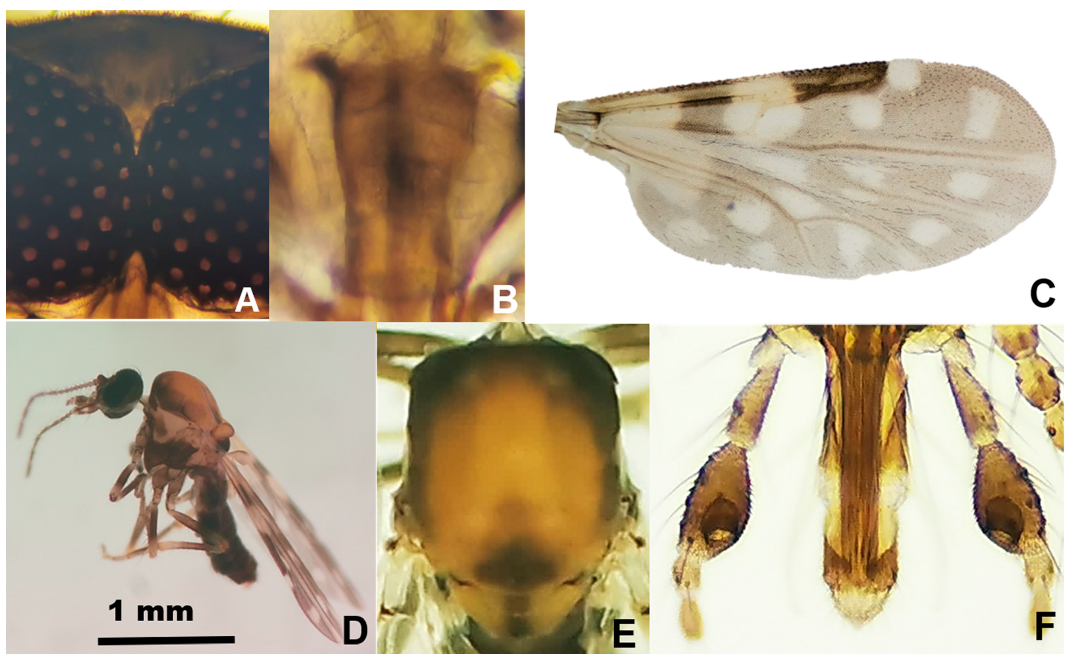
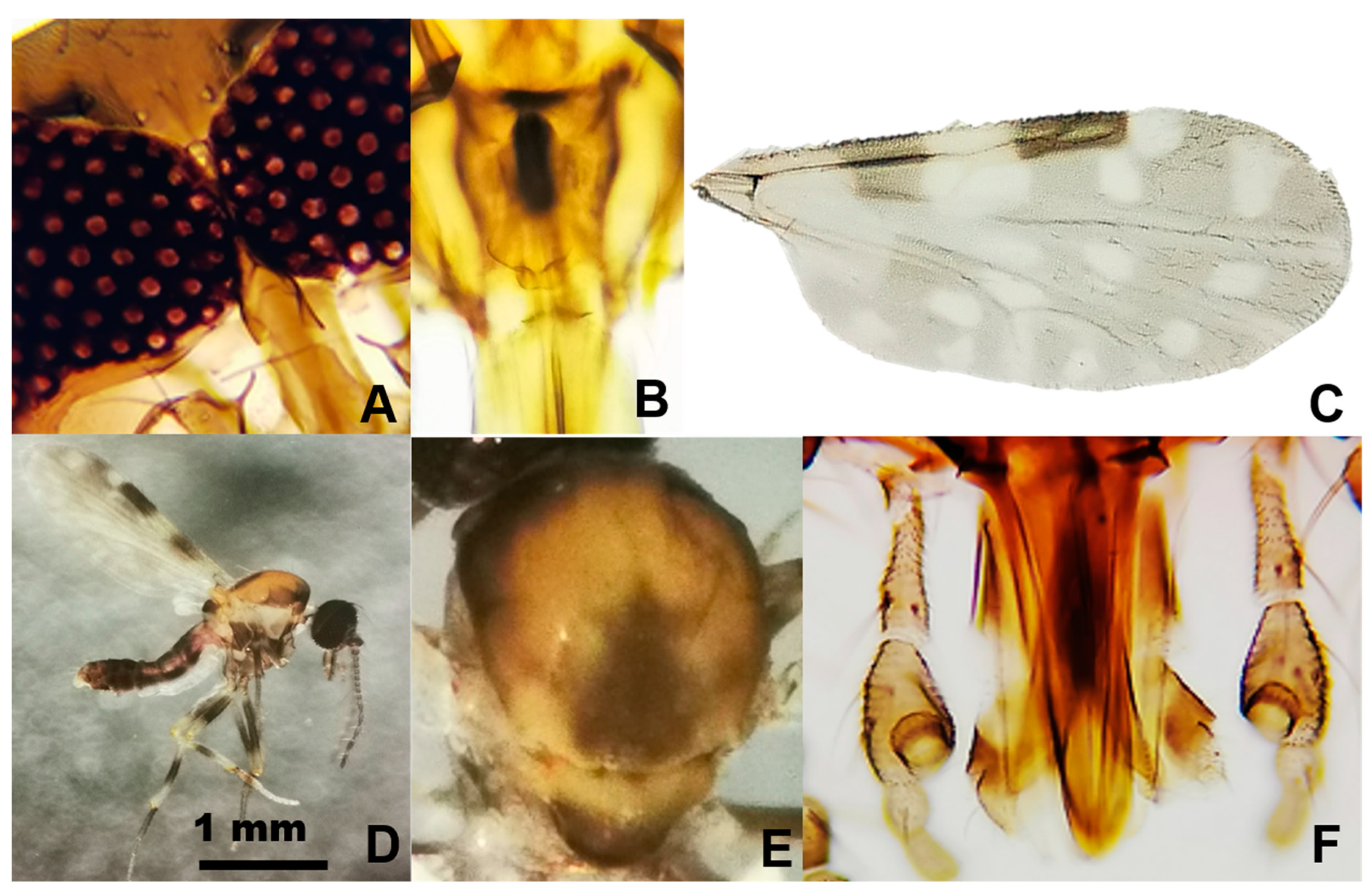
2.1. Species Composition Based on Morphological Features

A total of 263 mosquito specimens of 11 species were identified morphologically. The following species were recorded in decreasing order of abundance: Cx. quinquefasciatus (145 females, 32 males), Aedes scapularis (Rondani, 1848) (22 females), Culex secutor Theobald, 1901 (13 females, 4 males), Wyeomyia mitchellii (Theobald, 1905) (10 females, 1 male), Anopheles albimanus Weidemann, 1820 (11 females), Anopheles crucians Wiedemann, 1828 (7 females), Ae. vittatus (4 females, 2 males), Psorophora confinnis (Lynch Arribálzaga, 1891) (4 females), Ae. albopictus (3 females), Uranotaenia sapphirina (Osten Sacken, 1868) (2 females, 1 male), and Anopheles grabhamii Theobald, 1901 (2 females). In addition, 11 specimens could be identified to genus-level: Culex spp. (8 females), Aedes sp. (1 female), Wyeomyia sp. (1 female), and Uranotaenia sp. (1 female) (Figure 1).
Among the 803 specimens of Culicoides, six species were reported in the following order of abundance: Culicoides insignis Lutz, 1913 (392 females, 32 males), Culicoides foxi (Ortiz, 1950) (300 females, 21 males), Culicoides pusillus Lutz, 1913 (36 females, 15 males), Culicoides borinqueni Fox and Hoffman, 1944 (4 females), Culicoides jamaicensis (Edwards, 1922) (2 females), and Culicoides haitiensis Delecolle, Raccurt, and Rebholtz, 1986 (1 female) (Figure 1). Culicoides haitiensis and C. borinqueni were recorded for the first time for the Dominican Republic, whereas C. jamaicensis was reported for the first time for Hispaniola. Illustrated diagnostic characters of C. haitiensis and C. borinqueni females are provided for the accurate separation of both sibling species (Figure 2 and Figure 3). Fresh specimens can be easily separated under the stereomicroscope according to the wing pattern and pale spot of the scutellum. Mounted specimens can be separated based on the presence of cibarial armature, which is only present in C. borinqueni. Specimens of C. foxi were allocated to three different morphotypes according to their wing pattern: (i) typical morphospecies with a spot on the distal part of m1 (n = 290), (ii) morphospecies without a spot on m1 (n = 27), and (iii) morphospecies with both spots on m1 joined (n = 3) (Figure 4A).
Other collected haematophagous insects, the black fly species (Diptera: Simuliidae) Simulium quadrivittatum Loew, 1862 (n = 1), and Simulium haematopotum Malloch, 1914 (n = 1), and Haematobia irritans (Linnaeus, 1758) (Diptera: Muscidae) (n = 25) were also recorded by suction traps. No sand flies (Diptera: Psychodidae) were collected.

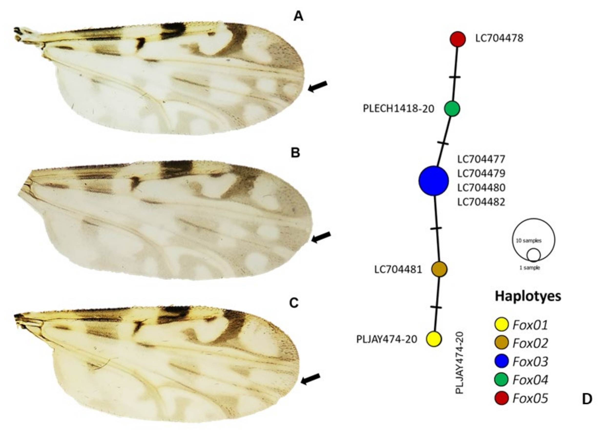
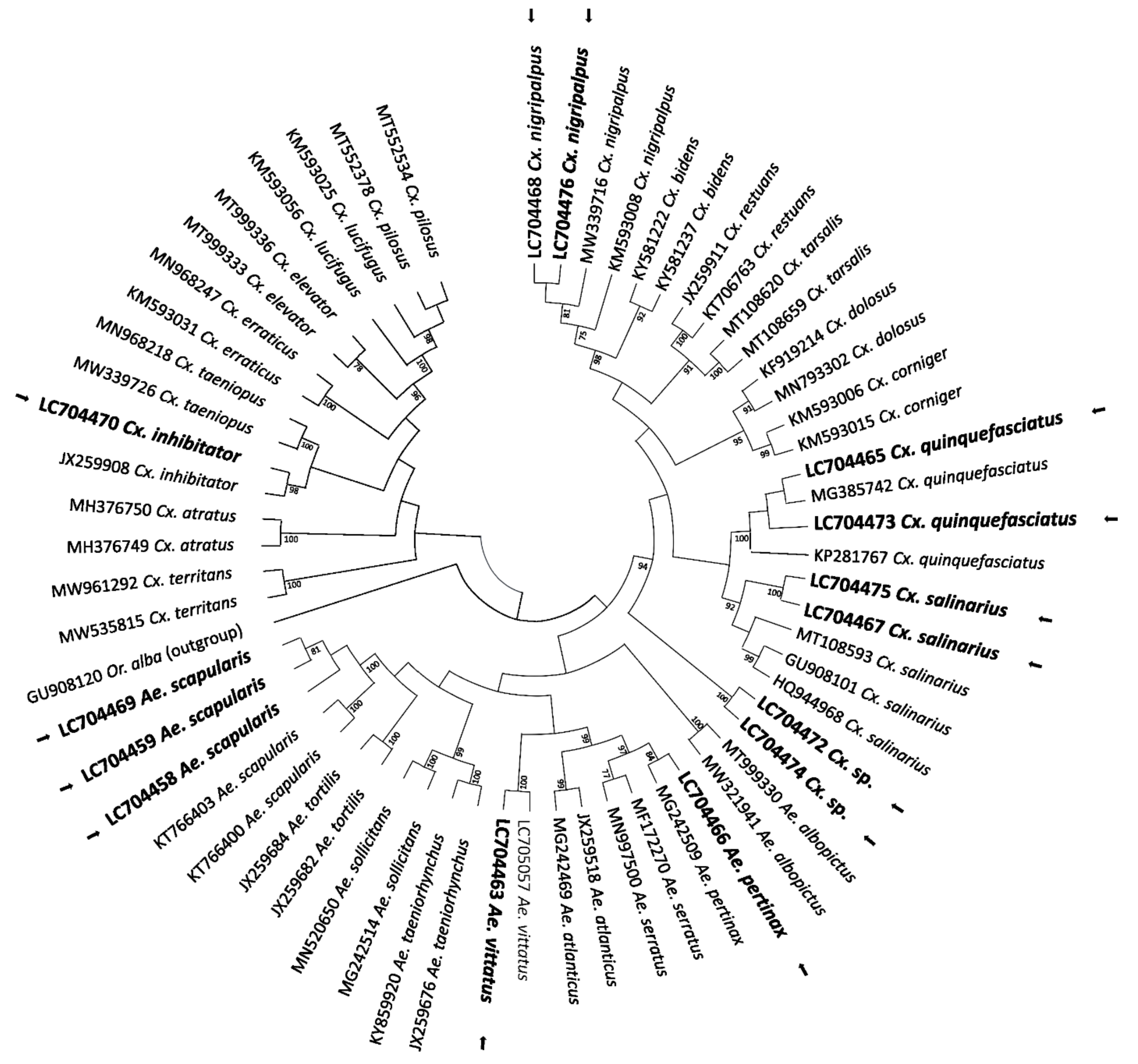
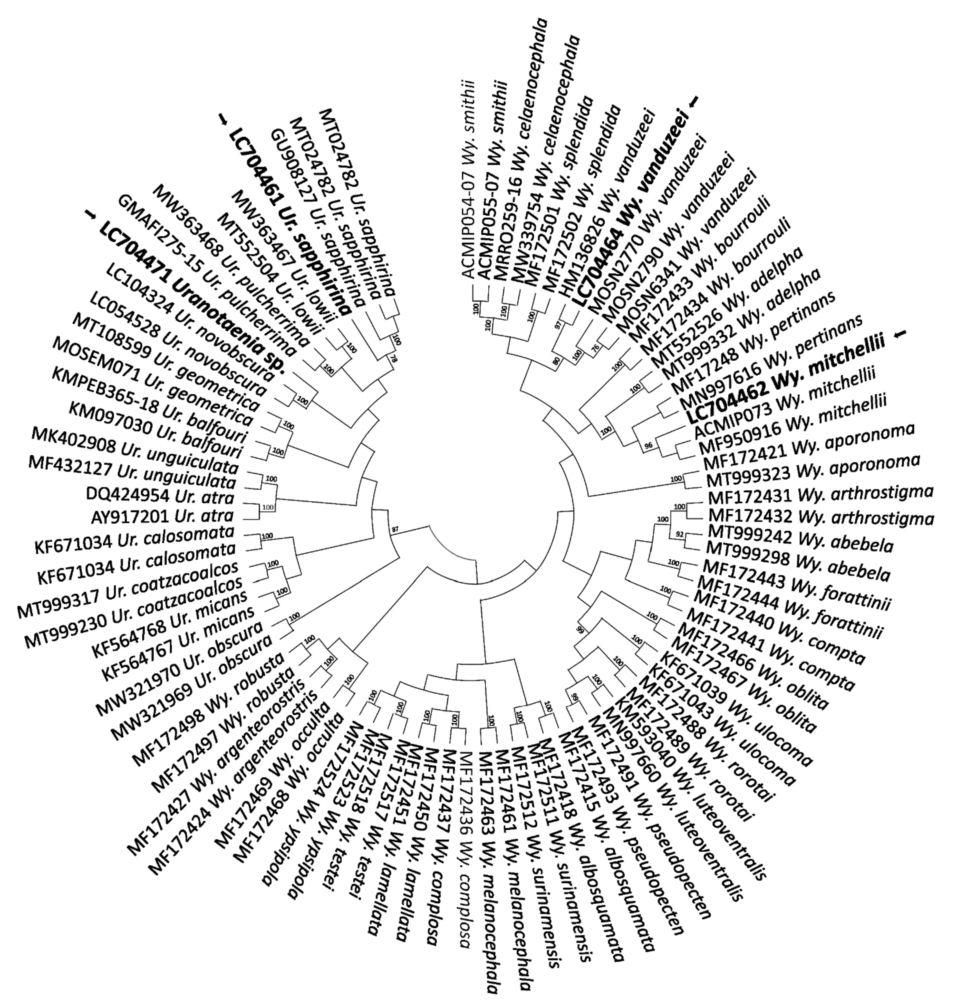
2.2. COI-Based DNA Barcodes and Phylogenetic Analysis

For mosquitoes, 18 COI DNA barcode sequences were analysed for phylogenetic reconstruction by comparing them with barcodes from public databases. The ML tree allowed to confirm the identification of 10 mosquito species at species-level, plus two at genus-level: Ae. scapularis (LC704458, LC704459, LC704469; n = 3), Ae. vittatus (LC704463; n = 1), Aedes pertinax Grabham, 1906 (LC704466; n = 1), Culex nigripalpus Theobald, 1901 (LC704468, LC704476; n = 2), Cx. quinquefasciatus (LC704465, LC704473; n = 2), Culex inhibitator (Dyar and Knab, 1906 (LC704470; n = 1), and Culex salinarius Coquillett, 1904 (LC704467, LC704475; n = 2), which represents the first report from the Greater Antilles. On the other hand, the samples referred to as Culex sp. (LC704472, LC704474; n = 2) could not be identified to the species-level based on homology and phylogenetic analysis (Figure 5). The nucleotide sequences of the Uranotaenia and Wyeomyia genera revealed an overall congruent topology with a consistent placement of the obtained sequences in the cluster with Wy. mitchellii (LC704462, n = 1), Wyeomyia vanduzeei Dyar and Knab, 1906 (LC704464, n = 1), and Ur. sapphirina (LC704461, n = 1). However, regardless of the strategy used for phylogenetic reconstruction, the sample Uranotaenia sp. (LC704471, n = 1) could not be identified to the species-level (Figure 6). The sequence homology value of the sample was <93%.
Regarding Culicoides, there were no available sequences for C. jamaicensis, C. haitiensis, and C. borinqueni; thus, our barcodes represent the first GenBank records of these species, and have been summited under the following accession numbers: C. jamaicensis (LC704483–LC704485), C. haitiensis (LC704939), and C. borinqueni (LC704940). We also obtained a total of six sequences of the three different morphotypes of C. foxi (Figure 4A–C). The analysis of pairwise distance values indicated that cox1 mtDNA sequences are very similar to one another, with a genetic diversity distance of 0 or close to 0 between Costa Rica and Dominican Republic samples (Table S1 in Supplementary File). Therefore, there are no phenotypic differences with the COI marker used. The Hd and Pi for all the C. foxi sequences analysed were 0.786 ± 0.022 and 0.002 ± < 0.000, respectively, and for the Dominican Republic samples, the values were 0.600 ± 0.046 and <0.000 ± < 0.000, respectively. A total of five haplotypes (Fox1–Fox5) were detected from eight aligned sequences (Figure 4D), although a larger study would be necessary in view of the lower intraspecific diversity. The most prevalent haplotype was Fox3, which represented 50% (4/8) of all C. foxi sequences (Figure 4D). Sequences from each haplotype were deposited in GenBank under accession numbers LC704477–LC704482.
On the other hand, the identification of H. irritans was confirmed molecularly, and showed 100% homology with public sequences from GenBank (e.g., accession number: KM669714).

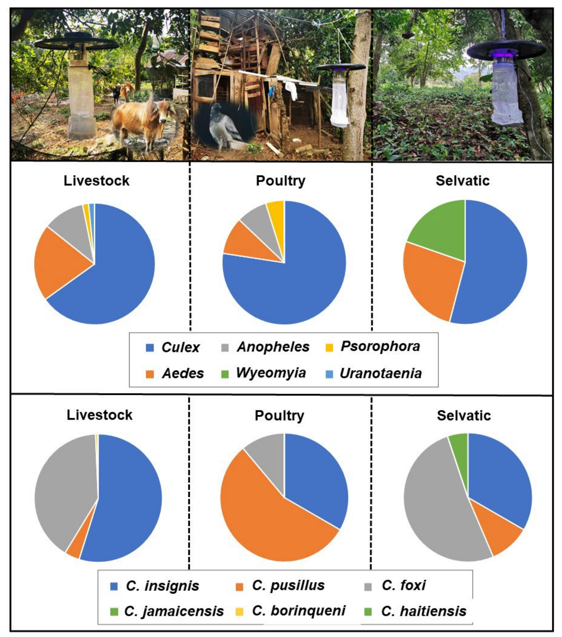
2.3. Spatial Distribution

The abundance of mosquitoes did not reveal significant differences among the three habitats (KW = 2.069, degrees of freedom [df] = 2, p = 0.355). The species richness was higher in the sylvatic habitats (n = 16) compared to livestock (n = 11) and poultry (n = 10) habitats. Culex quinquefasciatus was the most common species, found in 21 out of the 30 sampled sites (Figure 1). The newly introduced species Ae. vittatus was spotted in four sites. Culicoides were significantly more abundant in livestock farms compared with both poultry and sylvatic habitats (KW = 17.818, df = 2, p ≤ 0.05). Livestock habitats comprised higher number of species (n = 5) compared to sylvatic (n = 4) and poultry (n = 3) habitats. Culicoides insignis was the most frequent species, appearing in 18 out of the 30 sites (Figure 1).

2.4. Blood Meal Analysis

DNA from host blood meal sources was amplified successfully in 100% of the mosquito specimens analysed. The study of haematic preferences indicated a diet based mainly on avian and mammalian hosts. Culex species fed on chicken, whereas Aedes spp. and Ps. confinnis fed on pigs (Table 1). The overall amplification success was 65.1% in biting midges, being notably lower in C. pusillus compared with C. insignis. The blood meals of these two species and of C. foxi were derived exclusively from cows (Table 1).

3. Discussion

This work is pioneering in applying molecular tools (COI-based DNA barcoding) for the identification of mosquitoes, Culicoides biting species, and their host blood meal sources in the insular Caribbean. Previously, DNA barcoding had only been used to confirm the first record of Ae. vittatus in the Americas [10]. To our knowledge, the last study on host-derived blood meals was carried out in the 1980s in Hispaniola during an eastern equine encephalitis outbreak; the investigation employed precipitin tests [19].
Approximately 52 species of living mosquitoes have been recorded from Hispaniola [3,4,10,20,21,22]. However, most of these records were based on the identification of mosquitoes in the III–IV larval stage. Our identification effort revealed the presence of at least 16 species. However, two other species (Culex sp. and Uranotaenia sp.) require further confirmation because COI barcoding is dependent on the availability of representative sequences for comparison, and this approach fails when insufficient reference sequences are deposited in databases [23,24]. In addition, there is a lack of molecular and taxonomist professionals (and, therefore, a lack of identification keys), which also leads to misidentifications, making the interpretation of COI sequences problematic and inaccurate [25]. This fact is aggravated because Culex and Aedes species recognition is mainly based on adult morphology, but the absence and overlap of morphological characters have often been identified as factors that lead to the misidentification of these mosquito species [26]. Nevertheless, COI-based DNA barcoding was a useful tool to identify the remaining species, and contributed to display the phylogenetic relationships among the species. It was also useful for the determination of the blood-fed Culex specimens that were initially classified as Cx. quinquefasciatus based on morphological features, but, subsequently, were characterised molecularly into Cx. nigripalpus. This is not a trivial matter, as blood-engorged specimens show modification of the entire body, and scales and bristles of terga rub off or are distorted. For all these reasons, COI-based DNA barcoding should be used for accurate and precise identification of sibling species, species complexes, damaged specimens, and also blood-fed specimens.
Exotic/invasive species are among the primary threats to biodiversity in Hispaniola because they can cause environmental and economic damage [27], as well as negatively impact human and animal health through disease transmission [28]. Our study also expands the distribution of Ae. vittatus to other sites, including urban and rural settings, increasing the spatial distribution of this newly discovered exotic species. It is interesting to note that this species was trapped by UV-suction traps, which is not a particularly useful tool to collect most diurnally active Aedes mosquitoes, given that they show little positive attraction to light traps [29]. However, Ae. vittatus seem to be an exception because they can also be collected sporadically in non-baited light traps [10].
Regarding the finding of Cx. salinarius, this species has been found in The Bahamas, Bermuda, Canada, Mexico, and the United States [30]; therefore, this is the first time it has been reported in the Greater Antilles. This species closely resembles both the adult and immature stages of Cx. quinquefasciatus, and may be found in cohabitation. It is plausible to think that Cx. salinarius has been misidentified as Cx. quinquefasciatus in previous studies undertaken in the country. This culicine species has been incriminated as a potential bridge vector of several encephalitis viruses, such as West Nile virus, St. Louis encephalitis virus, and eastern equine encephalitis virus, among others [31]. The most relevant mosquito species in relation to the transmission of equine encephalitis in the Dominican Republic is Cx. quinquefasciatus, due to its feeding habits, and also to its great synanthropy and ubiquity [32], as seen in our study. However, Cx. salinarius should also be taken into account in vector control programmes from now on, especially in rural areas of the country, such as the municipality of Jarabacoa.
It should be pointed out that the type of collection method employed (i.e., CDC UV light suction traps) is not considered an appropriate tool for the collection of some mosquito species, particularly diurnal species, and thus, the checklist recorded in this manuscript might represent only part of the mosquito diversity present in the study area.
Only 12 species of extant Culicoides have been recorded in Haiti, and four in the Dominican Republic in the early 1990s [19,33,34,35]. However, this list requires further revision because Mitchell [19] recorded a single specimen of Culicoides obsoletus (Meigen, 1818) in Haiti, a species only found in the Palearctic region. With the incorporation of C. haitiensis, C. borinqueni, and C. jamaicensis, the number of living species in the Dominican Republic and Hispaniola has increased to seven and 13, respectively. Our study also provides the first pictures of the thorax of C. haitiensis, which possesses a characteristic dark spot on the scutum and scutellum that was not recorded in the original descriptions of Delecolle et al. [34] because the specimens were damaged. These characteristics allow unequivocal separation of this species from other related species without the need to mount specimens. We have also provided additional pictures of other important diagnostic characteristics, and the first COI barcoding sequences of the three mentioned Culicoides species. The molecular information provided for C. jamaicensis shed light on the unresolved question about the origin of this species in the Americas. Specifically, Meiswinkel et al. [36] postulated that C. jamaicensis and Culicoides paolae (Boorman, 1996) are morphologically similar, suggesting the possibility that the former was introduced into the Mediterranean Region at the time of Columbus. A deep molecular analysis coupled with a detailed morphometric study might help to resolve this mystery. It also interesting to note that, most likely, the C. foxi phenotypic differences respond to intraspecific variations within the same species as a consequence of different environmental conditions in breeding sites, or other unknown reasons. The number of haplotypes found suggests a relatively high diversity compared with other Culicoides species [37].
The analysis of field-collected engorged females through PCR amplification of host DNA present in a blood meal is a valuable method to determine host use of many blood-feeding arthropods [17]. Unfortunately, in our study, blood-engorged specimens were very scarce; hence, we could only draw preliminary conclusions. Culex quinquefasciatus was quite abundant, particularly in poultry shelters. Culex spp. are known to feed on a wide range of vertebrate species, including avian and mammalian hosts [38]. Culex quinquefasciatus has shown a wide host preference range in the insular Caribbean, feeding mainly on different avian species (51%), but also on a high number of humans [39]. It is interesting to note the specimen of Ae. albopictus, which fed on pigs. This species feeds primarily on humans, other mammals, and birds, and less commonly on other hosts, such as reptiles, amphibians, and fish [40,41]. Other collected species, such as Ae. scapularis and Psorophora sp., were also predominant, and feed on a wide variety of avian and mammalian hosts [41].
Though host–range interactions for most Culicoides species in Europe have been relatively well studied [42], so far, Neotropical Culicoides midges have been studied very little [43]. In the present investigation, a high number of Culicoides midges were collected in the traps located near livestock farms, with free access to the blood of domestic animals, compared with both sylvatic habitats and poultry habitats. The presence of permanent and available domestic hosts might explain the higher diversity in accordance with other studies, as Culicoides density increases as the host availability increases [44]. DNA barcoding data indicated a strong affinity of Culicoides to feed on Bos taurus, which is in agreement with the preliminary results recorded in Culicoides spp. in the Dominican Republic [19]. These data are quite surprising, considering the large number of other mammals and birds available in the vicinity of the sampling sites. Our results might indicate that both C. insignis and C. pusillus are strictly mammophilic species. Culicoides insignis, the most commonly trapped species in our study, is a widespread species often associated with farm environments, and has been confirmed as the main BTV vector in the Caribbean [45,46,47,48]. Similarly, although with less information available, C. pusillus is also a possible BTV vector [46], commonly found in farm holdings. Both species are often trapped in association with cattle and pigs in pasture environments [49,50], and to a lesser extent, in poultry environments [51,52].
A few other interesting Diptera species were also accidentally collected by UV-suction traps. The two species of simulids trapped in our study have recently been recorded affecting outdoor activities, and, in particular, Simulium quadrivittatum was reported to be the predominant anthropophilic species in the proximity of flowing water courses of La Vega Province (Dominican Republic) [53]. It is worth mentioning that the traps employed are not a suitable method for capturing black flies, as they are mostly attracted by CO2 traps, human landing, and/or by visual attraction with colour traps [54]. Although the Dominican Republic is free of onchocerciasis, the great density of black flies found in the proximity of some water courses justifies the implementation of specific control measures. On the other hand, a few specimens of H. irritans, commonly known as the horn fly, were trapped inside a cow barn on a mountainside. Haematobia irritans is considered one of the most troublesome species within bovine production systems, due to the intense stress it imposes on the animals; however, this blood-sucking fly has been poorly recorded in the Dominican Republic [55]. Interestingly, no sand flies were captured in our study, even though CDC light traps are effective tools for the surveillance of this family [56]. Lutzomyia cayennensis (Floch and Abonnenc, 1941) and Lutzomyia christophei (Fairchild and Trapido, 1950) were collected in the early 1980s by Johnson [57]. Since then, there has been no evidence of their existence in the country, even though Leishmania waltoni (Shaw et al., 2015) (a member of the Leishmania mexicana complex), the parasite responsible for diffuse cutaneous leishmaniasis, is present in the Dominican Republic [58].

4. Materials and Methods

4.1. Study Area

The study was conducted in the municipality of Jarabacoa (19°06′29.25″ N, 70°39′00.34″ W), La Vega Province (Dominican Republic). Jarabacoa is a town of ca. 72,000 inhabitants located in the heart of the Central Cordillera, with great economic and social development, offering ecological and adventure tourism as attractions [59]. This Caribbean municipality is characterised by a tropical rainforest climate according to the Köppen climate classification [60], with an average annual temperature of 22 °C, with abundant rain through most of the year (average precipitation of 2000 mm). Due to its location and elevation (an average elevation of 529 m above sea level [a.s.l.]), the temperature varies depending on the season.

4.2. Entomological Survey

CDC-miniature traps equipped with UV light (BioQuip Products Inc., Rancho Dominguez, CA, USA) were operated from sunset to early morning for a minimum of 12 h. Collection jars containing insects were stored at −20 °C until further processing. The sampling sites were allocated within three categories: livestock habitats (n = 10), poultry habitats (n = 10), and sylvatic habitats (n = 10). Livestock habitats were farming holdings composed of domestic animals, such as cows, pigs, horses, goats, dogs, and sheep. Poultry habitats were peridomestic settings composed of pigeons, chickens, goose, and guinea fowls. Sylvatic habitats were pristine settings with little human intervention, such as banana plantations, mixed forests, pastures, and riverbanks. Trapping was carried out once in each of the 30 sampling sites from 22 March to 2 April 2021. Excluding the weekend, three traps were run each day, corresponding with each of the three habitats, thus completing the trapping in 10 days (10 days × 3 habitats = 30 collections). The weather conditions over the trapping period were constant (average diurnal T° = 22.5 °C, and average nocturnal T° = 17.4 °C, without precipitations during the night period). Sampling sites were recruited door-to-door after getting permission of the owners and/or the corresponding authorities.

4.3. Morphological Species Identification

Haematophagous Diptera were separated from other insects, enumerated, sorted into families, and identified morphologically at the species-level following published taxonomic keys [34,53,61,62]. The terminalia of each male mosquito was mounted on Canadian Balsam and observed under a compound microscope. Rare or little-known species of Culicoides females were also mounted, and diagnostic features were photographed with a Leica S9 stereomicroscope coupled to a digital system.

4.4. Molecular Species Identification

Damaged or unconclusive specimens were analysed by COI-based DNA barcoding (mosquitoes, n = 18; Culicoides sp., n = 11; Haematobia sp., n = 1) to generate COI sequences. Genomic DNA (gDNA) from the insect legs of each specimen (or whole specimens in Culicoides) was extracted using a NZY tissue gDNA kit (NZYTech, Lisbon, Portugal) following the manufacturer’s instructions. Maceration of the tissues was carried out manually with a sterilised piston pellet. The 658 bp region flanking of the mitochondrial COI gene was amplified by polymerase chain reaction (PCR) using the primer set LCO1490 and HCO2198, following PCR protocols described by Folmer et al. [63].

4.5. Identification of Host Blood Meal Sources

Blood-engorged female mosquitoes and Culicoides midges were separated for additional analysis. Universal vertebrate-specific primers (cytB1-F: forward 5′-CCATCMAACATCTCAGCATGATGAAA-3′ and cytB2-R: reverse 5′-GCHCCTCAGAATGATATTTGTCCTCA-3′) were used to amplify the 350 bp segment of the host mitochondrial cytochrome b gene (cyt-b) already described previously [64]. PCR was carried out in a 25 µL final volume using NZYTaq 2× Green Master Mix (NZYTech), 0.4µM of each primer, and 2 µL of template DNA.

4.6. Analysis of Sequences and Statistical Analysis

Amplified PCR products were purified and sequenced at STAB Vida (Monte da Caparica, Portugal) in both senses using the same primers as for PCR. Chromatogram inspection and the assembly of forward and reverse sequences edition was carried out by BioEdit Sequence Alignment Editor (version 7.2.5, Carlsbad, CA, USA). The identity at the species-level was assessed based on the analysis of the generated cox1 sequences, considering both the higher similarity in the BOLD Systems identification tool [65] and the results of homology searches from the sequences available in GenBank [66]. The respective nucleotide sequences obtained were deposited in the DNA Data Bank of Japan [67]. The identity of blood meals was assessed based on the analysis of cyt-b sequences, taking into account the closest database matches at the species-level of vertebrate hosts (identity ≥ 99%). Statistical analysis (the Kruskal–Wallis [KW] test) was carried to compare the abundance of Culicoides and mosquitoes across the different habitats using IBM SPSS Statistics for Windows (Version 27.0, Armonk, NY, USA). The Kolmogorov–Smirnov (Lilliefors modification) and Shapiro–Wilk tests were used to test for normality, and Levene’s test was used to assess the homogeneity of variance. Due to the lack of normality of the data, large standard deviations, and a lack of homogeneity of variance, non-parametric tests were employed [68]. Significant differences were considered when p ≤ 0.05.

4.7. Evolutionary Analysis by the Maximum Likelihood Method

Available cox1 mtDNA sequences with >600 bp of mosquito species present in the Caribbean and nearby locations were retrieved from GenBank and BOLD Systems; sequences with potential errors (flagged sequences and incorrect identifications) were excluded. Multiple alignments were carried out with MAFFT version 7 [69]. For maximum likelihood (ML) phylogenetic analyses, the choice of the best-fitting evolutionary model was based on those defined using JModeltest2 [70] on the basis of the Akaike information criterion. Tree reconstruction was carried out with Mega 11 [71]. The evolutionary history was inferred by using the ML method and the General Time Reversible model [72]. Initial tree(s) for the heuristic search were obtained automatically by applying the Neighbor-Join and BioNJ algorithms to a pairwise distances matrix estimated using the maximum composite likelihood (MCL) approach, and then selecting the topology with a higher log likelihood value. Bootstrap coefficients were calculated for 1000 replicates, and only those with >75% support are shown in the tree. The phylogenetic trees were manipulated for display using FigTree v.1.4.2 [73].
The evolutionary pairwise divergence among all C. foxi sequences between and within the defined groups was estimated using the Tamura 3-parameter model [74]. The haplotype diversity (Hd) and nucleotide diversity (Pi) parameters were estimated with the program DnaSP v.6.12. [75]. Haplotype networks were illustrated with a median-joining network (MJN) algorithm (ε = 0) using the software PopART v. 1.7 [76] to analyse haplotype genealogy.

5. Conclusions

Our study provides new insights about the fauna and host interactions of adult mosquitoes and Culicoides in a touristic region of the Dominican Republic. New COI-based DNA barcoding sequences of some Diptera species are provided for the first time, improving the database library of DNA barcodes, and helping to identify unknown organisms. However, the usefulness of molecular-genetic analysis and DNA markers as a tool for identification at the species-level is hampered by the limited availability of DNA barcode sequences of Neotropical mosquito species in public repositories, which has affected the accurate identification of some of the mosquito species collected. Mosquito species such as Ae. vittatus, Ae. albopictus, Ae. scapularis, An. albimanus, Ps. confinnis, Cx. quinquefasciatus, and Cx. salinarius, among others, are potential vectors of human pathogens, and they have been found across different types of Dominican habitats. Culicoides insignis, the most predominant Culicoides species, is considered a proven vector of BTV. The Diptera species collected showed a strong affinity to feed on domestic animals, particularly, pigs, cows, and chickens. This result contributes to deepening the knowledge on the transmission cycles of the pathogens of human and veterinary concern in the insular Caribbean.

Supplementary Materials

The following supporting information can be downloaded at: https://0-www-mdpi-com.brum.beds.ac.uk/article/10.3390/pathogens11070714/s1, Table S1: Estimates of evolutionary divergence of cox1 over sequence pairs between groups.

Author Contributions

Conceptualization, M.A.G. and P.M.A.-E.; methodology, formal analysis, and data curation, M.A.G., P.M.A.-E., M.A.R.-S., D.B.-B. and E.F.; writing—original draft preparation, M.A.G. and P.M.A.-E.; writing—review and editing, M.A.G., P.M.A.-E., D.B.-B. and J.R.; supervision, D.B.-B., M.A.G. and E.F.; project administration, P.M.A.-E.; funding acquisition, M.A.G. and P.M.A.-E. All authors have read and agreed to the published version of the manuscript.

Funding

This work is part of the project: ‘Búsqueda, caracterización y evaluación de agentes ecológicamente amigables para el control de mosquitos (Diptera: Culicidae) de importancia médica en República Dominicana’, supported by the Fondo Nacional de Innovación y Desarrollo Científico y Tecnológico (FONDOCyT), Ministerio de Educación Superior, Ciencia y Tecnología (MESCyT), Project No. 2018-19-2B2-043.

Institutional Review Board Statement

Not applicable.

Informed Consent Statement

Not applicable.

Data Availability Statement

Not applicable.

Acknowledgments

The authors would like to thank the owners of the livestock and poultry farms for allowing us to set up the traps on their properties. The authors are also grateful to Ricardo Parreira of Instituto de Higiene e Medicina Tropical (IHMT) from Lisbon for technical support with the molecular part of this work.

Conflicts of Interest

The authors declare no conflict of interest.

References

  1. Alarcón-Elbal, P.M.; Paulino-Ramírez, R.; Diéguez-Fernández, L.; Fimia-Duarte, R.; Guerrero, K.A.; González, M. Arbovirosis transmitidas por mosquitos (Diptera: Culicidae) en la República Dominicana: Una revisión. Biologist 2017, 1, 193–219. [Google Scholar] [CrossRef] [Green Version]
  2. Marcondes, C.B.; Contigiani, M.; Gleiser, R.M.; Reisen, W. Emergent and reemergent arboviruses in South America and the Caribbean: Why so many and why now? J. Med. Entomol. 2017, 54, 509–532. [Google Scholar] [CrossRef] [PubMed]
  3. Sosa, M.A.R.; Rueda, J.; Bautista, Y.E.V.; Fimia-Duarte, R.; de Prada, M.B.; Guerrero, K.A.; Alarcón-Elbal, P.M. Diversidad de mosquitos (Diptera: Culicidae) de Jarabacoa, República Dominicana. Graellsia 2019, 75, e084. [Google Scholar]
  4. Rodríguez Sosa, M.A.; Rueda, J.; Pichardo Rodríguez, R.J.; Vásquez Bautista, Y.E.; Durán Tiburcio, J.C.; Fimia-Duarte, R.; Alarcón-Elbal, P.M. Primera cita de Culex interrogator (Diptera: Culicidae) para La Hispaniola y actualización del listado de mosquitos de Jarabacoa, República Dominicana. Novit. Caribaea 2020, 16, 110–121. [Google Scholar] [CrossRef]
  5. Becker, N.; Petrić, D.; Zgomba, M.; Boase, C.; Madon, M.B.; Dahl, C.; Kaiser, A. Mosquitoes. Identification, Ecology and Control, 3rd ed.; Springer International Publishing: Cham, Switzerland, 2020. [Google Scholar]
  6. Francis, S.; Frank, C.; Buchanan, L.; Green, S.; Stennett-Brown, R.; Gordon-Strachan, G.; Rubio-Palis, Y.; Grant, C.; Alexander-Lindo, R.L.; Nwokocha, C.; et al. Challenges in the control of neglected insect vector diseases of human importance in the Anglo-Caribbean. One Health 2021, 13, 100316. [Google Scholar] [CrossRef]
  7. Diéguez Fernández, L.; Monzón Muñoz, M.V.; Juárez Sandoval, J.A.; Barrios Barrios, D.Y.; Barrientos Juárez, M.E.; Barrios, Y.H.; Rodríguez-Sosa, M.A.; Vásquez-Bautista, Y.E.; Alarcón-Elbal, P.M. Caracterización de la infestación de viviendas por tres especies de mosquitos (Diptera: Culicidae) de importancia médica en países de bajos y medios ingresos: Recomendaciones para su control doméstico. Ciencia y Salud 2021, 5, 49–66. [Google Scholar] [CrossRef]
  8. Sudeep, A.B.; Shil, P. Aedes vittatus (Bigot) Mosquito: An emerging threat to hublic Health. J. Vector Borne Dis. 2017, 54, 295–300. [Google Scholar] [CrossRef]
  9. Díaz-Martínez, I.; Diéguez-Fernández, L.; Santana-Águila, B.; de la Paz, E.M.A.; Ruiz-Domínguez, D.; Alarcón-Elbal, P.M. New introduction of Aedes vittatus (Diptera: Culicidae) into the East-Central region of Cuba: Ecological characterization and medical relevance. Interam. J. Med. Health 2021, 4, 1–6. [Google Scholar]
  10. Alarcón-Elbal, P.M.; Rodríguez-Sosa, M.A.; Newman, B.C.; Sutton, W.B. The First record of Aedes vittatus (Diptera: Culicidae) in the Dominican Republic: Public health implications of a potential invasive mosquito species in the Americas. J. Med. Entomol. 2020, 57, 2016–2021. [Google Scholar] [CrossRef]
  11. Raccurt, C.P. Mansonella ozzardi and Its Vectors in the New World: An update with emphasis on the current situation in Haiti. J. Helminthol. 2018, 92, 655–661. [Google Scholar] [CrossRef]
  12. Lowrie, R.C.; Raccurt, C.P. Assessment of Culicoides barbosai as a vector of Mansonella ozzardi in Haiti. Am. J. Trop. Med. Hyg. 1984, 33, 1275–1277. [Google Scholar] [CrossRef]
  13. Raccurt, C.P.; Brasseur, P.; Boncy, J. Mansonelliasis, a neglected parasitic disease in Haiti. Mem. Inst. Oswaldo Cruz. 2014, 109, 709–711. [Google Scholar] [CrossRef] [Green Version]
  14. Elbadry, M.A.; Durães-Carvalho, R.; Blohm, G.M.; Stephenson, C.J.; Loeb, J.C.; White, S.K.; Telisma, T.; Chavannes, S.; Beau De Rochars, V.M.; Salemi, M.; et al. Orthobunyaviruses in the Caribbean: Melao and Oropouche virus infections in school children in Haiti in 2014. PLoS Negl. Trop. Dis. 2021, 15, e009494. [Google Scholar] [CrossRef]
  15. Thompson, L.H.; Mo, C.L.; Oviedo, M.T.; Homan, E.J. Prevalence and Incidence of Bluetongue Viruses in the Caribbean Basin: Serologic and Virologic Findings. In Proceedings of the Second International Symposium, Paris, France, 17–21 June 1992; Walton, T.E., Osburn, B.I., Eds.; CRC Press Inc.: Boca Raton, FL, USA; pp. 106–113. [Google Scholar]
  16. Garcia-Rejon, J.E.; Blitvich, B.J.; Farfan-Ale, J.A.; Loroño-Pino, M.A.; Chi Chim, W.A.; Flores-Flores, L.F.; Rosado-Paredes, E.; Baak-Baak, C.; Perez-Mutul, J.; Suarez-Solis, V.; et al. Host-feeding preference of the mosquito, Culex quinquefasciatus, in Yucatan State, Mexico. J. Insect Sci. 2010, 10, 32. [Google Scholar] [CrossRef] [Green Version]
  17. Kent, R.J. Molecular methods for arthropod bloodmeal identification and applications to ecological and vector-borne disease studies. Mol. Ecol. Resour. 2009, 9, 4–18. [Google Scholar] [CrossRef]
  18. Alarcón-Elbal, P.M.; Sandiford, S.L. One health entomology in the Insular Caribbean: Time to bet on prevention. Interam. J. Med. Health 2021, 4, e202101001. [Google Scholar] [CrossRef]
  19. Mitchell, C.J.; Taylor, S.A.; Christensen, H.A. Identification of blood meals from engorged dipterans collected in the Dominican Republic during an Eastern Equine encephalitis outbreak. J. Med. Entomol. 1983, 20, 686–687. [Google Scholar] [CrossRef]
  20. Rodríguez-Sosa, M.A.; Vásquez-Bautista, Y.E.; Fimia-Duarte, R.; Guerrero, K.A.; Alarcón-Elbal, P.M.; Rodríguez-Sosa, M.A.; Vásquez-Bautista, Y.E.; Fimia-Duarte, R.; Guerrero, K.A.; Alarcón-Elbal, P.M. Primer registro de Uranotaenia cooki (Diptera: Culicidae) para República Dominicana. Rev. Colomb. Entomol. 2018, 44, 141–144. [Google Scholar] [CrossRef]
  21. Rueda, J.; Rodríguez-sosa, M.A.; Vásquez-bautista, Y.E.; Guerrero, K.A. Primera cita de Culex (Culex) garciai González Broche, 2000 (Diptera: Culicidae) para La Española. An. Biol. 2018, 40, 95–101. [Google Scholar] [CrossRef]
  22. Bisset Lazcano, J.A.; Marquetti Fernández, M.C.; Rodriguez Coto, M.M. Contribución de estudios entomológicos sobre Aedes aegypti y Aedes albopictus. Retrospectiva y retos para su control en Cuba. Rev. Cuba. Med. Trop. 2017, 69, 1–20. [Google Scholar]
  23. Jinbo, U.; Kato, T.; Ito, M. Current progress in DNA barcoding and future implications for entomology. Entomol. Sci. 2011, 14, 107–124. [Google Scholar] [CrossRef]
  24. Chan, A.; Chiang, L.P.; Hapuarachchi, H.C.; Tan, C.H.; Pang, S.C.; Lee, R.; Lee, K.S.; Ng, L.C.; Lam-Phua, S.G. DNA Barcoding: Complementing morphological identification of mosquito species in Singapore. Parasit. Vectors 2014, 7, 569. [Google Scholar] [CrossRef]
  25. Buhay, J.E. “COI-like” sequences are becoming problematic in molecular systematic and DNA barcodeng studies. J. Crustac. Biol. 2009, 29, 96–110. [Google Scholar] [CrossRef]
  26. Weeraratne, T.C.; Surendran, S.N.; Parakrama Karunaratne, S.H.P. DNA barcoding of morphologically characterized mosquitoes belonging to the subfamily Culicinae from Sri Lanka. Parasit. Vectors 2018, 11, 266. [Google Scholar] [CrossRef] [PubMed] [Green Version]
  27. McCreless, E.E.; Huff, D.D.; Croll, D.A.; Tershy, B.R.; Spatz, D.R.; Holmes, N.D.; Butchart, S.H.M.; Wilcox, C. Past and estimated future impact of invasive alien mammals on insular threatened vertebrate populations. Nat. Commun. 2016, 7, 12488. [Google Scholar] [CrossRef] [PubMed] [Green Version]
  28. Russell, J.C.; Meyer, J.Y.; Holmes, N.D.; Pagad, S. Invasive alien species on Islands: Impacts, distribution, interactions and management. Environ. Conserv. 2017, 44, 359–370. [Google Scholar] [CrossRef] [Green Version]
  29. Thurman, E.B. Report of the initial operation of a light trap in northern Thailand. Mosq. News 1955, 15, 215–224. [Google Scholar]
  30. Walter Reed Biosystematics Unit (WRBU). 2021. Culex salinarius Coquillett. 1904. Available online: https://wrbu.si.edu/vectorspecies/mosquitoes/salinarius (accessed on 10 May 2022).
  31. Sardelis, M.R.; Turell, M.J.; Dohm, D.J.; O’Guinn, M.L. Vector competence of selected north american Culex and Coquillettidia mosquitoes for West Nile virus. Emerg. Infect. Dis. 2001, 7, 1018–1022. [Google Scholar] [CrossRef]
  32. Diéguez-Fernández, L.; Rodríguez-Sosa, M.A.; Vásquez-Bautista, Y.E.; Borge-de Prada, M.; Alarcón-Elbal, P.M. Aportes a la bioecología de Culex quinquefasciatus (Diptera: Culicidae), vector de encefalitis, en Jarabacoa, República Dominicana. Folia Entomol. Mex. 2020, 6, 26–32. [Google Scholar]
  33. Raccurt, C.P.; Rebholtz, C.; Delécolle, J.C. Culicoides haitiensis et Culicoides borinqueni en Haïti. 2. Notes Écologiques et Chorologiques [Dipt. Ceratopogonidae]. Bull. Société Entomol. Fr. 1996, 91, 125–130. [Google Scholar] [CrossRef]
  34. Delecolle, J.C.; Raccurt, C.P.; Rebholtz, C. Culicoides haitiensis n. sp. et C. borinqueni Fox et Hoffman en Haiti. Etude morphologique (Diptera, Ceratopogonidae). Nouv. Rev. Entomol. 1986, 3, 107–115. [Google Scholar]
  35. Perez-Gelabert, D.E. Checklist, bibliography and quantitative data of the arthropods of Hispaniola. Zootaxa 2020, 4749, zootaxa.4749.1.1. [Google Scholar] [CrossRef]
  36. Meiswinkel, R.; Labuschagne, K.; Goffredo, M. Christopher. Columbus and Culicoides: Was C. jamaicensis Edwards, 1922 introduced into the Mediterranean 500 years ago and later re-named C. paolae Boorman 1996? Vet. Ital. 2004, 40, 340–344. [Google Scholar]
  37. Aguilar-Vega, C.; Rivera, B.; Lucientes, J.; Gutiérrez-Boada, I.; Sánchez-Vizcaíno, J.M. A Study of the composition of the Obsoletus complex and genetic diversity of Culicoides obsoletus populations in Spain. Parasit. Vectors 2021, 14, 1–13. [Google Scholar] [CrossRef]
  38. Jansen, C.C.; Webb, C.E.; Graham, G.C.; Craig, S.B.; Zborowski, P.; Ritchie, S.A.; Russell, R.C.; Van Den Hurk, A.F. Blood sources of mosquitoes collected from urban and peri-urban environments in Eastern Australia with species-specific molecular analysis of avian blood meals. Am. J. Trop. Med. Hyg. 2009, 81, 849–857. [Google Scholar] [CrossRef] [Green Version]
  39. Fitzpatrick, D.M.; Hattaway, L.M.; Hsueh, A.N.; Ramos-Niño, M.E.; Cheetham, S.M. PCR-based bloodmeal analysis of Aedes aegypti and Culex quinquefasciatus (Diptera: Culicidae) in St. George Parish, Grenada. J. Med. Entomol. 2009, 56, 1170–1175. [Google Scholar] [CrossRef] [Green Version]
  40. Cebrián-Camisón, S.; de la Puente, J.M.; Figuerola, J. A literature review of host feeding patterns of invasive Aedes mosquitoes in Europe. Insects 2020, 11, 848. [Google Scholar] [CrossRef]
  41. Santos, C.S.; Pie, M.R.; da Rocha, T.C.; Navarro-Silva, M.A. Molecular identification of blood meals in mosquitoes (Diptera, Culicidae) in urban and forested habitats in southern Brazil. PLoS ONE 2019, 14, e0212517. [Google Scholar] [CrossRef]
  42. Martínez-de la Puente, J.; Figuerola, J.; Soriguer, R. Fur or feather? Feeding preferences of species of Culicoides biting midges in Europe. Trends Parasitol. 2015, 31, 16–22. [Google Scholar] [CrossRef] [Green Version]
  43. McGregor, B.L.; Stenn, T.; Sayler, K.A.; Blosser, E.M.; Blackburn, J.K.; Wisely, S.M.; Burkett-Cadena, N.D. Host use patterns of Culicoides spp. biting midges at a big game preserve in Florida, U.S.A., and implications for the transmission of Orbiviruses. Med. Vet. Entomol. 2019, 33, 110–120. [Google Scholar] [CrossRef] [Green Version]
  44. Garcia-Saenz, A.; Mccarter, P.; Baylis, M. The influence of host number on the attraction of biting midges, Culicoides spp., to light traps. Med. Vet. Entomol. 2011, 25, 113–115. [Google Scholar] [CrossRef]
  45. Davies, J.F. The distribution of sandflies (Culicoides spp.) Breeding in a tidal mangrove swamp in Jamaica and the effect of tides on the emergence of C. furens Poey and C. barbosai (Wirth & Blanton). West Indian Med. J. 1967, 16, 39–50. [Google Scholar] [PubMed]
  46. Mo, C.L.; Thompson, L.H.; Homan, E.J.; Oviedo, M.T.; Greiner, E.C.; González, J.; Sáenz, M.R. Bluetongue virus isolations from vectors and ruminants in Central America and the Caribbean. Interamerican Bluetongue Team. Am. J. Vet. Res. 1994, 55, 211–215. [Google Scholar] [PubMed]
  47. Tanya, V.N.; Greiner, E.C.; Gibbs, E.P.J. Evaluation of Culicoides insignis (Diptera: Ceratopogonidae) as a vector of Bluetongue virus. Vet. Microbiol. 1992, 32, 1–14. [Google Scholar] [CrossRef]
  48. Rios, R.R.S.; Santarém, M.C.A.; Ribeiro Júnior, K.A.L.; de Melo, B.A.; da Silva, S.G.M.; da Silva, N.C.; Dos Santos, V.R.V.; Dos Santos, J.M.; Santana, A.E.G.; Fraga, A.B. Culicoides insignis Lutz, 1913 (Diptera: Ceratopogonidae) biting midges in Northeast of Brazil. Insects 2021, 12, 366. [Google Scholar] [CrossRef]
  49. Bandeira, M.C.A.; Da Penha, A.; Moraes, J.L.P.; Brito, G.A.; Rebêlo, J.M.M. Infestation of brazilian peridomiciliary areas by Culicoides (Diptera: Ceratopogonidae) in humid and semihumid climates. J. Med. Entomol. 2016, 53, 1163–1168. [Google Scholar] [CrossRef] [PubMed]
  50. Carvalho, L.P.C.; Silva, F.S. Seasonal abundance of livestock-associated Culicoides species in northeastern Brazil. Med. Vet. Entomol. 2014, 28, 228–231. [Google Scholar] [CrossRef]
  51. Costa, J.C.; Lorosa, E.S.; Moraes, J.L.P.; Rebêlo, J.M.M. Espécies de Culicoides (Diptera; Ceratopogonidae) e hospedeiros potenciais em área de ecoturismo do Parque Nacional dos Lençóis Maranhenses, Brasil. Rev. Pan-Amazônica Saúde 2013, 4, 11–18. [Google Scholar] [CrossRef]
  52. González, M.A.; Dilger, E.; Ronderos, M.M.; Spinelli, G.R.; Courtenay, O.; Hamilton, J.G.C. Significant reduction in abundance of peridomestic mosquitoes (Culicidae) and Culicoides Midges (Ceratopogonidae) after chemical intervention in Western São Paulo, Brazil. Parasit. Vectors 2020, 13, 549. [Google Scholar] [CrossRef]
  53. González, M.A.; Ruiz-Arrondo, I.; Rodríguez-Sosa, M.A.; Rueda, J.; Oteo, J.A.; Alarcón-Elbal, P.M. New insights on black Flies (Diptera: Simuliidae) from a touristic destination of Hispaniola. Int. J. Trop. Insect Sci. 2022, 42, 2281–2295. [Google Scholar] [CrossRef]
  54. Rodríguez-Pérez, M.A.; Adeleke, M.A.; Burkett-Cadena, N.D.; Garza-Hernández, J.A.; Reyes-Villanueva, F.; Cupp, E.W.; Toé, L.; Salinas-Carmona, M.C.; Rodríguez-Ramírez, A.D.; Katholi, C.R.; et al. Development of a novel trap for the collection of black flies of the Simulium ochraceum complex. PLoS ONE 2013, 8, e76814. [Google Scholar] [CrossRef]
  55. Brito, L.G.; Regitano, L.C.d.A.; Huacca, M.E.F.; Carrilho, E.; Paes, M.J.; Moya-Borja, G.E. Genotype characterization of the Haematobia irritans (Diptera: Muscidae) from Brazil, Dominican Republic and Colombia based on randomly amplified polymorphic DNA (RAPD) analysis. Rev. Bras. Parasitol. Vet. 2008, 17, 179–184. [Google Scholar] [CrossRef] [PubMed]
  56. Rodríguez-Rojas, J.J.; Arque-Chunga, W.; Fernández-Salas, I.; Rebollar-Téllez, E.A. Comparative field evaluation of different traps for collecting adult Phlebotomine sand flies (Diptera: Psychodidae) in an endemic area of cutaneous leishmaniasis in Quintana Roo, Mexico. J. Am. Mosq. Control Assoc. 2016, 32, 103–116. [Google Scholar] [CrossRef] [PubMed]
  57. Johnson, R.N. Phlebotominae sand Flies (Diptera: Psychodidae) and diffuse cutaneous leishmaniasis in the Dominican Republic. Ph.D. Thesis, University of Florida, Gainesville, FL, USA, 1984. [Google Scholar]
  58. Shaw, J.; Pratlong, F.; Floeter-Winter, L.; Ishikawa, E.; El Baidouri, F.; Ravel, C.; Dedet, J.P. Characterization of Leishmania (Leishmania) waltoni n. nsp. (Kinetoplastida: Trypanosomatidae), the parasite responsible for diffuse cutaneous leishmaniasis in the Dominican Republic. Am. J. Trop. Med. Hyg. 2015, 93, 552. [Google Scholar] [CrossRef] [PubMed] [Green Version]
  59. Alvarado, J.; Oddone, N.; Gil, L. Fortalecimiento de la Cadena de Valor de Turismo en Pedernales, República Dominicana. Publicación de las Naciones Unidas: CEPAL, México, 2017. Available online: https://www.cepal.org/es/publicaciones/41644-fortalecimiento-la-cadena-valor-turismo-pedernales-republica-dominicana (accessed on 22 October 2021).
  60. Beck, H.E.; Zimmermann, N.E.; McVicar, T.R.; Vergopolan, N.; Berg, A.; Wood, E.F. Present and Future Köppen-Geiger Climate Classification Maps at 1-Km Resolution. Sci. Data 2018, 5, 180214. [Google Scholar] [CrossRef] [PubMed] [Green Version]
  61. Wirth, W.W.; Dyce, A.L.; Spinelli, G. An atlas of wing photographs, with a summary of the numerical characters of the neotropical species of Culicoides (Diptera: Ceratopogonidae). Contrib. Am. Entomol. Inst. 1998, 22, 4. [Google Scholar]
  62. González Broche, R. Culícidos de Cuba; Editorial Científico Técnica: La Habana, Cuba, 2006. [Google Scholar]
  63. Folmer, O.; Black, M.; Hoeh, W.; Lutz, R.; Vrijenhoek, R. DNA primers for amplification of mitochondrial cytochrome c oxidase subunit I from diverse metazoan invertebrates. Mol. Mar. Biol. Biotechnol. 1994, 3, 294–299. [Google Scholar] [PubMed]
  64. Bravo-Barriga, D.; Parreira, R.; Maia, C.; Afonso, M.O.; Blanco-Ciudad, J.; Serrano, F.J.; Pérez-Martín, J.E.; Gómez-Gordo, L.; Campino, L.; Reina, D.; et al. Detection of Leishmania DNA and blood meal sources in Phlebotomine sand flies (Diptera: Psychodidae) in western of Spain: Update on distribution and risk factors associated. Acta Trop. 2016, 164, 414–424. [Google Scholar] [CrossRef]
  65. Barcode of Life Data System (BOLD). Available online: http://www.boldsystems.org/index.php/IDS_OpenIdEngine (accessed on 15 May 2022).
  66. Benson, D.A.; Cavanaugh, M.; Clark, K.; Karsch-Mizrachi, I.; Lipman, D.J.; Ostell, J.; Sayers, E.W. GenBank. Nucleic Acids Res. 2013, 41, D36–D42. [Google Scholar] [CrossRef] [Green Version]
  67. DNA Data Bank of Japan (DDBJ). Available online: https://www.ddbj.nig.ac.jp/index-e.html (accessed on 15 May 2022).
  68. Siegel, S.; Castellan, N.J., Jr. Nonparametric Statistics for the Behavioral, Sciences, 2nd ed.; McGraw-Hill Humanities: New York, NY, USA, 1988; pp. 262–272. [Google Scholar]
  69. Katoh, K.; Rozewicki, J.; Yamada, K.D. MAFFT online service: Multiple sequence alignment, interactive sequence choice and visualization. Brief. Bioinform. 2019, 20, 1160–1166. [Google Scholar] [CrossRef] [Green Version]
  70. Darriba, D.; Taboada, G.L.; Doallo, R.; Posada, D. jModelTest 2: More models, new heuristics and parallel computing. Nat. Methods 2012, 9, 772. [Google Scholar] [CrossRef] [Green Version]
  71. Tamura, K.; Stecher, G.; Kumar, S. MEGA11: Molecular Evolutionary Genetics Analysis version 11. Mol. Biol. Evol. 2021, 38, 3022–3027. [Google Scholar] [CrossRef]
  72. Nei, M.; Kumar, S. Molecular Evolution and Phylogenetics, Oxford University Press: New York, NY, USA, 2000.
  73. FigTree Version-v1.4.2. Available online: http://tree.bio.ed.ac.uk/software/figtree/ (accessed on 15 May 2022).
  74. Tamura, K. Estimation of the number of nucleotide substitutions when there are strong Transition-Transversion and G+C-Content Biases. Mol. Biol. Evol. 1992, 9, 678–687. [Google Scholar]
  75. Rozas, J.; Ferrer-Mata, A.; Sanchez-del Barrio, J.C.; Guirao-Rico, S.; Librado, P.; Ramos-Onsins, S.E.; Sanchez-Gracia, A. DnaSP 6: DNA Sequence polymorphism analysis of large data sets. Mol. Biol. Evol. 2017, 34, 3299–3302. [Google Scholar] [CrossRef]
  76. Leigh, J.W.; Bryant, D. POPART: Full-feature software for haplotype network construction. Methods Ecol. Evol. 2015, 6, 1110–1116. [Google Scholar] [CrossRef]
Figure 1. Schematic representation of the distribution of mosquitoes (above, by genera) and Culicoides (below, by species) across the three habitats sampled in Jarabacoa (Dominican Republic) by CDC UV light traps.
Pathogens 11 00714 g001
Figure 2. Culicoides haitiensis: (A) eyes, (B) cibarium, (C) wing, (D) general aspect, (E) scutum and scutellum, and (F) palps.
Pathogens 11 00714 g002
Figure 3. Culicoides borinqueni: (A) eyes, (B) cibarial armature, (C) wing, (D) general aspect, (E) scutum and scutellum, and (F) palps.
Pathogens 11 00714 g003
Figure 4. Culicoides foxi morphospecies and haplotypes. (A) The typical morphospecies with a pale spot on m1; (B) the morphospecies without a spot on m1; and (C) the morphospecies with both spots on m1 joined. The arrows indicate the position of the spots. (D) Median joining network of the haplotypes. Note: each circle represents a haplotype.
Pathogens 11 00714 g004
Figure 5. Evolutionary history of Culex and Aedes genus samples. The tree with the highest log likelihood (−2981.46) is shown. Initial tree(s) for the heuristic search were obtained automatically by applying the Neighbor-Join and BioNJ algorithms to a matrix of pairwise distances estimated using the maximum composite likelihood (MCL) approach, and then selecting the topology with superior log likelihood value. A discrete gamma distribution was used to model evolutionary rate differences among sites (five categories; +G, parameter = 0.3796). The rate variation model allowed for some sites to be evolutionarily invariable (+I, 30.86% sites). This analysis involved 63 nucleotide sequences. All positions containing gaps and missing data were eliminated (complete deletion option). There were 405 positions in the final dataset. At specific branch nodes, bootstrap values >75% in 1000 repetitions are indicated. Orthopodomyia alba Baker, 1936 (GU908120) was used as an outgroup sequence. The sequences of our study are presented in bold with an arrow.
Pathogens 11 00714 g005
Figure 6. Evolutionary history of Uranotaenia and Wyeomyia samples. The tree with the highest log likelihood (−4386.72) is shown. Initial tree(s) for the heuristic search were obtained automatically by applying the Neighbor-Join and BioNJ algorithms to a matrix of pairwise distances estimated using the maximum composite likelihood (MCL) approach, and then selecting the topology with superior log likelihood value. A discrete gamma distribution was used to model evolutionary rate differences among sites (five categories; +G, parameter = 0.4512). The rate variation model allowed for some sites to be evolutionarily invariable (+I, 28.79% sites). This analysis involved 87 nucleotide sequences. All positions containing gaps and missing data were eliminated (complete deletion option). There was a total of 356 positions in the final dataset. At specific branch nodes, bootstrap values >75% of 1000 repetitions are indicated. Our sequences are shown in bold with an arrow.
Pathogens 11 00714 g006
Table 1. Summary of host-derived blood meals in mosquitoes and Culicoides midges collected in Jarabacoa (Dominican Republic) during 2021.
Diptera SpeciesNumber AnalysedNumber Amplified (%) 1Host
Culex quinquefasciatus11 (100)Gallus gallus
Culex nigripalpus *11 (100)Gallus gallus
Aedes albopictus11 (100)Sus scrofa
Aedes scapularis11 (100)Sus scrofa
Psorophora confinnis11 (100)Sus scrofa
Culicoides insignis3022 (73.3)Bos taurus
Culicoides pusillus125 (41.6)Bos taurus
Culicoides foxi *11 (100)Bos taurus
Total4833 (68.7)
1 Percentage calculated as (the number of the blood-fed specimens analysed/the number of blood-fed specimens with amplification host) × 100. * Confirmed by DNA barcoding (accession numbers LC704468 and LC704477, respectively).
Publisher’s Note: MDPI stays neutral with regard to jurisdictional claims in published maps and institutional affiliations.

