
Serological Evidence of Q Fever among Dairy Cattle and Buffalo Populations in the Campania Region, Italy

 , , and
Abstract
:1. Introduction
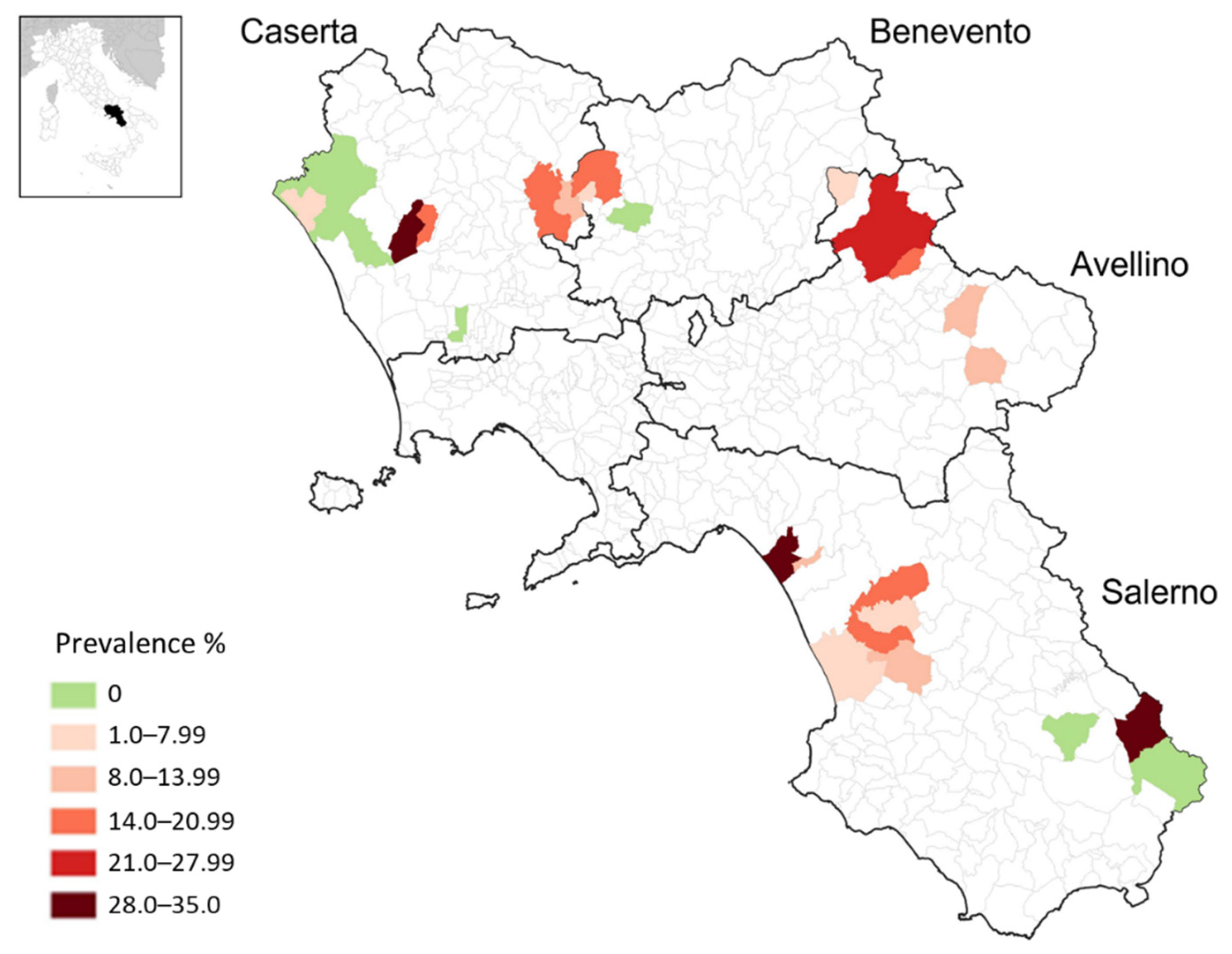
2. Results
3. Discussion
4. Materials and Methods
4.1. Study Area and Sampling
4.2. Commercial ELISA Assay
4.3. Statistical Analysis
5. Conclusions
Supplementary Materials
Author Contributions
Funding
Institutional Review Board Statement
Informed Consent Statement
Data Availability Statement
Acknowledgments
Conflicts of Interest
References
- Maurin, M.; Raoult, D. Q Fever. Clin. Microbiol. Rev. 1999, 12, 518–553. [Google Scholar] [CrossRef] [PubMed] [Green Version]
- Angelakis, E.; Raoult, D. Q Fever. Vet. Microbiol. 2010, 140, 297–309. [Google Scholar] [CrossRef] [PubMed] [Green Version]
- Arricau-Bouvery, N.; Rodolakis, A. Is Q Fever an Emerging or Re-Emerging Zoonosis? Vet. Res. 2005, 36, 327–349. [Google Scholar] [CrossRef] [PubMed] [Green Version]
- Eldin, C.; Mélenotte, C.; Mediannikov, O.; Ghigo, E.; Million, M.; Edouard, S.; Mege, J.L.; Maurin, M.; Raoult, D. From Q Fever to Coxiella Burnetii Infection: A Paradigm Change. Clin. Microbiol. Rev. 2017, 30, 115–190. [Google Scholar] [CrossRef] [Green Version]
- van Asseldonk, M.A.P.M.; Prins, J.; Bergevoet, R.H.M. Economic Assessment of Q Fever in the Netherlands. Prev. Vet. Med. 2013, 112, 27–34. [Google Scholar] [CrossRef]
- Saegerman, C.; Grégoire, F.; Delooz, L. Diagnosis of Coxiella burnetii Cattle Abortion: A One-Year Observational Study. Pathogens 2022, 11, 429. [Google Scholar] [CrossRef]
- Roest, H.I.J.; Tilburg, J.J.H.C.; van der Hoek, W.; Vellema, P.; van Zijderveld, F.G.; Klaassen, C.H.W.; Raoult, D. The Q Fever Epidemic in the Netherlands: History, Onset, Response and Reflection. Epidemiol. Infect. 2011, 139, 1–12. [Google Scholar] [CrossRef] [Green Version]
- Pouquet, M.; Bareille, N.; Guatteo, R.; Moret, L.; Beaudeau, F. Coxiella Burnetii Infection in Humans: To What Extent Do Cattle in Infected Areas Free from Small Ruminants Play a Role? Epidemiol. Infect. 2020, 148, E232. [Google Scholar] [CrossRef]
- Huang, M.; Ma, J.; Jiao, J.; Li, C.; Chen, L.; Zhu, Z.; Ruan, F.; Xing, L.; Zheng, X.; Fu, M.; et al. The Epidemic of q Fever in 2018 to 2019 in Zhuhai City of China Determined by Metagenomic Next-Generation Sequencing. PLoS Negl. Trop. Dis. 2021, 15, e0009520. [Google Scholar] [CrossRef]
- European Centre for Disease Prevention and Control. Q fever. In Annual Epidemiological Report for 2019; ECDC: Stockholm, Sweden, 2021. [Google Scholar]
- Starnini, G.; Caccamo, F.; Farchi, F.; Babudieri, S.; Brunetti, B.; Rezza, G. An Outbreak of Q Fever in a Prison in Italy. Epidemiol. Infect. 2005, 133, 377–380. [Google Scholar] [CrossRef]
- Selvaggi, T.M.; Rezza, G.; Scagnelli, M.; Rigoli, R.; Rassu, M.; de Lalla, F.; Pellizzer, G.P.; Tramarin, A.; Bettini, C.; Zampieri, L. Investigation of a Q-fever outbreak in northern Italy. Eur. J. Epidemiol. 1996, 12, 403–408. [Google Scholar] [CrossRef] [PubMed]
- Rizzo, F.; Vitale, N.; Ballardini, M.; Borromeo, V.; Luzzago, C.; Chiavacci, L.; Mandola, M.L. Q Fever Seroprevalence and Risk Factors in Sheep and Goats in Northwest Italy. Prev. Vet. Med. 2016, 130, 10–17. [Google Scholar] [CrossRef] [PubMed]
- Barlozzari, G.; Sala, M.; Iacoponi, F.; Volpi, C.; Polinori, N.; Rombolà, P.; Vairo, F.; Macrì, G.; Scarpulla, M. Cross-Sectional Serosurvey of Coxiella burnetii in Healthy Cattle and Sheep from Extensive Grazing System in Central Italy. Epidemiol. Infect. 2020, 148, 9. [Google Scholar] [CrossRef] [PubMed] [Green Version]
- Masala, G.; Porcu, R.; Sanna, G.; Chessa, G.; Cillara, G.; Chisu, V.; Tola, S. Occurrence, Distribution, and Role in Abortion of Coxiella burnetii in Sheep and Goats in Sardinia, Italy. Vet. Microbiol. 2004, 99, 301–305. [Google Scholar] [CrossRef]
- Pexara, A.; Solomakos, N.; Govaris, A. Q Fever and Seroprevalence of Coxiella burnetii in Domestic Ruminants. Vet. Ital. 2018, 54, 265–279. [Google Scholar]
- Guatteo, R.; Seegers, H.; Taurel, A.F.; Joly, A.; Beaudeau, F. Prevalence of Coxiella burnetii Infection in Domestic Ruminants: A Critical Review. Vet. Microbiol. 2011, 149, 1–16. [Google Scholar] [CrossRef]
- Galluzzo, P.; Villari, S.; Geraci, F.; Sciacca, C.; Grippi, F.; Currò, V.; Chiarenza, G. Seroprevalence of Coxiella burnetii in Dairy Cattle from Sicily. Vet. Ital. 2019, 55, 247–252. [Google Scholar]
- Perugini, A.G.; Capuano, F.; Esposito, A.; Marianelli, C.; Martucciello, A.; Iovane, G.; Galiero, G. Detection of Coxiella burnetii in Buffaloes Aborted Fetuses by IS111 DNA Amplification: A Preliminary Report. Res. Vet. Sci. 2009, 87, 189–191. [Google Scholar] [CrossRef]
- Capuano, F.; Landolfi, M.C.; Monetti, D.M. Influence of Three Types of Farm Management on the Seroprevalence of Q Fever as Assessed by an Indirect Immunofluorescence Assay. Vet. Rec. 2001, 149, 669–671. [Google Scholar] [CrossRef]
- Ferrara, G.; Colitti, B.; Pagnini, U.; Iovane, G.; Rosati, S.; Montagnaro, S. Characterization of Recombinant Ybgf Protein for the Detection of Coxiella Antibodies in Ruminants. J. Vet. Diagn. Investig. 2022, 34, 646–653. [Google Scholar] [CrossRef]
- Wolf, A.; Prüfer, T.L.; Schoneberg, C.; Campe, A.; Runge, M.; Ganter, M.; Bauer, B.U. Risk Factors for an Infection with Coxiella burnetii in German Sheep Flocks. Epidemiol. Infect. 2020, 148, e260. [Google Scholar] [CrossRef] [PubMed]
- Ruiz-Fons, F.; Astobiza, I.; Barandika, J.F.; Hurtado, A.; Atxaerandio, R.; Juste, R.A.; García-Pérez, A.L. Seroepidemiological study of Q fever in domestic ruminants in semi-extensive grazing systems. BMC Vet. Res. 2010, 6, 3. [Google Scholar] [CrossRef] [PubMed] [Green Version]
- Çekani, M.; Papa, A.; Kota, M.; Velo, E.; Berxholi, K. Report of a Serological Study of Coxiella Burnetii in Domestic Animals in Albania. Vet. J. 2008, 175, 276–278. [Google Scholar] [CrossRef] [PubMed]
- Mccaughey, C.; Murray, L.J.; Mckenna, J.P.; Menzies, F.D.; Mccullough, S.J.; O’neill, H.J.; Wyatt, D.E.; Cardwell, C.R.; Coyle, P.V. Coxiella burnetii (Q Fever) Seroprevalence in Cattle. Epidemiol. Infect. 2010, 138, 21–27. [Google Scholar] [CrossRef] [Green Version]
- Szymańska-Czerwińska, M.; Jodełko, A.; Pluta, M.; Kowalik, S.; Niemczuk, K. Seroprevalence of Coxiella burnetii among domestic ruminants and horses in Poland. Acta Virol. 2017, 61, 369–371. [Google Scholar] [CrossRef] [Green Version]
- Muskens, J.; van Engelen, E.; van Maanen, C.; Bartels, C.; Lam, T.J.G.M. Paper: Prevalence of Coxiella burnetii Infection in Dutch Dairy Herds Based on Testing Bulk Tank Milk and Individual Samples by PCR and ELISA. Vet. Rec. 2011, 168, 79. [Google Scholar] [CrossRef]
- Klemmer, J.; Njeru, J.; Emam, A.; El-Sayed, A.; Moawad, A.A.; Henning, K.; Elbeskawy, M.A.; Sauter-Louis, C.; Straubinger, R.K.; Neubauer, H.; et al. Q Fever in Egypt: Epidemiological Survey of Coxiella burnetii Specific Antibodies in Cattle, Buffaloes, Sheep, Goats and Camels. PLoS ONE 2018, 13, e0192188. [Google Scholar] [CrossRef] [Green Version]
- Shome, R.; Deka, R.P.; Milesh, L.; Sahay, S.; Grace, D.; Lindahl, J.F. Coxiella Seroprevalence and Risk Factors in Large Ruminants in Bihar and Assam, India. Acta Trop. 2019, 194, 41–46. [Google Scholar] [CrossRef]
- Kidsin, K.; Panjai, D.; Boonmar, S. The First Report of Seroprevalence of Q Fever in Water Buffaloes (Bubalus bubalis) in Phatthalung, Thailand. Vet. World 2021, 14, 2574–2578. [Google Scholar] [CrossRef]
- Keshavamurthy, R.; Singh, B.B.; Kalambhe, D.G.; Aulakh, R.S.; Dhand, N.K. Prevalence of Coxiella burnetii in Cattle and Buffalo Populations in Punjab, India. Prev. Vet. Med. 2019, 166, 16–20. [Google Scholar] [CrossRef]
- Dhaka, P.; Malik, S.V.S.; Yadav, J.P.; Kumar, M.; Barbuddhe, S.B.; Rawool, D.B. Apparent Prevalence and Risk Factors of Coxiellosis (Q Fever) among Dairy Herds in India. PLoS ONE 2020, 15, e0239260. [Google Scholar] [CrossRef] [PubMed]
- Agger, J.F.; Paul, S. Increasing Prevalence of Coxiella burnetii Seropositive Danish Dairy Cattle Herds. Acta Vet. Scand. 2014, 56, 46. [Google Scholar] [CrossRef] [PubMed] [Green Version]
- Schimmer, B. Dutch Q Fever Epidemic in a “One Health” Context: Outbreaks, Seroprevalence and Occupational Risks. Master’s Thesis, Utrecht University, Utrecht, The Netherlands, April 2018. [Google Scholar]
- Di Domenico, M.; Curini, V.; Di Lollo, V.; Massimini, M.; Di Gialleonardo, L.; Franco, A.; Caprioli, A.; Battisti, A.; Cammà, C. Genetic diversity of Coxiella burnetii in domestic ruminants in central Italy. BMC Vet. Res. 2018, 14, 171. [Google Scholar] [CrossRef] [PubMed] [Green Version]
- Chisu, V.; Mura, L.; Foxi, C.; Masala, G. Coxiellaceae in Ticks from Human, Domestic and Wild Hosts from Sardinia, Italy: High Diversity of Coxiella-like Endosymbionts. Acta Parasitol. 2021, 66, 654–663. [Google Scholar] [CrossRef] [PubMed]
- Ebani, V.V. Retrospective Study on the Occurrence of Antibodies against Coxiella burnetii in Dogs from Central Italy. Pathogens 2020, 9, 1068. [Google Scholar] [CrossRef]
- Ebani, V.V.; Nardoni, S.; Giani, M.; Rocchigiani, G.; Archin, T.; Altomonte, I.; Poli, A.; Mancianti, F. Molecular survey on the occurrence of avian haemosporidia, Coxiella burnetii and Francisella tularensis in waterfowl from central Italy. Int. J. Parasitol. Parasites Wildl. 2019, 10, 87–92. [Google Scholar] [CrossRef]
- Valkovska, L.; Mališevs, A.; Kovaļenko, K.; Bērziņš, A.; Grantiņa-Ieviņa, L. Coxiella burnetii DNA in Milk, Milk Products, and Fermented Dairy Products. J. Vet. Res. 2021, 65, 441–447. [Google Scholar] [CrossRef]
- D’Ugo, E.; Sdanganelli, M.; Grasso, C.; Magurano, F.; Marcheggiani, S.; Boots, B.; Baggieri, M.; Mancini, L. Detection of Coxiella burnetii in Urban River Water. Vector Borne Zoonotic Dis. 2017, 17, 514–516. [Google Scholar] [CrossRef]
- Torina, A.; Naranjo, V.; Pennisi, M.G.; Patania, T.; Vitale, F.; Laricchiuta, P.; Alongi, A.; Scimeca, S.; Kocan, K.M.; de la Fuente, J. Serologic and molecular characterization of tickborne pathogens in lions (Panthera leo) from the Fasano Safari Park, Italy. J. Zoo Wildl. Med. 2007, 38, 591–593. [Google Scholar] [CrossRef]
- Fenga, C.; Gangemi, S.; De Luca, A.; Calimeri, S.; Lo Giudice, D.; Pugliese, M.; Licitra, F.; Alibrandi, A.; Costa, C. Seroprevalence and occupational risk survey for Coxiella burnetii among exposed workers in Sicily, Southern Italy. Int. J. Occup. Med. Environ. Health 2015, 28, 901–907. [Google Scholar] [CrossRef]
- Stevenson, M.A. Sample Size Estimation in Veterinary Epidemiologic Research. Front. Vet. Sci. 2021, 7, 539573. [Google Scholar] [CrossRef] [PubMed]


| Variable and Level | n | Positive | % | 95% CI | χ2 | p |
|---|---|---|---|---|---|---|
| Total | 626 | 73 | 11.7 | 9.15–14.2 | ||
| Species | ||||||
| Cattle | 412 | 59 | 14.3 | 10.9–17.7 | ||
| 8.27 | 0.004 | |||||
| Buffalo | 214 | 14 | 6.5 | 3.2–9.8 | ||
| Province | ||||||
| Avellino | 100 | 13 | 13 | 6.4–19.5 | ||
| Benevento | 100 | 8 | 8 | 2.7–13.3 | 1.62 | 0.66 |
| Salerno | 233 | 28 | 12 | 7.8–16.2 | ||
| Caserta | 193 | 24 | 12.4 | 7.8–17.1 | ||
| Age | ||||||
| ≤24 months | 128 | 8 | 6.2 | 2–10.5 | ||
| 4.57 | 0.032 | |||||
| >24 months | 498 | 65 | 13 | 10.1–16 | ||
| Housing | ||||||
| Partly grazed | 120 | 10 | 8.3 | 3.4–13.3 | ||
| 1.57 | 0.2 | |||||
| Stallfed | 506 | 63 | 12.4 | 9.6–15.3 | ||
| Coexistence with other ruminant species | ||||||
| Yes | 273 | 41 | 15 | 10.8–19.3 | ||
| 5.3 | 0.021 | |||||
| No | 353 | 32 | 9 | 6.1–12.1 |
| Variable | Exp (B) | SE | OR | CI OR% | p-Value |
|---|---|---|---|---|---|
| Species (cattle) | 1.37 | 0.65 | 3.95 | 1.1–14.2 | 0.036 |
| Age | 0.25 | 0.05 | 1.3 | 1.2–1.4 | <0.001 |
Publisher’s Note: MDPI stays neutral with regard to jurisdictional claims in published maps and institutional affiliations. |
© 2022 by the authors. Licensee MDPI, Basel, Switzerland. This article is an open access article distributed under the terms and conditions of the Creative Commons Attribution (CC BY) license (https://creativecommons.org/licenses/by/4.0/).
Share and Cite
Ferrara, G.; Colitti, B.; Pagnini, U.; D’Angelo, D.; Iovane, G.; Rosati, S.; Montagnaro, S. Serological Evidence of Q Fever among Dairy Cattle and Buffalo Populations in the Campania Region, Italy. Pathogens 2022, 11, 901. https://0-doi-org.brum.beds.ac.uk/10.3390/pathogens11080901
Ferrara G, Colitti B, Pagnini U, D’Angelo D, Iovane G, Rosati S, Montagnaro S. Serological Evidence of Q Fever among Dairy Cattle and Buffalo Populations in the Campania Region, Italy. Pathogens. 2022; 11(8):901. https://0-doi-org.brum.beds.ac.uk/10.3390/pathogens11080901
Chicago/Turabian StyleFerrara, Gianmarco, Barbara Colitti, Ugo Pagnini, Danila D’Angelo, Giuseppe Iovane, Sergio Rosati, and Serena Montagnaro. 2022. "Serological Evidence of Q Fever among Dairy Cattle and Buffalo Populations in the Campania Region, Italy" Pathogens 11, no. 8: 901. https://0-doi-org.brum.beds.ac.uk/10.3390/pathogens11080901