
Recent Methods for the Viability Assessment of Bacterial Pathogens: Advances, Challenges, and Future Perspectives

1
Department of Industrial Environmental Engineering, Gachon University, 1342 Seongnam-daero, Sujeong-gu, Seongnam-si 13120, Korea
2
Department of BioNano Technology, Gachon University, 1342 Seongnam-daero, Sujeong-gu, Seongnam-si 13120, Korea
*
Author to whom correspondence should be addressed.
Pathogens 2022, 11(9), 1057; https://0-doi-org.brum.beds.ac.uk/10.3390/pathogens11091057
Submission received: 30 August 2022
/
Revised: 14 September 2022
/
Accepted: 15 September 2022
/
Published: 16 September 2022
(This article belongs to the Special Issue Prevalence and Antimicrobial Resistance of Foodborne Pathogens)
Abstract
:Viability assessment is a critical step in evaluating bacterial pathogens to determine infectious risks to public health. Based on three accepted viable criteria (culturability, metabolic activity, and membrane integrity), current viability assessments are categorized into three main strategies. The first strategy relies on the culturability of bacteria. The major limitation of this strategy is that it cannot detect viable but nonculturable (VBNC) bacteria. As the second strategy, based on the metabolic activity of bacteria, VBNC bacteria can be detected. However, VBNC bacteria sometimes can enter a dormant state that allows them to silence reproduction and metabolism; therefore, they cannot be detected based on culturability and metabolic activity. In order to overcome this drawback, viability assessments based on membrane integrity (third strategy) have been developed. However, these techniques generally require multiple steps, bulky machines, and laboratory technicians to conduct the tests, making them less attractive and popular applications. With significant advances in microfluidic technology, these limitations of current technologies for viability assessment can be improved. This review summarized and discussed the advances, challenges, and future perspectives of current methods for the viability assessment of bacterial pathogens.
1. Introduction
Throughout history, humanity is continuing the fight against bacterial pathogens, but it has been met with challenges, as bacterial infectious diseases are among the leading causes of mortality worldwide [1,2]. Along with the development and improvement of food processing, drinking water treatment, and sanitation, the threats of infectious diseases from the environment have been significantly reduced. However, many bacteria persist in food, water, and environmental samples even when these samples have been treated to remove the contamination [3,4]. Therefore, a method that can evaluate the viability of bacterial pathogens in food, water, and environmental samples is critical in decreasing the risks of microbial infections.
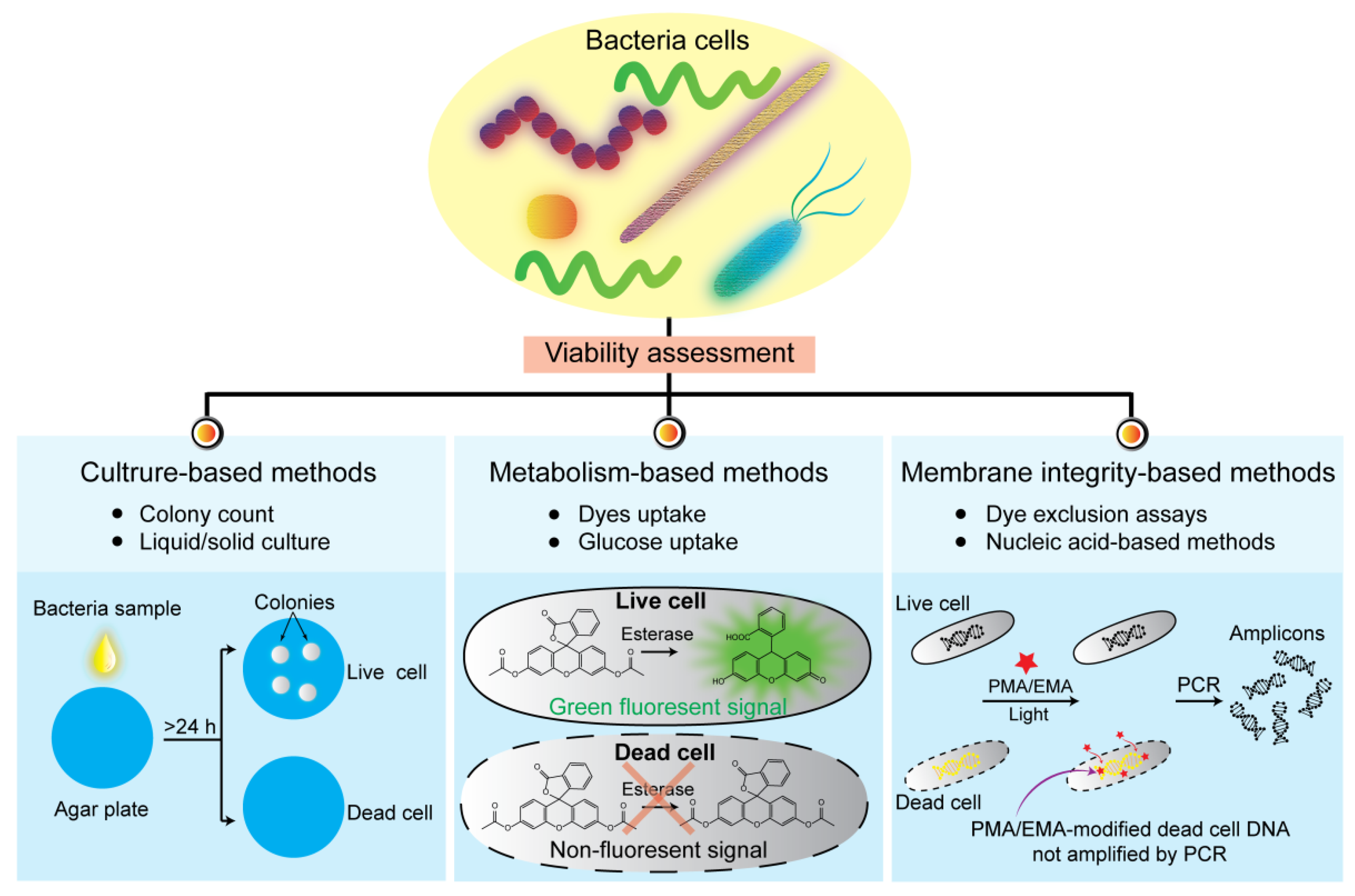
To evaluate the viability of bacterial pathogens, culturability, metabolic activity, and membrane integrity are three widespread and accepted criteria [5]. The bacterial culturability can be measured by determining their ability to produce a colony—A visible mass of bacteria originating from a single mother cell when plated on an appropriate solid media. In order to form a colony, bacteria must be reproducible and metabolically active and have an intact bacterial membrane [6]. However, sometimes bacteria can enter the viable but nonculturable (VBNC) state due to unfavorable conditions, such as low temperatures, low-nutrient environments, and high antibiotic concentrations [7,8,9,10,11,12]. When bacterial pathogens enter the VBNC state, they cannot be evaluated based on culturability criteria. As an alternative strategy, VBNC bacteria can be detected by measuring their metabolic activity. Numerous studies have reported that VBNC bacteria can be evaluated by measuring the uptake of substrates, such as fluorescent dyes and glucose [13,14,15,16]. However, VBNC bacteria can enter the dormant state in which the metabolic activities of VBNC bacteria are inactive [17,18]. As a result, bacteria in the dormant state cannot be detected by measuring metabolic substrates. Therefore, another criterion for viability assessment has been introduced, relying on membrane integrity. In this approach, a dead bacterium would have a disrupted and/or broken membrane, whereas a live bacterium has an intact membrane [19,20,21]. Based on three accepted criteria (culturability, metabolic activity, and membrane integrity) for bacterial viability, various methods have been developed for evaluating bacterial viability (Figure 1). This review summarized the current methods for the viability assessment of bacterial pathogens and discussed their advances, challenges, and future perspectives.
2. Viability Assessments Based on Culturability
As a traditional method, the plate culture method has been widely accepted for detecting bacterial viability for >100 years [22]. This technique was first discovered by Robert Koch in 1881 for culturing, detecting, and quantifying viable bacteria [23]. A contaminated sample can be plated on an agar plate, followed by incubation for various times at various temperatures depending on the bacterial species. After incubation, viable bacteria form colonies, whereas nonviable bacteria do not [24,25]. Different bacterial types can form colonies with different shapes, sizes, and colors. Culture-dependent methods not only provide information about bacterial viability but can also be helpful in identifying bacteria [26]. However, the culture-dependent method must combine with other technologies, such as biochemical tests, Gram staining, catalase test, and sporulation test, for bacterial identification [27,28]. Quantifying viable bacteria using the culture-dependent method requires manual steps, such as spreading samples on an agar plate and counting colonies. Recently, automated instruments for spreading have been well developed and are even available in the market, such as Microstreak® and commercial spiral platers [29,30]. For counting bacterial colonies, various automatic systems have been published. For example, Zhu et al. reported an automatic analysis system for counting bacterial colonies based on images captured with near-infrared light [31]. This system is convenient and cost-effective for counting colonies automatically by processing images. It takes 11–21 s to count colonies on each agar, with an average relative error of 0.2%. In another study, Molina et al. used Scan® 500 (Interscience) to capture images and digitalize them to count the number of Escherichia coli colonies [32]. Therefore, an automated system reduces time consumption and manual steps. Although automatic systems could improve the efficiency of culture-dependent methods, the process requires 2–3 days for bacterial isolation and up to 1 week to obtain the final results of viability and quantification. Furthermore, as stated earlier, the most serious limitation of culture-dependent methods is that they cannot detect VBNC bacteria.
3. Viability Assessments Based on Metabolic Activities
3.1. Dyes Uptake Assay
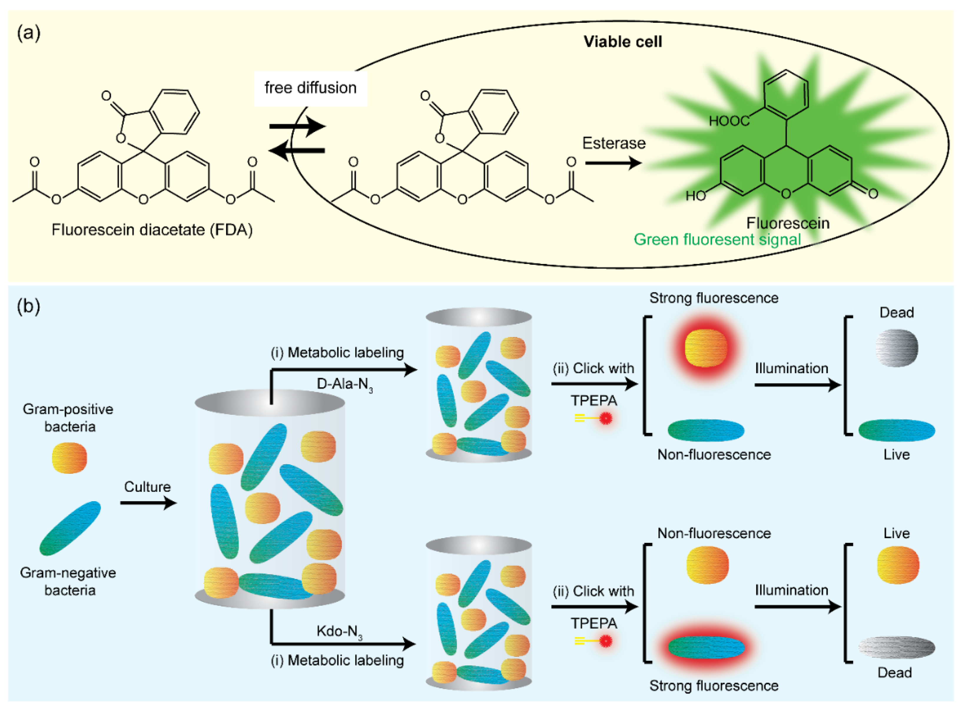
The metabolic activity of viable bacteria can be detected based on their uptake of dyes through bacterial membranes. When the dyes enter the bacterial membrane, they are hydrolyzed by an active enzyme system, such as esterases, lipases, and proteases, to convert nonfluorescent to detectable fluorescent signals. Fluorescein diacetate (FDA) is a common dye used for viability assessment based on the activity of nonspecific enzymes, as mentioned above, and it is a nonpolar and nonfluorescent dye. FDA has lipophilic properties because it comprises two acetate groups and is, therefore, permeable to lipid bilayer membranes of bacteria [33]. After the transportation of FDA into bacteria, it is hydrolyzed to fluorescein by nonspecific intracellular enzymes and releases measurable fluorescent signals (Figure 2a) [34]. Fluorescein is a polar molecule and, therefore, cannot move across bacterial lipid membranes. As a result, fluorescent signals are accumulated inside bacteria. The advantages of this technology are as follows: (1) FDA uptake does not require any specific transport pathway through the membranes because FDA can enter bacteria via a passive transport mechanism, and (2) extracellular FDA does not produce any background signal [33]. However, FDA carries severe disadvantages [35]. First, the quenching effect can occur when the fluorescein concentration inside bacteria is too elevated. Second, the FDA-based method is highly sensitive to pH. The acidic environment can enhance the protonation of fluorescein, which can enhance the efflux of fluorescein via passive efflux, decreasing the fluorescent signal. The product of FDA hydrolysis is acetic acid; therefore, it can decrease the intracellular pH. The pH also affects intracellular enzyme activity; each enzyme has different optimal pH conditions. Because plenty of intracellular enzymes catalyze FDA hydrolysis, optimizing the pH condition for FDA hydrolysis is difficult [36,37,38]. Nevertheless, biological metabolism-based fluorescent labeling can greatly improve bacteria labeling using the combination of a fluorescent probe and a bio-orthogonal group. Furthermore, it can be employed as an effective approach for the discrimination of peptidoglycan of gram-positive bacteria and lipopolysaccharide of gram-negative bacteria [39,40,41,42]. For example, a bacteria-metabolizable dual-functional probe TPEPy-d-Ala was developed for fluorescence turn-on imaging of bacteria based on aggregation-induced emission [39]. When metabolically bound with bacterial peptidoglycan, the mobility of the TPEPy-d-Ala probe is inhibited, resulting in clear visualization of intracellular bacteria by fluorescence signal enhancements. In another study, Wu et al. designed a bio-orthogonal fluorescence dye (TPEPA) for discriminating gram-positive (Staphylococcus aureus) and gram-negative (E. coli) bacteria by metabolic engineering (Figure 2b) [42]. Due to the different structures of bacteria morphology, this synthesized TPEPA dye could distinguish live and dead bacteria via selective imaging of metabolically decorated gram-negative bacteria with Kdo-N3 and gram-positive bacteria with D-Ala-N3 under a fluorescence microscope, respectively.
3.2. Glucose Uptake Assay
Glucose is a monosaccharide composed of an aldehyde group (–CHO) and six carbon atoms. Compared to other carbon sources, such as fructose, sucrose, and lactose, glucose is more abundant and can be found in most beverages. In energy metabolism, almost all organisms, including bacteria, use glucose as the main source of energy and the building block for biopolymers in all kingdoms of life [43]. Some viable bacteria would consume glucose from the environment into their cytoplasm through the membrane transport system. Once glucose is imported into the cytoplasm, it is metabolized through different pathways to become ready-to-use energy [44]. Therefore, glucose content has become one of the most important parameters for evaluating the metabolic activity of bacteria.
Methods for viability assessment based on glucose can be categorized into two main strategies: using artificial fluorescent glucose and using enzymatic assays. For the first strategy, glucose uptake can be measured using artificial fluorescent glucose, namely, 2-[N-(7-nitrobenz-2-oxa-1,3-diazol-4-yl)amino]-2-deoxy-D-glucose (2-NBDG). Only viable bacteria with active metabolisms can consume 2-NBDG via a glucose transporting system. Once 2-NBDG is incorporated into bacteria, it is decomposed as a nonfluorescent compound. Meanwhile, dead bacteria cannot degrade 2-NBDG, resulting in the remaining fluorescent signals. However, not all bacteria can consume 2-NBDG. Vibrio mimicus 10393, Bacillus cereus JCM 2152, Plesiomonas shigelloides NP321, Aeromonas hydrophila JCM 1027, and E. coli W539 could not take in 2-NBDG [45,46,47]. Moreover, a fluorescent spectrophotometer or a fluorescence microscope is usually required to analyze fluorescent signals that could limit their application in low-resource settings.
For the second strategy, the remaining glucose can be measured by enzymatic assays. In the presence of glucose oxidase, glucose is oxidized to form D-gluconic acid and H2O2. After that, the H2O2 level is measured by a colorimetric reaction with o-dianisidine under the catalyzation of peroxidase to switch o-dianisidine from a colorless to a colored compound [48,49,50]. Generally, enzymatic assays are expensive, and natural enzymes have low stability and are difficult to store. With the development of nanotechnology, nanozymes have been developed to address the limitations of natural enzymes. Nanozymes are nanomaterials with enzyme-like activities. Unlike natural enzymes, nanozymes are low-cost and easy to store and have high stability and tunable catalytic activities [51]. Given these advantages, glucose oxidase- and peroxidase-mimicking nanozymes for glucose assays have proven their potential for viability assessment [52,53]. In general, detecting viable bacteria using enzymatic assays, along with significant advances in nanozymes, obviously have many advantages over the fluorescent glucose-based method, including (1) the method is suitable for almost all bacteria, (2) the results can be observed by the naked eye, and (3) the method is cost-saving and portable because it does not require bulky machines, such as a fluorescence microscope or fluorescent spectrophotometer.
3.3. Adenosine Triphosphate (ATP) Assay
ATP Bioluminescence assay is inspired by the enzymatic reaction in fireflies, which releases detectable light by converting luciferin to oxyluciferin in the presence of ATP and luciferase [54]. ATP is an energy-carrying molecule that is essential for the metabolic activities of living organisms [55]. Therefore, ATP is widely used as a marker for viable cells, which are detected by luciferin–luciferase luminescence reactions with increasing light intensity correlating to a higher number of live cells [56,57]. The reaction can be performed within minutes and does not require heavy equipment, having assay kits with portable luminescence detectors that are already available on the market by many suppliers [57]. However, since ATP is a common energy currency for all living cells, using ATP as a marker for live bacteria might result in misinterpretation if the samples contain non-bacterial or extracellular ATP [58,59]. In addition, the ATP level can vary between bacterial species and depends on the physiological states, making direct interpretation of ATP levels to bacteria counts unreliable [60,61].
4. Viability Assessments Based on Membrane Integrity
As mentioned earlier, among the three criteria (culturability, metabolic activity, and membrane integrity) for viability assessments, membrane integrity is the most reliable criterion. Bacteria can enter the states allowing them to silence the reproducibility and metabolic activity; therefore, some bacteria cannot be detected using the culturability and metabolic activity criteria. However, membrane integrity is critical for bacterial function and survival [62].
4.1. Dye Exclusion Assays
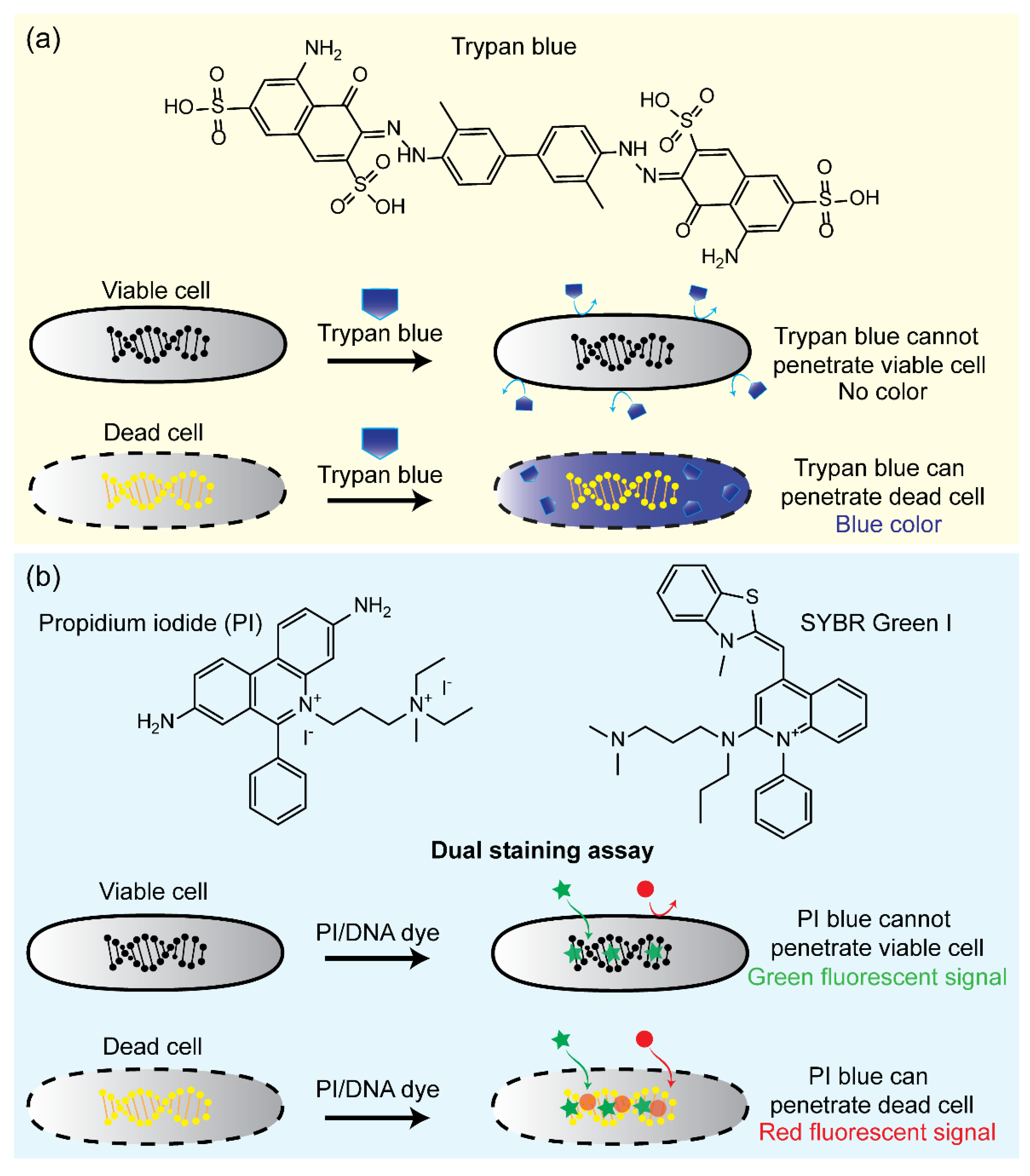
For the viability assessment of bacteria based on membrane integrity criteria, the dye exclusion assay is one of the most common and useful techniques. The mechanism of the dye exclusion assay is based on the fact that bacteria with intact membranes are highly selective concerning the dyes that can move across their membrane, whereas a compromised membrane permits easy access. When dyes enter bacteria with damaged membranes, they interact with intracellular proteins or nucleic acids and release detectable fluorescent signals [63,64,65]. As a typical example, trypan blue is an anionic hydrophilic azo dye widely used to stain dead cells (Figure 3a). Due to its high negative charge, it is excluded from the bacteria with intact membranes. In contrast, dead bacteria can take up trypan blue because they lose membrane selectivity. After entering the bacterial cytoplasm, trypan blue interacts with cytoplasmic proteins and emits blue fluorescent signals [66,67]. The relative number of live and dead bacteria can be measured using a fluorescence microscope [68,69], light microscope [70,71,72], or flow cytometer [71,73,74] by counting unstained and stained bacteria.
As another example of dye exclusion assays, propidium iodide (PI) is also widely used for the viability assessment of bacteria, especially after the report of Boulos in 1999 [75]. Like trypan blue, PI only stains dead bacteria because it can only penetrate bacteria with compromised membranes. However, PI does not interact with intracellular proteins as trypan blue; instead, PI intercalates to DNA and RNA inside dead bacteria. The reaction with nucleic acids enhances ~30-fold the fluorescence of PI and shifts the excitation/emission maximum of PI to 535/617 nm, whereas free PI has an excitation/emission maximum of 493/636 nm [76]. The fluorescent signal can be analyzed using fluorescence microscopy, flow cytometry, or confocal laser scanning. For viability assessments, PI is usually coupled with dyes that can penetrate and stain nucleic acids of live and dead bacteria, thereby obtaining total bacteria counts. Khan et al. optimized the staining protocol and flow cytometry to detect VBNC and VC bacteria within 70 min [77]. Various fluorescent probes, such as SYTO 9, SYTO 13, SYTO 17, SYTO 40, and PI, were performed to qualify VBNC and VC E. coli O157:H7, Pseudomonas aeruginosa, Pseudomonas syringae, and Salmonella enterica. Recently, a highly sensitive approach using DNA dyes for bacterial viability was suggested by Feng et al., who used SYBR Green I and PI dyes for identifying S. aureus, E. coli, Klebsiella pneumoniae, Mycobacterium tuberculosis, and Acinetobacter baumannii in <30 min (Figure 3b) [78]. Another protocol was based on the dual SYTO9/PI staining assay to rapidly detect Staphylococcus and P. aeruginosa, and fluorescent signals were observed by fluorescence microscopy [76]. SYTO 9 can penetrate live and dead bacteria regardless of their membrane integrity, intercalate to DNA and RNA, and release a green fluorescent signal. Because PI exhibits a stronger affinity toward nucleic acids than SYTO 9, PI can replace SYTO 9 when both dyes are exposed to the same nucleic acid. As a result, dead bacteria are stained by PI with a red fluorescent signal, whereas a green fluorescent signal released from SYTO 9 represents live bacteria [79,80,81,82].
Although dye exclusion assays are one of the most common methods among viability assessments mentioned above, a major disadvantage of this approach is it cannot distinguish different bacterial species. In other words, dye exclusion assays can only evaluate the ratio of viable and nonviable bacteria; they cannot provide information about which bacterial species are present in the samples. This drawback may limit the application of dye exclusion assays in identifying viable pathogens.
Figure 3.
(a) Schematic illustration of the working principle of bacterial viability using trypan blue dye. (b) Schematic illustration of the working principle of bacterial viability using PI/SYBR Green I assay [78]. Green star, DNA dye; Red circle, PI.
Figure 3.
(a) Schematic illustration of the working principle of bacterial viability using trypan blue dye. (b) Schematic illustration of the working principle of bacterial viability using PI/SYBR Green I assay [78]. Green star, DNA dye; Red circle, PI.

4.2. Nucleic Acid-Based Methods
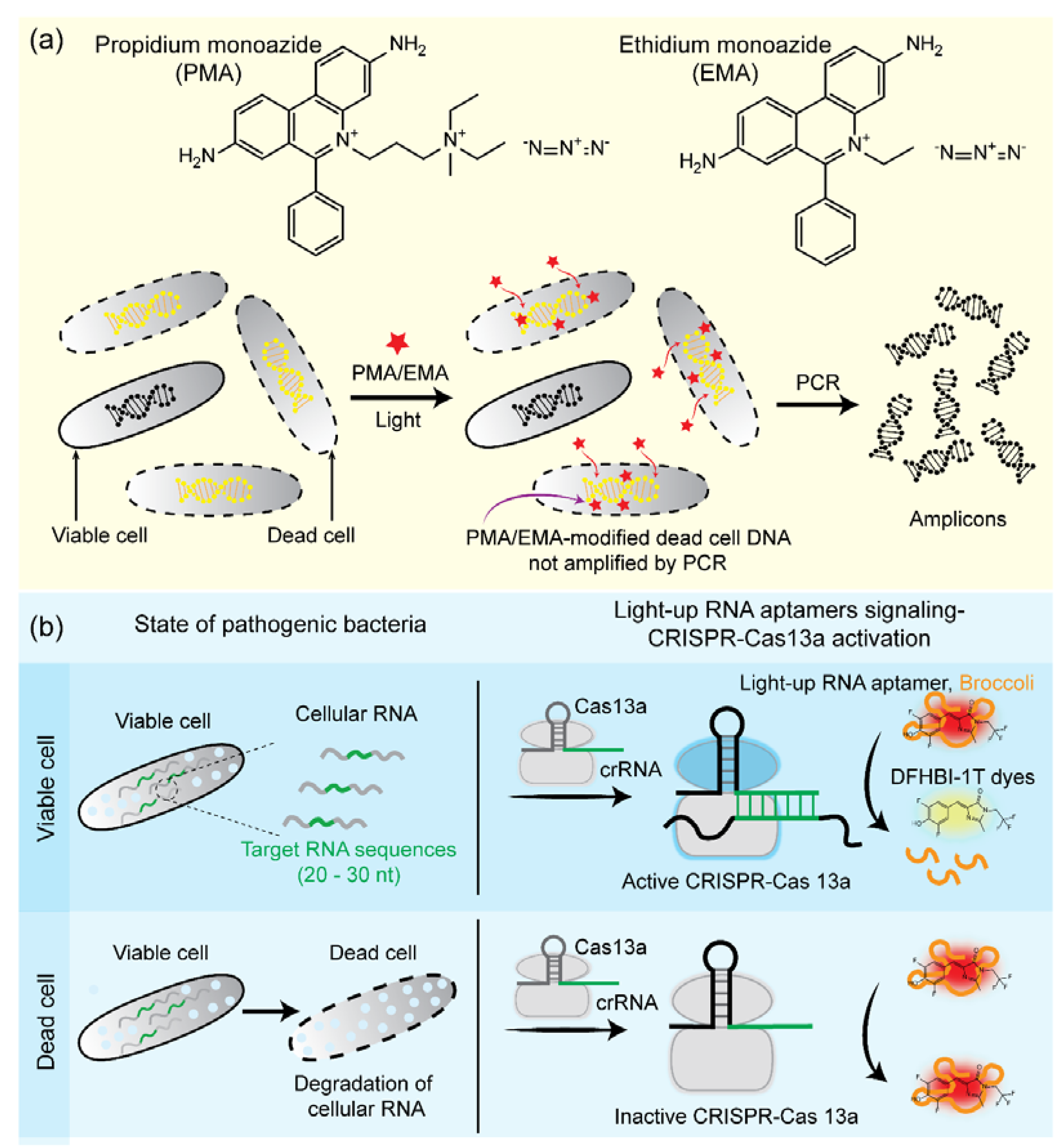
In recent years, by coupling photoreactive DNA-intercalating dyes, biomolecular techniques [e.g., polymerase chain reaction (PCR), loop-mediated isothermal amplification (LAMP), and recombinase-aided amplification (RAA)] have been widely applied for bacterial viability assays (Figure 4a) [83,84,85,86]. In this approach, DNA-intercalating dyes, such as ethidium monoazide (EMA) and propidium monoazide (PMA), are used to penetrate only nonviable bacteria with compromised membranes, whereas viable bacteria with intact membranes should pose a barrier for DNA-intercalating dyes [87,88]. Vondrakova et al. suggested that the killing methods and species-specific differences can affect EMA/PMA-quantitative PCR (qPCR) efficacies because some bacterial species are resistant to the EMA/PMA pretreatment technique [89]. In addition, to enhance the selectivity and sensitivity, a new DNA modification dye (named PMAxx, an improved version of PMA) has been developed and applied for bacterial viability assays [90,91,92]. After the penetration of dyes, they are exposed to bright visible light to stimulate the interaction between dyes and DNA [93,94]. EMA and PMA contain an azide group that can be converted into a highly active nitrene radical under exposure to bright visible light. The active nitrene radicals can bind covalently to DNA from nonviable bacteria, whereas unbound nitrene radicals are simultaneously inactivated by reacting with H2O in samples. The covalent bond between DNA and nitrene radical changes the DNA structure in nucleotide angle, inhibiting DNA elongation by polymerases. Nitrene radicals also reduce the solubility of DNA, enabling DNA removal by the DNA extraction process. Dye-treated samples undergo DNA extraction, followed by PCR to amplify DNA from viable bacteria. In contrast, DNA from nonviable bacteria cannot be amplified because of the covalent bond between DNA and nitrene radicals [95,96,97,98,99]. A combination of PMA dye and qPCR is the most popularly studied for the viability assay of various bacteria, such as S. aureus, E. coli O157, P. aeruginosa, Lactobacillus spp., M. tuberculosis, B. cereus, etc. [100,101,102,103,104,105,106]. For example, Li et al. introduced viability PCR with PMA and DyeTox13-qPCR methods for detecting the invA gene from Salmonella typhimurium [107]. In this study, by optimizing the DyeTox13 assay with EMA, the PCR signal from dead cells was reduced, which helped to overcome the main limitation of the PCR approach concerning its inability to discriminate dead from live bacteria. In other words, false-positive results from dead bacteria were eliminated using this method. In 2019, Cao et al. developed real-time PCR and LAMP approaches to detect Vibrio parahaemolyticus in shrimp samples, which achieved the limit of detection (LOD) of ~10.5 colony-forming units (CFU)/mL [90]. Using the PMA-LAMP approach, VBNC E. coli O157:H7 and S. enterica were successfully detected and quantified in fresh produce [108]. In another study, Xu et al. proposed a modified PMAxx dye combined with RAA to detect viable S. aureus in milk samples, and the LOD was ~102 CFU/mL [86]. Recently, apart from DNA-based methods, RNA-based methods are also being used for bacterial viability assays using RNA as an indicator [109,110,111]. Viable vancomycin-resistant Enterococcus was successfully discriminated against using reverse transcription LAMP for RNA amplification combined with colorimetric detection within 1 h [111]. As an alternative platform for bacterial viability, many studies tried to detect live bacteria without requiring nucleic acid extraction and amplification as usual [112,113]. Remarkably, with the advancement of clustered regularly interspaced short palindromic repeats (CRISPR)/CRISPR-associated proteins (Cas), Zhang et al. recently introduced a light-up RNA aptamer signaling-CRISPR/Cas13a principle for identifying live B. cereus without requiring transcription and amplification (Figure 4b) [112]. The system could detect ~10 CFU of B. cereus in spoiled food. Adapting the same concept, Wei et al. developed a highly specific and sensitive detection method based on the aptamer-based Cas14a1 to determine live S. aureus with a LOD of ~400 CFU/mL live cells [113]. Therefore, this new approach allows live bacterial detection without amplification based on Cas14a1 and a pathogenic aptamer. Although this new approach could open a new way for live bacteria with high sensitivity and specificity compared to other amplification approaches, the LOD is higher, and the total time was >150 min.
4.3. Microfluidic Technology for Viability Assessments
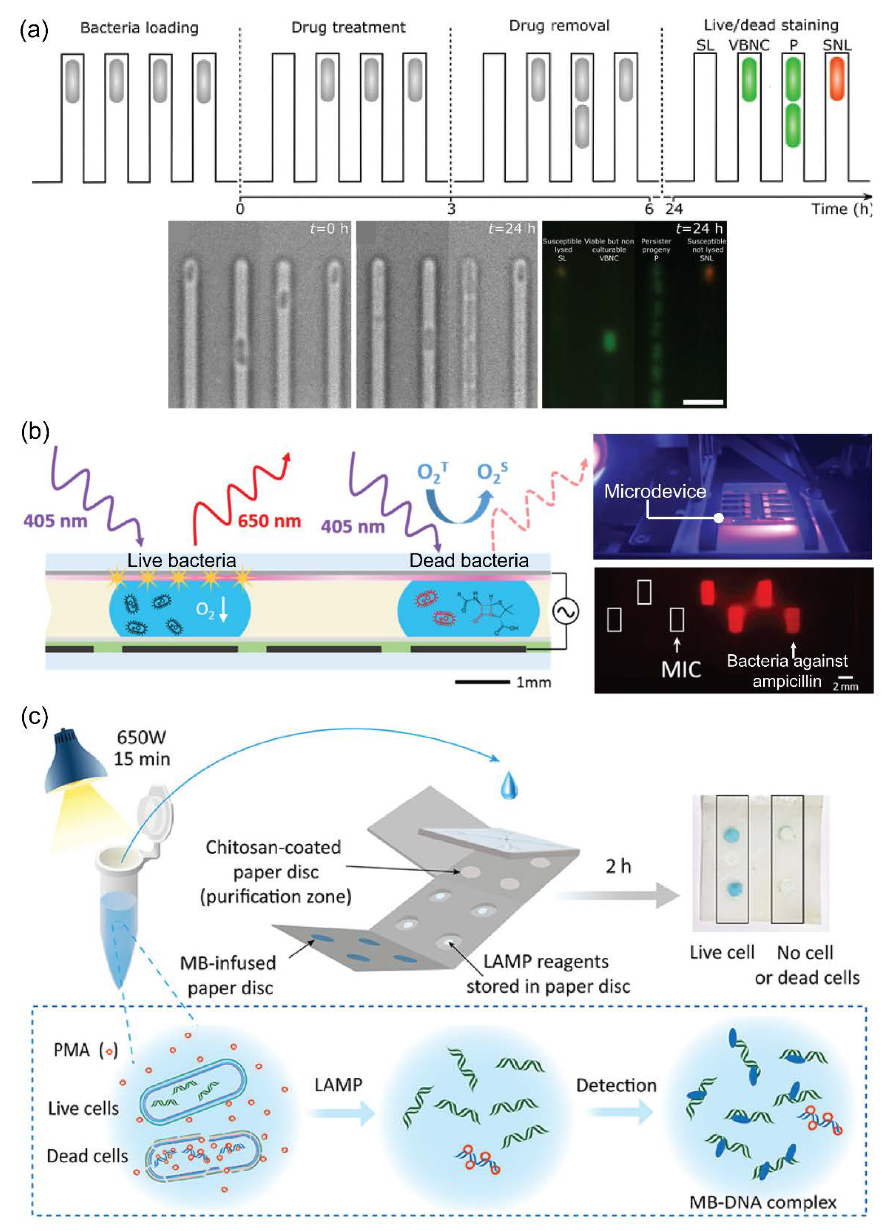
In recent years, microfluidics, also known as lab-on-a-chip, has been introduced and gained much attention from the public due to its wide-ranging applications in different fields, especially in cell biological research [114,115,116,117]. Generally, microfluidic technology is a fast-rising system that offers the integration of various processes into a single microdevice (micrometers to centimeters in size). Microdevices can offer highly efficient, sensitive, and rapid analysis with low energy consumption [118]. Due to these advantages, various viability assessments based on microfluidics have emerged for discriminating between live and dead bacterial cells [119,120]. For example, Bamford et al. introduced a combined system, including microfluidic channels and time-lapse microscopy, for observing VBNC cells before, during, and after drug treatment based on a fluorescent signal from SYTO 9 dyes (Figure 5a) [121]. Using this microdevice, a series of actions, such as culturing bacteria, treating drugs, and staining live/dead cells, could be performed simultaneously under microscopy and could capture images to investigate phenotypic or genotypic heterogeneity. In contrast, Qiu et al. fabricated a digital microfluidic device for an antimicrobial susceptibility test using an optical oxygen sensor film (Figure 5b) [122]. By measuring extracellular dissolved oxygen, this device could allow on-chip culturing and monitoring of E. coli growth (red fluorescent signals) with minimal sample handling and lower-volume cultures. In 2015, Chang et al. invested an integrated microfluidic system to rapidly detect live Staphylococcus from joint fluid as a medical application, helping with immediate medical decisions and antibiotic choices [123]. The bacterial sample was incubated with EMA and vancomycin-conjugated magnetic beads for distinguishing live bacteria and amplified 16S for bacterial typing by PCR. Using a PDMS integrated device, live bacteria were successfully detected within 30 min, and the LOD was ~102 CFU per reaction. For foodborne pathogen detection, Etayash et al. used the microfluidic cantilever for the in situ detection and discrimination of Listeria monocytogenes and E. coli, and the microdevice could detect bacteria at a concentration of single-cell per microliter [124]. Briefly, a biomaterial microcantilever embeds a microfluidic channel where the internal surfaces are chemically or physically functionalized with receptors that can selectively capture bacteria. Moreover, this device also can serve as a high-throughput device for the real-time detection of bacteria and allows discrimination between intact and dead E. coli and their metabolic response to antibiotics based on the presence of metabolic activity. In another study, Tung et al. introduced a paper-based microfluidic device that integrated treatment and molecular biology to detect viable E. coli O157:H7 and Salmonella spp. (Figure 5c) [125]. The paper-based device employed the most advanced techniques, such as chitosan-based DNA extraction, isothermal amplification (LAMP), and colorimetric detection, for screening multiple pathogens. In another study, E. coli was successfully discriminated against using microfluidics based on a centrifuge platform to perform a LIVE/DEAD BacLight bacterial viability assay [126]. For environmental application, Zhu et al. reported a high-resolution three-dimensional printed microdevice for E. coli detection using an integrated PMA-PCR device [127]. Especially, an on-chip PMMA pretreatment was used to improve the accuracy by eliminating the need for pipetting steps. As a global public health issue, M. tuberculosis is a bacterium that causes serious disease, namely tuberculosis, and is slower in growth than other infectious bacteria; thus, making it difficult and challenging for early detection [128]. Recently, Wang et al. introduced an integrated microfluidic system to automatically detect live M. tuberculosis and distinguish dead bacteria from clinical samples [129]. In this study, using this fully integrated microdevice (including bacterial capture, PMA treatment, lysis, and PCR quantification), M. tuberculosis could achieve automated detection in a single chip within 90 min, and the LOD was as low as 100 CFU. From these examples, numerous microfluidic devices have been fabricated to integrate various processes required for viability assessment, such as dye treatment, DNA extraction, amplification, and detection. DNA-based analyses using a microdevice reduce the time and cost of the bacterial viability test. Therefore, the obvious advantages provided by microfluidic technology make this approach powerful for viability assessment.
5. Conclusions
This review summarized the current techniques for viability assessment. Based on three viable criteria (culturability, metabolic activity, and membrane integrity), current viability assessments were categorized into three main strategies (Table 1). The earliest published viability assessment is the plate culturing method, which relies on culturability. However, this technique cannot detect VBNC bacteria. In order to evaluate VBNC bacteria, the next generation for viability assessment has been developed, which relies on metabolic activity. Although VBNC bacteria reproduce poorly, they still maintain metabolic activity by the uptake of nutrients through the bacterial membrane. Dye and glucose uptake are the most common viability assessments relying on metabolic activity. Only metabolically active bacteria can uptake and convert artificial fluorescent or natural glucose and emit detectable signals. However, bacteria can enter the dormant state in which the metabolic activities of VBNC bacteria are inactive and, therefore, cannot be detected when using metabolic activity criteria. In order to overcome this limitation, dye exclusion assays relying on membrane integrity have been developed. In this approach, fluorescent dyes, such as trypan blue and PI, are used to penetrate and stain only nonviable bacteria. The major disadvantage of this approach is that it cannot distinguish different bacteria species. With the combination between dye exclusion assays and PCR, technology for viability assessment took a giant step, which allows the determination of certain viable bacteria in samples. Generally, all listed technologies require multiple processes, bulky machines, and laboratory technicians to conduct the whole process and analyze the results. Along with significant advances in microfluidic technology, almost all processes required for detecting viable bacteria have been simply integrated into a single microfluidic device. The obvious advantages of microfluidic devices, such as cost-effectiveness, high automation, and user-friendliness, make them a potential technology for viability assessment.
Author Contributions
Conceptualization, K.T.L.T. and N.Y.L.; writing—original draft preparation, K.T.L.T.; writing—review and editing, K.T.L.T. and N.Y.L.; supervision, N.Y.L.; project administration, N.Y.L.; funding acquisition, N.Y.L. All authors have read and agreed to the published version of the manuscript.
Funding
This work was supported by the National Research Foundation of Korea (NRF) grant funded by the Korea government (MSIT) (No. NRF2020R1A2B5B01001971) and also by Basic Science Research Program through the National Research Foundation of Korea (NRF) funded by the Ministry of Education (2021R1A6A1A03038996).
Institutional Review Board Statement
Not applicable.
Informed Consent Statement
Not applicable.
Data Availability Statement
Not applicable.
Conflicts of Interest
The authors declare no conflict of interest.
References
- Nii-Trebi, N.I. Emerging and neglected infectious diseases: Insights, advances, and challenges. BioMed Res. Int. 2017, 2017, 5245021. [Google Scholar] [PubMed]
- Streicher, L.M. Exploring the future of infectious disease treatment in a post-antibiotic era: A comparative review of alternative therapeutics. J. Glob. Antimicrob. Resist. 2021, 24, 285–295. [Google Scholar] [CrossRef] [PubMed]
- Wesche, A.M.; Gurtler, J.B.; Marks, B.P.; Ryser, E.T. Stress, sublethal injury, resuscitation, and virulence of bacterial foodborne pathogens. J. Food Prot. 2009, 72, 1121–1138. [Google Scholar] [CrossRef] [PubMed]
- Verraes, C.; Boxstael, S.V.; Meervenne, E.V.; Coillie, E.V.; Butaye, P.; Catry, B.; Schaetzen, M.-A.; Huffel, X.V.; Imberechts, H.; Dierick, K.; et al. Antimicrobial resistance in the food chain: A review. Int. J. Environ. Res. Public Health 2013, 10, 2643–2669. [Google Scholar] [CrossRef] [PubMed]
- Nocker, A.; Camper, A.K. Novel approaches toward preferential detection of viable cells using nucleic acid amplifcation techniques. FEMS Microbiol. Lett. 2009, 291, 137–142. [Google Scholar] [CrossRef]
- Barbau-Piednoir, E.; Mahillon, J.; Pillyser, J.; Coucke, W.; Nancy, H.; Roosens, N.H.; Botteldoorn, N. Evaluation of viability-qPCR detection system on viable and dead Salmonella serovar Enteritidis. J. Microbiol. Methods 2014, 103, 131–137. [Google Scholar] [CrossRef]
- Biosca, E.G.; Amaro, C.; Marco-Noales, E.; Oliver, J.D. Effect of low temperature on starvation-survival of the eel pathogen Vibrio vulnificus Biotype 2. Appl. Environ. Microbiol. 1996, 62, 450–455. [Google Scholar] [CrossRef]
- Du, M.; Chen, J.; Zhang, X.; Li, A.; Li, Y.; Wang, Y. Retention of virulence in a viable but nonculturable edwardsiella tarda isolate. Appl. Environ. Microbiol. 2007, 73, 1349–1354. [Google Scholar] [CrossRef]
- Fleischmann, S.; Robben, C.; Alter, T.; Rossmanith, P.; Mester, P. How to evaluate non-growing cells—current strategies for determining antimicrobial resistance of VBNC bacteria. Antibiotics 2021, 10, 115. [Google Scholar] [CrossRef]
- Li, L.; Mendis, N.; Trigui, H.; Oliver, J.D.; Sebastien, P.; Faucher, S.P. The importance of the viable but non-culturable state in human bacterial pathogens. Front. Microbiol. 2014, 5, 258. [Google Scholar] [CrossRef] [Green Version]
- Ou, A.; Wang, K.; Mao, Y.; Yuan, L.; Ye, Y.; Chen, L.; Zou, Y.; Huang, T. First report on the rapid detection and identification of methicillin-resistant Staphylococcus aureus (MRSA) in viable but non-culturable (VBNC) under food storage conditions. Front. Microbiol. 2021, 11, 615875. [Google Scholar] [CrossRef] [PubMed]
- Xu, H.-S.; Roberts, N.; Singleton, F.L.; Attwell, R.W.; Grimes, D.J.; Colwell, R.R. Survival and viability of nonculturable Escherichia coli and Vibrio cholerae in the estuarine and marine environment. Microb. Ecol. 1982, 8, 313–323. [Google Scholar] [CrossRef] [PubMed]
- Guo, L.; Yea, C.; Cui, L.; Wan, K.; Chen, S.; Zhang, S.; Yu, X. Population and single cell metabolic activity of UV-induced VBNC bacteria determined by CTC-FCM and D2O-labeled Raman spectroscopy. Environ. Int. 2019, 130, 104883. [Google Scholar] [CrossRef] [PubMed]
- Rahman, I.; Shahamat, M.; Kirchman, P.A.; Russek-Cohen, E.; Colwell, R.R. Methionine uptake and cytopathogenicity of viable but nonculturable Shigella dysenteriae Type 1. Appl. Environ. Microbiol. 1994, 60, 3573–3578. [Google Scholar] [CrossRef]
- Wideman, N.E.; Oliver, J.D.; Crandall, P.G.; Jarvis, N.A. Detection and potential virulence of viable but non-culturable (VBNC) Listeria monocytogenes: A review. Microorganisms 2021, 9, 194. [Google Scholar] [CrossRef]
- Zeng, B.; Zhao, G.; Cao, X.; Yang, Z.; Wang, C.; Hou, L. Formation and resuscitation of viable but nonculturable Salmonella typhi. BioMed Res. Int. 2013, 2013, 907170. [Google Scholar] [CrossRef]
- Fisher, R.A.; Gollan, B.; Helaine, S. Persistent bacterial infections and persister cells. Nat. Rev. Microbiol. 2017, 15, 453–464. [Google Scholar] [CrossRef]
- Wood, T.K.; Knabel, S.J.; Kwan, B.W. Bacterial persister cell formation and dormancy. Appl. Environ. Microbiol. 2013, 79, 7116–7121. [Google Scholar] [CrossRef]
- Grégori, G.; Citterio, S.; Ghiani, A.; Labra, M.; Sgorbati, S.; Brown, S.; Denis, M. Resolution of viable and membrane-compromised bacteria in freshwater and marine waters based on analytical flow cytometry and nucleic acid double staining. Appl. Environ. Microbiol. 2001, 67, 4662–4670. [Google Scholar] [CrossRef]
- Lv, X.-C.; Li, Y.; Qiu, W.-W.; Wu, X.-Q.; Xu, B.-X.; Liang, Y.-T.; Liu, B.; Chen, S.-J.; Rao, P.-F.; Ni, L. Development of propidium monoazide combined with real-time quantitative PCR (PMA-qPCR) assays to quantify viable dominant microorganisms responsible for the traditional brewing of Hong Qu glutinous rice wine. Food Control 2016, 66, 69–78. [Google Scholar] [CrossRef]
- Tan, G.; Zhou, R.; Zhang, W.; Hu, Y.; Ruan, Z.; Li, J.; Zhang, C.; Shen, D.; Peng, N.; Liang, Y.; et al. Detection of viable and total bacterial community in the pit mud of chinese strong-flavor liquor using propidium monoazide combined with quantitative PCR and 16S rRNA gene sequencing. Front. Microbiol. 2020, 11, 896. [Google Scholar] [CrossRef] [PubMed]
- Yun, K.; Park, D.; Kang, M.; Song, J.; Chung, Y.; Bang, H.; Jeon, N.L. A petri-dish with micromolded pattern as a coordinate indicator for live-cell time lapse microscopy. BioChip J. 2022, 16, 27–32. [Google Scholar] [CrossRef]
- Bull, A.T.; Quayle, J.R. New dimensions in microbiology: An introduction. Phil. Trans. R. Soc. Lond. B 1982, 297, 447–457. [Google Scholar]
- Lehnig, M.; Glass, S.; Lippmann, N.; Ziganshyna, S.; Eulenburg, V.; Werdehausen, R. Evaluation of a luminometric cell counting system in context of antimicrobial photodynamic inactivation. Microorganisms 2022, 10, 950. [Google Scholar] [CrossRef]
- Sanders, E.R. Aseptic laboratory techniques: Plating methods. J. Vis. Exp. 2012, 63, e3064. [Google Scholar] [CrossRef] [PubMed]
- Sousa, A.M.; Machado, I.; Nicolau, A.; Pereira, M.O. Improvements on colony morphology identification towards bacterial profiling. J. Microbiol. Methods 2013, 95, 327–335. [Google Scholar] [CrossRef] [PubMed]
- Franco-Duarte, R.; Černáková, L.; Kadam, S.; Kaushik, K.S.; Salehi, B.; Bevilacqua, A.; Corbo, M.R.; Antolak, H.; Dybka-Stępién, K.; Leszczewicz, M.; et al. Advances in chemical and biological methods to identify microorganisms—from past to present. Microorganisms 2019, 7, 130. [Google Scholar] [CrossRef]
- Pradhan, P.; Tamang, J.P. Phenotypic and genotypic identification of bacteria isolated from traditionally prepared dry starters of the Eastern Himalayas. Front. Microbiol. 2019, 10, 2526. [Google Scholar] [CrossRef]
- Glasson, J.H.; Guthrie, L.H.; Nielsen, D.J.; Bethell, F.A. Evaluation of an automated instrument for inoculating and spreading samples onto agar plates. J. Clin. Microbiol. 2008, 46, 1281–1284. [Google Scholar] [CrossRef]
- King, G.W.; Kath, G.S.; Siciliano, S.; Simpson, N.; Masurekar, P.; Sigmund, J.; Polishook, J.; Skwish, S.; Bills, G.; Genilloud, O.; et al. Automated agar plate streaker: A linear plater on society for biomolecular sciences standard plates. J. Biomol. Screen. 2006, 11, 704–711. [Google Scholar] [CrossRef]
- Zhu, G.; Yan, B.; Xing, M.; Tian, C. Automated counting of bacterial colonies on agar plates based on images captured at near-infrared light. J. Microbiol. Methods 2018, 153, 66–73. [Google Scholar] [CrossRef] [PubMed]
- Molina, F.; Simancas, A.; Ramírez, M.; Tabla, R.; Roa, I.; Rebollo, J.E. A new pipeline for designing phage cocktails based on phage-bacteria infection networks. Front. Microbiol. 2021, 12, 564532. [Google Scholar] [CrossRef] [PubMed]
- Nitsch, A.; Haralambiev, L.; Einenkel, R.; Muzzio, D.O.; Zygmunt, M.T.; Ekkernkamp, A.; Burchardt, M.; Stope, M.B. Determination of in vitro membrane permeability by analysis of intracellular and extracellular fluorescein signals in renal cells. In Vivo 2019, 33, 1767–1771. [Google Scholar] [CrossRef]
- Hong, D.; Lee, G.; Jung, N.C.; Jeon, M. Fast automated yeast cell counting algorithm using bright-field and fluorescence microscopic images. Biol. Proced. Online 2013, 15, 13. [Google Scholar] [CrossRef]
- Sträuber, H.; Müller, S. Viability states of bacteria—specific mechanisms of selected probes. Cytom. Part A 2010, 77, 623–634. [Google Scholar] [CrossRef]
- Breeuwer, P.; Drocourt, J.-L.; Bunschoten, N.; Zwietering, M.H.; Rombouts, F.M.; Abee, T. Characterization of uptake and hydrolysis of fluorescein diacetate and carboxyfluorescein diacetate by intracellular esterases in saccharomyces cerevisiae, which result in accumulation of fluorescent product. Appl. Environ. Microbiol. 1995, 61, 1614–1619. [Google Scholar] [CrossRef] [PubMed]
- Dzionek, A.; Dzik, J.; Wojcieszyńska, D.; Guzik, U. Fluorescein diacetate hydrolysis using the whole biofilm as a sensitive tool to evaluate the physiological state of immobilized bacterial cells. Catalysts 2018, 8, 434. [Google Scholar] [CrossRef]
- Schnürer, J.; Rosswall, T. Fluorescein diacetate hydrolysis as a measure of total microbial activity in soil and litter. Appl. Environ. Microbiol. 1982, 43, 1256–1261. [Google Scholar] [CrossRef] [PubMed]
- Hu, F.; Qi, G.; Kenry; Mao, D.; Zhou, S.; Wu, M.; Wu, W.; Liu, B. Visualization and in situ ablation of intracellular bacterial pathogens through metabolic labeling. Angew. Chem. 2020, 59, 9288–9292. [Google Scholar] [CrossRef] [PubMed]
- Row, R.D.; Prescher, J.A. Constructing new biorthogonal reagents and reactions. Acc. Chem. Res. 2018, 51, 1073–1081. [Google Scholar] [CrossRef] [PubMed]
- Liang, J.; Tang, B.Z.; Liu, B. Specific light-up bioprobes based on AlEgen conjugates. Chem. Soc. Rev. 2015, 44, 2798–2811. [Google Scholar] [CrossRef] [PubMed] [Green Version]
- Wu, M.; Qi, G.; Liu, X.; Duan, Y.; Liu, J.; Liu, B. Bio-othogonal alegen for specific discrimination and elimination of bacteria pathogens via metabolic engineering. Chem. Mater. 2020, 32, 858–865. [Google Scholar] [CrossRef]
- Jeckelmann, J.-M.; Erni, B. Transporters of glucose and other carbohydrates in bacteria. Pflugers Arch. 2020, 472, 1129–1153. [Google Scholar] [CrossRef] [PubMed]
- Sundar, G.S.; Islam, E.; Braza, R.D.; Silver, A.B.; Breton, Y.L.; McIver, K.S. Route of glucose uptake in the group a streptococcus impacts SLS-mediated hemolysis and survival in human blood. Front. Cell. Infect. Microbiol. 2018, 8, 71. [Google Scholar] [CrossRef]
- Matsuoka, H.; Oishi, K.; Watanabe, M.; Kozone, I.; Saito, M.; Igimi, S. Viable cell detection by the combined use of fluorescent glucose and fluorescent glycine. Biosci. Biotechnol. Biochem. 2003, 67, 2459–2462. [Google Scholar] [CrossRef]
- Tao, J.; McCourt, C.; Sultana, H.; Nelson, C.; Driver, J.; Hackmann, T.J. Use of a fluorescent analog of glucose (2-NBDG) to identify uncultured rumen bacteria that take up glucose. Appl. Environ. Microbiol. 2019, 85, e03018-18. [Google Scholar] [CrossRef]
- Yoshioka, K.; Oh, K.-B.; Saito, M.; Nemoto, Y.; Matsuoka, H. Evaluation of 2-[N-(7-nitrobenz-2-oxa-l,3-diazol-4-yl)amino]-2-deoxy-D. glucose, a new fluorescent derivative of glucose, for viability assessment of yeast Candida albicans. Appl. Microbic. Biotechnol. 1996, 46, 400–404. [Google Scholar]
- Braissant, O.; Astasov-Frauenhoffer, M.; Waltimo, T.; Bonkat, G. A review of methods to determine viability, vitality, and metabolic rates in microbiology. Front. Microbiol. 2020, 11, 547458. [Google Scholar] [CrossRef]
- Silva, P.B.M.; Oliveira, K.A.; Coltro, W.K.T. Colorimetric detection of glucose in biological fluids using toner-based microzone plates. J. Braz. Chem. Soc. 2017, 28, 197–201. [Google Scholar] [CrossRef]
- Xiao, J.; Liu, Y.; Su, L.; Zhao, D.; Zhao, L.; Zhang, X. Microfluidic chip-based wearable colorimetric sensor for simple and facile detection of sweat glucose. Anal. Chem. 2019, 91, 14803–14807. [Google Scholar] [CrossRef]
- Liang, M.; Yan, X. Nanozymes: From new concepts, mechanisms, and standards to applications. Acc. Chem. Res. 2019, 52, 2190–2200. [Google Scholar] [CrossRef] [PubMed]
- Wang, Q.; Zhang, X.; Huang, L.; Zhang, Z.; Dong, S. GOx@ZIF-8(NiPd) nanoflower: An artificial enzyme system for tandem catalysis. Angew. Chem. 2017, 129, 16298–16301. [Google Scholar] [CrossRef]
- Zhang, Q.; Wang, X.; Kang, Y.; Sun, H.; Liang, Y.; Liu, J.; Su, Z.; Dan, J.; Luo, L.; Yue, T.; et al. Natural products self-assembled nanozyme for cascade detection of glucose and bacterial viability in food. Foods 2021, 10, 2596. [Google Scholar] [CrossRef]
- McElroy, W.D. The energy source for bioluminescence in an isolated system. Proc. Natl. Acad. Sci. USA 1947, 33, 342–345. [Google Scholar] [CrossRef]
- Knowles, J.R. Enzyme-catalyzed phosphoryl transfer reactions. Annu. Rev. Biochem. 1980, 49, 877–919. [Google Scholar] [CrossRef] [PubMed]
- Bottari, B.; Santarelli, M.; Neviani, E. Determination of microbial load for different beverages and foodstuff by assessment of intracellular ATP. Trends Food Sci. Technol. 2015, 44, 36–48. [Google Scholar] [CrossRef]
- Hammes, F.; Goldschmidt, F.; Vital, M.; Wang, Y.; Egli, T. Measurement and interpretation of microbial adenosine tri-phosphate (ATP) in aquatic environments. Water Res. 2010, 44, 3915–3923. [Google Scholar] [CrossRef]
- Siragusa, G.R.; Cutter, C.N.; Dorsa, W.J.; Koohmaraie, M. Use of a rapid microbial ATP bioluminescence assay to detect contamination on beef and pork carcasses. J. Food Prot. 1995, 58, 770–775. [Google Scholar] [CrossRef]
- Sakakibara, T.; Murakami, S.; Hattori, N.; Nakajima, M.O.; Imai, K. Enzymatic treatment to eliminate the extracellular ATP for improving the detectability of bacterial intracellular ATP. Anal. Biochem. 1997, 250, 157–161. [Google Scholar] [CrossRef]
- Selan, L.; Berlutti, F.; Passariello, C.; Thaller, M.C.; Renzini, G. Reliability of a bioluminescence ATP assay for detection of bacteria. J. Clin. Microbiol. 1992, 30, 1739–1742. [Google Scholar] [CrossRef]
- Yaginuma, H.; Kawai, S.; Tabata, K.V.; Tomiyama, K.; Kakizuka, A.; Komatsuzaki, T.; Noji, H.; Imamura, H. Diversity in ATP concentrations in a single bacterial cell population revealed by quantitative single-cell imaging. Sci. Rep. 2014, 4, 6522. [Google Scholar] [CrossRef] [PubMed]
- Ammendolia, D.A.; Bement, W.M.; Brumell, J.H. Plasma membrane integrity: Implications for health and disease. BMC Biology 2021, 19, 71. [Google Scholar] [CrossRef] [PubMed]
- Breeuwer, P.; Abee, T. Assessment of viability of microorganisms employing fluorescence techniques. Int. J. Food Microbiol. 2000, 55, 193–200. [Google Scholar] [CrossRef]
- Kwizera, R.; Akampurira, A.; Kandole, T.K.; Nielsen, K.; Kambugu, A.; Meya, D.B.; Boulware, D.R.; Rhein, J. Evaluation of trypan blue stain in a haemocytometer for rapid detection of cerebrospinal fluid sterility in HIV patients with cryptococcal meningitis. BMC Microbiol. 2017, 17, 182. [Google Scholar] [CrossRef] [PubMed]
- Ramirez, C.N.; Antczak, C.; Djaballah, H. Cell viability assessment: Toward content-rich platforms. Expert Opin. Drug Discov. 2010, 5, 223–233. [Google Scholar] [CrossRef]
- Kerschbauma, H.H.; Tasa, B.A.; Schürz, M.; Oberascher, K.; Bresgen, N. Trypan blue—Adapting a dye used for labelling dead cells to visualize pinocytosis in viable cells. Cell. Physiol. Biochem. 2021, 55, 171–184. [Google Scholar]
- Tran, S.-L.; Puhar, A.; Ngo-Camus, M.; Ramarao, N. Trypan blue dye enters viable cells incubated with the pore-forming toxin HlyII of Bacillus cereus. PLoS ONE 2011, 6, e22876. [Google Scholar] [CrossRef]
- Bresgen, N.; Ohlenschläger, I.; Wacht, N.; Afazel, S.; Ladurner, G.; Eckl, P.M. Ferritin and FasL (CD95L) mediate density dependent apoptosis in primary rat hepatocytes. J. Cell. Physiol. 2008, 217, 800–808. [Google Scholar] [CrossRef]
- Hu, C.; He, S.; Lee, Y.J.; He, Y.; Kong, E.M.; Li, H.; Anastasio, M.A.; Popescu, G. Live-dead assay on unlabeled cells using phase imaging with computational specificity. Nat. Commun. 2022, 13, 713. [Google Scholar] [CrossRef]
- Louis, K.S.; Siegel, A.C. Cell viability analysis using trypan blue: Manual and automated methods. Methods Mol. Biol. 2011, 740, 7–12. [Google Scholar]
- Franke, J.D.; Braverman, A.L.; Cunningham, A.M.; Eberhard, E.E.; Perry, G.A. Erythrosin B: A versatile colorimetric and fluorescent vital dye for bacteria. BioTechniques 2019, 68, 7–13. [Google Scholar] [CrossRef] [PubMed]
- Silva, K.J.S.; Sabogal-Paz, L.P. Cryptosporidium spp. and Giardia spp. (oo)cysts as target-organism in sanitation and environmental monitoring: A review in microscopy-based viability assays. Water Res. 2021, 189, 116590. [Google Scholar] [CrossRef] [PubMed]
- Davey, H.; Guyot, S. Estimation of microbial viability using flow cytometry. Curr. Protoc. Cytom. 2020, 93, e72. [Google Scholar] [CrossRef] [PubMed]
- McEvoy, B.; Lynch, M.; Rowan, N.J. Opportunities for the application of real-time bacterial cell analysis using flow cytometry for the advancement of sterilization microbiology. J. Appl. Microbiol. 2020, 130, 1794–1812. [Google Scholar] [CrossRef] [PubMed]
- Boulos, L.; Prevost, M.; Barbeau, B.; Coallier, J.; Desjardins, R. LIVE/DEAD® BacLight™: Application of a new rapid staining method for direct enumeration of viable and total bacteria in drinking water. J. Microbiol. Methods 1999, 37, 77–86. [Google Scholar] [CrossRef]
- Stiefel, P.; Schmidt-Emrich, S.; Maniura-Weber, K.; Ren, Q. Critical aspects of using bacterial cell viability assays with the fluorophores SYTO9 and propidium iodide. BMC Microbiol. 2015, 15, 36. [Google Scholar] [CrossRef]
- Khan, M.M.T.; Pyle, B.H.; Camper, A.K. Specific and rapid enumeration of viable but nonculturable and viable-culturable gram-negative bacteria by using flow cytometry. Appl. Environ. Microbiol. 2010, 76, 5088–5096. [Google Scholar] [CrossRef]
- Feng, J.; Yee, R.; Zhang, S.; Tian, L.; Shi, W.; Zhang, W.-H.; Zhang, Y. A Rapid growth-independent antibiotic resistance detection test by SYBR Green/propidium iodide viability assay. Front. Med. 2018, 5, 127. [Google Scholar] [CrossRef]
- Freire, J.M.; Gaspar, D.; Torre, B.G.; Veiga, A.S.; Andreu, D.; Castanho, M.A.R.B. Monitoring antibacterial permeabilization in real time using time-resolved flow cytometry. Biochim. Biophys. Acta 2015, 1848, 554–560. [Google Scholar] [CrossRef]
- Kolek, J.; Branska, B.; Drahokoupil, M.; Patakova, P.; Melzoch, K. Evaluation of viability, metabolic activity and spore quantity in clostridial cultures during ABE fermentation. FEMS Microbiol. Lett. 2016, 363, fnw031. [Google Scholar] [CrossRef]
- Rosenberg, M.; Azevedo, N.F.; Ivask, A. Propidium iodide staining underestimates viability of adherent bacterial cells. Sci. Rep. 2019, 9, 6483. [Google Scholar] [CrossRef] [PubMed]
- Wang, M.; Ateia, M.; Hatano, Y.; Miyanagad, K.; Yoshimura, C. Novel fluorescence-based method for rapid quantification of live bacteria in river water and treated wastewater. Environ. Sci. Adv. 2022, 1, 30. [Google Scholar] [CrossRef]
- Kontchou, J.A.; Nocker, A. Optimization of viability qPCR for selective detection of membrane-intact Legionella pneumophila. J. Microbiol. Methods 2019, 156, 68–76. [Google Scholar] [CrossRef] [PubMed]
- Rosenberg, J.N.; Cady, N.C. Surveilling cellular vital signs: Toward label-free biosensors and real-time viability assays for bioprocessing. Curr. Opin. Biotechnol. 2021, 71, 123–129. [Google Scholar] [CrossRef] [PubMed]
- Leifels, M.; Cheng, D.; Sozzi, E.; Shoults, D.C.; Wuertz, S.; Mongkolsuk, S.; Sirikanchana, K. Capsid integrity quantitative PCR to determine virus infectivity in environmental and food applications—A systematic review. Water Res. X 2021, 11, 100080. [Google Scholar] [CrossRef]
- Xie, G.; Zhou, D.; Zhao, G.; Feng, X.; Aguilar, Z.P.; Xu, H. Recombinase aided amplification with photoreactive DNA-binding dye for rapid detection of viable Staphylococcus aureus. LWT 2021, 135, 110249. [Google Scholar] [CrossRef]
- Cawthorn, D.M.; Witthuhn, R.C. Selective PCR detection of viable Enterobacter sakazakii cells utilizing propidium monoazide or ethidium bromide monoazide. J. Appl. Microbiol. 2008, 105, 1178–1185. [Google Scholar] [CrossRef]
- Stinson, L.F.; Keelan, J.A.; Payne, M.S. Characterization of the bacterial microbiome in first-pass meconium using propidium monoazide (PMA) to exclude nonviable bacterial DNA. Lett. Appl. Microbiol. 2019, 68, 378–385. [Google Scholar] [CrossRef]
- Vondrakova, L.; Turonova, H.; Scholtz, V.; Pazlarova, J.; Demnerova, K. Impact of various killing methods on EMA/PMA qPCR efficacy. Food Control 2018, 85, 23–28. [Google Scholar] [CrossRef]
- Cao, X.; Zhao, L.; Zhang, J.; Chen, X.; Shi, L.; Fang, X.; Xie, H.; Chang, Y.; Wang, L. Detection of viable but nonculturable Vibrio parahaemolyticus in shrimp samples using improved real-time PCR and real-time LAMP methods. Food Control 2019, 103, 145–152. [Google Scholar] [CrossRef]
- Gao, S.; Sun, C.; Hong, H.; Gooneratne, R.; Mutukumira, A.; Wu, X. Rapid detection of viable Cronobacter sakazakii in powdered infant formula using improved propidium monoazide (PMAxx) and quantitative recombinase polymerase amplification (qRPA) assay. Food Control 2021, 124, 107899. [Google Scholar] [CrossRef]
- Mu, D.; Zhou, D.; Xie, G.; Liu, J.; Wang, Z.; Xiong, Q.; Xu, H. Real-time recombinase-aided amplification with improved propidium monoazide for the rapid detection of viable Escherichia coli O157:H7 in milk. J. Dairy Sci. 2022, 105, 1028–1038. [Google Scholar] [CrossRef] [PubMed]
- Hein, I.; Schneeweiss, W.; Stanek, C.; Wagner, M. Ethidium monoazide and propidium monoazide for elimination of unspecific DNA background in quantitative universal real-time PCR. J. Microbiol. Methods 2007, 71, 336–339. [Google Scholar] [CrossRef]
- Nocker, A.; Cheung, C.-Y.; Camper, A.K. Comparison of propidium monoazide with ethidium monoazide for differentiation of live vs. dead bacteria by selective removal of DNA from dead cells. J. Microbiol. Methods 2006, 67, 310–320. [Google Scholar] [CrossRef]
- Lee, A.S.; Lamanna, O.K.; Ishida, K.; Hill, E.; Nguyen, A.; Hsieh, M.H. A novel propidium monoazide-based PCR assay can measure viable uropathogenic E. coli in vitro and in vivo. Front. Cell. Infect. Microbiol. 2022, 12, 794323. [Google Scholar] [CrossRef] [PubMed]
- Taylor, M.J.; Bentham, R.H.; Ross, K.E. Limitations of using propidium monoazide with qPCR to discriminate between live and dead legionella in biofilm samples. Microbiol. Insights 2014, 4, 15–24. [Google Scholar] [CrossRef] [PubMed]
- Santander, R.D.; Meredith, C.L.; Aćimović, S.G. Development of a viability digital PCR protocol for the selective detection and quantification of live Erwinia amylovora cells in cankers. Sci. Rep. 2019, 9, 11530. [Google Scholar] [CrossRef]
- Zhang, Z.; Liu, W.; Xu, H.; Aguilar, Z.P.; Shah, N.P.; Wei, H. Propidium monoazide combined with real-time PCR for selective detection of viable Staphylococcus aureus in milk powder and meat products. Int. J. Dairy Sci. 2015, 98, 1625–1633. [Google Scholar] [CrossRef]
- Zhang, Z.; Feng, L.; Xu, H.; Liu, C.; Shah, N.P.; Wei, H. Detection of viable enterotoxin-producing Bacillus cereus and analysis of toxigenicity from ready-to-eat foods and infant formula milk powder by multiplex PCR. J. Dairy Sci. 2016, 99, 1047–1055. [Google Scholar] [CrossRef]
- Chang, C.-W.; Lin, M.-H. Optimization of PMA-qPCR for Staphylococcus aureus and determination of viable bacteria in indoor air. Int. J. Indoor Environ. Health 2018, 28, 64–72. [Google Scholar]
- Rey, M.d.l.A.; Racca, A.R.; Ribeiro, L.R.; Cruz, F.D.S.; Cap, M.; Mozgovoj, M.V.; Cristianini, M.; Vaudagna, S.R. High-pressure processing treatment of beef burgers: Effect on Escherichia coli O157 inactivation evaluated by plate count and PMA-qPCR. J. Food Sci. 2022, 87, 2324–2335. [Google Scholar] [CrossRef] [PubMed]
- Golpayegani, A.; Douraghi, M.; Rezaei, F.; Alimohammadi, M.; Nodehi, R.N. Propidium monoazide-quantitative polymerase chain reaction (PMA-qPCR) assay for rapid detection of viable and viable but non-culturable (VBNC) Pseudomonas aeruginosa in swimming pools. J. Environ. Health Sci. Eng. 2019, 17, 407–416. [Google Scholar] [CrossRef] [PubMed]
- Yang, Y.; Liu, Y.; Shu, Y.; Xia, W.; Xu, R.; Chen, Y. Modified PMA-qPCR method for repaid quantification of viable Lastobacillus spp. in fermented dairy products. Food Anal. Methods 2021, 14, 1908–1918. [Google Scholar] [CrossRef]
- Dorn-In, S.; Gareis, M.; Schwaiger, K. Differentiation of live and dead Mycobacterium tuberculosis complex in meat samples using PMA-qPCR. Food Microbiol. 2019, 84, 103275. [Google Scholar] [CrossRef]
- Zhou, P.; Xie, G.; Liang, T.; Aguilar, Z.; Xu, H. Rapid and quantitative detection of viable emetic Bacillus cereus by PMA-qPCR assay in milk. Mol. Cell. Probes 2019, 47, 101437. [Google Scholar] [CrossRef]
- Gou, J.; Wang, W.; Zhao, H.; Luo, Y.; Wan, M.; Li, Y. A new PMA-qPCR method for rapid and accurate detection of viable bacteria and spores of marine-derived Bacillus velezensis B-9987. J. Microbiol. Methods 2022, 199, 106537. [Google Scholar]
- Li, L.; Fu, J.; Bae, S. Changes in physiology states of Salmonella Typhimurium measured by qPCR with PMA and DyeTox13 Green Azide after pasteurization and UV treatment. Appl. Microbiol. Biotechnol. 2022, 106, 2739–2750. [Google Scholar] [CrossRef]
- Han, L.; Wang, K.; Ma, L.; Delaquis, P.; Bach, S.; Feng, J.; Lu, X. Viable but nonculturable Escherichia coli O157:H7 and Salmonella enterica in fresh produce: Rapid determination by loop-mediated isothermal amplification couple with a propidium monoazide treatment. Food Microbiol. 2020, 86, e02566-19. [Google Scholar] [CrossRef]
- Li, R.; Tun, H.M.; Jahan, M.; Zhang, Z.; Kumar, A.; Dilantha Fernando, W.G.; Farenhorst, A.; Khafpour, E. Comparison of DNA-, PMA-, and RNA-based 16S rRNA illumine sequencing for detection of live bacteria in water. Sci. Res. 2017, 7, 5752. [Google Scholar]
- Lee, S.; Bae, S. Molecular viability testing of viable by non-culturable bacteria induced by antibiotic exposure. Microb. Biotechnol. 2018, 11, 1008–1016. [Google Scholar] [CrossRef]
- Trinh, T.N.D.; Lee, N.Y. Colorimetric detection of viable antibiotic resistant Enterococcus mediated by cordless operation of reverse transcription loop-mediated isothermal amplification. J. Biotechnol. 2022, 357, 92–99. [Google Scholar] [CrossRef] [PubMed]
- Zhang, T.; Zhou, W.; Lin, X.; Khan, M.R.; Deng, S.; Zhou, M.; He, G.; Wu, C.; Deng, R.; He, Q. Light-up RNA aptamer signaling-CRISPE-Cas13a-based mix-and-read assays for profiling viable pathogenic bacteria. Biosens. Bioelectron. 2021, 176, 112906. [Google Scholar] [CrossRef] [PubMed]
- Wei, Y.; Tao, Z.; Wan, L.; Zong, C.; Wu, J.; Tan, X.; Wang, B.; Gou, Z.; Zhang, L.; Yuan, H.; et al. Aptamer-based Cas14a1 biosensor for amplification-free live pathogenic detection. Biosens. Bioelectron. 2022, 211, 114282. [Google Scholar] [CrossRef] [PubMed]
- Yue, M.; Schmieder, R.; Edwards, R.A.; Rankin, S.C.; Schifferli, D.M. Microfluidic PCR combined with pyrosequencing for identification of allelic variants with phenotypic associations among targeted Salmonella genes. Appl. Environ. Microbiol. 2012, 78, 7480–7482. [Google Scholar] [CrossRef] [PubMed]
- Han, X.; Liu, Y.; Yin, J.; Yue, M.; Mu, Y. Microfluidic devices for multiplexed detection of foodborne pathogens. Food Res. Int. 2021, 143, 110246. [Google Scholar] [CrossRef]
- Zhao, X.; Li, M.; Liu, Y. Microfluidic-based approaches for foodborne pathogen detection. Microorganisms 2019, 7, 381. [Google Scholar] [CrossRef]
- Zhang, D.; Bi, H.; Liu, B.; Qiao, L. Detection of pathogenic microorganisms by microfluidics based analytical methods. Anal. Chem. 2018, 90, 5512–5520. [Google Scholar] [CrossRef]
- Kang, S.-M. Recent advances in microfluidic-based microphysiological systems. BioChip J. 2022, 16, 13–26. [Google Scholar] [CrossRef]
- Chang, W.-H.; Wang, C.-H.; Yang, S.-Y.; Lin, Y.-C.; Wu, J.-J.; Lee, M.S.; Lee, G.-B. Rapid isolation and diagnosis of live bacteria from human joint fluids by using an integrated microfluidic system. Lab Chip 2014, 14, 3376–3384. [Google Scholar] [CrossRef]
- Wang, C.-H.; Chang, C.-J.; Wu, J.-J.; Lee, G.-B. An integrated microfluidic device utilizing vancomycin conjugated magnetic beads and nanogold-labeled specific nucleotide probes for rapid pathogen diagnosis. Nanomedicine 2014, 10, 809–818. [Google Scholar] [CrossRef]
- Bamford, R.A.; Smith, A.; Metz, J.; Glover, G.; Titball, R.W.; Pagliara, S. Investigating the physiology of viable but non-culturable bacteria by microfluidics and time-lapse microscopy. BMC Biol. 2017, 15, 121. [Google Scholar] [CrossRef] [PubMed]
- Qiu, W.; Nagl, S. Automated miniaturized digital microfluidic antimicrobial susceptibility test using a chip-integrated optical oxygen sensor. ACS Sens. 2021, 6, 1147–1156. [Google Scholar] [CrossRef] [PubMed]
- Chang, W.-H.; Wang, C.-H.; Lin, C.-L.; Wu, J.-J.; Lee, M.S.; Lee, G.-B. Rapid detection and typing of live bacteria from human joint fluid samples by utilizing an integrated microfluidic system. Biosens. Bioelectron. 2015, 66, 148–154. [Google Scholar] [CrossRef] [PubMed]
- Etayash, H.; Khan, M.F.; Kaur, K.; Thundat, T. Microfluidic cantilever detects bacteria and measures their susceptibility to antibiotics in small confined volumes. Nat. Commun. 2016, 7, 12947. [Google Scholar] [CrossRef] [PubMed]
- Trieu, P.T.; Lee, N.Y. Paper-based all-in-one origami microdevice for nucleic acid amplification testing for rapid colorimetric identification of live cells for point-of-care testing. Anal. Chem. 2019, 91, 11013–11022. [Google Scholar] [CrossRef]
- Zoval, J.V.; Madou, M.J. Centrifuge-based fluidic platforms. Proc. IEEE 2004, 92, 140–153. [Google Scholar] [CrossRef]
- Zhu, Y.; Huang, X.; Xie, X.; Bahnemann, J.; Lin, X.; Wu, X.; Wang, S.; Hoffmann, M.R. Propidium monoazide pretreatment on a 3D-printed microfluidic device for efficient PCR determination of “live versus dead” microbial cells. Environ. Sci. Water Res. Technol. 2018, 4, 956–963. [Google Scholar] [CrossRef] [Green Version]
- Iacobino, A.; Plccaro, G.; Glannonl, F.; Mustazzolu, A.; Fattorlnl, L. Mycobacterium tuberculosis is selectively killed by rifampin and rifapentine in hypoxia at neutral pH. Antimicrob. Agents Chemother. 2017, 61, e02296-16. [Google Scholar] [CrossRef]
- Wang, C.-H.; Chang, J.-R.; Hung, S.-C.; Dou, H.-Y.; Lee, G.-B. Rapid molecular diagnosis of live Mycobacterium tuberculosis on an integrated microfluidic system. Sens. Actuators B Chem. 2022, 365, 131968. [Google Scholar] [CrossRef]
- Ou, F.; McGoverin, C.; Swift, S.; Vanholsbeeck, F. Rapid and cost-effective evaluation of bacterial viability using fluorescence spectroscopy. Anal. Bioanal. Chem. 2019, 411, 3653–3663. [Google Scholar] [CrossRef] [Green Version]
Figure 1.
Summary of the representative methods for determining bacterial viability including culture-based methods, metabolism-based methods, and membrane integrity-based methods. PMA, propidium monoazide; EMA, ethidium monoazide; PCR, polymerase chain reaction. Red star, PMA/EMA.
Figure 1.
Summary of the representative methods for determining bacterial viability including culture-based methods, metabolism-based methods, and membrane integrity-based methods. PMA, propidium monoazide; EMA, ethidium monoazide; PCR, polymerase chain reaction. Red star, PMA/EMA.

Figure 2.
(a) Schematic illustration of the working principle of bacteria counting using FDA [34]. (b) Schematic illustration of the synthesized fluorescence turn-on TPEPA dye for discrimination and precise ablation of pathogens [42].

Figure 4.
(a) Schematic illustration of the working principle of bacterial viability using the combination of PMA/EMA staining with polymerase chain reaction (PCR). Red star, PMA/EMA. (b) Schematic illustration of light−up RNA aptamer signaling−clustered regularly interspaced short palindromic repeats (CRISPR)/Cas13a for mix−and−read detection of viable pathogenic bacteria. Broccoli is a special RNA aptamer sequence that can bind and turn on specific dyes, and it was designed to serve as the signal reporter for CRISPR−Cas13a [112].

Figure 5.
Representative microfluidic devices for the bacterial viability test. (a) A novel single-cell approach to study VBNC E. coli cells using microfluidic channels combined with time-lapse microscopy. The schematic illustrates a step-by-step procedure to distinguish VBNC cells from susceptible nonlysed (SNL), susceptible lysed (SL), and persister (P) cells. Adapted with permission from Ref. [121]. Copyright 2017, Springer Nature. (b) An integrated digital microfluidic chip with an oxygen sensor for an E. coli culture droplet applied in antimicrobial susceptibility test. The photos show a real image of a microdevice observed under ultraviolet light after a 16 h on-chip culture of E. coli. MIC, minimum inhibitory concentration. Adapted with permission from Ref. [122]. Copyright 2021, American Chemical Society. (c) A fully integrated origami microdevice for live bacterial identification based on nucleic acid analysis. MB, methylene blue; LAMP, loop-mediated isothermal amplification; PMA, propidium monoazide. Adapted with permission from Ref. [125]. Copyright 2019, American Chemical Society.


Table 1.
Advantages and disadvantages of current viability assessments.
| Method | Principle | Advantages | Disadvantages | Cost Estimation | Ref. |
|---|---|---|---|---|---|
| Culture-based method |
|
|
|
| [24,25,26] |
| Metabolism-based method |
|
|
|
| [33,42,45,46,47,130] |
| Membrane integrity-based method |
|
|
|
| [70,71,72,73,74,97,98,99,100,101,102,103,130] |
Publisher’s Note: MDPI stays neutral with regard to jurisdictional claims in published maps and institutional affiliations. |
© 2022 by the authors. Licensee MDPI, Basel, Switzerland. This article is an open access article distributed under the terms and conditions of the Creative Commons Attribution (CC BY) license (https://creativecommons.org/licenses/by/4.0/).
Share and Cite
MDPI and ACS Style
Trinh, K.T.L.; Lee, N.Y. Recent Methods for the Viability Assessment of Bacterial Pathogens: Advances, Challenges, and Future Perspectives. Pathogens 2022, 11, 1057. https://0-doi-org.brum.beds.ac.uk/10.3390/pathogens11091057
AMA Style
Trinh KTL, Lee NY. Recent Methods for the Viability Assessment of Bacterial Pathogens: Advances, Challenges, and Future Perspectives. Pathogens. 2022; 11(9):1057. https://0-doi-org.brum.beds.ac.uk/10.3390/pathogens11091057
Chicago/Turabian StyleTrinh, Kieu The Loan, and Nae Yoon Lee. 2022. "Recent Methods for the Viability Assessment of Bacterial Pathogens: Advances, Challenges, and Future Perspectives" Pathogens 11, no. 9: 1057. https://0-doi-org.brum.beds.ac.uk/10.3390/pathogens11091057
Note that from the first issue of 2016, this journal uses article numbers instead of page numbers. See further details here.