Article

Antimicrobial and Biofilm-Preventing Activity of l-Borneol Possessing 2(5H)-Furanone Derivative F131 against S. aureusC. albicans Mixed Cultures

1
Laboratory of Molecular Genetics of Microorganisms, Institute of Fundamental Medicine and Biology, Kazan Federal University, 420008 Kazan, Russia
2
Biofunctional Chemistry Laboratory, Alexander Butlerov Institute of Chemistry, Kazan Federal University, 420008 Kazan, Russia
3
Biomedical Engineering Research Centre, St. Petersburg Electrotechnical University, 197022 St. Petersburg, Russia
*
Author to whom correspondence should be addressed.
Submission received: 29 November 2022 / Revised: 15 December 2022 / Accepted: 21 December 2022 / Published: 23 December 2022
(This article belongs to the Special Issue Advances in Treatment of Biofilm Infections)

Abstract

:
Candida albicans and Staphylococcus aureus are human pathogens that are able to form mixed biofilms on the surface of mucous membranes, implants and catheters. In biofilms, these pathogens have increased resistance to antimicrobials, leading to extreme difficulties in the treatment of mixed infections. The growing frequency of mixed infections caused by S. aureus and C. albicans requires either the development of new antimicrobials or the proposal of alternative approaches to increase the efficiency of conventional ones. Here, we show the antimicrobial, biofilm-preventing and biofilm-eradicating activity of 2(5H)-furanone derivative F131, containing an l-borneol fragment against S. aureus–C. albicans mixed biofilms. Furanone F131 is also capable of inhibiting the formation of monospecies and mixed biofilms by S. aureus and C. albicans. The minimal biofilm-prevention concentration (MBPC) of this compound was 8–16 μg/mL for S. aureus and C. albicans mono- and two-species biofilms. While the compound demonstrates slightly lower activity compared to conventional antimicrobials (gentamicin, amikacin, fluconazole, terbinafine and benzalkonium chloride), F131 also increases the antimicrobial activity of fluconazole–gentamicin and benzalkonium chloride against mixed biofilms of S. aureus–C. albicans, thus reducing MBPC of fluconazole–gentamicin by 4–16 times and benzalkonium chloride twofold. F131 does not affect the transcription of the MDR1, CDR1 and CDR2 genes, thus suggesting a low risk of micromycete resistance to this compound. Altogether, combined use of antibiotics with a F131 could be a promising option to reduce the concentration of fluconazole used in antiseptic compositions and reduce the toxic effect of benzalkonium chloride and gentamicin. This makes them an attractive starting point for the development of alternative antimicrobials for the treatment of skin infections caused by S. aureus–C. albicans mixed biofilms.

1. Introduction

Staphylococcus aureus is a common opportunistic microorganism causing various infectious diseases in human and animals. Together with various virulence factors allowing host colonization, tissue invasion, and host immune system evasion, this bacterium also possesses different tools for overcoming the treatment with antimicrobials [1,2], that makes S. aureus one of the most dangerous nosocomial pathogens [3,4]. Thus, based on its susceptibility to antibiotics, S. aureus is classified into methicillin-susceptible S. aureus (MSSA) and methicillin-resistant S. aureus (MRSA) [5,6]. In the last decade, MRSA quickly became the most common drug-resistant pathogen identified in many parts of the world [7,8,9].
The biofilm formation by S. aureus on various immunogenic and non-immunogenic surfaces of the human body represents an additional challenge in medicine [10]. While in the biofilm, cells intensively produce factors of virulence, induce horizontal genes transfers and remain inaccessible for antimicrobials and the immune system of the host due to a physical barrier formed by an extracellular matrix [11,12,13,14,15]. Consequently, rapid development of multiple resistances to antimicrobials occurs, as well as the acute infection transforms to persistent, chronic and recurrent infection [16,17,18].
In immune-compromised patients, S. aureus is often associated with other nosocomial pathogens, such as E. coli, P. aeruginosa, K. pneumoniae and C. albicans [19,20,21,22,23]. The microscopic fungi C. albicans colonizes many niches of the human body, and has a wide range of tools to survive under the conditions of the treatment, including morphological transition between yeast and hyphal forms, adhesins expression, biofilm formation and secretion of hydrolytic enzymes [24]. Moreover, in response to treatment, the increased synthesis of membrane efflux transporters Cdr2p and Mdr1p is induced, in addition to constitutively expressed Cdr1p, and leads to extremely high resistance development [25,26]. In humans, it causes various infections of the mucous membranes, skin, nails and the gastrointestinal tract [27,28,29,30,31,32].
C. albicans also often forms biofilms, and their emergence is positively correlated with increased virulence and higher patient mortality [33,34]. In mixed communities, S. aureus and C. albicans form rigid biofilms on medical implants and catheters [35] in which the fungi provide a scaffold for the bacteria [36,37]. Thus, S. aureus exhibits high affinity for the hyphae shape of C. albicans because these species are able to interlock and interact synergistically, forming a dense and architecturally complex biofilm [37,38]. For example, the onset of oral candidiasis can lead to systemic staphylococcal infection resulting from a thick matrix of mixed biofilm formed on the tongue tissue [39,40]. In vitro studies have shown that in C. albicansS. aureus biofilm, microorganisms exhibit an increased resistance due to the high content of polysaccharides synthesized by C. albicans, which provide low permeability of antimicrobials into the biofilm [21]. As a result, the complexity of these polymicrobial infections creates an additional challenge for finding effective treatment strategies, and the development of new approaches to treat polymicrobial communities remains the most urgent task of medical microbiology.
Among various compounds with antibiofilm activity, 2(5H)-furanone derivatives have been suggested [41,42,43]. These compounds are the natural tool of the red algae Delisea pulchra, suppressing their biofouling [44,45,46]. Various furanones can be either synthesized in natural conditions by various microorganisms and plants or obtained chemically [47,48]. In cells, furanones are involved in intraspecific and interspecific signaling and communication, and also they act as attractants, pheromones and antimicrobials [49]. For natural furanones, high antimicrobial activity was shown, as well as the ability to reduce the formation of biofilms by E. coli, P. aeruginosa, B. subtilis, B. cereus, Staphylococcus spp. and C. albicans [41,43,44,50,51,52,53,54,55]. Beyond their ability to prevent the formation of biofilms, some 2(5H)-furanone derivatives also exhibit antibacterial activity against S. aureus [41,56] and S. epidermidis [57]. In addition, some studies have shown that furanones are able to increase microbial susceptibility to antibiotics [41,53,58,59]. In addition, non-toxic concentrations of furanones can increase the efficiency of fluconazole and terbinafine against resistant strains of C. albicans [53]. The 2(5H)-furanone derivative possessing menthol moiety and sulfonyl group has been reported to enhance the activity of some antimicrobials against S. aureus and C. albicans [41,53]. Here, we aim to evaluate the ability of novel 2(5H)-furanone derivative F131, possessing two pharmacophores (2(5H)-furanone cycle and l-borneol moiety) to exhibit an antimicrobial and biofilm-preventing activity and to potentiate conventional antibiotics and antifungal agents against mixed cultures of S. aureus and C. albicans.

2. Materials and Methods

2.1. Chemistry (General Information)
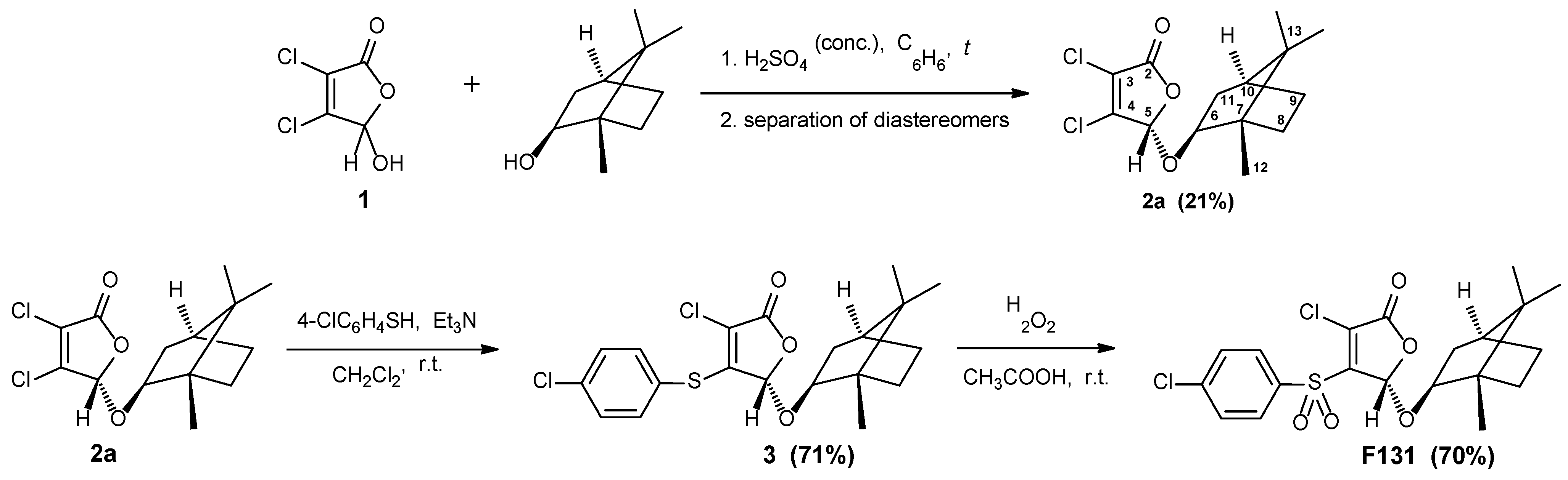
The 3,4-Dichloro-5-hydroxyfuran-2(5H)-one (mucochloric acid, 1) (Vekton, Russia) was commercially available and was further recrystallized from water, mp 127 °C. The (1S,2R,4S)-(–)-Borneol and 4-chlorothiophenol (Acros Organics) were used without further purification. All solvents were purified and distilled by standard procedures. IR spectra were recorded on a Bruker Tensor-27 spectrometer fitted with a Pike MIRacle ATR accessory (diamond/ZnSe crystal plate). IR spectra were recorded of solids with characteristic absorption wavenumbers (νmax) reported in cm−1. NMR spectra were measured on a Bruker Avance III 400 spectrometer at 400.17 MHz (1H) and 100.62 MHz (13C) at 20 °C in the deuterated chloroform. The chemical shifts (δ) are expressed in parts per million (ppm) and are calibrated using residual undeuterated solvent peak as the internal reference (CDCl3: δH 7.26, δC 77.16). All coupling constants (J) are reported in Hertz (Hz) and multiplicities are indicated as: s (singlet), m (multiplet). High-resolution mass spectra (HRMS) were obtained by electrospray ionisation (ESI) with positive (+) ion detection on a Bruker micrOTOF–III quadrupole time-of-flight mass spectrometer. Analytical thin layer chromatography (TLC) was carried out on Sorbfil PTLC-AF-A-UF plates using dichloromethane as the eluent and UV light as the visualizing agent. The melting points were measured on an OptiMelt Stanford Research Systems MPA100 automated melting point apparatus and were not corrected. Optical rotations were measured on a Perkin-Elmer model 341 polarimeter at λ 589 nm and at 20 °C in chloroform (concentration c is given as g/100 mL).

2.2. Synthesis

The 3,4-Dichloro-5(S)-[(1S,2R,4S)-1,7,7-trimethylbicyclo[2.2.1]heptan-2-yloxy]-2(5H)-furanone (2a) was synthesized according to the known method [52,60].
5(S)-3-Chloro-4-[(4-chlorophenyl)sulfanyl]-5-[(1S,2R,4S)-1,7,7-trimethylbicyclo[2.2.1]-heptan-2-yloxy]-2(5H)-furanone(3). To a solution of furanone 2a (0.40 g, 1.3 mmol) in dichloromethane (20 mL) with intense stirring was added dropwise a solution of 4-chlorothiophenol (0.19 g, 1.3 mmol) in dichloromethane (3 mL), and a solution of triethylamine (0.18 mL, 1.3 mmol) in dichloromethane (1 mL). The reaction mixture was stirred at room temperature for 2 h (monitored by TLC). After the completion of the reaction, the mixture was washed with water (100 mL), the organic layer was evaporated to dryness. The obtained solid residue was recrystallized from hexane to afford thioether 3. Colorless solid (0.38 g, 71%); mp 128 °C; Rf 0.61; [α] D 20 = –49.3 (CHCl3, c = 1.0). IR (ATR) νmax 2979, 2949, 2882 (C–H), 1776 (C=O), 1599, 1477, 1455 (C=Carom) cm−1. 1H NMR (CDCl3, 400 MHz) δ 7.50, 7.39 (4H, AA′BB′, 3JAB = 3JA′B′ = 8.2, 4JAA′ = 4JBB′ = 2.2, 5JAB′ = 5JA′B = 0.3 Hz, Harom), 5.73 (1H, s, H-5), 3.77–3.67 (1H, m, H-6), 2.24–2.12 (1H, m, H-11), 1.85–1.75 (1H, m, H-8 or H-9), 1.74–1.60 (2H, m, H-8 or H-9, H-10), 1.29–1.11 (3H, m, H-8, H-9, H-11), 0.82, 0.79 (6H, s, CH3 (iPr)), 0.63 (3H, s, H-12). 13C{1H} NMR (CDCl3, 100 MHz) δ 164.8 (C-2), 153.5 (C-4), 136.9, 135.9, 130.0, 124.9, 120.5 (C-3, Carom), 102.2 (C-5), 88.3 (C-6), 49.6 (C-13), 47.8 (C-7), 44.9 (C-10), 36.9 (C-11), 28.1, 26.6 (C-8, C-9), 19.7, 18.8 (CH3 (iPr)), 13.6 (C-12). HRMS (ESI) m/z 435.0566 (calcd for C20H22Cl2NaO3S, 435.0559).
5(S)-3-Chloro-4-[(4-chlorophenyl)sulfonyl]-5-[(1S,2R,4S)-1,7,7-trimethylbicyclo[2.2.1]-heptan-2-yloxy]-2(5H)-furanone (F131). Thioether 3 (0.29 g, 0.7 mmol) was dissolved in 10 mL of glacial acetic acid, 33% hydrogen peroxide (0.74 mL, 7.3 mmol) was added under stirring, and the mixture was stirred for 5 days at room temperature (monitored by TLC). When the reaction was complete, the mixture was evaporated to dryness, and the white solid residue was recrystallized from hexane to give sulfone F131. Colorless crystals (0.22 g, 70%); mp 142 °C; Rf 0.49; [α] D 20 = +158.6 (c 1.0, CHCl3). IR (ATR) νmax 2991, 2956, 2931, 2889 (C–H), 1786 (C=O), 1622 (C=Clactone), 1579, 1475 (C=Carom), 1344 (SO2 asym), 1163 (SO2 sym) cm–1. 1H NMR (CDCl3, 400 MHz) δ 8.03, 7.60 (4H, AA′BB′, N = 3JAB + 5JAB′ = 8.6 Hz, Harom), 6.23 (1H, s, H-5), 4.16–4.07 (1H, m, H-6), 2.33–2.22 (1H, m, H-11), 1.78–1.63 (3H, m, H-8, H-9, H-10), 1.36–1.23 (1H, m, H-8 or H-9), 1.23–1.05 (2H, m, H-8 or H-9, H-11), 1.04 (3H, s, H-12), 0.89, 0.88 (6H, s, CH3 (iPr)). 13C{1H} NMR (CDCl3, 100 MHz) δ 162.8 (C-2), 150.6 (C-4), 142.5, 137.0, 134.1, 130.3, 130.0 (C-3, Carom), 102.7 (C-5), 90.7 (C-6), 49.9 (C-13), 47.9 (C-7), 44.9 (C-10), 36.8 (C-11), 28.2, 26.7 (C-8, C-9), 19.7, 18.9 (CH3 (iPr)), 13.9 (C-12). HRMS (ESI) m/z 467.0449 (calcd for C20H22Cl2NaO5S, 467.0457).

2.3. Antimicrobials

Amikacin, gentamicin, benzalkonium chloride, terbinafine and fluconazole were purchased from Sigma, (St. Louis, MO, USA). Solutions were prepared at a concentration of 20 mg/mL in deionized water, or in dimethyl sulfoxide (DMSO) for fluconazole. Solutions of F131 were prepared at a concentration of 10 mg/mL in DMSO.

2.4. Strains and Growth Conditions

The antimicrobial activity of the compounds was assessed on methicillin-susceptible Staphylococcus aureus (MSSA) ATCC 29213 and 9 clinical isolates of S. aureus (3 MSSA and 6 MRSA) provided by the Pharmaceuticals Research Center of Kazan Federal University. Antimycotic activity was assessed on 12 clinical isolates of Candida albicans (6 fluconazole-resistant strains, and 6 fluconazole-susceptible strains) obtained from the Kazan Research Institute of Epidemiology and Microbiology. The bacterial strains were stored in 10% (v/v) glycerol stocks, fungal strains in 50% glycerol stock at −80 °C, and freshly streaked on Luria Agar (LA) plates following by their overnight growth at 37 °C before use. To obtain a mature biofilm, bacterial and fungal cells were grown in basal medium (BM broth) [54] in tissue-culture-treated (TC-treated) plates under static conditions for 48 h at 37 °C. A mannitol salt agar (peptone 10 g, meat extract 1 g, NaCl 75 g, D-mannitol 10 g, agar–agar 12 g in 1.0 L tap water, Oxoid) and Sabouraud’s agar (Difco) were used for differential count of S. aureus and C. albicans, respectively, from S. aureus–C. albicans mixed cultures.

2.5. Determination of the Minimal Inhibitory (MIC) and the Minimal Bactericidal/Fungicidal Concentrations (MBC/MFC) of F131 and Reference Antimicrobials

The minimum inhibitory concentration (MIC) was determined by serial 2-fold microdilution in 96-well plates according to the EUCAST rules for antimicrobial susceptibility testing [61] with some modifications. The maximal final concentration of each tested compound was 512 µg/mL. The next well contained two-fold decreasing concentration of the antimicrobial in the range of 0.5–512 μg/mL. The wells were seeded with microbial culture with a final density of 106 CFU/mL in a BM broth [54] as universal medium for all further experiments. The plates were incubated for 24 hours at 37 °C without shaking. The minimal inhibitory concentration of compounds was defined as the concentration that ensures complete suppression of the visible growth of the strains. The minimal bactericidal/fungicidal concentration (MBC/MFC) was determined by inoculation in 3 mL of nutrient broth with 3 μL of culture fluid from wells in which no visible growth was observed. MBC/MFC was considered as the minimum concentration of the compound, which ensures the complete absence of microorganisms growth.

2.6. Analysis of the Biofilm–Prevention Concentration (BPC)

To determine the minimal biofilm-prevention concentration, bacterial and fungal cells were grown in 96-well adhesive plastic plates for 48 hours without shaking at 37 °C in 200 μL BM broth with an initial density 3 × 10⁷ CFU/mL in the presence of the test compounds with concentration in the range of 0.5–512 μg/mL. Next, wells were washed with water and subjected to a staining with crystal violet as described in [62]. The minimal biofilm inhibitory concentration was defined as the lowest concentration at which the residual biofilm was two-fold less compared to untreated wells.

2.7. Assessment of Synergy between F131 and Conventional Antimicrobials

To assess a synergy between F131 and antimicrobials, a checkerboard assay was performed as described previously [41]. Each plate contained serial dilutions of a F131 and various antimicrobials in a checkerboard pattern. One of the antimicrobial substances [A] was diluted horizontally, and the F131 [B] vertically on a 96-well plate. The extreme lines and columns contained only one of the considered compounds to determine their MICs in each experiment. The initial concentration of each of the studied antimicrobial agents was 2 × MIC. The plates were incubated at 37 °C for 24 h. The experiments were performed in a triplicate, a growth control without the addition of any antimicrobial agent was included in each plate. The fractional inhibitory concentration index (FICI) for each double combination was calculated as follows:
FICI   =   MIC A   in   combination MIC   A + MIC B   in   combination MIC B
Interpretation of the obtained FICI values was carried out according to Odds [63]; FICI ≤ 0.5 corresponded to synergy, 0.5 < FICI ≤ 4 an additive effect, while FICI > 4 corresponded to antagonism.

2.8. Quantification of Viable Cells

To assess the viability of detached cells and cells in the biofilm in the presence of antimicrobials, a drop plate assay [64] with modifications was used. Briefly, a series of ten-fold dilutions of cell suspension from each well were prepared in 3 technical repeats and 5 μL from each dilution were dropped onto both mannitol salt agar and Sabouraud agar with ciprofloxacin (10 μg/mL) to differentiate S. aureus and C. albicans cells, respectively. CFUs were counted from the two last drops where 5–15 colonies were grown and averaged. In the case of the biofilm-embedded cells, the wells were pre-washed twice with sterile 0.9% NaCl and biofilms were suspended in 0.9% NaCl by scratching the well bottoms. Additionally, the wells were sonicated for 2 min to disintegrate the remained cell clumps [65]. The dilutions of obtained suspension were prepared and CFUs were counted as described above.

2.9. Confocal laser Scanning Microscopy (CLSM)

To assess the localization of F131 in bacterial and fungal cells, confocal laser scanning microscopy was performed using an Olympus IX83 inverted microscope supplemented with a STEDYCON ultrawide extension platform. Both monospecies and bacterial-fungal cultures of S. aureus and C. albicans were grown on cell imaging cover slips (Eppendorf) for 48 h in BM broth. The fluorescent derivative of 2(5H)-furanone F145 [53] was added to the cells at a final concentration of 20 μg/mL and biofilms were analyzed at blue (405/410–508 nm) channel.

2.10. RNA Isolation and Real-Time One-Step qRT-PCR

To analyze the transcription of the efflux system genes MDR1, CDR1, and CDR2, the PCR was performed using PCR amplifier "BioRad CFX96" (BioRad, Singapore) using Extra Mix for reverse transcription and quantitative real-time PCR in a one-step method (BioLabMix, Novosibirsk, Russia) with SYBR Blue under conditions recommended by the manufacturer. The oligonucleotides used for the qRT-PCR are shown in Table 1 [66,67,68]. A total RNA was extracted from planktonic C. albicans 688 cells subjected to 48-h treatment with either fluconazole or F131. Reaction mixture (50 µL) contained 1× RT-qPCR SYBR Blue buffer, 0.1 µM of each primers, 0.1 μM of each dNTPs, 2.5% DMSO, 5% RT-qPCR extra-mix and nuclease-free water (DEPC). The RT–PCR program included reverse transcription at 45 °C for 30 min, followed by 37 cycles of primer annealing–elongation–melting. Primer annealing temperatures were calculated using the Tm Calculator service (https://tmcalculator.neb.com, accessed on 6 January 2022). The elongation time was calculated based on the size of the synthesized fragment and the speed of the DNA polymerase. Real-time PCR monitoring was carried out at the stage of final synthesis at 72 °C. The 18s rRNA and GAPDH genes were used as references; the transcription level of qCDR1, qCDR1 and MDR1 genes was normalized by the 18s rRNA transcription level.

2.11. Data Analysis

All experiments were performed in three biological replicates with three technical replicates. The data was analyzed using GraphPad Prism version 6.0 for Windows (GraphPad Software, San Diego, CA, USA). The significance of difference has been checked by using the Kruskal–Wallis test. Significant differences from control were considered at p < 0.05.

3. Results

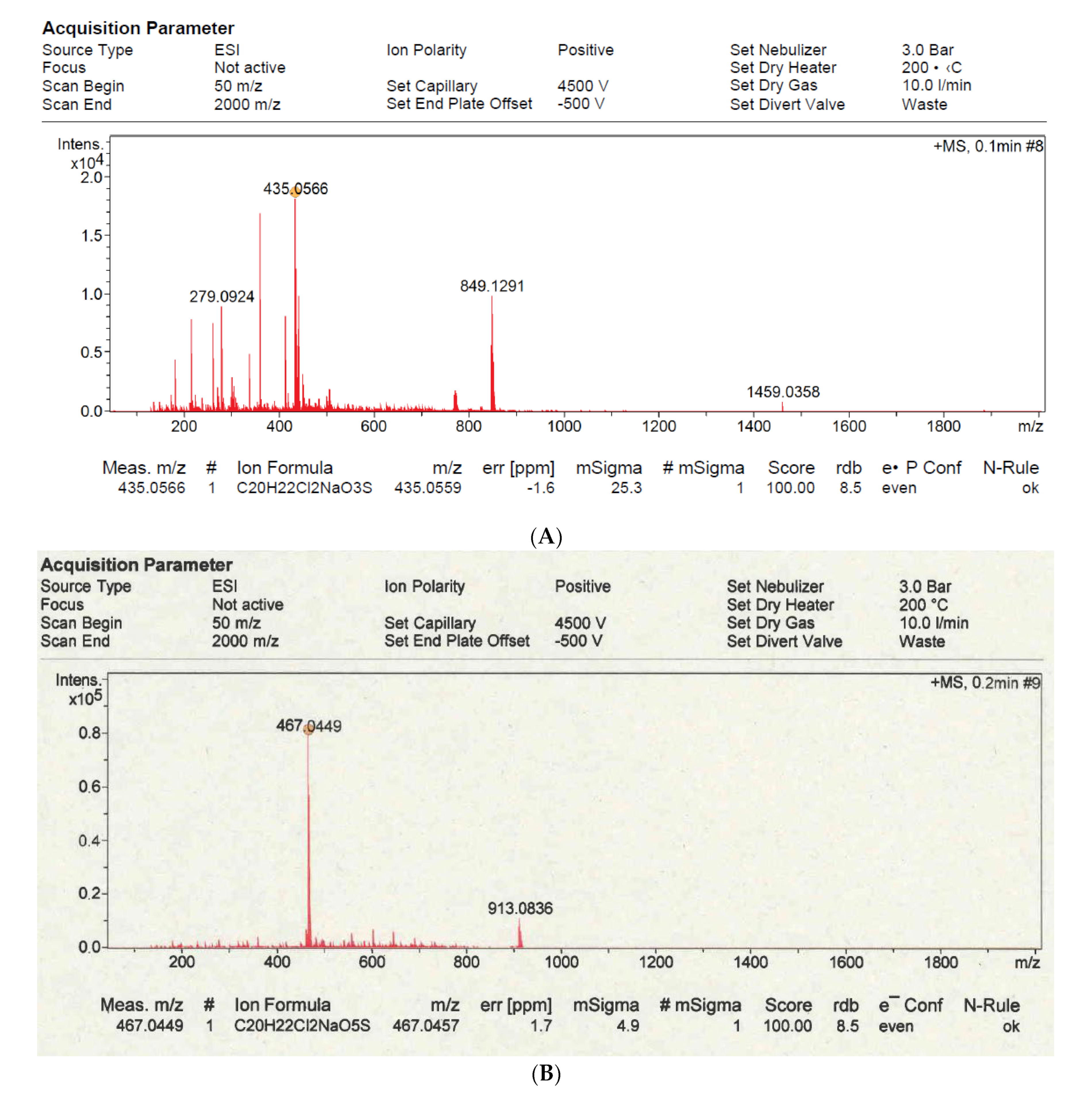
3.1. Synthesis of Furanone F131

Synthesis of novel 2(5H)-furanone derivative F131 possessing sulfonyl group and l-borneol moiety was achieved via three steps from commercially available mucochloric acid 1 (Figure 1). Following a previously published procedure, mucochloric acid 1 was first converted to the corresponding 5-(l-bornyloxy) derivative, obtained as a mixture of two diastereomers 2a + 2b [52,60]. The pure stereoisomer 2a with S-configuration of the carbon atom C5 of the γ-lactone cycle was isolated after two recrystallizations from hexane.
Next, regioselective introduction of 4-chlorothiophenol fragment into the molecule of the isolated stereoisomer 2a was carried out. It is well-known that interaction of 3,4-dihaloderivatives of 2(5H)-furanone with different thiols in the presence of basic compounds goes with the substitution of halogen atom in the fourth position of the lactone cycle [69,70]. The thiilation reaction was performed in dichloromethane at room temperature using the equimolar ratio of furanone 2a, thiol and triethylamine. Chiral thioether 3 was obtained with 71% yield and subjected to a further oxidation reaction. We used a simple and convenient method of the synthesis of furanone sulfonyl derivatives, based on the treatment of thioethers with excess of hydrogen peroxide in acetic acid at room temperature [70,71]. Thus, the desired novel sulfone F131 in a stereomerically pure form was isolated with 70% yield as colorless crystals (Figure 1). The structure of borneol-containing furanones 3 and F131 has been characterized by IR and NMR spectroscopy (Figure A1, Figure A2, Figure A3 and Figure A4).

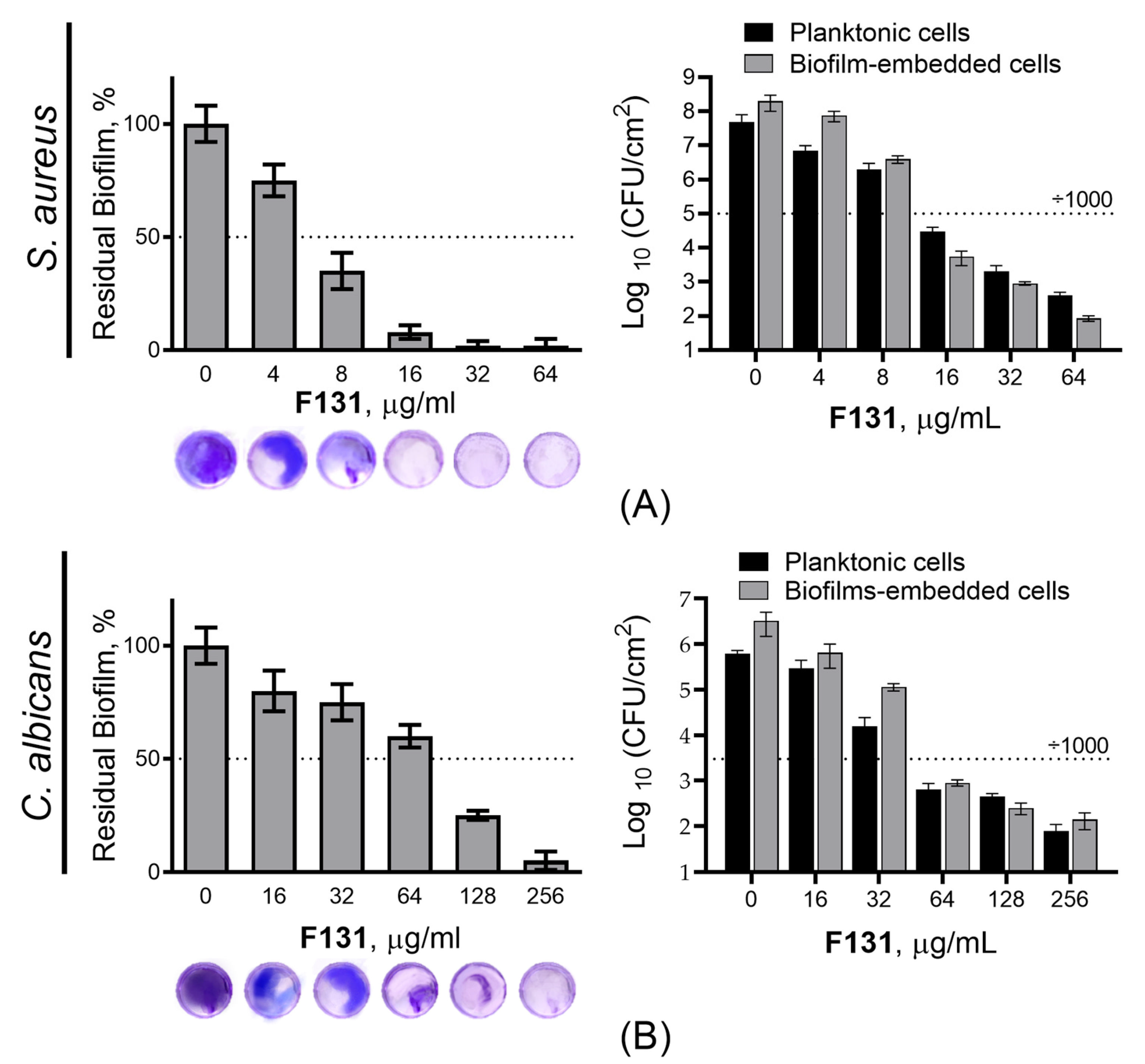
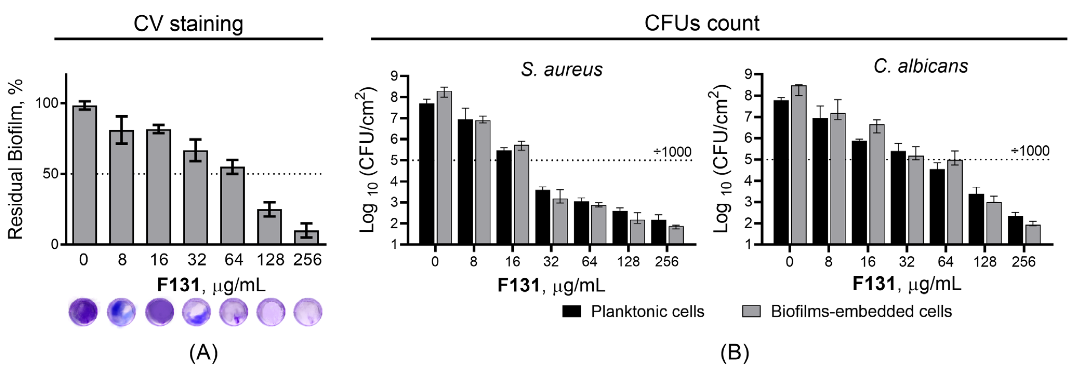
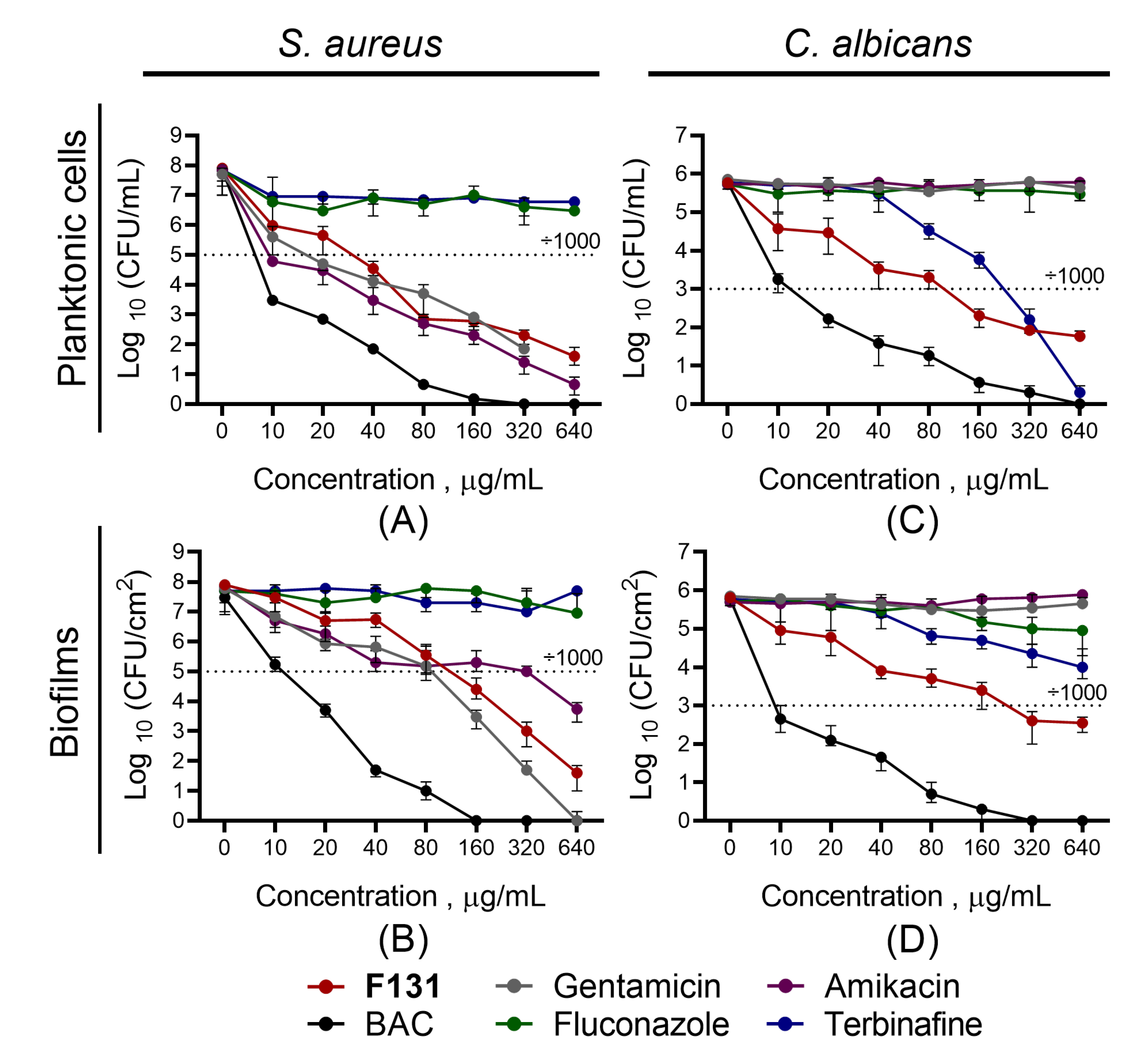
3.2. Antimicrobial and Biofilm Preventing Activity of F131

The antimicrobial and antifungal properties of F131 were evaluated using a range of S. aureus clinical isolates (n = 9) and methicillin-sensitive S. aureus ATCC 29213 (Table 2), as well as twelve C. albicans clinical isolates with different susceptibility to fluconazole (Table 3). All clinical isolates of S. aureus were resistant to gentamicin. MRSA strains were also resistant to amikacin, while MSSA strains remained sensitive to the antibiotic. Six of the twelve clinical isolates of C. albicans were resistant to fluconazole. MIC of F131 ranged within 8–16 µg/mL for S. aureus isolates, and 32–128 µg/mL for C. albicans isolates (Table 2 and Table 3) suggesting that susceptibility of isolates to F131 does not correlate with resistance to conventional antimicrobials. Of note, the l-borneol itself was ineffective against both S. aureus and C. albicans (MIC > 1024 for all isolates). The minimal bactericidal concentration (MBC) and the minimal fungicidal concentration (MFC) values of F131 ranged within 32–128 µg/mL and 128–1024 µg/mL respectively (Table 2 and Table 3). These data indicate a considerable bactericidal and fungicidal activity of F131, as the MIC/MBC ratio lies within 2–4, with a few exceptions.
Further, since the biofilm-inhibiting properties for many 2(5H)-furanone derivatives have been reported, the ability of F131 to repress the biofilm formation by S. aureus and C. albicans was assessed. For that S. aureus and C. albicans were grown for 48h under static conditions in the presence of F131. Then, the biofilm was quantified by both crystal violet staining and direct count of viable cells. The compound led to two-fold reduction of the S. aureus biofilm biomass at the concentration of 8 µg/mL, whereas the three-log decrease of viable cells in the biofilm was observed at 16 µg/mL (Figure 2A). For fluconazole-resistant C. albicans 688, the biofilm biomass was reduced twice at 128 µg/mL, and 64 µg/mL led to a 1000-fold decrease of viable cell in the biofilm (Figure 2B). Of note, the dose-dependent dynamics of viable cells decrease was similar for both planktonic and adherent (biofilm-forming) ones, suggesting that the observed biofilm prevention activity is driven, apparently, rather by the reduction of viable cells than the true inhibition of the biofilm formation. This hypothesis is also supported by fact; that the conventional antimicrobials such as gentamicin, amikacin, fluconazole, terbinafine and benzalkonium chloride also reduced the amount of viable cells in the biofilm at their respective two-fold MICs (Figure 3 and Figure 4).

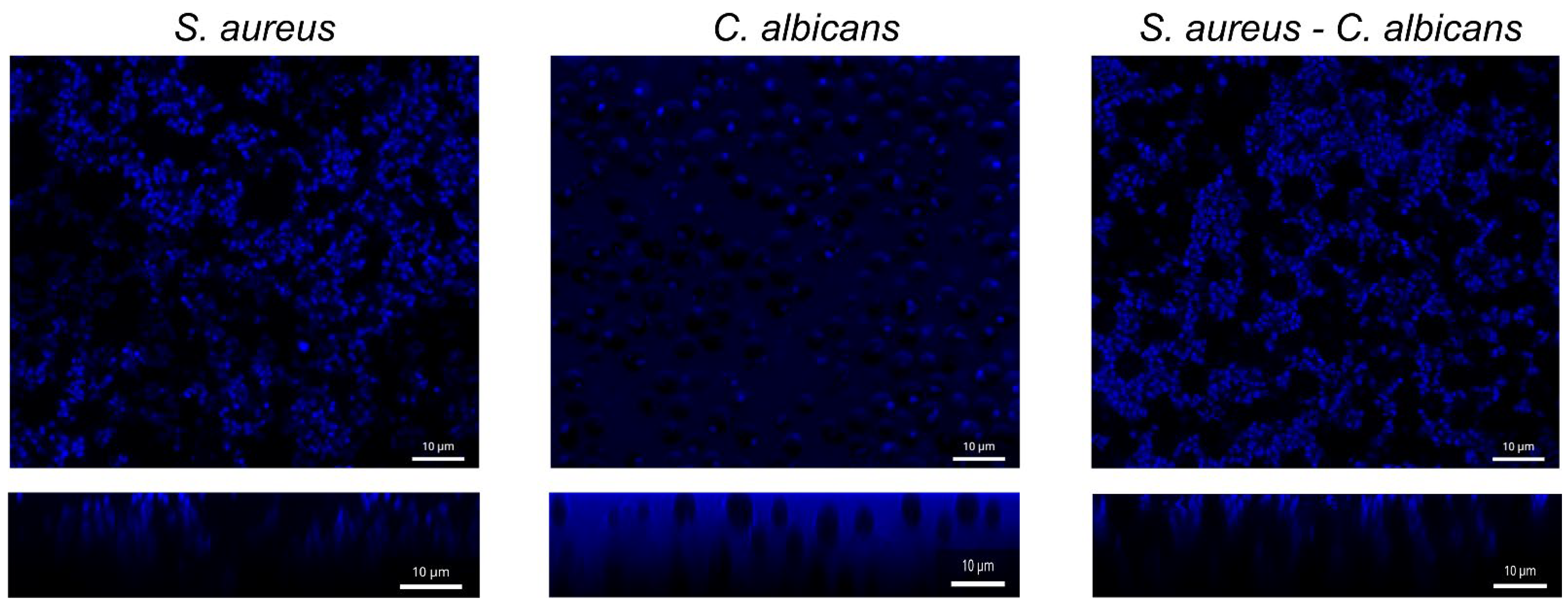
3.3. Antibiofilm Activity of F131 on S. aureus and C. albicans Mixed Biofilms

S. aureus ATCC 29213 and C. albicans 688 fluconazole-resistant isolates were chosen to apply tests on mixed bacterial–fungal biofilms. The S. aureusC. albicans mixed culture was grown under static conditions in BM broth in the presence of F131 in concentrations varying from 2 to 256 µg/mL. After 48h, the biofilms were quantified by the crystal violet staining and CFUs count by plating on mannitol salt agar, and Sabouraud agar with ciprofloxacin (10 μg/mL) to differentiate S. aureus and C. albicans cells, respectively. The F131 at 128 μg/mL completely inhibited the biofilms formation in mixed cultures (Figure 5a). The growth of S. aureus and C. albicans in mixed culture was repressed at 16 µg/mL and 64 µg/mL, respectively (Figure 5b), similarly to monocultures.
To further analyze whether F131 not only prevents biofilm formation but also eradicates already established ones, different concentrations (5–320 μg/mL) of F131 or reference antimicrobials were added to the established 24 h old mixed biofilms. After 24 h, biofilms and detached cells clumps in culture liquid were quantified by differential CFUs counting. For the detached S. aureus cell clumps, the efficiency of F131 was comparable with those of both aminoglycosides. Further, both F131 and aminoglycosides reduced the CFUs number of S. aureus in biofilm by three orders of magnitude at a concentration of 80 μg/mL (2.5–5 × MBC), whereas 10 μg/mL (5 × MBC) of benzalkonium chloride was required to achieve the same effect (Figure 6B). On C. albicans, the efficiency of F131 was comparable with those of terbinafine. Of note, neither terbinafine nor F131 led to three-order reduction in the CFUs count even at concentrations up to 640 μg/mL (Figure 6D).
These findings raised questions about the penetration possibility of furanone derivatives into the biofilm matrix formed by S. aureus–C. albicans. To confirm these suggestions, the fluorescent analogue of 2(5H)-furanone named F145, possessing the fluorescent moiety instead of l-borneol and described earlier [72], was added to established 24-hours-old mono- and mixed-biofilms of S. aureus–C. albicans, and after 20 min, incubation samples were analyzed by confocal laser scanning microscopy. The fluorescence of F145 was observed throughout the whole biofilm (Figure 7), confirming its fast penetration through the S. aureusC. albicans biofilm matrix.

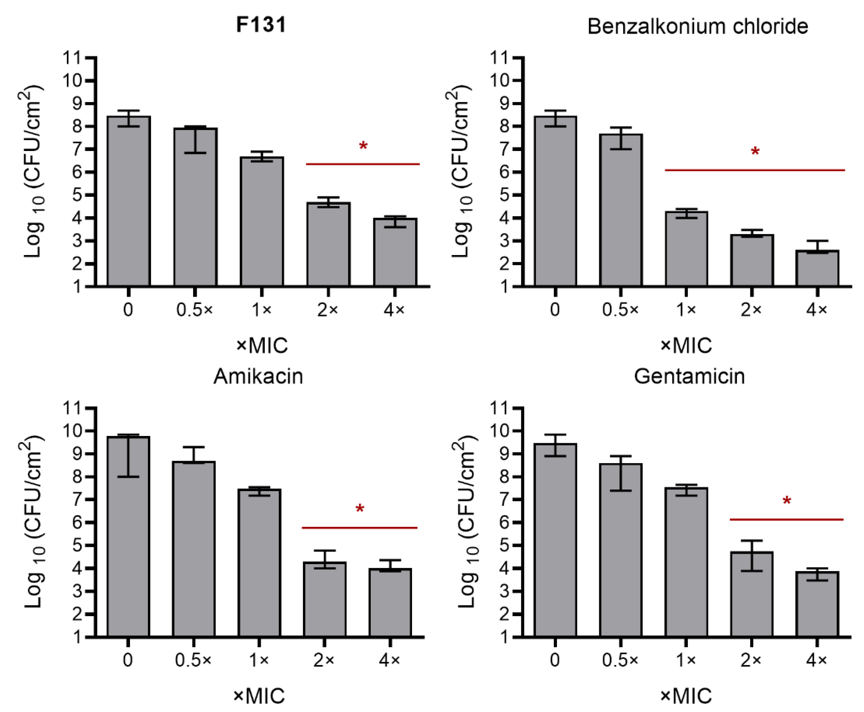
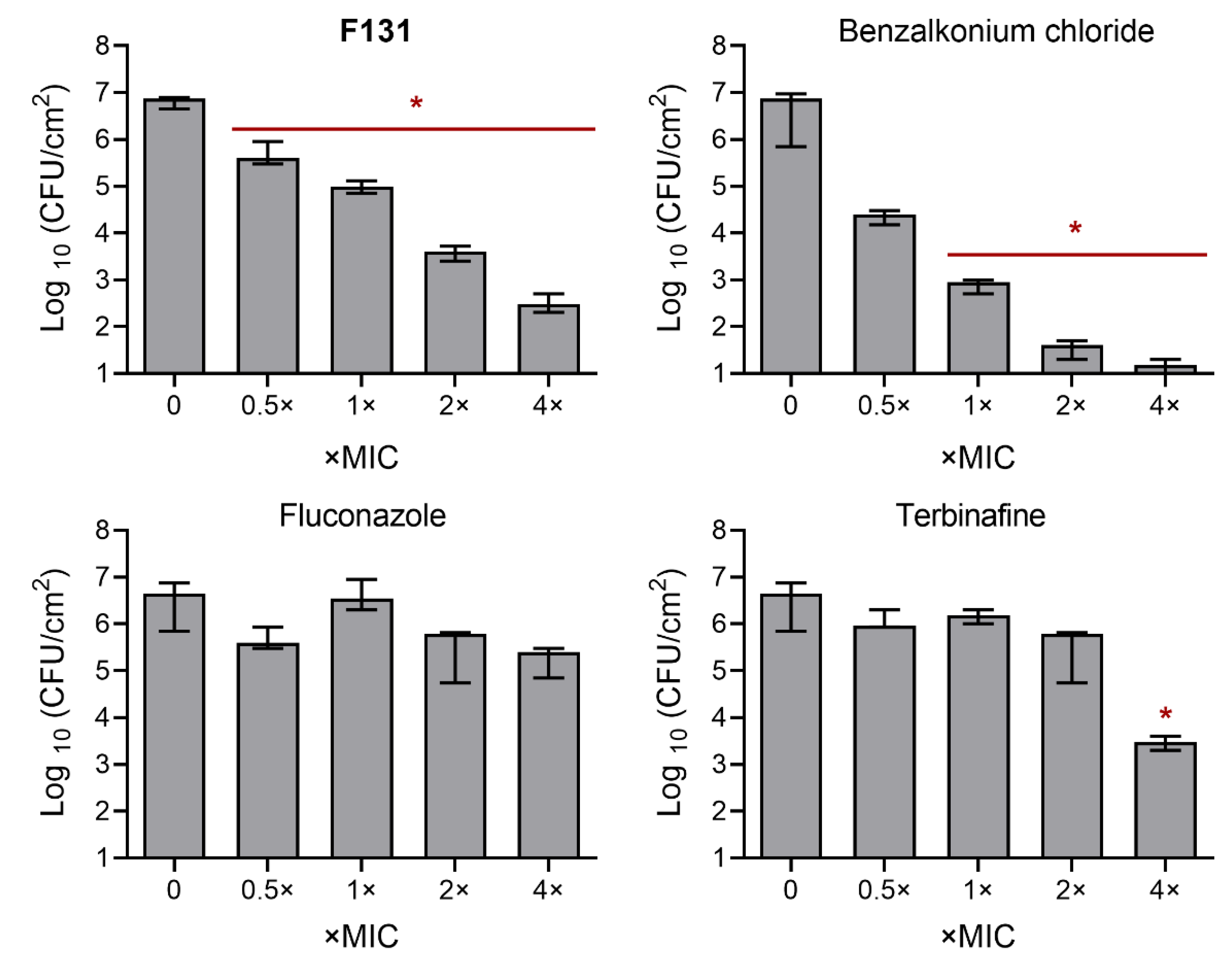
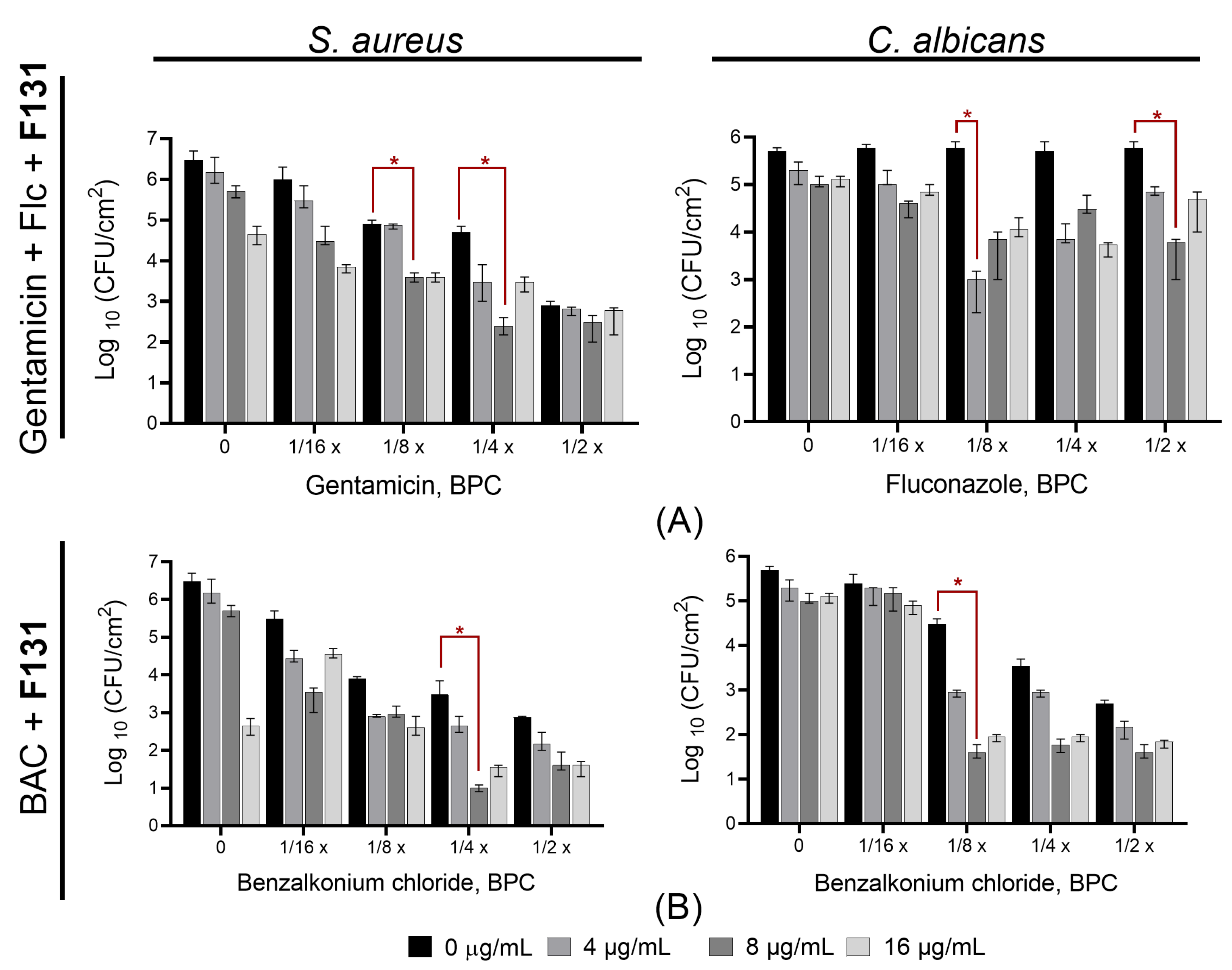
3.4. Synergistic Effects of F131 with Other Antimicrobials on S. aureus–C. albicans Mixed Biofilms

Previously, it has been shown that the 2(5H)-furanone derivative F105 containing l-menthol and sulfonyl moieties, exhibits a synergy with aminoglycosides and benzalkonium chloride against S. aureus [41] and potentiates the antifungal activity of fluconazole and terbinafine against C. albicans cells [53]. Taking into account the similar structures of compounds F105 and F131, which are both chiral furanone sulfones, possessing a fragment of terpene alcohol in the fifth position of the lactone cycle, we assumed similar properties for the latter.
Therefore, the synergism of F131 in combination with conventional antimicrobials was analyzed by the checkerboard assay on S. aureus ATCC 29213 and C. albicans 688 fluconazole-resistant isolate. The values of the fractional inhibitory concentrations (FICI) were determined in checkerboard assay and the median was calculated. For bacterial and fungal mono-species cultures, F131 demonstrated marked synergism with gentamicin with FICI of 0.1875, and fluconazole with FICI of 0.375. Additionally, pronounced synergy was observed also for benzalkonium chloride for S. aureus (FICI = 0.25) and C. albicans (FICI = 0.1875) (Table 4 and Table 5). Thus, the presence of F131 significantly lowered the MIC of gentamicin by 16-fold, and the MIC of benzalkonium chloride by eight-fold. Of note, the presence of F131 altered fluconazole susceptibility in fluconazole-resistant isolate (C. albicans 688), as it became more sensitive to fluconazole with FIC value of 128 (Table 5). In turn, FIC values of F131 decreased up to eight-fold when combining with gentamicin, fluconazole, and benzalkonium chloride.
Further, we questioned whether the synergistic effect of 2(5H)-furanone derivatives could be used to eradicate mixed S. aureus–C. albicans infection. Therefore, we tested the effect of combination of F131 with conventional antimicrobials on mixed S. aureus–C. albicans biofilms formation and eradication, since the biofilm-embedded consortium became extremely resistant to treatment compared to planktonic cells. Firstly, the S. aureus–C. albicans mixed cultures were inoculated in BM broth in the presence of various concentrations of antimicrobials as indicated. F131 concentrations were 4, 8, and 16 μg/mL, which correspond to ¼–1.0 BPCs for S. aureus and 1/16–1/4 BPCs for C. albicans. In bacterial–fungal mixed biofilms, a considerable synergism between F131 and gentamicin–fluconazole mix was detected at 1/4 BPC of gentamicin and 1/8 BPC of fluconazole against S. aureus–C. albicans, as judged from the observed three-order reduction of the viable cell number per cm2 for the untreated control (Figure 8A). Moreover, the combination of F131 with sub-BPCs of benzalkonium chloride (1/4 BPC for S. aureus and 1/8 BPC for C. albicans) decreased the viable cell number in mixed biofilms by three orders of magnitude, indicating a strong synergistic effect (Figure 8B).
For convenience, we plotted a graph representing the antifungal and antibacterial effect of fluconazole–gentamicin mix and benzalkonium chloride alone and in combination with fixed concentration of F131 (8 µg/mL) against S. aureusC. albicans mixed biofilms (Figure 9). As could be seen from the Figure 9, in the presence of 8 µg/mL F131, the concentrations of fluconazole, gentamicin and benzalkonium chloride, required to decreases CFUs number by three orders of magnitude are four-fold lower when compared with solely antimicrobials. Taken together, these data clearly indicate that F131 at a concentration of 8 µg/mL (1/2 BPC for S. aureus, and 1/8 BPC for C. albicans) is able to significantly reduce the number of viable cells when combining with sub-BPCs of conventional antifungals and antibacterial drugs.

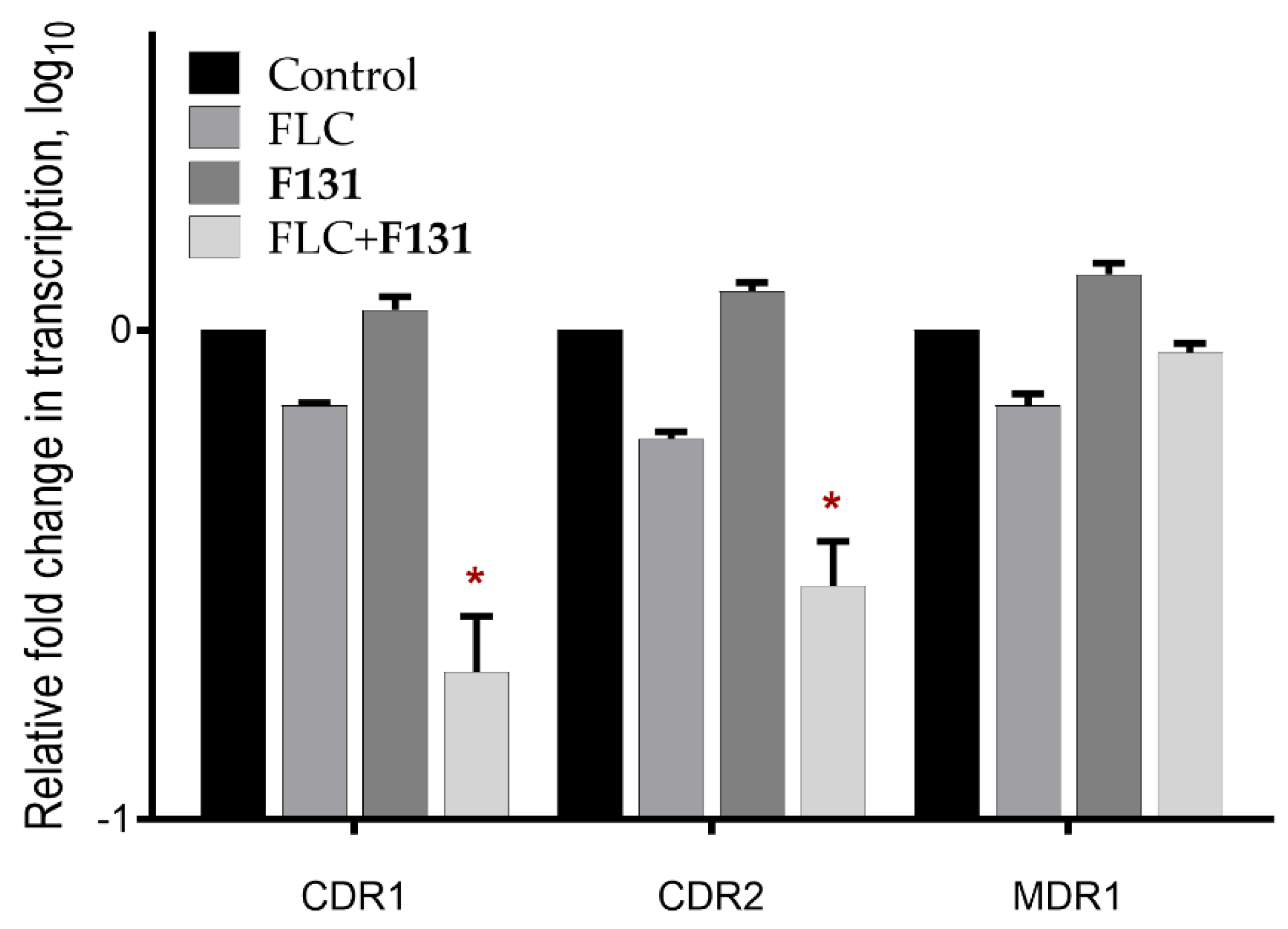
3.5. The Combination of F131 with Fluconazole Significantly Decreased the Expression Level of CDR1 and CDR2 Genes

The mRNA levels of the efflux system genes MDR1, CDR1, and CDR2 were analyzed by quantitative real-time RT-PCR in C. albicans 688 isolate (resistant isolate). Cells were grown in the presence of 32 µg/mL fluconazole and/or 8 µg/mL F131 for 24 hours. The same volume of solvents (DMSO and ethanol) was added to the untreated control group of cells. The results showed that F131 solely did not affect the expression of the mentioned genes, since the coefficient of relative expression of MDR1, CDR1 and CDR2 were 1.12, 1.29 and 1.31, respectively. When treating with fluconazole, the expression level was suppressed of the MDR1, CDR1 and CDR2 genes, although underestimated (relative expression 0.71, 0.62 and 0.69, respectively). It is noteworthy that the expression of CDR1 and CDR2 genes decreased significantly after the combined treatment with fluconazole and F131 (relative expression 0.22 and 0.31, respectively) (Figure 10).

4. Discussion

Taking into account the high mortality and morbidity rates associated with S. aureusC. albicans mixed infections, there is an always pursuit to find new potential alternatives that ensure better efficiency and lower toxicity during the management of candida mixed infections, especially in the course of strains that are resistant to fluconazole. Here, we show that the novel 2(5H)-furanone derivative F131, consisting of two pharmacophores (2(5H)-furanone cycle and l-borneol moiety), exhibits an antimicrobial and biofilm-preventing activity as well as potentiates conventional antibiotics and antifungal agents against S. aureus and C. albicans and their mixed culture. Earlier, we showed that the structural analogue of F131 with l-menthol fragment (3-chloro-5(S)-[(1R,2S,5R)-2-isopropyl-5-methylcyclohexyloxy]-4-[4-methylphenylsulfonyl]-2(5H)-furanone F105) exhibited comparable antibacterial and antifungal activity against S. aureus [41] and C. albicans [53]. F131 possesses the l-borneol instead of l-menthol fragment and demonstrates higher antimicrobial activity. Interestingly, many previous studies reported the anti-inflammatory and antimicrobial activity of borneol derivatives [73,74,75]. Some aromatic borneol compounds were significantly active with MIC50 = 125 µg/mL for both S. aureus and C. albicans [76]. In our previous study we examined the relationship between the chemotype and antimicrobial activity of 2(5H) derivatives. We found that the l-borneol moiety is an essential part of the molecule of 3,4-dichloro-5(S)-[(1S,2R,4S)-1,7,7-trimethylbicyclo[2.2.1]heptan-2-yloxy]-2(5H)-furanone (F123), with respect to demonstrating its antibacterial activity against gram-positive bacterium B. cereus [52]. This data raised a question whether the borneol fragment is responsible for the observed antimicrobial activity of F131. For that, we tested the activity of terpene alcohol l-borneol against both S. aureus and C. albicans in mono and mixed cultures. However, our data showed that l-borneol itself is not able to inhibit the growth of either S. aureus or C. albicans, suggesting that this terpene alcohol apparently facilitates the penetration of the multipharmacophore drug into both the cells, probably thereby also providing the synergy with other antimicrobials (Table 2 and Table 3), and into biofilms (Figure 7) thus exhibiting biofilm-eradicating activity (Figure 6).
Our data show that F131 demonstrates bactericidal and fungicidal activity, as the MIC/MBC ratio lies within 2–4, with a few exceptions (Table 2 and Table 3). Moreover, the susceptibility of isolates to F131 does not correlate with resistance to conventional antimicrobials, making this compound suitable to the treatment of infections caused by resistant strains, including mixed C. albicansS. aureus infections. In addition, F131 was able to prevent the biofilm formation by both pathogens, either separately or in the mixed culture (Figure 2 and Figure 5). Generally, BPCs of F131 were found to approximately correspond to MICs, suggesting indirect biofilm-preventing activity of F131, apparently because of the reduction of viable cells rather than the true inhibition of the biofilm formation. Additionally, this assumption is supported by noting that the amount of viable planktonic cells also decreased by three orders of magnitude at the corresponding BPCs of F131 against both S. aureus and C. albicans (Figure 2). Thus, it could be suggested that the biofilm suppression was rather the consequence of cell growth repression. Interestingly, for C. albicans the BPCs were a little higher than MICs. This phenomenon might be attributed to the induction of biofilm formation by the fungi under sub-lethal concentrations of furanone derivatives [53]. Having a closer look at the F131 chemotype, F131 is a halogenated furanone containing a chlorine atom in the third position of a five-membered cycle, but our findings are not in agreement with previous studies that reported a direct biofilm-inhibiting activity of some halogenated furanones against gram-positive bacteria such as Bacilli and Staphylococci [48,54].

5. Conclusions

Taken together, our in vitro data allows the assumption of the F131 chemotype as a biofilm-preventing agent, especially in the course of skin infections caused by CandidaStaphylococcus mixed biofilms. In addition, due to its synergistic effect with conventional antimicrobials, the combination of the latter with F131 or F131-like compounds would give rise to the minimization of the required therapeutic doses and thus reduction of side effects, toxicity and tolerance development by pathogens. Nevertheless, further in vitro assays for the biosafety of F131 and in vivo studies are required to understand further perspectives to managing CandidaStaphylococcus mixed infections.

Author Contributions

Conceptualization, A.K. (Airat Kayumov) and E.T.; methodology, E.T. and A.K. (Alsu Khabibrakhmanova); software, M.B.; validation, M.B., A.K. (Almira Kurbangalieva) and A.K. (Airat Kayumov); formal analysis, A.K. (Almira Kurbangalieva) and A.K. (Airat Kayumov); investigation, R.S., E.T., A.K. (Alena Kolesnikova) and A.K. (Alsu Khabibrakhmanova); resources, A.K. (Almira Kurbangalieva) and A.K. (Airat Kayumov); data curation, M.B.; writing—original draft preparation, R.S., A.K. (Almira Kurbangalieva) and A.K. (Airat Kayumov); writing—review and editing, E.T., M.B., A.K. (Almira Kurbangalieva) and A.K. (Airat Kayumov); visualization, R.S., E.T., A.K. (Alsu Khabibrakhmanova) and A.K. (Alena Kolesnikova); supervision, A.K. (Almira Kurbangalieva) and A.K. (Airat Kayumov); project administration, A.K. (Airat Kayumov); funding acquisition, E.T. and A.K. (Almira Kurbangalieva). All authors have read and agreed to the published version of the manuscript.

Funding

This work was funded by the subsidy allocated to Kazan Federal University for the state assignment in the sphere of scientific activities, project №FZSM-2022-0017. The synthetic part of this work was funded by the subsidy allocated to Kazan Federal University for the state assignment in the sphere of scientific activities (no. 0671-2020-0063).

Institutional Review Board Statement

Not applicable.

Informed Consent Statement

Not applicable.

Data Availability Statement

All data are included in the manuscript.

Conflicts of Interest

The authors declare no conflict of interest.

Appendix A

Figure A1. 1H (A) and 13C{1H} (B) NMR spectra of compound 3 (CDCl3).
Pathogens 12 00026 g0a1
Figure A2. 1H (A) and 13C{1H} (B) NMR spectra of compound F131 (CDCl3).
Pathogens 12 00026 g0a2
Figure A3. IR spectra of thioether 3 (A) and sulfone F131 (B).
Pathogens 12 00026 g0a3
Figure A4. Mass spectra of thioether 3 (A) and sulfone F131 (B).
Pathogens 12 00026 g0a4

References

  1. Muthukrishnan, G.; Masters, E.A.; Daiss, J.L.; Schwarz, E.M. Mechanisms of Immune Evasion and Bone Tissue Colonization That Make Staphylococcus aureus the Primary Pathogen in Osteomyelitis. Curr. Osteoporos. Rep. 2019, 17, 395–404. [Google Scholar] [CrossRef] [PubMed]
  2. Budzyńska, A.; Skowron, K.; Kaczmarek, A.; Wietlicka-Piszcz, M.; Gospodarek-Komkowska, E. Virulence Factor Genes and Antimicrobial Susceptibility of Staphylococcus aureus Strains Isolated from Blood and Chronic Wounds. Toxins 2021, 13, 491. [Google Scholar] [CrossRef] [PubMed]
  3. Pichereau, S.; Rose, W.E. Invasive community-associated MRSA infections: Epidemiology and antimicrobial management. Expert Opin. Pharmacother. 2010, 11, 3009–3025. [Google Scholar] [CrossRef] [PubMed]
  4. Sharma, N.K.; Garg, R.; Baliga, S.; Bhat, K.G. Nosocomial Infections and Drug Susceptibility Patterns in Methicillin Sensitive and Methicillin Resistant Staphylococcus aureus. J. Clin. Diagn. Res. 2013, 7, 2178–2180. [Google Scholar] [CrossRef]
  5. Turner, N.A.; Sharma-Kuinkel, B.K.; Maskarinec, S.A.; Eichenberger, E.M.; Shah, P.P.; Carugati, M.; Holland, T.L.; Fowler, V.G., Jr. Methicillin-resistant Staphylococcus aureus: An overview of basic and clinical research. Nat. Rev. Genet. 2019, 17, 203–218. [Google Scholar] [CrossRef]
  6. Cheung, G.Y.C.; Bae, J.S.; Otto, M. Pathogenicity and virulence of Staphylococcus aureus. Virulence 2021, 12, 547–569. [Google Scholar] [CrossRef]
  7. Miao, J.; Chen, L.; Wang, J.; Wang, W.; Chen, D.; Li, L.; Li, B.; Deng, Y.; Xu, Z. Current methodologies on genotyping for nosocomial pathogen methicillin-resistant Staphylococcus aureus (MRSA). Microb. Pathog. 2017, 107, 17–28. [Google Scholar] [CrossRef]
  8. Khan, A.; Wilson, B.; Gould, I.M. Current and future treatment options for community-associated MRSA infection. Expert Opin. Pharmacother. 2018, 19, 457–470. [Google Scholar] [CrossRef]
  9. Nataraj, B.H.; Mallappa, R.H. Antibiotic Resistance Crisis: An Update on Antagonistic Interactions between Probiotics and Methicillin-Resistant Staphylococcus aureus (MRSA). Curr. Microbiol. 2021, 78, 2194–2211. [Google Scholar] [CrossRef]
  10. Archer, N.K.; Mazaitis, M.J.; Costerton, J.W.; Leid, J.G.; Powers, M.E.; Shirtliff, M.E. Staphylococcus aureus biofilms: Properties, Regulation, and Roles in Human Disease. Virulence 2011, 2, 445–459. [Google Scholar] [CrossRef]
  11. Kaplan, J.B.; Mlynek, K.D.; Hettiarachchi, H.; Alamneh, Y.A.; Biggemann, L.; Zurawski, D.V.; Black, C.C.; Bane, C.E.; Kim, R.K.; Granick, M.S. Extracellular polymeric substance (EPS)-degrading enzymes reduce staphylococcal surface attachment and biocide resistance on pig skin in vivo. PLoS ONE 2018, 13, e0205526. [Google Scholar] [CrossRef]
  12. Lister, J.L.; Horswill, A.R. Staphylococcus aureus biofilms: Recent developments in biofilm dispersal. Front. Cell. Infect. Microbiol. 2014, 4, 178. [Google Scholar] [CrossRef] [Green Version]
  13. Schilcher, K.; Horswill, A.R. Staphylococcal Biofilm Development: Structure, Regulation, and Treatment Strategies. Microbiol. Mol. Biol. Rev. 2020, 84, e00026-19. [Google Scholar] [CrossRef]
  14. Idrees, M.; Sawant, S.; Karodia, N.; Rahman, A. Staphylococcus aureus Biofilm: Morphology, Genetics, Pathogenesis and Treatment Strategies. Int. J. Environ. Res. Public Health 2021, 18, 7602. [Google Scholar] [CrossRef]
  15. Kranjec, C.; Angeles, D.M.; Mårli, M.T.; Fernández, L.; García, P.; Kjos, M.; Diep, D. Staphylococcal Biofilms: Challenges and Novel Therapeutic Perspectives. Antibiotics 2021, 10, 131. [Google Scholar] [CrossRef]
  16. Cassat, J.E.; Thomsen, I. Staphylococcus aureus infections in children. Curr. Opin. Infect. Dis. 2021, 34, 510–518. [Google Scholar] [CrossRef]
  17. Liu, T.; Chen, Y.; Bi, G.; Luo, J.; Du, Z.; Kong, J.; Chen, Y. Generation of Methicillin-Resistant Staphylococcus Aureus Biofilm Infection in an Immunosuppressed Rat Model. J. Pharmacol. Exp. Ther. 2017, 23, 5803–5811. [Google Scholar] [CrossRef] [Green Version]
  18. Arciola, C.R.; Campoccia, D.; Montanaro, L. Implant infections: Adhesion, biofilm formation and immune evasion. Nat. Rev. Microbiol. 2018, 16, 397–409. [Google Scholar] [CrossRef]
  19. Balasubramanian, D.; Harper, L.; Shopsin, B.; Torres, V.J. Staphylococcus aureus pathogenesis in diverse host environments. Pathog. Dis. 2017, 75, ftx005. [Google Scholar] [CrossRef] [Green Version]
  20. Carolus, H.; Van Dyck, K.; Van Dijck, P. Candida albicans and Staphylococcus Species: A Threatening Twosome. Front. Microbiol. 2019, 10, 2162. [Google Scholar] [CrossRef]
  21. Vila, T.; Kong, E.F.; Montelongo-Jauregui, D.; Van Dijck, P.; Shetty, A.C.; McCracken, C.; Bruno, V.M.; Jabra-Rizk, M.A. Therapeutic implications of C. albicans-S. aureus mixed biofilm in a murine subcutaneous catheter model of polymicrobial infection. Virulence 2021, 12, 835–851. [Google Scholar] [CrossRef] [PubMed]
  22. Fox, E.P.; Cowley, E.S.; Nobile, C.J.; Hartooni, N.; Newman, D.K.; Johnson, A.D. Anaerobic Bacteria Grow within Candida albicans Biofilms and Induce Biofilm Formation in Suspension Cultures. Curr. Biol. 2014, 24, 2411–2416. [Google Scholar] [CrossRef] [PubMed] [Green Version]
  23. Rodrigues, M.E.; Gomes, F.; Rodrigues, C.F. Candida spp./Bacteria Mixed Biofilms. J. Fungi 2019, 6, 5. [Google Scholar] [CrossRef] [PubMed] [Green Version]
  24. Mayer, F.L.; Wilson, D.; Hube, B. Candida albicanspathogenicity mechanisms. Virulence 2013, 4, 119–128. [Google Scholar] [CrossRef] [PubMed] [Green Version]
  25. Suchodolski, J.; Krasowska, A. Fructose Induces Fluconazole Resistance in Candida albicans through Activation of Mdr1 and Cdr1 Transporters. Int. J. Mol. Sci. 2021, 22, 2127. [Google Scholar] [CrossRef]
  26. Cavalheiro, M.; Pais, P.; Galocha, M.; Teixeira, M.C. Host-Pathogen Interactions Mediated by MDR Transporters in Fungi: As Pleiotropic as it Gets! Genes 2018, 9, 332. [Google Scholar] [CrossRef] [Green Version]
  27. Nami, S.; Mohammadi, R.; Vakili, M.; Khezripour, K.; Mirzaei, H.; Morovati, H. Fungal vaccines, mechanism of actions and immunology: A comprehensive review. Biomed. Pharmacother. 2018, 109, 333–344. [Google Scholar] [CrossRef]
  28. Achkar, J.M.; Fries, B.C. Candida Infections of the Genitourinary Tract. Clin. Microbiol. Rev. 2010, 23, 253–273. [Google Scholar] [CrossRef] [Green Version]
  29. Ganguly, S.; Mitchell, A.P. Mucosal biofilms of Candida albicans. Curr. Opin. Microbiol. 2011, 14, 380–385. [Google Scholar] [CrossRef] [Green Version]
  30. Nobile, C.J.; Johnson, A.D. Candida albicans Biofilms and Human Disease. Annu. Rev. Microbiol. 2015, 69, 71–92. [Google Scholar] [CrossRef]
  31. D’Enfert, C.; Kaune, A.-K.; Alaban, L.-R.; Chakraborty, S.; Cole, N.; Delavy, M.; Kosmala, D.; Marsaux, B.; Fróis-Martins, R.; Morelli, M.; et al. The impact of the Fungus-Host-Microbiota interplay upon Candida albicans infections: Current knowledge and new perspectives. FEMS Microbiol. Rev. 2020, 45, fuaa060. [Google Scholar] [CrossRef]
  32. Lopes, J.P.; Lionakis, M.S. Pathogenesis and virulence of Candida albicans. Virulence 2021, 13, 89–121. [Google Scholar] [CrossRef]
  33. Tumbarello, M.; Fiori, B.; Trecarichi, E.M.; Posteraro, P.; Losito, A.R.; De Luca, A.; Sanguinetti, M.; Fadda, G.; Cauda, R.; Posteraro, B. Risk Factors and Outcomes of Candidemia Caused by Biofilm-Forming Isolates in a Tertiary Care Hospital. PLoS ONE 2012, 7, e33705. [Google Scholar] [CrossRef] [Green Version]
  34. Rajendran, R.; Sherry, L.; Nile, C.J.; Sherriff, A.; Johnson, E.M.; Hanson, M.F.; Williams, C.; Munro, C.A.; Jones, B.J.; Ramage, G. Biofilm formation is a risk factor for mortality in patients with Candida albicans bloodstream infection—Scotland, 2012–2013. Clin. Microbiol. Infect. 2016, 22, 87–93. [Google Scholar] [CrossRef] [Green Version]
  35. Pfaller, M.; Neofytos, D.; Diekema, D.; Azie, N.; Meier-Kriesche, H.-U.; Quan, S.-P.; Horn, D. Epidemiology and outcomes of candidemia in 3648 patients: Data from the Prospective Antifungal Therapy (PATH Alliance®) registry, 2004–2008. Diagn. Microbiol. Infect. Dis. 2012, 74, 323–331. [Google Scholar] [CrossRef]
  36. Harriott, M.M.; Noverr, M.C. Candida albicans and Staphylococcus aureus Form Polymicrobial Biofilms: Effects on Antimicrobial Resistance. Antimicrob. Agents Chemother. 2009, 53, 3914–3922. [Google Scholar] [CrossRef] [Green Version]
  37. Peters, B.M.; Ovchinnikova, E.S.; Krom, B.P.; Schlecht, L.M.; Zhou, H.; Hoyer, L.L.; Busscher, H.J.; van der Mei, H.C.; Jabra-Rizk, M.A.; Shirtliff, M.E. Staphylococcus aureus adherence to Candida albicans hyphae is mediated by the hyphal adhesin Als3p. Microbiology 2012, 158, 2975–2986. [Google Scholar] [CrossRef] [Green Version]
  38. Kong, E.F.; Tsui, C.; Kucharíková, S.; Andes, D.; Van Dijck, P.; Jabra-Rizk, M.A. Commensal Protection of Staphylococcus aureus against Antimicrobials by Candida albicans Biofilm Matrix. Mbio 2016, 7, e01365-16. [Google Scholar] [CrossRef] [Green Version]
  39. Kong, E.F.; Kucharíková, S.; Van Dijck, P.; Peters, B.M.; Shirtliff, M.E.; Jabra-Rizk, M.A. Clinical Implications of Oral Candidiasis: Host Tissue Damage and Disseminated Bacterial Disease. Infect. Immun. 2015, 83, 604–613. [Google Scholar] [CrossRef] [Green Version]
  40. Schlecht, L.M.; Peters, B.M.; Krom, B.P.; Freiberg, J.A.; Hänsch, G.M.; Filler, S.G.; Jabra-Rizk, M.A.; Shirtliff, M.E. Systemic Staphylococcus aureus infection mediated by Candida albicans hyphal invasion of mucosal tissue. Microbiology 2015, 161, 168–181. [Google Scholar] [CrossRef]
  41. Sharafutdinov, I.S.; Trizna, E.Y.; Baidamshina, D.R.; Ryzhikova, M.N.; Sibgatullina, R.R.; Khabibrakhmanova, A.M.; Latypova, L.Z.; Kurbangalieva, A.R.; Rozhina, E.V.; Klinger-Strobel, M.; et al. Antimicrobial Effects of Sulfonyl Derivative of 2(5H)-Furanone against Planktonic and Biofilm Associated Methicillin-Resistant and -Susceptible Staphylococcus aureus. Front. Microbiol. 2017, 8, 2246. [Google Scholar] [CrossRef] [PubMed]
  42. Janssens, J.C.A.; Steenackers, H.; Robijns, S.; Gellens, E.; Levin, J.; Zhao, H.; Hermans, K.; De Coster, D.; Verhoeven, T.L.; Marchal, K.; et al. Brominated Furanones Inhibit Biofilm Formation by Salmonella enterica Serovar Typhimurium. Appl. Environ. Microbiol. 2008, 74, 6639–6648. [Google Scholar] [CrossRef] [PubMed] [Green Version]
  43. Proctor, C.R.; McCarron, P.A.; Ternan, N.G. Furanone quorum-sensing inhibitors with potential as novel therapeutics against Pseudomonas aeruginosa. J. Med. Microbiol. 2020, 69, 195–206. [Google Scholar] [CrossRef] [PubMed]
  44. Ren, D.; Sims, J.J.; Wood, T.K. Inhibition of biofilm formation and swarming of Escherichia coli by (5Z)-4-bromo-5-(bromomethylene)-3-butyl-2(5H)-furanone. Environ. Microbiol. 2001, 3, 731–736. [Google Scholar] [CrossRef] [PubMed]
  45. Ren, D.; Bedzyk, L.A.; Ye, R.W.; Thomas, S.M.; Wood, T.K. Differential gene expression shows natural brominated furanones interfere with the autoinducer-2 bacterial signaling system of Escherichia coli. Biotechnol. Bioeng. 2004, 88, 630–642. [Google Scholar] [CrossRef]
  46. Ren, D.; Bedzyk, L.A.; Setlow, P.; England, D.F.; Kjelleberg, S.; Thomas, S.M.; Ye, R.W.; Wood, T.K. Differential Gene Expression to Investigate the Effect of (5Z)-4-Bromo- 5-(Bromomethylene)-3-Butyl-2(5H)-Furanone on Bacillus subtilis. Appl. Environ. Microbiol. 2004, 70, 4941–4949. [Google Scholar] [CrossRef] [Green Version]
  47. Ngwane, A.H.; Panayides, J.-L.; Chouteau, F.; Macingwana, L.; Viljoen, A.; Baker, B.; Madikane, E.; de Kock, C.; Wiesner, L.; Chibale, K.; et al. Design, synthesis, and in vitro antituberculosis activity of 2(5H)-Furanone derivatives. IUBMB Life 2016, 68, 612–620. [Google Scholar] [CrossRef] [Green Version]
  48. Yujie, L.; Geng, X.; Huang, Y.-C.; Li, Y.; Yang, K.; Ye, L.; Chen, X.; Zhao, G.; Yin, C. The Effect of Brominated Furanones on the Formation of Staphylococcus aureus Biofilm on PVC. Cell Biophys. 2013, 67, 1501–1505. [Google Scholar] [CrossRef]
  49. Gondela, E.; Walczak, K.Z. Synthesis and preliminary bioactivity assays of 3,4-dichloro-5-(ω-hydroxyalkylamino)-2(5H)-furanones. Eur. J. Med. Chem. 2010, 45, 3993–3997. [Google Scholar] [CrossRef]
  50. Hentzer, M.; Riedel, K.; Rasmussen, T.B.; Heydorn, A.; Andersen, J.B.; Parsek, M.R.; Rice, S.A.; Eberl, L.; Molin, S.; Høiby, N.; et al. Inhibition of quorum sensing in Pseudomonas aeruginosa biofilm bacteria by a halogenated furanone compound. Microbiology 2002, 148, 87–102. [Google Scholar] [CrossRef]
  51. Hentzer, M.; Wu, H.; Andersen, J.B.; Riedel, K.; Rasmussen, T.B.; Bagge, N.; Kumar, N.; Schembri, M.; Song, Z.; Kristoffersen, P.; et al. Attenuation of Pseudomonas aeruginosa virulence by quorum sensing inhibitors. EMBO J. 2003, 22, 3803–3815. [Google Scholar] [CrossRef]
  52. Sharafutdinov, I.S.; Pavlova, A.S.; Khabibrakhmanova, A.M.; Faizova, G.M.; Kurbangalieva, A.R.; Tanaka, K.; Trizna, E.Y.; Baidamshina, D.R.; Bogachev, M.I.; Kayumov, A.R. Targeting Bacillus cereus Cells: Increasing Efficiency of Antimicrobials by the Bornyl-Possessing 2(5H)-furanone Derivative. New Microbiol. 2019, 42, 29–36. [Google Scholar]
  53. Sharafutdinov, I.S.; Ozhegov, G.D.; Sabirova, A.E.; Novikova, V.V.; Lisovskaya, S.A.; Khabibrakhmanova, A.M.; Kurbangalieva, A.R.; Bogachev, M.I.; Kayumov, A.R. Increasing Susceptibility of Drug-Resistant Candida albicans to Fluconazole and Terbinafine by 2(5H)-Furanone Derivative. Molecules 2020, 25, 642. [Google Scholar] [CrossRef] [Green Version]
  54. Kayumov, A.; Khakimullina, E.N.; Sharafutdinov, I.; Trizna, E.Y.; Latypova, L.Z.; Lien, H.T.; Margulis, A.B.; Bogachev, M.; Kurbangalieva, A.R. Inhibition of biofilm formation in Bacillus subtilis by new halogenated furanones. J. Antibiot. 2014, 68, 297–301. [Google Scholar] [CrossRef] [Green Version]
  55. Buchta, V.; Pour, M.; Kubanová, P.; Silva, L.; Votruba, I.; Vopršálová, M.; Schiller, R.; Fáková, H.; Špulák, M. In Vitro Activities of 3-(Halogenated Phenyl)-5-Acyloxymethyl- 2,5-Dihydrofuran-2-ones against Common and Emerging Yeasts and Molds. Antimicrob. Agents Chemother. 2004, 48, 873–878. [Google Scholar] [CrossRef] [Green Version]
  56. Trizna, E.; Latypova, L.; Kurbangalieva, A.; Bogachev, M.I.; Kayumov, A. 2(5H)-Furanone Derivatives as Inhibitors of Staphylococcal Biofilms. BioNanoScience 2016, 6, 423–426. [Google Scholar] [CrossRef]
  57. Lonn-Stensrud, J.; Landin, M.A.; Benneche, T.; Petersen, F.C.; Scheie, A.A. Furanones, potential agents for preventing Staphylococcus epidermidis biofilm infections? J. Antimicrob. Chemother. 2008, 63, 309–316. [Google Scholar] [CrossRef] [Green Version]
  58. Pan, J.; Bahar, A.A.; Syed, H.; Ren, D. Reverting Antibiotic Tolerance of Pseudomonas aeruginosa PAO1 Persister Cells by (Z)-4-bromo-5-(bromomethylene)-3-methylfuran-2(5H)-one. PLoS ONE 2012, 7, e45778. [Google Scholar] [CrossRef] [Green Version]
  59. Pan, J.; Ren, D. Structural effects on persister control by brominated furanones. Bioorganic Med. Chem. Lett. 2013, 23, 6559–6562. [Google Scholar] [CrossRef]
  60. Chen, Q.; Huang, B. A Novel Chiral 5((-)-bornyloxy)-3,4-dichloro-2(5 H)-furanone the Efficient Optically Pure Synthesis and Stereospecific Tandem Michael Addition Elimination Reaction. Chin. Chem. Lett. 1993, 4, 675–678. [Google Scholar]
  61. Leclercq, R.; Cantón, R.; Brown, D.F.J.; Giske, C.G.; Heisig, P.; MacGowan, A.P.; Mouton, J.W.; Nordmann, P.; Rodloff, A.C.; Rossolini, G.M.; et al. EUCAST expert rules in antimicrobial susceptibility testing. Clin. Microbiol. Infect. 2013, 19, 141–160. [Google Scholar] [CrossRef] [PubMed] [Green Version]
  62. Merritt, J.H.; Kadouri, D.E.; O’Toole, G.A. Growing and Analyzing Static Biofilms. Curr. Protoc. Microbiol. 2005, 22, 1B-1. [Google Scholar] [CrossRef]
  63. Odds, F.C. Synergy, antagonism, and what the chequerboard puts between them. J. Antimicrob. Chemother. 2003, 52, 1. [Google Scholar] [CrossRef]
  64. Herigstad, B.; Hamilton, M.; Heersink, J. How to optimize the drop plate method for enumerating bacteria. J. Microbiol. Methods 2001, 44, 121–129. [Google Scholar] [CrossRef] [PubMed]
  65. Kayumov, A.R.; Nureeva, A.A.; Trizna, E.Y.; Gazizova, G.R.; Bogachev, M.; Shtyrlin, N.; Pugachev, M.; Sapozhnikov, S.V.; Shtyrlin, Y. New Derivatives of Pyridoxine Exhibit High Antibacterial Activity against Biofilm-Embedded Staphylococcus Cells. BioMed Res. Int. 2015, 2015, 890968. [Google Scholar] [CrossRef]
  66. Watamoto, T.; Samaranayake, L.P.; Egusa, H.; Yatani, H.; Seneviratne, C.J. Transcriptional regulation of drug-resistance genes in Candida albicans biofilms in response to antifungals. J. Med. Microbiol. 2011, 60, 1241–1247. [Google Scholar] [CrossRef]
  67. Jia, W.; Zhang, H.; Li, C.; Li, G.; Liu, X.; Wei, J. The calcineruin inhibitor cyclosporine a synergistically enhances the susceptibility of Candida albicans biofilms to fluconazole by multiple mechanisms. BMC Microbiol. 2016, 16, 113. [Google Scholar] [CrossRef] [Green Version]
  68. Zheng, L.; Duan, Z.; Tang, D.; He, Y.; Chen, X.; Chen, Q.; Li, M. GP IIb/IIIa-Mediated Platelet Activation and Its Modulation of the Immune Response of Monocytes against Candida albicans. Front. Cell. Infect. Microbiol. 2021, 11, 783085. [Google Scholar] [CrossRef]
  69. Kurbangalieva, A.R.; Devyatova, N.F.; Bogdanov, A.V.; Berdnikov, E.A.; Mannafov, T.G.; Krivolapov, D.B.; Litvinov, I.A.; Chmutova, G.A. Synthesis of Novel Arylthio Derivatives of Mucochloric Acid. Phosphorus Sulfur Silicon Relat. Elem. 2007, 182, 607–630. [Google Scholar] [CrossRef]
  70. Latypova, L.Z.; Saigitbatalova, E.S.; Chulakova, D.R.; Lodochnikova, O.A.; Kurbangalieva, A.; Berdnikov, E.A.; Chmutova, G.A. Sulfides, sulfones, and sulfoxides of the furan-2(5H)-one series. synthesis and structure. Russ. J. Org. Chem. 2014, 50, 521–534. [Google Scholar] [CrossRef]
  71. Khabibrakhmanova, A.M.; Rabbanieva, E.S.; Gerasimova, D.P.; Islamov, D.R.; Latypova, L.Z.; Lodochnikova, O.A.; Kurbangalieva, A.R. Optically Active Bisthioethers and Disulfones Derived from Furan-2(5H)-one and Dithiols: Synthesis and Structure. Russ. J. Org. Chem. 2022, 58, 1160–1169. [Google Scholar] [CrossRef]
  72. Sharafutdinov, I.S.; Pavlova, A.S.; Akhatova, F.S.; Khabibrakhmanova, A.M.; Rozhina, E.V.; Romanova, Y.J.; Fakhrullin, R.; Lodochnikova, O.A.; Kurbangalieva, A.R.; Bogachev, M.I.; et al. Unraveling the Molecular Mechanism of Selective Antimicrobial Activity of 2(5H)-Furanone Derivative against Staphylococcus aureus. Int. J. Mol. Sci. 2019, 20, 694. [Google Scholar] [CrossRef]
  73. Huang, D.; Zheng, S.; Zhang, T.; Cheng, Y.-X. Design, synthesis, fungicidal activities and structure–activity relationship studies of (−)-borneol derivatives containing 2-aryl-thiazole scaffold. Bioorganic Med. Chem. Lett. 2021, 45, 128006. [Google Scholar] [CrossRef]
  74. Zhang, X.; Fang, J.; Li, C.; Zhang, J.; Yang, S.; Deng, B.; Tu, S. Design, Synthesis, and Fungicidal Activities of Indole-Modified Cinnamamide Derivatives. Chem. Biodivers. 2022, 19, e2022009. [Google Scholar] [CrossRef]
  75. Yang, W.; Chen, X.; Xiuling, Y.; Guo, S.; Wang, Z.; Yu, X. Advances in Pharmacological Activities of Terpenoids. Nat. Prod. Commun. 2020, 15, 1934578X20903555. [Google Scholar] [CrossRef] [Green Version]
  76. Silva, A.T.M.E.; Pereira, V.V.; Takahashi, J.A.; Silva, R.R.; Duarte, L.P. Microwave-assisted synthesis of borneol esters and their antimicrobial activity. Nat. Prod. Res. 2017, 32, 1714–1720. [Google Scholar] [CrossRef]
Figure 1. Synthesis of 2(5H)-furanone derivative F131.
Pathogens 12 00026 g001
Figure 2. The biofilm-preventing activity (BPC) of F131 against biofilms of (A) S. aureus ATCC 29213, and (B) C. albicans 688FR. F131 was added to the broth and cells were growing for 24 h. Next the biofilms were subjected to crystal-violet staining, the viable planktonic and biofilm-embedded cells were assessed by CFUs counting.
Pathogens 12 00026 g002
Figure 3. Comparison of antibiofilm activity of antibiotics against S. aureus ATCC29213 isolate. S. aureus ATCC 29213 isolate was grown for 48 h without agitation in a 96-well flat-bottom plate in the presence of amikacin, gentamicin, benzalkonium and F131 in concentrations of 0.5-4-fold MIC, which were as follows: 8 mg/L for amikacin, 4 mg/L for gentamicin, 0.5 mg/L for benzalkonium, and 8 mg/L for F131. The asterisks (*) denote a statistically significant difference in cells viability in the untreated wells and treated ones in Kruskal–Wallis test (p < 0.05).
Pathogens 12 00026 g003
Figure 4. Comparison of antibiofilm activity of antimycotics against C. albicans isolate 688FR. C. albicans isolate was grown for 48 h under static conditions in a 96-well flat-bottom plate in the presence of sub-MICs of fluconazole, terbinafine, benzalkonium and F131. The concentrations are given in relative units as X-fold MIC, which were as follows: 1024 mg/L for fluconazole, 128 mg/L for terbinafine, 4 mg/L for benzalkonium, and 64 mg/L for F131. The asterisks (*) denote a statistically significant difference in cells viability in the untreated wells and treated ones in Kruskal–Wallis test (p < 0.05).
Pathogens 12 00026 g004
Figure 5. The biofilm-preventing activity (BPC) of F131 against S. aureus–C. albicans mixed biofilms. F131 was added prior the inoculation with following cell growth for 24 h. The biofilms were quantified after (A) crystal violet staining, (B) CFU count.
Pathogens 12 00026 g005
Figure 6. Biofilm eradication activity of F131 and reference drugs on S. aureus–C. albicans mixed biofilms. Different concentrations (5–320 μg/mL) of F131 or reference antimicrobials and antimycotics were added to the established biofilms. After 24 h, biofilms-viable S. aureus and C. albicans cells were differentially counted on mannitol salt agar and Sabouraud agar, respectively, with ciprofloxacin (10 μg/mL). Point 0 corresponds to untreated cells (negative control). BAC—benzalkonium chloride. Data are presented as medians from four independent experiments with IQR. Dotted line shows a three-log-decrease of viable cells.
Pathogens 12 00026 g006
Figure 7. The CLSM-analysis of F145 penetration into S. aureus, C. albicans and S. aureus–C. albicans mixed biofilms. The images show a plan view on a basal biofilm layer (indicated by X and Y axis) and a cross section thought the biofilm (Z axis). The scale bars indicate 10 µm.
Pathogens 12 00026 g007
Figure 8. Quantification of the synergistic effects of F131 on mixed S. aureusC. albicans biofilm formation. Three concentrations of F131 (4, 8, 16 µg/mL) were combined with sub-BPC concentrations of either benzalkonium chloride (BAC) or gentamicin + fluconazole (Flc) (in 1:1 ratio of respective BPC). The viable cell counts were determined by CFU counting by plating on mannitol salt agar and Sabouraud agar with ciprofloxacin (10 μg/mL) to differentiate S. aureus and C. albicans cells, respectively. The experiments were performed in triplicate and the median and error are shown. The asterisks (*) denote a statistically significant difference in cells viability in the untreated wells and treated ones in Kruskal–Wallis test (p < 0.05).
Pathogens 12 00026 g008
Figure 9. Antifungal and antibacterial effect of fluconazole–gentamicin mixture and benzalkonium chloride alone and in combination with 8 µg/mL F131 against S. aureusC. albicans mixed biofilms. Sub-BPCs of fluconazole–gentamicin mixture or benzalkonium chloride were added prior the inoculation with following cell growth for 48 h. After 24 h, biofilms-viable S. aureus and C. albicans cells in biofilms were differentially counted on mannitol salt agar and Sabouraud agar with ciprofloxacin (10 μg/mL), respectively. Point 0 corresponds to untreated cells (negative control). BAC—benzalkonium chloride. Data are presented as medians from four independent experiments with IQR. Dotted line shows three-log-decrease of viable cells count. (*) denote a statistically significant difference in cells viability in the untreated wells and treated ones in Kruskal–Wallis test (p < 0.05).
Pathogens 12 00026 g009
Figure 10. Relative expression levels of efflux system genes CDR1, CDR2 and MDR1 in fluconazole-resistant mixed culture C. albicans and S. aureus. Cells were treated with fluconazole (FLC) (32 μg/mL), F131 (8 μg/mL) and their combination, respectively. Data normalized for control group. The 18s rRNA gene was used as reference. (*) denote a statistically significant difference in cells viability in the untreated wells and treated ones in Kruskal–Wallis test (p < 0.05).
Pathogens 12 00026 g010
Table 1. The oligonucleotides used for the qRT-PCR.
NameSequence
q18s for5’ GGATTTACTGAAGACTAACTACTG 3’
q18s rev5’ GAACAACAACCGATCCCTAGT 3’
qGAPDH for5’ GTCTCCTCTGACTTCAACAGCG 3’
qGAPDH rev5’ ACCACCCTGTTGCTGTAGCCAA 3’
qCDR1 for5’ GTACTATCCATCAACCATCAGCACTT 3’
qCDR1 rev5’ GCCGTTCTTCCACCTTTTTGTA 3’
qCDR1 for5’ TGCTGAACCGACAGACTCAGTT 3’
qCDR2 rev5’ AAGAGATTGCCAATTGTCCCATA 3’
qMDR1 for5’ TCAGTCCGATGTCAGAAAATGC 3’
qMDR2 rev5’ GCAGTGGGAATTTGTAGTATGACAA 3’
Table 2. Susceptibility of S. aureus to F131, gentamicin, amikacin and benzalkonium chloride. MIC and MBC values are shown in µg/mL.
S. aureus Strains and IsolatesMIC, µg/mLMBC, µg/mL
F131Gen
(** Ecoff = 2)
Ami
(** Ecoff = 16)
BAC *F131GenAmiBAC *
ATCC 29213 (MSSA)8480.53216322
18 (MSSA)8480.53216322
25 (MSSA)8880.56432322
26 (MSSA)16880.5 32 32324
1053 (MRSA)816320.56432324
1065 (MRSA)84320.5 32 321282
1130 (MRSA) 8 8320.2564162564
1145 (MRSA)164640.2532322564
1167 (MRSA) 8 32320.532642562
1168 (MRSA)1616320.25128161284
* BAC—benzalkonium chloride, Gen—gentamicin, Ami—amikacin; ** Ecoff—epidemiological cut-off value; the MIC value is above the ECOFF defines that the bacterium is likely to be resistant [61].
Table 3. Susceptibility of C. albicans clinical isolates to F131, fluconazole, terbinafine and benzalkonium chloride. MIC and MBC values are shown in µg/mL.
C. albicans IsolatesMICMFC
F131FLCTRBBACF131FLCTRBBAC
K4940 FR 64 >5121284>512>5122564
K5050 FR 64 >5121284>512>5122564
K4074 FR64>5122564256>5125124
K3957 FR64>5121284256>5125124
661FR32>5122562256>5125124
688 FR64>5121282128>5122564
701128161282128>5125124
70332161284256>5122568
76132642562256>5125128
76264321282256>5122564
72232128644128>5122564
748641281282128>5122564
FLC—fluconazole, TRB—terbinafine, BAC—benzalkonium chloride.
Table 4. Biofilm-prevention concentration, fractional inhibitory concentration and FICI values of F131 on S. aureus ATCC 21239 biofilms.
BPCBPC (F131)FICFIC (F131)FICImin
Gentamicin1616 1 20.1875
Amikacin1616840.75
Benzalkonium chloride0.5160.062520.25
BPC—biofilm-prevention concentration, FIC—fractional inhibitory concentration.
Table 5. Biofilm-prevention concentration, fractional inhibitory concentration and FICI values of F131 on C. albicans 688FR biofilms.
BPCBPC (F131)FICFIC (F131)FICImin
Fluconazole512<128128160.375
Terbinafine512128256641
Benzalkonium chloride 4 1280.580.1875
BPC—Biofilm-prevention concentration, FIC—fractional inhibitory concentration.
Disclaimer/Publisher’s Note: The statements, opinions and data contained in all publications are solely those of the individual author(s) and contributor(s) and not of MDPI and/or the editor(s). MDPI and/or the editor(s) disclaim responsibility for any injury to people or property resulting from any ideas, methods, instructions or products referred to in the content.

