Article

Indoor Bacterial and Fungal Burden in “Moldy” versus “Non-Moldy” Homes: A Case Study Employing Advanced Sequencing Techniques in a US Metropolitan Area

by
Bhavin V. Chauhan
1,†,
Daleniece Higgins Jones
2,†,
Goutam Banerjee
3,
Saumya Agrawal
3,
Irshad M. Sulaiman
4,
Chunrong Jia
1 and
Pratik Banerjee
3,*
1
Division of Epidemiology, Biostatistics, and Environmental Health, School of Public Health, University of Memphis, Memphis, TN 38152, USA
2
Department of Public Health, University of Tennessee, Knoxville, TN 37996, USA
3
Department of Food Science and Human Nutrition, University of Illinois at Urbana-Champaign, Urbana, IL 61820, USA
4
Southeast Regional Laboratory, U.S. Food and Drug Administration, Atlanta, GA 30309, USA
*
Author to whom correspondence should be addressed.
These authors contributed equally to this work.
Submission received: 6 June 2023 / Revised: 28 July 2023 / Accepted: 28 July 2023 / Published: 1 August 2023
(This article belongs to the Special Issue Detection of Indoor Fungi)

Abstract

:
The presence of fungi in the indoor environment is associated with allergies and other respiratory symptoms. The aim of this study was to use sequencing and molecular methods, including next-generation sequencing (NGS) approaches, to explore the bacterial and fungal communities and their abundance in the indoor environment of houses (n = 20) with visible “moldy” (HVM) and nonvisible “non-moldy” (HNM) in Memphis, TN, USA. Dust samples were collected from air vents and ground surfaces, and the total DNA was analyzed for bacteria and fungi by amplifying 16S rRNA and ITS genes on the Illumina Miseq. Results indicated that Leptosphaerulina was the most abundant fungal genus present in the air vent and ground samples from HNM and HVM. At the same time, the most abundant bacterial genera in the air vent and ground samples were Propionibacterium and Streptococcus. The fungi community diversity was significantly different in the air vent samples. The abundance of fungal species known to be associated with respiratory diseases in indoor dust samples was similar, regardless of the visibility of fungi in the houses. The existence of fungi associated with respiratory symptoms was compared with several parameters like dust particulate matter (PM), CO2 level, temperature, and humidity. Most of these parameters are either positively or negatively correlated with the existence of fungi associated with respiratory diseases; however, none of these correlations were significant at p = 0.05. Our results indicate that implementing molecular methods for detecting indoor fungi may strengthen common exposure and risk assessment practices.

Graphical Abstract

1. Introduction

The indoor environment is believed to have a significant health impact as people spend the majority of their time in a built environment. Past research has gone so far as to estimate that humans in industrialized countries spend up to 90% of their lives inside [1,2]. Several research articles have demonstrated the indoor microbial environment, specifically the microbial entities we contact primarily, like bacteria or fungi, and their health impacts on human occupants [3,4,5]. For example, an increased presence of fungi and bacteria in the home has been correlated with adverse health effects such as disease transmission, asthma, allergies, and flu-like symptoms [6,7,8]. In addition, various environmental factors (including temperature, moisture, and ventilation) can change the abundance and diversity of bacteria or fungi in the indoor microbiome. Likewise, an increase in humidity has been shown to impact fungal and bacterial abundance in indoor environments [9].
Emerging evidence suggests that damp houses favor the growth of fungal spores formed by Penicillium, Aspergillus, and Cladosporium, which may be associated with breathing problems [3,10]. The concentrations of fungal spores in the ambient air vary greatly depending on the environment, the climate, the time of day, and the season [11]. Fortunately, fungal contamination and dust mites can be controlled by reducing the humidity [12]. However, reducing humidity in homes with poor ventilation or humid climates has proven difficult. Fungi such as Penicillium sp., associated with asthma, and Aspergillus sp., associated with atopy, are the primary sources of fungal loads in these indoor spaces due to their ability to grow at high concentrations on substrates such as wallpaper or upholstery [11,13,14,15,16]. The global prevalence of “allergic fungal airway disease” (AFAD) is uncertain; however, it has been estimated that 4.8 million people worldwide with asthma have Allergic Bronchopulmonary Aspergillosis (ABPA), a pulmonary disease caused by Aspergillus fumigatus (AF) [17,18,19]. AF can cause a detrimental response in susceptible hosts, such as repeated wheezing and dyspnea, that can become life-threatening in acute cases [18,20]. In addition, AF-sensitized asthma can develop into ABPA via molecular allergens, as AF produces a multitude of factors, some of which can act as allergens that can aggravate asthma symptoms [18,21].
In the past, microbial analyses were predominately conducted by culturing. However, the vast majority (estimated at 99% or more) of microorganisms cannot be cultured [6]. It is also difficult to differentiate between species using culturing or direct visual observation-based methodologies due to morphological similarities. For fungi, research has traditionally focused on the visible detection of “molds” (which typically indicate only filamentous fungi) in homes, followed by microscopic identifications. However, the visual and microscopic identification method of fungi (or mold) is error-prone due to subjectivity and person-to-person variations [22,23]. It is also impossible to detect hundreds to thousands of different fungal taxa (often at species levels) by microscopic methods [24]. Moreover, there are no standard methods for fungal detection at the species level, nor are there any evaluation criteria for assessing fungal loads in indoor spaces [11]. These challenges call for more advanced approaches for a comprehensive look into the indoor microbiome of homes.
Molecular culture-independent approaches, such as quantitative polymerase chain reaction (qPCR), can assess the abundance and bypass the bias of morphological similarities in fungal communities [6]. However, PCR is limited to pre-determined target microorganisms and may not be suitable for the broad-spectrum detection of non-target microbes [25,26]. High-throughput sequencing methods, such as next-generation sequencing (NGS) and metagenomics approaches, overcome the limitations of traditional methods by offering target-independent identification of microbial taxa in complex samples [25,27]. Thus, by employing the NGS method, not only fungi but a diverse microbial community, including pathogens and hundreds or even thousands of other taxa, can be identified [28]. Furthermore, exposure to these microorganisms often occurs through airborne transmission. Therefore, assessment of the relative abundance of microorganisms is possible by analyzing dust particles to which the airborne microorganisms have attached [29].
In this study, we used NGS along with qRT-PCR to investigate the indoor microbiome, explicitly focusing on the fungal and bacterial communities of homes with visible mold (HVM) contamination and homes with no visible mold (HNM) in Memphis, TN, USA. Memphis is located near the running edge of the Mississippi River with humidity levels that often range between the 1950s and mid-1980s, which is 20% higher than the EPAs standards to reduce fungal presence [12], which makes Memphis a prime location to assess both fungal and bacterial communities. We address two specific questions in this study: (a) Is there any association between the diversity of fungi and bacteria communities found in homes? (b) Are there any differences in the relative abundance of pathogenic fungi found in homes with visible mold contamination vs. homes with no visible mold contamination?

2. Materials and Methods

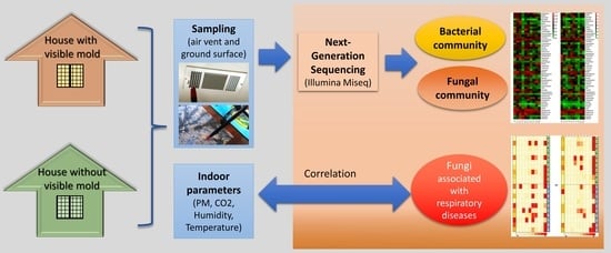
2.1. Study Area and Sampling Plan

This study was performed in greater Memphis, TN, USA. Samples (described below) were collected using two recruitment methods. The first recruitment utilized the citizens’ mold remediation hotline of the Shelby County Health Department (SCHD). The second method utilized friends’ participation. The friend recruitment was based on known citizens in Memphis who requested participation because of fungal concerns and general air quality in their homes. These subjects did not have any visible fungi before or during the sampling period and therefore were categorized as the control group.
A total of twenty houses were sampled during the study. Ten of the twenty houses had visible fungi (H2, H3, H4, H5, H6, H7, H8, H9, H10, and H18), and ten houses did not (H1, H11, H12, H13, H14, H15, H16, H17, H19, and H20). Field sampling was a one-week test in the respective homes. Samples were taken from the home by vacuuming content, deploying environmental instruments/samplers, and filling out a field sheet and questionnaire. The instruments included a PM counter and a CO2 logger placed at a central location in the living room. During the home setup, the field sheet logged the date, time, location, and other important parameters. The questionnaire asked about relevant information used to gather specific data that could help the project’s objectives and aims. Samples were collected using an ORECK vacuum and respected filter attachment (DUSTREAM Collector) that captured the vacuumed dust content following the Vacuum Dust Sample Collection Protocol recommended by the U.S. Department of Housing and Urban Development (HUD) [30] with minor modifications. Briefly, dust content was captured from two different areas: air vents (return vents) and the ground. Air vents were vacuumed in the room or adjacent room of fungal growth (or the main living rooms and bedrooms for HNM) and categorized as short-term exposure. Ground samples were captured using the same method and categorized as long-term exposure. Based on the home setup, the number of air vents that were vacuumed ranged from 1 to 4. Ground samples were collected from either carpet or a hard floor. Carpet samples were vacuumed over a one-square-meter area. Hard floor samples were vacuumed in a two-square-meter area. Thus, each home provided two types of samples: one ground-collected sample and one air-vent-collected sample. The vacuum collection was conducted on the initial visit to the sampling home. The vacuum filters were transported to the lab and stored at 4 °C until extraction.

2.2. Measurement of Indoor Environment Parameters

The microbial community largely depends on different types of environmental factors, and thus we have measured dust particulate matter (PM), carbon dioxide (CO2), humidity, and temperature in all the sampled houses (both HNM and HVM). We have used a continuous PM counter (OPC-N2, Alphasense Inc., Great Notley, Braintree, UK) that could measure mass concentrations of small particles. Similarly, the CO2 datalogger (1% CO2 + RH/T Data Logger, Model: K-33 ELG, CO2Meter.com, Ormond Beach, FL, USA) could continuously measure the level of CO2 (ppm), humidity (%), and temperature (°C) using Gaslab software. The PM counter is already deployable after removing any excel sheets from its memory bank; no calibrations are needed for the PM counter as the software is built into the machine, whereas CO2 dataloggers were configured by clearing all previous logs, properly setting the time/date, and adjusting the collection interval to every 5 min.

2.3. DNA Extraction, Purification, and Sequencing

Genomic DNA (gDNA) was extracted from each sample (n = 40) using the Qiagen DNA PowerSoil kit, following the manufacturer’s protocol. Purification of the extracted DNA was performed using the Epigentek DNA Concentrator kit (P-1006). The V4 hypervariable region of the 16S rRNA of the metagenomic DNA was amplified using a set of universal primers 515F (5′-GTGYCAGCMGCCGCGGTAA-3′) and 806R (5′-GGACTACNVGGGTWTCTAAT-3′). Similarly, ITS1F (5′-CTTGGTCATTTAGAGGAAGTAA-3′) and ITS2R (5′-GCTGCGTTCTTCATCGATGC-3′) were used to amplify the ITS region of fungi. The forward primer was tagged with a barcode sequence. The PCR (28 cycles) was conducted with the HotStarTaq Plus Master Mix Kit (Qiagen, Germantown, MD, USA), following the manufacturer’s instructions. The confirmation of the amplification was conducted by checking the PCR product on a 2% agarose gel. In order to prepare the Illumina DNA library, an equal proportion of the PCR products (in terms of concentration and molecular weight) were pooled and purified by Ampure XP beads. The paired-end sequencing was conducted at MR DNA (Shallowater, TX, USA) on a MiSeq, following the manufacturer’s guidelines.

2.4. Sequence Processing and Analysis

The processing and analysis of the sequences were conducted following the pipeline described by Mukherjee et al. (2016) [31], with some modifications. In brief, the quality of both the sequence files was checked (Phred score 25 or >25), followed by the removal of barcodes and other short (<150 bp) sequences along with sequences with ambiguous base calls using fastp v0.23.0 developed and reported by Chen et al. (2018) [32]. The chimeras were removed from the obtained sequences and denoised using DADA2 [4]. The final fasta file containing the cleaned sequences was used to construct the Operational Taxonomic Units (OTUs) following the MR DNA analysis pipeline (MR DNA, Shallowater, TX, USA) [33] for the downstream analysis of the bacterial and fungal composition. The final fasta file containing the cleaned sequences was used to construct the Operational Taxonomic Units (OTUs) using the Ribosomal Database Project [34,35,36,37] for bacteria and the UNITE database for fungi and was clustered at a 3% divergence level (97% similarity). The generated BIOM file was used to calculate bacterial and fungal composition, and visualization was conducted in the Microbiomeanalyst platform [38] and R packages (ggplot and vegan).

2.5. PCR Detection of Fungal Species Known to Be Associated with Respiratory Complications

Fungal species targets were selected based on their inclusion in a panel called the Environmental Relative Moldiness Index (ERMI) –developed by the EPA- to evaluate mold issues in water-damaged homes. Most of the species selected in this study are from ERMI Group 1 and Group 2, known to be associated with allergic respiratory diseases, nosocomial infections, and allergic respiratory reactions [39,40,41]. They were detected using qPCR, considering the threshold cycle (CT) of 27 as the detection limit [42,43]. Each PCR mixture included the following: PCR master mix (12.5 µL), nuclease-free water (8 µL), primers (2 µL), and the respected DNA –home sample or positive control (2.5 uL) for a total value of 25 µL. The PCR conditions and primer sequences were according to previous reports by Haugland and Vesper [44] and Haugland et al. (2004) [45].

2.6. Statistical Analysis

The statistical analysis was conducted using R (Version 4.0.0, R Foundation, Indianapolis, IN, USA). R packages ‘Vegan’ [46] were used for ecological data analysis and visualization using ‘ggplots2’ [47]. We explored and visualized the differences in community patterns in houses with and without visible fungal growths using the two-dimensional (2D) nonmetric multidimensional scaling (NMDS) ordination method, in which a stress function assessed the goodness of fit of the ordination compared with the original sample ranking [48]. In this process, the OTU counts of fungi and bacteria (at the genus level) were rarefied. The OTU table was square-root transformed, and the ‘gower’ dissimilarity matrix was used as the best method for calculating the distance matrix for the gradient analysis. It was followed by quantitative evaluation by permutational multivariate analysis of variance (PERMANOVA) [49] with 999 permutations using the adonis function from ‘Vegan’ package to test if houses with visible fungal growth and houses with no-fungal growth harbor significantly different taxonomic compositions. The network plots are generated using a statistical software called PAST v 4.0 [50]. The Venn diagram is prepared using a web-based platform provided by Bioinformatics & Evolutionary Genomics (available at: http://bioinforma tics.psb.ugent.be/webtools/Venn/, accessed on 12 April 2023). All heatmaps are generated using TBtools software [51].

3. Results

3.1. Data Showed the Presence of Several Pathogenic Bacterial and Fungal Genera with Distinctive Patterns in the Ground versus Air Vent Dust

We have analyzed a total of 40 samples for both fungi and bacteria (ten samples each from the air vent and ground from HNM and ten samples each from the air vent and ground from HVM). The percentage abundance of bacterial and fungal genera in the air vent and ground samples of both HNM and HVM are shown in Figure 1 and Figure 2, respectively. The bacterial genera that were highly abundant across the air vent samples of HNM and HVM were Propionibacterium, Sphingomonas, Staphylococcus, Corynebacterium, Methylobacterium, and Halospirulina (Figure 1A,B). However, the most abundant bacterial genera in the ground surface samples of both HVM and HNM were Streptococcus, Candidatus, Bacteroides, Sphingomonas, Massilia, Halospirulina, Staphylococcus, and Corynebacterium (Figure 1C,D). Similarly, across HNM, the most abundant fungal genera in air vent samples were Leptosphaerulina, Cladosporium, Curvularia, and Aspergillus (Figure 2A,B). On the other hand, the fungal genera that were highly abundant on ground surfaces across HVM were Leptosphaerulina, Cladosporium, and Aureobasidium (Figure 2C,D).
The bacterial and fungal diversity in ground and air vent samples was measured using the Shannon diversity index (Figure S1). The comparison of bacterial diversity between ground and air vent samples in HNM and HVM is presented in Figure S1A,B, respectively. The result indicated that the ground samples have higher diversity in both house types compared to the air vent samples; however, the difference was not significant at p < 0.05. Similarly, the diversity of fungal communities in HNM and HVM was calculated and presented in Figure S1C,D, respectively. In both house types, the ground sample exhibited higher diversity compared to the air vent sample; however, none of these differences were significant at the p < 0.05 level.
Based on the Operational Taxonomic Units (OTUs) data, a total of 58 and 53 bacterial genera were identified from the air vent and ground surfaces of both HNM and HVM and presented in Figure 3 using a heatmap diagram. The genus Halospirulina was observed to be the most abundant in the air vent samples of two HNM (H20 and H17, Figure 3A), while Methylobacterium was identified as the most abundant bacterial genera in the air vent sample of H8 (Figure 3B). The genus Veillonella was recorded to be the maximum in the ground sample of one HNM (H12, Figure 3C). However, in HVM (H2), the highest load of the bacterial genus belongs to Ignatzschineria, followed by Flavobacterium (Figure 3D).
Similarly, a total of 91 fungal genera were recorded from OTU data and presented in Figure 4. In the air vent samples of both HNM and HVM, the maximum abundance of fungal genera belongs to Saccharomyces in H17 and Phaeoisaria in H6 (Figure 4A,B). At the same time, the maximum load of fungi in the ground samples of HNM and HVM belongs to the genera Apiognomonia and Leptosphaerulina (Figure 4C,D), respectively.

3.2. Network Analysis Shows Higher Variability in Fungal Than Bacterial Communities in Dust Samples from Air Vents and Ground

Network plots display the similarities in bacterial and fungal communities in HNM and HVM (Figure 5). The houses with similar bacterial and fungal genera’ compositions were connected with different numbers of nodes. However, there will be no network connection if the difference is significant. In the case of air vent samples collected from HNM and HVM, the composition of the bacterial candidates at the genus level shares similarities, except for H17 and H20 (Figure 5A) and H7 (Figure 5B), respectively. Interestingly, the bacterial communities in the ground samples obtained from HNM are less common compared to air vent samples (Figure 5C). However, in the case of ground samples obtained from HVM, the bacterial community composition is very close to each other except for H2 (Figure 5D). The fungal community compositions in air vent samples collected from HNM and HVM are nearly similar, except for H17 (Figure 5E), H2, and H6 (Figure 5F). In contrast, the fungal communities in the ground samples collected from H13, H12, and H19 (HNM) are not close to any other houses (Figure 5G). However, for ground samples collected from HVM, all the houses have similar fungal community compositions except for H4 (Figure 5H).
Furthermore, the bacterial and fungal genus composition differences in the ground and air vent samples were analyzed and presented in a Venn diagram (Figure 6). Ground and air vent samples contain 29 and 34 unique bacterial genera, respectively (Figure 6A); however, 24 bacterial genera such as Cloacibacterium, Pseudomonas, Pinus, Dioscorea, Peptoclostridium, Flavobacterium, Halospirulina, etc. are recorded to be common. Similarly, the unique fungal genus numbers are 37 and 36 in the ground and air vent samples, respectively. The shared fungal genera between these sample types belong to Aureobasidium, Curvularia, Exophiala, Scolecobasidium, Stereum, Cladosporium, Ramulispora, Powellomyces, Phyllactinia, Trametes, Malassezia, Candida, Pleurotus, Rhizophlyctis, Aspergillus, Leptosphaerulina, Saccharomyces, Alternaria, and Paraphoma (Figure 6B).

3.3. The Fungal Communities in Air Vent Samples Differ Significantly between Home Types

To visualize the differences in taxonomic compositions between fungal and bacterial communities, we generated NMDS ordination plots (Figure 7), which provided information on observed fungal and bacterial communities (at the genus level) in HNM and HVM. No significant differences were observed between the air vent bacterial communities in HNM and HVM (Figure 7A, PERMANOVA, R2 = 0.07, p = 0.13). Similarly, no significant difference was observed among bacterial communities in ground samples collected from HNM and HVM (Figure 7B, PERMANOVA, R2 = 0.05, p = 0.51). No significant differences were observed in the ground dust samples from HNM and HVM (Figure 7C, PERMANOVA, R2 = 0.04, p = 0.60). However, a significant difference in fungal communities in air vent samples of HNM and HVM was observed (Figure 7D, PERMANOVA, R2 = 0.07, p < 0.05). Along with house types, the difference in the composition of bacterial and fungal communities in ground and air vent samples was also calculated. The difference in bacterial communities between ground and air vent samples in HNM (Figure 7E) and HVM (Figure 7F) showed a good fit (stress 0.153 and 0.166, respectively) but was not significant at the p < 0.05 level (R2 = 0.07 and 0.06, respectively). Similarly, the difference in fungal communities between ground and air vent samples in HNM and HVM was presented in Figure 7G,H, respectively. In both houses, the difference in fungal compositions in ground and air vent samples was not significant at the p < 0.05 level (R2 = 0.04 and 0.06, respectively).

3.4. Respiratory Disease-Related Fungal Species Were Detected in Both Home Types, Regardless of the Visibility of Fungi (Molds) in the Home

The presence and absence of a selected group of fungi known to be associated with respiratory complications [39,40,41] have been screened using qPCR from indoor dust samples and presented in Figure 8. The abundance of fungal species known to be associated with respiratory diseases is comparable, regardless of the visibility status of fungi/mold in the home. Alternaria alternata was recorded as the most abundant fungal species in ground and air vent dust samples from both HNM and HVM, with some exceptions. For example, it was not detected in the ground sample collected from H9 or the air vent samples collected from H1 and d H5. Aspergillus niger and Aspergillus versicolor were also found in some houses’ ground and air vent samples; however, Aspergillus sydowii was not recorded in any of those houses.

3.5. Respiratory Disease-Associated Fungi Did Not Show Any Significant Correlation with Indoor Environmental Parameters

Furthermore, to find the relationship between fungal communities known to be associated with respiratory disease and the indoor environment, we measured indoor environmental parameters, including particulate dust matter, CO2 concentration, temperature, and relative humidity. The relationships were examined using Spearman correlations for ground (Table 1) and air vent samples (Table 2). The result depicted a weak positive correlation between all indoor parameters and fungal communities in the ground samples collected from HNM (Table 1). However, the correlation was negative in ground samples collected from HVM, except for PM10 (Table 1). Similarly, in the air vent sample, the correlation was positive for PM, CO2 level, and humidity and negative for temperature (Table 2). In HVM, the correlation was negative between fungal communities and indoor parameters like PM and CO2 levels. However, temperature and humidity exhibited a very weak positive correlation (Table 2). Overall, all the correlations were not statistically significant at p < 0.05.

4. Discussion

Microorganisms are ubiquitous in environments, including indoor buildings. Phenotype-based identification has been successfully used to identify fungi in the past but can be unhelpful for atypical morphologies or fungi that fail to sporulate. It is time-consuming and laborious. Rapid identification of molds can facilitate a more timely and accurate assessment of exposure to pathogenic fungi. Thus, our study used molecular approaches to identify and enumerate the bacterial and fungal communities found in HNM and HVM. In the past, few studies have explored microbial composition in homes and home appliances like freezers, TVs, pillowcases, kitchen cutting boards, etc. [3,28,52]. Interestingly, the microbial composition and diversity (mostly bacterial and fungi) vary significantly from one house to another depending on the place, latitude, and humidity [52]. Several previous studies have reported variability in indoor fungal diversity based on factors such as house types, sampling types, indoor household locations, and items sampled [53,54,55]. In this study, we collected dust samples from different houses in Memphis, TN, USA, and explored the bacterial and fungal diversity and abundance. We found that the diversity of fungal communities varied in contrast to the consistency of bacterial communities. Given the geographic location of Memphis-Shelby County and the history of the high prevalence of pediatric asthma and other allergic diseases in this metropolitan area [56,57], our study provides the first analytical information on the indoor fungal and bacterial loads in this region of the USA.
In our experiment, the bacterial genere Rickettsia and Halospirulina were found to be common in both HNM and HVM. The genus Rickettsia is a Gram-negative, non-spore-forming bacteria and is reported to be responsible for causing spotted fever and typhus in humans [58]. Conversely, information regarding the interaction of Halospirulina with humans is lacking. In HNM, one of the most abundant genera was Streptococcus, while in HVM, Staphylococcus was abundant. This agrees with several earlier studies showing that Streptococcus and Staphylococcus are the two most abundant bacteria found in indoor environments [59,60,61].
The most abundant fungal genus in all these houses was found to be Leptosphaerulina. The genus Leptosphaerulina comprises roughly twenty-five species of filamentous ascomycetes that produce dark-colored pseudothecia and are primarily associated with plant diseases [62,63]. It may be noted that the ascospores of Leptosphaerulina are rarely reported from indoor air samples. On the other hand, its anamorph, Pithomyces [64], is common in the outdoor air worldwide, including in two North American cities (New York and Toronto) [65]. Similar to our results, Leptosphaerulina was found to be one of the most abundant genera in a previous study utilizing culture-independent molecular methods to evaluate the effect of water damage in buildings and subsequent renovation/remediation on indoor fungal communities [66]. On the other hand, Alternaria alternata, a major allergen and one of the most common fungi often associated with asthma development, persistence, and exacerbation [67,68,69], was found abundant in HNM and HVM. In addition, Chaetomium globosum (abundant in both home types) is a fungus with type I and III allergens associated with skin and nail infections in humans and infrequently causes cerebral and systemic infections in immunocompromised individuals [70,71,72,73]. In a pilot study, Andersen et al. investigated the air quality in different houses (with no visible fungi) in Denmark. They reported that Alternaria, Pseudopithomyces, and Cladosporium were the dominant fungus genera in an indoor environment, which is consistent with our finding [74]. Alternaria, Cladosporium, and Leptosphaerulina (Pithomyces) are common outdoor fungi; thus, they may have originated and been transmitted from outdoor to indoor environments. Interestingly, in our metataxonomic analyses, the abundance of Penicillium was found to be low, which is contrary to several previous reports showing Penicillium as one of the most abundant airborne fungal genera in the indoor air of moldy homes [9,53,74,75]. However, our qPCR assays detected Penicillium brevicompactum in the ground and air vent samples from at least one moldy home, while air vent samples from two moldy homes yielded positive results for Penicillium citrinum. The low detection rates of Penicillium could be attributed to the assay design (NGS versus qPCR), differences in environmental conditions, or other unknown factors.
The NMDS gradient analysis produces ordinations based on the dissimilarity matrix, or distance between different data sets. In this investigation, the NMDS analysis (Figure 7) revealed that the composition of bacterial communities in HNM and HVM is similar. This agrees with past studies that found bacterial communities differ depending on the surface type [2,8,28,76]. Although the NMDS analysis displayed no significant differences in ground fungal communities regardless of visible fungal presence or absence in homes, there is a significant difference in fungal communities in air vent samples collected from HNM and HVM. Visible fungi indicate an active fungal (“mold”) problem; however, there is also a chance of a nonvisible prevalence of pathogenic fungi in homes. This further confirms the importance of analyzing fungal diversity in homes in order to detect hidden mold using adequate techniques, e.g., molecular approaches [24,25,77].
The diversity of microbial communities in an indoor environment highly depends on the building’s occupants and the surrounding outdoor environment [78]. There is always an exchange between indoor and outdoor environments through human activities, water through the plumbing system, and air passages (windows, ventilation system, heating system, etc.), which determine the shape of the microbial community. Furthermore, humidity varies significantly from one season (winter) to another (summer) and is considered a critical regulatory factor in airborne microbial (especially fungi) growth and pathogenic agents [78]. In this investigation, we have tried to correlate the fungal species that are known to be associated with respiratory illnesses with their indoor environments, like dust particle size (PM), CO2 level, humidity, and temperature, using Sperman’s Rho statistics. Depending on the house status (HNM and HVM) and sampling site (ground and air vent), fungal communities exhibited some positive and negative relationships; however, none of these correlations were statistically significant (p < 0.05). This could be due to a number of factors, such as sample size, variability in environmental conditions, or other unknown factors that may have influenced the results. Environmental factors are known to be associated with asthma and other respiratory illnesses [79]. To this end, the agents, fungi, or bacteria that may prompt adverse health reactions need further consideration. Indoor fungal exposure has been studied countless times with scientific data backing it, acknowledging that exposure can lead to increased severity of asthma, as shown by human intervention and mouse trials [7,80].
Exposures to pathogenic fungi, including molds, are reported to be associated with several health problems, such as allergies, shortness of breath, chest tightness, and other respiratory complications [40,41]. Using qPCR analysis, a previous study investigated the occurrence of ten variable fungal and three bacterial groups in about 3000 homes in different geographical locations [52]. Their result demonstrated that the abundance of fungal and bacterial candidates largely depends on geographical location. In this study, we have assessed the presence of fungal targets from Group 1 and Group 2 of the ERMI-panel (developed by the US EPA) and fungi that are reported to be associated with allergic respiratory diseases, infections, and allergic respiratory reactions in both house types (HVM and HNM) using qPCR (Figure 8). The most common fungi found in both ground and air vent dust samples in both house types were Alternaria alternata, followed by Chaetomium globosum. The spore produced by A. alternata is a well-known biological contaminant and has been reported to be an inducer of several respiratory illnesses [81]. Chaetomium globosum is a mesophilic mold ubiquitously distributed in nature and was reported to be associated with onychomycosis [82]. This filamentous indoor fungus is often found on damp gypsum boards or plywood [83,84]. Therefore, Chaetomium abundance in both house types (moldy and non-moldy) in the present study may indicate the existence of dampness on gypsum boards or plywood wallboards. Furthermore, Aspergillus versicolor was also detected in many homes (both HVM and HNM), which is associated with several health problems, including neurological issues and pulmonary infections [85,86]. Interestingly, we have detected a broad spectrum of fungi associated with several respiratory complications in homes designated as “no visible mold” (HNM). Furthermore, there was no distinct demarcation in response to the presence and absence of mold species associated with health issues between HVM and HNM. So, our observation strongly suggests that homes without visible mold are not risk-free and may induce health complications. This study provides two new contributions to our understanding of the attribution of microbial risk factors in indoor environments. First, by employing genomics/sequencing-based methods, we empirically proved that molecular techniques that utilize the detection of microbial nucleic acids and genetic materials provide a significantly higher resolution in identifying potential microbial hazards in subjects that the visual and qualitative identification modalities failed to uncover. We unearthed how non-moldy homes may pose “hidden” risks of pathogenic microbial exposures, which could be statistically similar to the ones from moldy homes. Second, our study contributes to the information on indoor microbial (fungal and bacterial) prevalence information, for the first time, from a metropolitan area in a geographic location (mid-south US) that reports higher rates of pediatric asthma and other respiratory diseases, coupled with poor housing conditions [56,57].
The limitations of the present study may include a lack of statistical power as we only sampled from a total of twenty homes. Nonetheless, there are reports of similar studies with a limited sample size (such as n = 28 homes) [75]. A larger sample size (home samples) and repeated sample collections might be valuable to establish a significant correlation between fungi status and indoor environments. However, despite the limited number of homes sampled, we could display in-depth molecular profiles of bacteria and fungi in these homes, showcasing the importance and utility of molecular methods. Moreover, our results indicate that fungal exposure does not depend on the presence of visible fungi/mold in a home. This means the exposure may also come from “invisible” fungi/molds, such as microscopic fungi spores, as reported previously [87].

5. Conclusions

In this study, we used molecular methods to report the bacterial and fungal communities, the presence of fungal species known to be associated with respiratory diseases in HNM and HVM, and their correlations with indoor environmental factors. We found that these molecular methods provide rich information and are very useful in fungal and bacterial diagnostics in indoor environments. Future research aimed at assessing indoor air quality in homes will need to use updated technologies to ascertain the correlation between the microbiota of the built environment and adverse health effects.

Supplementary Materials

The following are available online at https://0-www-mdpi-com.brum.beds.ac.uk/article/10.3390/pathogens12081006/s1, Figure S1: Represents the Shannon alpha diversity in HNM and HVM. (A) and (B) indicate the bacterial diversity in ground and air vent samples in HNM and HVM, respectively. (C) and (D) demonstrate the fungal diversity in ground and air vent samples in HNM and HVM, respectively.

Author Contributions

Conceptualization, P.B., C.J. and D.H.J.; methodology, B.V.C., D.H.J., G.B., S.A. and I.M.S.; software, G.B.; formal analysis, B.V.C., D.H.J. and G.B.; resources, P.B. and I.M.S.; data curation, B.V.C., D.H.J. and G.B.; writing—original draft preparation, B.V.C., D.H.J. and G.B.; writing—review and editing, P.B., C.J. and I.M.S.; supervision, P.B. and C.J.; funding acquisition, P.B. All authors have read and agreed to the published version of the manuscript.

Funding

This research was partially funded by the FedEx Institute of Technology (Smart City Program) at the University of Memphis and a JPB Environmental Health Fellowship award granted by the JPB Foundation and managed by the Harvard T.H. Chan School of Public Health.

Institutional Review Board Statement

Not applicable. This study does not include any humans or animals, and thus Institutional Review Board permission is not required.

Informed Consent Statement

Not applicable.

Data Availability Statement

This manuscript contains metagenomic sequence data from both bacteria (40) and fungi (40). The metataxonomic sequence datasets associated with this paper are archived at the NCBI Sequence Read Archive (SRA) under the BioProject accession number PRJNA816110.

Acknowledgments

The authors thank Xianqiang Fu and Ashley Robinson of the University of Memphis for their technical assistance.

Conflicts of Interest

The authors declare no conflict of interest. Disclaimer: The findings and conclusions made in this presentation are those of the presenter and do not necessarily represent the views or official position of U.S. Food and Drug Administration (FDA) or the U.S. Department of Health and Human Services (DHHS). The names of vendors or manufacturers are provided as examples of available product sources; it does not imply endorsement of vendors, manufacturers, or products by FDA or DHHS.

References

  1. Kelley, S.T.; Gilbert, J.A. Studying the microbiology of the indoor environment. Genome Biol. 2013, 14, 202. [Google Scholar] [CrossRef] [Green Version]
  2. Horve, P.F.; Lloyd, S.; Mhuireach, G.A.; Dietz, L.; Fretz, M.; MacCrone, G.; Van Den Wymelenberg, K.; Ishaq, S.L. Building upon current knowledge and techniques of indoor microbiology to construct the next era of theory into microorganisms, health, and the built environment. J. Expo. Sci. Environ. Epidemiol. 2020, 30, 219–235. [Google Scholar] [CrossRef] [Green Version]
  3. Leung, M.H.; Tong, X.; Lee, P.K. Indoor microbiome and airborne pathogens. Compr. Biotechnol. 2019, 6, 96–106. [Google Scholar]
  4. Cox, J.; Stone, T.; Ryan, P.; Burkle, J.; Jandarov, R.; Mendell, M.J.; Adams, R.I.; Niemeier-Walsh, C.; Reponen, T. Associations of observed home dampness and mold with the fungal and bacterial dust microbiomes. Environ. Sci. Process. Impacts 2021, 23, 491–500. [Google Scholar] [CrossRef]
  5. Pausan, M.-R.; Blohs, M.; Mahnert, A.; Moissl-Eichinger, C. The sanitary indoor environment—A potential source for intact human-associated anaerobes. npj Biofilms Microbiomes 2022, 8, 44. [Google Scholar] [CrossRef]
  6. Emerson, J.B.; Keady, P.B.; Brewer, T.E.; Clements, N.; Morgan, E.E.; Awerbuch, J.; Miller, S.L.; Fierer, N. Impacts of flood damage on airborne bacteria and fungi in homes after the 2013 colorado front range flood. Environ. Sci. Technol. 2015, 49, 2675–2684. [Google Scholar] [CrossRef]
  7. Karvonen, A.M.; Hyvärinen, A.; Korppi, M.; Haverinen-Shaughnessy, U.; Renz, H.; Pfefferle, P.I.; Remes, S.; Genuneit, J.; Pekkanen, J. Moisture damage and asthma: A birth cohort study. Pediatrics 2015, 135, e598–e606. [Google Scholar] [CrossRef] [Green Version]
  8. Lax, S.; Smith, D.P.; Hampton-Marcell, J.; Owens, S.M.; Handley, K.M.; Scott, N.M.; Gibbons, S.M.; Larsen, P.; Shogan, B.D.; Weiss, S. Longitudinal analysis of microbial interaction between humans and the indoor environment. Science 2014, 345, 1048–1052. [Google Scholar] [CrossRef] [Green Version]
  9. Adams, R.I.; Sylvain, I.; Spilak, M.P.; Taylor, J.W.; Waring, M.S.; Mendell, M.J. Fungal signature of moisture damage in buildings: Identification by targeted and untargeted approaches with mycobiome data. Appl. Environ. Microbiol. 2020, 86, e01047-20. [Google Scholar] [CrossRef]
  10. Cabral, J.P. Can we use indoor fungi as bioindicators of indoor air quality? Historical perspectives and open questions. Sci. Total Environ. 2010, 408, 4285–4295. [Google Scholar] [CrossRef]
  11. Haas, D.; Habib, J.; Galler, H.; Buzina, W.; Schlacher, R.; Marth, E.; Reinthaler, F. Assessment of indoor air in austrian apartments with and without visible mold growth. Atmos. Environ. 2007, 41, 5192–5201. [Google Scholar] [CrossRef]
  12. EPA Ten Things You Should Know About Mold. Available online: https://www.epa.gov/mold/ten-things-you-should-know-about-mold (accessed on 28 September 2022).
  13. Morey, P.; Andrew, M.; Ligman, B.; Jarvis, J. Hidden Mold Sometimes Enters the Indoor Air. In Proceedings of the 9th International Conference on Indoor Air Quality and Climate. Monterey, Calif.; Levine, H., Bendy, G., Eds.; International Academy of Indoor Air Sciences: Monterey, CA, USA, 2002; pp. 455–460. [Google Scholar]
  14. Gravesen, S.; Nielsen, P.A.; Iversen, R.; Nielsen, K.F. Microfungal contamination of damp buildings—Examples of risk constructions and risk materials. Environ. Health Perspect. 1999, 107, 505–508. [Google Scholar] [CrossRef]
  15. Garrett, M.H.; Rayment, P.R.; Hooper, M.A.; Abramson, M.J.; Hooper, B.M. Indoor airborne fungal spores, house dampness and associations with environmental factors and respiratory health in children. Clin. Exp. Allergy 1998, 28, 459–467. [Google Scholar] [CrossRef]
  16. Miller, J.D.; Haisley, P.D.; Reinhardt, J.H. Air sampling results in relation to extent of fungal colonization of building materials in some water-damaged buildings. Indoor Air 2000, 10, 146–151. [Google Scholar] [CrossRef] [Green Version]
  17. Mistry, H.; Soberanis, H.M.A.; Kyyaly, M.A.; Azim, A.; Barber, C.; Knight, D.; Newell, C.; Haitchi, H.M.; Wilkinson, T.; Howarth, P. The clinical implications of Aspergillus fumigatus sensitization in difficult-to-treat asthma patients. J. Allergy Clin. Immunol. Pract. 2021, 9, 4254–4267.e4210. [Google Scholar] [CrossRef]
  18. Luo, W.; Hu, H.; Wu, Z.; Wei, N.; Huang, H.; Zheng, P.; Liu, Y.; Sun, B. Molecular allergen sensitization of Aspergillus fumigatus between allergic bronchopulmonary aspergillosis and a fumigatus-sensitized asthma in guangzhou, southern china. J. Clin. Lab. Anal. 2020, 34, e23448. [Google Scholar] [CrossRef]
  19. Druey, K.M.; McCullough, M.; Krishnan, R. Aspergillus fumigatus protease alkaline protease 1 (alp1): A new therapeutic target for fungal asthma. J. Fungi 2020, 6, 88. [Google Scholar] [CrossRef] [PubMed]
  20. Sullivan, A.; Hunt, E.B.; Ward, C.; Lapthorne, S.; Eustace, J.A.; Fanning, L.J.; Plant, B.J.; O’Byrne, P.M.; MacSharry, J.A.; Murphy, D.M. The presence of Aspergillus fumigatus in asthmatic airways is not clearly related to clinical disease severity. Allergy 2020, 75, 1146–1154. [Google Scholar] [CrossRef] [PubMed]
  21. Namvar, S.; Labram, B.; Rowley, J.; Herrick, S. Aspergillus fumigatus—Host interactions mediating airway wall remodelling in asthma. J. Fungi 2022, 8, 159. [Google Scholar] [CrossRef] [PubMed]
  22. Godish, T.; Godish, D. Inter-laboratory variability in total mould particle counts based on co-located, concurrently collected total airborne samples. In Proceedings of the Indoor Environmental Quality, Problems, Research and Solutions, Durham, NC, USA, 17–19 July 2006. [Google Scholar]
  23. Robertson, L.D.; Brandys, R. A multi-laboratory comparative study of spore trap analyses. Mycologia 2011, 103, 226–231. [Google Scholar] [CrossRef]
  24. Vesper, S. Traditional mould analysis compared to a DNA-based method of mould analysis. Crit. Rev. Microbiol. 2011, 37, 15–24. [Google Scholar] [CrossRef] [PubMed]
  25. Nonnenmann, M.W.; Coronado, G.; Thompson, B.; Griffith, W.C.; Hanson, J.D.; Vesper, S.; Faustman, E.M. Utilizing pyrosequencing and quantitative pcr to characterize fungal populations among house dust samples. J. Environ. Monit. 2012, 14, 2038–2043. [Google Scholar] [CrossRef] [PubMed] [Green Version]
  26. Banerjee, P.; Bhunia, A.K. Mammalian cell-based biosensors for pathogens and toxins. Trends Biotechnol. 2009, 27, 179–188. [Google Scholar] [CrossRef]
  27. Bahram, M.; Netherway, T.; Frioux, C.; Ferretti, P.; Coelho, L.P.; Geisen, S.; Bork, P.; Hildebrand, F. Metagenomic assessment of the global diversity and distribution of bacteria and fungi. Environ. Microbiol. 2021, 23, 316–326. [Google Scholar] [CrossRef]
  28. Dunn, R.R.; Fierer, N.; Henley, J.B.; Leff, J.W.; Menninger, H.L. Home life: Factors structuring the bacterial diversity found within and between homes. PLoS ONE 2013, 8, e64133. [Google Scholar] [CrossRef] [Green Version]
  29. Tringe, S.G.; Zhang, T.; Liu, X.; Yu, Y.; Lee, W.H.; Yap, J.; Yao, F.; Suan, S.T.; Ing, S.K.; Haynes, M. The airborne metagenome in an indoor urban environment. PLoS ONE 2008, 3, e1862. [Google Scholar] [CrossRef] [PubMed]
  30. Ashley, P.; Dewalt, G.; Hamilton, R.; Jones, J.; Pinzer, E. Vacuum Dust Sample Collection Protocol for Allergens. HUD Office of Healthy Homes and Lead Hazard Control: 2008. Available online: https://www.hud.gov/sites/documents/DOC_12539.PDF (accessed on 9 May 2023).
  31. Mukherjee, N.; Bartelli, D.; Patra, C.; Chauhan, B.V.; Dowd, S.E.; Banerjee, P. Microbial diversity of source and point-of-use water in rural haiti–a pyrosequencing-based metagenomic survey. PLoS ONE 2016, 11, e0167353. [Google Scholar] [CrossRef] [Green Version]
  32. Chen, S.; Zhou, Y.; Chen, Y.; Gu, J. Fastp: An ultra-fast all-in-one fastq preprocessor. Bioinformatics 2018, 34, i884–i890. [Google Scholar] [CrossRef]
  33. Dowd, S.E.; Callaway, T.R.; Wolcott, R.D.; Sun, Y.; McKeehan, T.; Hagevoort, R.G.; Edrington, T.S. Evaluation of the bacterial diversity in the feces of cattle using 16s rdna bacterial tag-encoded flx amplicon pyrosequencing (btefap). BMC Microbiol. 2008, 8, 125. [Google Scholar] [CrossRef] [Green Version]
  34. Edgar, R.C. Search and clustering orders of magnitude faster than blast. Bioinformatics 2010, 26, 2460–2461. [Google Scholar] [CrossRef] [Green Version]
  35. Mukherjee, N.; Dowd, S.E.; Wise, A.; Kedia, S.; Vohra, V.; Banerjee, P. Diversity of bacterial communities of fitness center surfaces in a us metropolitan area. Int. J. Environ. Res. Public Health 2014, 11, 12544–12561. [Google Scholar] [CrossRef] [Green Version]
  36. Swanson, K.S.; Dowd, S.E.; Suchodolski, J.S.; Middelbos, I.S.; Vester, B.M.; Barry, K.A.; Nelson, K.E.; Torralba, M.; Henrissat, B.; Coutinho, P.M. Phylogenetic and gene-centric metagenomics of the canine intestinal microbiome reveals similarities with humans and mice. ISME J. 2011, 5, 639–649. [Google Scholar] [CrossRef]
  37. Cole, J.R.; Wang, Q.; Fish, J.A.; Chai, B.; McGarrell, D.M.; Sun, Y.; Brown, C.T.; Porras-Alfaro, A.; Kuske, C.R.; Tiedje, J.M. Ribosomal database project: Data and tools for high throughput rrna analysis. Nucleic Acids Res. 2014, 42, D633–D642. [Google Scholar] [CrossRef] [Green Version]
  38. Chong, J.; Liu, P.; Zhou, G.; Xia, J. Using microbiomeanalyst for comprehensive statistical, functional, and meta-analysis of microbiome data. Nat. Protoc. 2020, 15, 799–821. [Google Scholar] [CrossRef]
  39. Vesper, S.J.; McKinstry, C.; Haugland, R.A.; Iossifova, Y.; Lemasters, G.; Levin, L.; Khurana Hershey, G.K.; Villareal, M.; Bernstein, D.I.; Lockey, J.; et al. Relative moldiness index as predictor of childhood respiratory illness. J. Expo. Sci. Env. Epidemiol. 2007, 17, 88–94. [Google Scholar] [CrossRef] [Green Version]
  40. Reponen, T.; Lockey, J.; Bernstein, D.I.; Vesper, S.J.; Levin, L.; Khurana Hershey, G.K.; Zheng, S.; Ryan, P.; Grinshpun, S.A.; Villareal, M.; et al. Infant origins of childhood asthma associated with specific molds. J. Allergy Clin. Immunol. 2012, 130, 639–644.e635. [Google Scholar] [CrossRef] [Green Version]
  41. Vesper, S.; McKinstry, C.; Haugland, R.; Wymer, L.; Bradham, K.; Ashley, P.; Cox, D.; Dewalt, G.; Friedman, W. Development of an environmental relative moldiness index for us homes. J. Occup. Env. Med. 2007, 49, 829–833. [Google Scholar] [CrossRef]
  42. Zhu, Y.-G.; Johnson, T.A.; Su, J.-Q.; Qiao, M.; Guo, G.-X.; Stedtfeld, R.D.; Hashsham, S.A.; Tiedje, J.M. Diverse and abundant antibiotic resistance genes in Chinese swine farms. Proc. Natl. Acad. Sci. USA 2013, 110, 3435–3440. [Google Scholar] [CrossRef]
  43. Looft, T.; Johnson, T.A.; Allen, H.K.; Bayles, D.O.; Alt, D.P.; Stedtfeld, R.D.; Sul, W.J.; Stedtfeld, T.M.; Chai, B.; Cole, J.R.; et al. In-feed antibiotic effects on the swine intestinal microbiome. Proc. Natl. Acad. Sci. USA 2012, 109, 1691–1696. [Google Scholar] [CrossRef]
  44. Haugland, R.; Vesper, S. Method of identifying and quantifying specific fungi and bacteria. U.S. Patent 6,387,652, 14 May 2002. [Google Scholar]
  45. Haugland, R.A.; Varma, M.; Wymer, L.J.; Vesper, S.J. Quantitative pcr analysis of selected Aspergillus, Penicillium and Paecilomyces species. Syst. Appl. Microbiol. 2004, 27, 198–210. [Google Scholar] [CrossRef]
  46. Dixon, P. Vegan, a package of r functions for community ecology. J. Veg. Sci. 2003, 14, 927–930. [Google Scholar] [CrossRef]
  47. Wickham, H. Ggplot: Using the Grammar of Graphics with r; Springer: New York, NY, USA, 2009. [Google Scholar]
  48. Knief, C.; Ramette, A.; Frances, L.; Alonso-Blanco, C.; Vorholt, J.A. Site and plant species are important determinants of the methylobacterium community composition in the plant phyllosphere. ISME J. 2010, 4, 719–728. [Google Scholar] [CrossRef] [Green Version]
  49. Anderson, M.J. A new method for non-parametric multivariate analysis of variance. Austral. Ecol. 2001, 26, 32–46. [Google Scholar]
  50. Hammer, O.; Harper, D.A.; Ryan, P.D. Palaeontological Statistics Software Package for Education and Data Analysis. Palaeontol. Electron. 2001, 4. Available online: http://palaeo-electronica.org/2001_1/past/issue1_01.htm (accessed on 28 September 2022).
  51. Chen, C.; Chen, H.; Zhang, Y.; Thomas, H.R.; Frank, M.H.; He, Y.; Xia, R. Tbtools: An integrative toolkit developed for interactive analyses of big biological data. Mol. Plant 2020, 13, 1194–1202. [Google Scholar] [CrossRef]
  52. Rocchi, S.; Reboux, G.; Scherer, E.; Laboissière, A.; Zaros, C.; Rouzet, A.; Valot, B.; Khan, S.; Dufourg, M.-N.; Leynaert, B. Indoor microbiome: Quantification of exposure and association with geographical location, meteorological factors, and land use in france. Microorganisms 2020, 8, 341. [Google Scholar] [CrossRef]
  53. Coombs, K.; Taft, D.; Ward, D.V.; Green, B.J.; Chew, G.L.; Shamsaei, B.; Meller, J.; Indugula, R.; Reponen, T. Variability of indoor fungal microbiome of green and non-green low-income homes in cincinnati, ohio. Sci. Total Env. 2018, 610–611, 212–218. [Google Scholar] [CrossRef]
  54. Chew, G.L.; Rogers, C.; Burge, H.A.; Muilenberg, M.L.; Gold, D.R. Dustborne and airborne fungal propagules represent a different spectrum of fungi with differing relations to home characteristics. Allergy 2003, 58, 13–20. [Google Scholar] [CrossRef]
  55. Coombs, K.C.; Chew, G.L.; Schaffer, C.; Ryan, P.H.; Brokamp, C.; Grinshpun, S.A.; Adamkiewicz, G.; Chillrud, S.; Hedman, C.; Colton, M.; et al. Indoor air quality in green-renovated vs. Non-green low-income homes of children living in a temperate region of us (ohio). Sci. Total Environ. 2016, 554–555, 178–185. [Google Scholar] [CrossRef] [Green Version]
  56. Shin, E.K.; Shaban-Nejad, A. Urban decay and pediatric asthma prevalence in memphis, tennessee: Urban data integration for efficient population health surveillance. IEEE Access 2018, 6, 46281–46289. [Google Scholar] [CrossRef]
  57. Shin, E.K.; Mahajan, R.; Akbilgic, O.; Shaban-Nejad, A. Sociomarkers and biomarkers: Predictive modeling in identifying pediatric asthma patients at risk of hospital revisits. NPJ Digit. Med. 2018, 1, 50. [Google Scholar] [CrossRef] [Green Version]
  58. Lokida, D.; Hadi, U.; Lau, C.-Y.; Kosasih, H.; Liang, C.J.; Rusli, M.; Sudarmono, P.; Lukman, N.; Laras, K.; Asdie, R.H. Underdiagnoses of rickettsia in patients hospitalized with acute fever in indonesia: Observational study results. BMC Infect. Dis. 2020, 20, 364. [Google Scholar] [CrossRef]
  59. Rintala, H.; Pitkäranta, M.; Toivola, M.; Paulin, L.; Nevalainen, A. Diversity and seasonal dynamics of bacterial community in indoor environment. BMC Microbiol. 2008, 8, 56. [Google Scholar] [CrossRef] [Green Version]
  60. Bouillard, L.; Michel, O.; Dramaix, M.; Devleeschouwer, M. Bacterial contamination of indoor air, surfaces, and settled dust, and related dust endotoxin concentrations in healthy office buildings. Ann. Agric. Environ. Med. 2005, 12, 187–192. [Google Scholar]
  61. Lee, L.; Tin, S.; Kelley, S.T. Culture-independent analysis of bacterial diversity in a child-care facility. BMC Microbiol. 2007, 7, 27. [Google Scholar] [CrossRef] [Green Version]
  62. Abler, S.W. Ecology and Taxonomy of Leptosphaerulina spp. Associated with Turfgrasses in the United States. Ph.D. Thesis, Virginia Tech, Blacksburg, VA, USA, 2003. [Google Scholar]
  63. Victoria, A.; Furtado, B.; Holz, M.; Romero-Arenas, O.; Dallagnol, L. First report of leptosphaerulina leaf spot caused by leptosphaerulina trifolii on trifolium repens in brazil. Plant Dis. 2020, 104, 972. [Google Scholar] [CrossRef]
  64. Roux, C. Leptosphaerulina chartarum sp. Nov., the teleomorph of Pithomyces chartarum. Trans. Br. Mycol. Soc. 1986, 86, 319–323. [Google Scholar] [CrossRef]
  65. Pakpour, S.; Li, D.-W.; Klironomos, J. Relationships of fungal spore concentrations in the air and meteorological factors. Fungal Ecol. 2015, 13, 130–134. [Google Scholar] [CrossRef]
  66. Pitkäranta, M.; Meklin, T.; Hyvärinen, A.; Nevalainen, A.; Paulin, L.; Auvinen, P.; Lignell, U.; Rintala, H. Molecular profiling of fungal communities in moisture damaged buildings before and after remediation—A comparison of culture-dependent and culture-independent methods. BMC Microbiol. 2011, 11, 235. [Google Scholar] [CrossRef] [Green Version]
  67. Salo, P.M.; Arbes, S.J., Jr.; Sever, M.; Jaramillo, R.; Cohn, R.D.; London, S.J.; Zeldin, D.C. Exposure to alternaria alternata in us homes is associated with asthma symptoms. J. Allergy Clin. Immunol. 2006, 118, 892–898. [Google Scholar] [CrossRef] [Green Version]
  68. Bush, R.K.; Prochnau, J.J. Alternaria-induced asthma. J. Allergy Clin. Immunol. 2004, 113, 227–234. [Google Scholar] [CrossRef]
  69. Causton, B.; Pardo-Saganta, A.; Gillis, J.; Discipio, K.; Kooistra, T.; Rajagopal, J.; Xavier, R.J.; Cho, J.L.; Medoff, B.D. Carma3 mediates allergic lung inflammation in response to alternaria alternata. Am. J. Respir. Cell Mol. Biol. 2018, 59, 684–694. [Google Scholar] [CrossRef]
  70. Piecková, E. In vitro toxicity of indoor chaetomium kunze ex fr. Ann. Agric. Environ. Med. 2003, 10, 9–14. [Google Scholar]
  71. Abbott, S.P.; Sigler, L.; McAleer, R.; McGough, D.A.; Rinaldi, M.; Mizell, G. Fatal cerebral mycoses caused by the ascomycete chaetomium strumarium. J. Clin. Microbiol. 1995, 33, 2692–2698. [Google Scholar] [CrossRef]
  72. Serena, C.; Ortoneda, M.; Capilla, J.; Pastor, F.J.; Sutton, D.A.; Rinaldi, M.G.; Guarro, J. In vitro activities of new antifungal agents against Chaetomium spp. And inoculum standardization. Antimicrob. Agents Chemother. 2003, 47, 3161–3164. [Google Scholar] [CrossRef] [Green Version]
  73. Salo, J.M.; Kedves, O.; Mikkola, R.; Kredics, L.; Andersson, M.A.; Kurnitski, J.; Salonen, H. Detection of Chaetomium globosum, ch. Cochliodes and ch. Rectangulare during the diversity tracking of mycotoxin-producing chaetomium-like isolates obtained in buildings in finland. Toxins 2020, 12, 443. [Google Scholar] [CrossRef]
  74. Andersen, B.; Frisvad, J.C.; Dunn, R.R.; Thrane, U. A pilot study on baseline fungi and moisture indicator fungi in danish homes. J. Fungi 2021, 7, 71. [Google Scholar] [CrossRef]
  75. Omebeyinje, M.H.; Adeluyi, A.; Mitra, C.; Chakraborty, P.; Gandee, G.M.; Patel, N.; Verghese, B.; Farrance, C.E.; Hull, M.; Basu, P.; et al. Increased prevalence of indoor Aspergillus and Penicillium species is associated with indoor flooding and coastal proximity: A case study of 28 moldy buildings. Env. Sci. Process Impacts 2021, 23, 1681–1687. [Google Scholar] [CrossRef]
  76. Flores, G.E.; Bates, S.T.; Caporaso, J.G.; Lauber, C.L.; Leff, J.W.; Knight, R.; Fierer, N. Diversity, distribution and sources of bacteria in residential kitchens. Environ. Microbiol. 2013, 15, 588–596. [Google Scholar] [CrossRef]
  77. Adams, R.I.; Miletto, M.; Taylor, J.W.; Bruns, T.D. The diversity and distribution of fungi on residential surfaces. PLoS ONE 2013, 8, e78866. [Google Scholar] [CrossRef]
  78. National Academies of Sciences, Engineering, and Medicine. Microbiomes of the Built Environment: A Research Agenda for Indoor Microbiology, Human Health, and Buildings; The National Academies Press: Washington, DC, USA, 2017. [Google Scholar]
  79. Cortes-Ramirez, J.; Wilches-Vega, J.D.; Paris-Pineda, O.M.; Rod, J.; Ayurzana, L.; Sly, P.D. Environmental risk factors associated with respiratory diseases in children with socioeconomic disadvantage. Heliyon 2021, 7, e06820. [Google Scholar] [CrossRef]
  80. Kumar, R.K.; Herbert, C.; Foster, P.S. Mouse models of acute exacerbations of allergic asthma. Respirology 2016, 21, 842–849. [Google Scholar] [CrossRef]
  81. Gabriel, M.F.; Postigo, I.; Tomaz, C.T.; Martínez, J. Alternaria alternata allergens: Markers of exposure, phylogeny and risk of fungi-induced respiratory allergy. Environ. Int. 2016, 89, 71–80. [Google Scholar] [CrossRef]
  82. Kim, D.M.; Lee, M.H.; Suh, M.K.; Ha, G.Y.; Kim, H.; Choi, J.S. Onychomycosis caused by Chaetomium globosum. Ann. Dermatol. 2013, 25, 232–236. [Google Scholar] [CrossRef] [Green Version]
  83. Lewinska, A.M.; Hoof, J.B.; Peuhkuri, R.H.; Rode, C.; Lilje, O.; Foley, M.; Trimby, P.; Andersen, B. Visualization of the structural changes in plywood and gypsum board during the growth of Chaetomium globosum and Stachybotrys chartarum. J. Microbiol. Methods 2016, 129, 28–38. [Google Scholar] [CrossRef]
  84. Andersen, B.; Dosen, I.; Lewinska, A.M.; Nielsen, K.F. Pre-contamination of new gypsum wallboard with potentially harmful fungal species. Indoor Air 2017, 27, 6–12. [Google Scholar] [CrossRef] [Green Version]
  85. Ladd, T.B.; Johnson, J.A., Jr.; Mumaw, C.L.; Greve, H.J.; Xuei, X.; Simpson, E.; Barnes, M.A.; Green, B.J.; Croston, T.L.; Ahmed, C. Aspergillus versicolor inhalation triggers neuroimmune, glial, and neuropeptide transcriptional changes. ASN Neuro 2021, 13. [Google Scholar] [CrossRef]
  86. Hodgson, M.J.; Morey, P.; Leung, W.-Y.; Morrow, L.; Miller, D.; Jarvis, B.B.; Robbins, H.; Halsey, J.F.; Storey, E. Building-associated pulmonary disease from exposure to Stachybotrys chartarum and Aspergillus versicolor. J. Occup. Environ. Med. 1998, 40, 241–249. [Google Scholar] [CrossRef]
  87. Reponen, T.; Singh, U.; Schaffer, C.; Vesper, S.; Johansson, E.; Adhikari, A.; Grinshpun, S.A.; Indugula, R.; Ryan, P.; Levin, L. Visually observed mold and moldy odor versus quantitatively measured microbial exposure in homes. Sci. Total Environ. 2010, 408, 5565–5574. [Google Scholar] [CrossRef] [Green Version]
Figure 1. Diversity of bacterial genera in indoor dust samples collected from homes with or without visible mold (HVM and HNM) in Memphis. Panels (A,B) show the relative abundance (%) of air vent bacteria in HNM and HVM, respectively. The percent abundance of ground surface bacterial genera in HNM and HVM is presented in panels (C,D), respectively.
Pathogens 12 01006 g001
Figure 2. Diversity of fungal genera in indoor dust samples collected from homes with or without visible mold (HVM and HNM) in Memphis. Panels (A,B) show the relative abundance (%) of air vent fungal genera in HNM and HVM, respectively. Panels (C,D) represent the abundance percentage of ground surface fungal genera in HNM and HVM.
Pathogens 12 01006 g002
Figure 3. The heatmap demonstrates the abundance of the bacterial genera in the air vent and ground samples of both HNM and HVM. (A,B) represent the bacterial load in air vent samples of HNM and HVM, respectively. Panels (C,D) represent the bacterial load in ground samples HNM and HVM, respectively. All OTU values are log converted.
Pathogens 12 01006 g003
Figure 4. The heatmap demonstrates the abundance of fungal genera in the air vent and ground samples of both HNM and HVM. Panels (A,B) represent the fungal load in air vent samples of HNM and HVM, respectively. Panels (C,D) represent the fungal load in ground samples of HNM and HVM, respectively. All OTU values are log converted.
Pathogens 12 01006 g004
Figure 5. Network plots demonstrate the connection between houses based on bacterial and fungal genera’ composition. (A,B) represent the network connections of houses based on the bacterial composition of the air vent samples of HNM and HVM, respectively. Panels (C,D) represent the network connections of houses based on the bacterial composition of the ground samples of HNM and HVM, respectively. Panels (E,F) represent the network connections of houses based on fungal compositions in the air vent samples of HNM and HVM, respectively. Finally, panels (G,H) represent the network connections of houses based on fungal composition in the ground samples of HNM and HVM, respectively.
Pathogens 12 01006 g005
Figure 6. The Venn diagram demonstrates the unique and shared bacterial (A) and fungal (B) compositions between the ground and air vent samples collected from all twenty houses.
Pathogens 12 01006 g006
Figure 7. Nonmetric multidimensional scaling (NMDS) ordination plot presenting taxonomic composition differences observed in bacterial and fungal communities in HNM and HVM. Each data point in the plots represents the bacterial or fungal community (at the genus level) identified in a home. In (A) (air vent bacteria) and (B) (ground bacteria), red dots represent HNM, while blue dots represent HVM. In (C) (air vent fungi) and (D) (ground fungi), red dots represent HNM, while blue dots represent HVM associated with respiratory diseases in indoor dust samples collected from HNM and HVM. (E,F) represent the bacterial communities in ground and air vent samples in HNM and HVM, respectively. (G,H) demonstrate the fungal composition in ground and air vent samples in HNM and HVM, respectively.
Pathogens 12 01006 g007
Figure 8. The checkered box demonstrates the presence of selected fungal species known to be associated with respiratory diseases in both HNM and HVM enumerated by qPCR. Black (present), white (absent). (A,B) indicate the ground and air vent samples.
Pathogens 12 01006 g008
Table 1. Spearman correlations of OTUs of fungal species known to be associated with respiratory diseases in ground dust samples with multiple indoor environmental parameters.
House TypeCorrelation BetweenCorrelation Coefficient, rsCorrelationp Value
Houses with no visible mold (HNM)OTU: PM 1.00.22Positive0.53
OTU: PM 2.50.22Positive0.53
OTU: PM 10.00.23Positive0.51
OTU: CO2 level0.35Positive0.38
OTU: Humidity0.28Positive0.49
OUT: Temperature0.38Positive0.27
Houses with visible mold (HVM)OTU: PM 1.0−0.05Negative0.87
OTU: PM 2.5−0.05Negative0.87
OTU: PM 10.00.01Positive0.96
OTU: CO2 level−0.28Negative0.42
OTU: Humidity−0.03Negative0.93
OTU: Temperature−0.21Negative0.55
Table 2. Spearman correlations of OTUs of fungal species known to be associated with respiratory diseases in air vent samples with multiple indoor environmental parameters.
House TypeCorrelation BetweenCorrelation Coefficient, rsCorrelationp Value
Houses with no visible mold (HNM)OTU: PM 1.00.62Positive0.05
OTU: PM 2.50.62Positive0.05
OTU: PM 10.00.46Positive0.17
OTU: CO2 level0.23Positive0.57
OTU: Humidity0.30Positive0.45
OTU: Temperature−0.22Negative0.52
Houses with visible mold (HVM)OTU: PM 1.0−0.48Negative0.17
OTU: PM 2.5−0.48Negative0.17
OTU: PM 10.0−0.45Negative0.19
OTU: CO2 level−0.45Negative0.18
OTU: Humidity0.10Positive0.77
OTU: Temperature0.11Positive0.75
Disclaimer/Publisher’s Note: The statements, opinions and data contained in all publications are solely those of the individual author(s) and contributor(s) and not of MDPI and/or the editor(s). MDPI and/or the editor(s) disclaim responsibility for any injury to people or property resulting from any ideas, methods, instructions or products referred to in the content.

