Article

Analysis of Ensifer aridi Mutants Affecting Regulation of Methionine, Trehalose, and Inositol Metabolisms Suggests a Role in Stress Adaptation and Symbiosis Development

by
Meryem Belfquih
1,2,
Abdelkarim Filali-Maltouf
1,* and
Antoine Le Quéré
1,2,*
1
Laboratory of Microbiology and Molecular Biology (LMBM), Department of Biology, Faculty of Sciences, University Mohammed V in Rabat, Rabat 10100, Morocco
2
Laboratoire des Symbioses Tropicales et Méditerranéennes (LSTM), University of Montpellier, IRD, CIRAD, INRAE, Institut Agro, 34398 Montpellier, France
*
Authors to whom correspondence should be addressed.
Microorganisms 2022, 10(2), 298; https://doi.org/10.3390/microorganisms10020298
Submission received: 20 December 2021 / Revised: 19 January 2022 / Accepted: 20 January 2022 / Published: 26 January 2022
(This article belongs to the Special Issue Genetics, Genomics, Physiology and Biochemistry of Rhizobium)

Abstract

:
Isolated from desert, the nitrogen-fixing bacterium Ensifer aridi LMR001 is capable of survival under particularly harsh environmental conditions. To obtain insights in molecular mechanisms involved in stress adaptation, a recent study using RNAseq revealed that the RpoE2-mediated general stress response was activated under mild saline stress but appeared non-essential for the bacterium to thrive under stress and develop the symbiosis. Functions associated with the stress response included the metabolisms of trehalose, methionine, and inositol. To explore the roles of these metabolisms in stress adaptation and symbiosis development, and the possible regulatory mechanisms involved, mutants were generated notably in regulators and their transcriptions were studied in various mutant backgrounds. We found that mutations in regulatory genes nesR and sahR of the methionine cycle generating S-adenosylmethionine negatively impacted symbiosis, tolerance to salt, and motility in the presence of NaCl. When both regulators were mutated, an increased tolerance to detergent, oxidative, and acid stresses was found, suggesting a modification of the cell wall components which may explain these phenotypes and support a major role of the fine-tuning methylation for symbiosis and stress adaptation of the bacterium. In contrast, we also found that mutations in the predicted trehalose transport and utilization regulator ThuR and the trehalose phosphate phosphatase OtsB-encoding genes improved symbiosis and growth in liquid medium containing 0.4 M of NaCl of LMR001ΔotsB, suggesting that trehalose metabolism control and possibly trehalose-6 phosphate cellular status may be biotechnologically engineered for improved symbiosis under stress. Finally, transcriptional fusions of gfp to promoters of selected genes and expression studies in the various mutant backgrounds suggest complex regulatory interplay between inositol, methionine, and trehalose metabolic pathways.

1. Introduction

Primarily found in deserts with an ability to develop the symbiosis of plants native and adapted to stress, Ensifer aridi represents an interesting species to study genes and functions that may explain its particular adaptation, broad host range, and phylo-geographic distribution [1,2,3,4,5]. Next-generation sequencing technologies (NGS) have become accessible to study any organism at the genome-wide level. Comparative genomic analyzes of 6 Ensifer aridi strains isolated in Asia, Africa, or America from diverse legumes showed that accessory genes were most probably located on large plasmids acquired through horizontal gene transfers that probably enabled the bacterium to develop the symbiosis with local hosts [6].
Among the most rapid methods to identify genes and functions associated to a particular stress, transcriptomics represents a method of choice. Genome-wide transcriptional responses resulting from abiotic stresses have been studied using microarray technology or NGS-based RNAseq in several rhizobial genera including, for example, Bradyrhizobium [7], Rhizobium [8,9], Mesorhizobium [10,11,12,13,14], or Sinorhizobium (Ensifer) [15,16,17,18,19,20]. The bacterial response to an osmotic upshift is complex and varies according to the strain and species. Nevertheless, to counteract osmotic pressure and the resulting oxidative stress, bacteria commonly accumulate osmoprotectants, compatible solutes, chaperones, and enzymes involved in ROS scavenging and they modify their cellular membranes and envelops [21].
According to a transcriptome analysis performed in E. aridi to explore the response of this bacterium to hyperosmotic stress, a number of genes were found differentially regulated, including the alternative extra-cytoplasmic function sigma factor RpoE2 which suggested activation of the general stress response (GSR) [15]. However, the mutation of the rpoE2 gene suggested that the response was complex and that alternative regulatory mechanisms may be recruited by the bacterium to cope with various stresses. Among genes that were found differentially regulated, a strong repression of motility genes could be identified and validated phenotypically by swimming assays [15]. We also found that salt stress altered regulation for a number of genes involved in the methionine cycle generating S-adenosylmethionine (SAM), the universal methyl donor, in inositol transport and catabolism or in trehalose de novo biosynthesis and transport [15]. To further explore the possible roles that these metabolisms have in stress adaptation and symbiosis of E. aridi, deletion mutants of regulatory genes involved in the methionine cycle (nesR, sahR), the trehalose transport and utilization (thuR), the inositol catabolism (iolR), or in the trehalose denovo biosynthesis (otsB) were generated and phenotyped. Finally, to explore the interdependency between regulated pathways, the transcriptional responses of studied genes were assessed in the various mutant backgrounds upon mobilization of vectors that harbored selected promoter fusions to green fluorescence protein (GFP)-encoding gene.

2. Materials and Methods

2.1. Bacterial Strains and Plasmids

The bacterial strains and plasmids used in this study are listed in Table 1. Ensifer strains and derivatives were cultured in tryptone yeast extract (TY) medium [22] at 28 °C. E. coli strains were grown at 37 °C in Luria–Bertani (LB) medium [23]. Agar at 2% (w/v) was added when needed. For selection of bacterial transformants and transconjugants, sucrose (5%, w/v) gentamycin (Gm, 10 μg mL−1), kanamycin (Km, 50 μg mL−1), ampicillin (Ap, 50 μg mL−1), rifampicin (Rif, 100 μg mL−1), and 5-bromo-4-chloro-3-indolyl-beta-D-galactosidase (X-gal, 80 μg mL−1) were added to the media when required.

2.2. Mutants’ Constructions

The cloning of iolR (PEG374), thuR (PEG6269), nesR (PEG5735), and otsB (PEG4868) 5′ and 3′ regions were performed using a two-step PCR using primers listed in Table 2. The PCR was carried out using as template genomic DNA extracts [29] from 10 mL of LMR001 liquid cultures at the early stationary phase s. First, PCR reactions were performed to amplify separately both the 5′ and the 3′ flanking regions using the proof-reading enzyme Phusion™ (Thermo-Fischer) and recommended chemistry, except for primers (1 µM final concentrations for forward and reverse primers). The cycling conditions included a primary denaturation step of 30 s at 98 °C followed by 35 cycles (98 °C, 10 s/56 °C, 30 s/72 °C, 45 s) and a final elongation step of 7 min at 72 °C. PCR products were purified using Illustra™ GFX PCR DNA and Gel Band Purification kits (GE Healthcare, Chicago, IL, USA), following manufacturer’s recommendations. Then, for each gene to be mutated, a second PCR reaction was set up with purified PCR products from the 5′ and 3′ regions obtained that contain complementary sequences included in the 5′ end of internal primers (see Table 2), thus enabling priming of the amplification. The 25 µL reactions contained 5 µL of 5× GoTaq buffer (Promega), 1.25 µL of MgCl2 (25 mM), 2.5 µL of dNTP mix (2.5 mM each), 0.125 µL GoTaq (5 U/µL), 2 µL of each purified PCR products obtained, and nuclease free water. A primary PCR consisting of a denaturation step of 95 °C for 5 min, followed by 5 cycles (95 °C, 30 s/50 °C, 45 s/72 °C, 50 s) and a final elongation step (5 min at 72 °C) was performed so as to generate chimeric DNA molecules consisting of merged 5′ and 3′ regions. Then, 15 µL of the mix (3 µL of 5× GoTaq buffer, 0.75 µL of MgCl2 (25 mM), 1.5 µL of dNTP mix, 0.125 µL of GoTaq (5 U/µL), 4 µL of external primers (10 µM), and 1.625 µL of nuclease free water) was added to the PCR reaction and subjected to the following cycling conditions: a denaturation step of 95 °C for 5 min followed by 25 cycles (95 °C, 30 s/55 °C, 45 s/72 °C, 50 s) and 5 min at 72 °C. The PCR products were purified from 1% agar gel using Promega Wizard® SV Gel and PCR Clean-Up System, following recommended protocol. As a result of the two amplifications’ process, the PCR-amplified and -purified products were cloned as chimeric fragments into pGEM-T Easy Vector (Promega kit) and transferred into XL2-Blue ultra-competent cells. The resulting positive clones were selected and the plasmids were extracted using the Wizard® Plus SV Minipreps DNA Purification System, digested by the XbaI enzyme, and ligated into suicide vector pJQ200SK [26]. Enzymes were purchased from Promega. The latter plasmid was used to transform Ensifer aridi LMR001 cells by the bipartite conjugation method using E. coli S17-1 as a donor (Simon et al. 1983) or by tri-parental mating procedure with E. coli DH5α [24] as a donor and the helper plasmid pRK2013 [27]. Transconjugants were selected on TY plates containing Rif and 5% sucrose to allow selection of knockout clones that had undergone a double cross-over. Double cross-over gene replacement in mutants was verified by screening for antibiotic-resistant sensibility phenotypes (Rif and Gm, respectively) and also PCR to verify deletion by using specific external and internal primers.
For the construction of the sahR (PEG2239) mutant, the 5′ and 3′ regions were first amplified using the primer pairs SAHR-P2239-A-AI/SAHR-P2239-B-PI and SAHR-P2239-C-PI/SAHR-P2239-D-XI (Table 2), respectively. PCR products were then cloned into pGEM-T Easy Vector following manufacturer’s recommendations. Ligations were used to transform competent DH5α cells. The plasmids were extracted and clones containing the inserts oriented from ApaI toward NsiI in the MCS were selected using plasmid and insert specific primers. DNA inserts in purified plasmids were sequenced and further digested with PstI. The clone that contained the 5′ region generated a linearized plasmid that was further dephosphorylated, and the plasmid that contained the 3′ region generated a band corresponding to the linearized vector and a second band that corresponded to the 3′ region cloned that was further extracted from an agarose gel. The latter insert was further ligated into the PstI digested and dephosphorylated vector containing the 5′ region. Ligation mix was used to transform the DH5α cells. A clone containing the 5′ and the 3′ regions in the correct orientation was selected by positive screening using specific primers. Finally, the insert was excised using external restriction sites ApaI and XbaI and cloned into pJQ200SK prior conjugation into LMR001 to generate the ΔsahR strain, as described above. Finally, the double mutant ΔsahRΔnesR was produced by mutating the ΔsahR strain upon double recombination of the pJQ200SK clone containing the nesR 5′ and 3′ regions, as described above.

2.3. Phenotypic Characterization of the Mutants

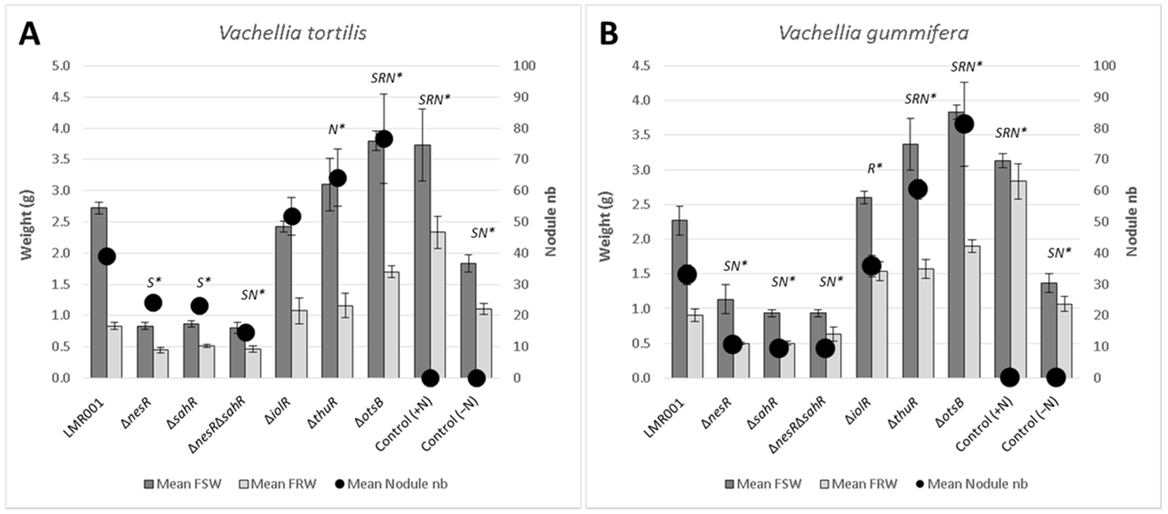
2.3.1. Plant Nodulation Assay

Seeds of Vachellia tortilis subsp. raddiana and endemic Moroccan Vachellia gummifera were surface sterilized, germinated, and cultivated, as described previously [15]. Briefly, after germination (5 days in the dark), young seedlings were transferred in pots containing sterilized sand and Ensifer aridi strains were inoculated using 1 mL of washed cells at an optical density (600 nm) ≈ 0.7 corresponding to exponential growth phase. Six individual plants were inoculated with each rhizobial strain and sterile water was added for the negative and positive controls. Plants were grown at 26 °C with 16 h day and 8 h night and watered using BD medium for watering [30] supplemented with 3 mM of KNO3 for positive controls. Plants were harvested after 6 months of growth, and the nodule number, plant shoot, and root fresh weights were recorded.

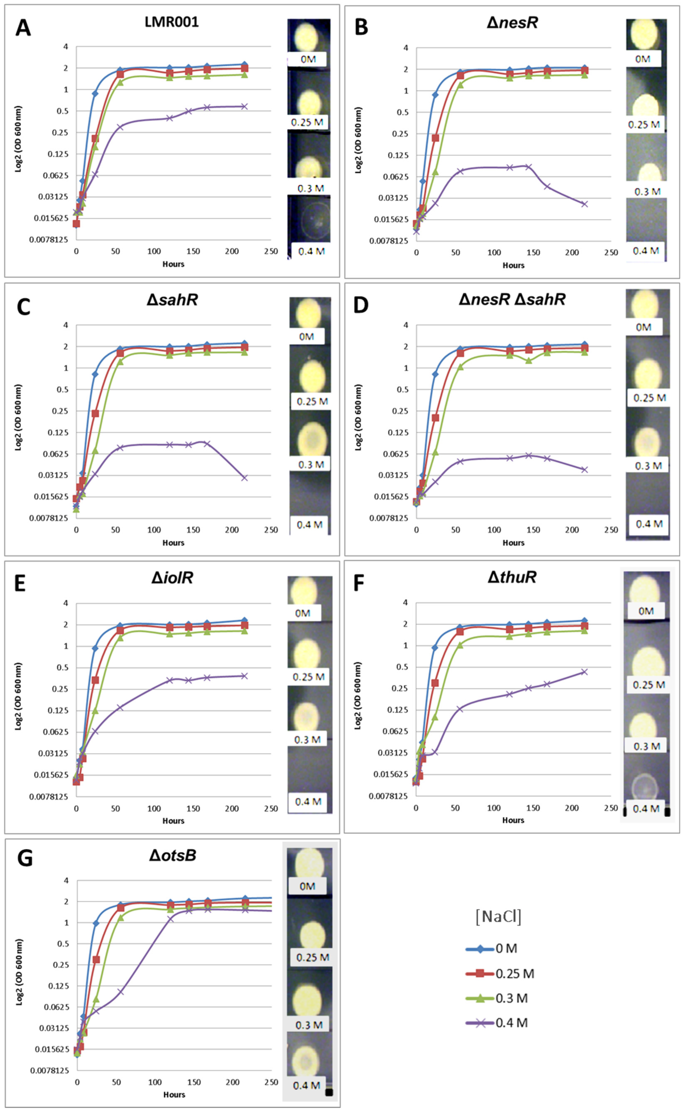
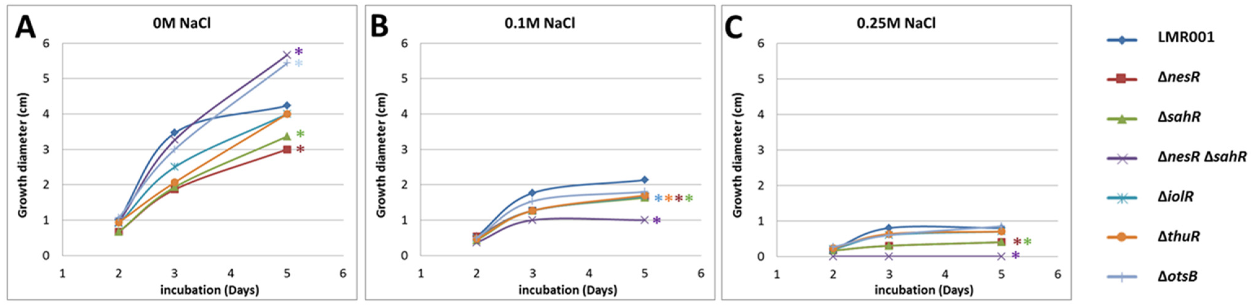
2.3.2. Motility, Tolerance to Salt, Detergent, Acid and Oxidative Stresses

The mutants were phenotyped as described previously [15]. Briefly, bacterial motility was calculated by measuring the growth diameter of motile bacteria after 2, 3, or 5 days of incubation at 28 °C in soft TY agar (0.2% w/v) complemented or not with NaCl at 0.1 or 0.25 M. Salt tolerance was estimated by growing bacteria in TY liquid medium and TY complemented with 0.25, 0.3, or 0.4 M of NaCl and the OD was recorded after 0, 4, 8, 24, 56, 120, 144, 168, and 216 h of growth at 28 °C with shaking. Then, 5 µL of 2-week-old cultures were spotted onto TY agar plates to estimate the long-term survival of bacteria in these different media. Sensitivities of the mutants to oxidative, acid, and detergent stresses were compared to those of the wild-type LMR001 strain. Briefly, bacterial cultures (pre-grown to OD 600 nm ≈ 0.7 in TY liquid medium) were added to 16 mL of soft TY agar containing 0.7% of agar precooled to 45 °C and poured onto TY agar plates. Once solidified, a paper disc (6 mm diameter) soaked with 5 µL of H2O2 (2 M), HCl (5.5 M), or SDS (10% w/v) was placed onto the surface of the plates that were incubated at 28 °C for three days prior to recording the diameters of the inhibition zones. The means of the three replicates were calculated.

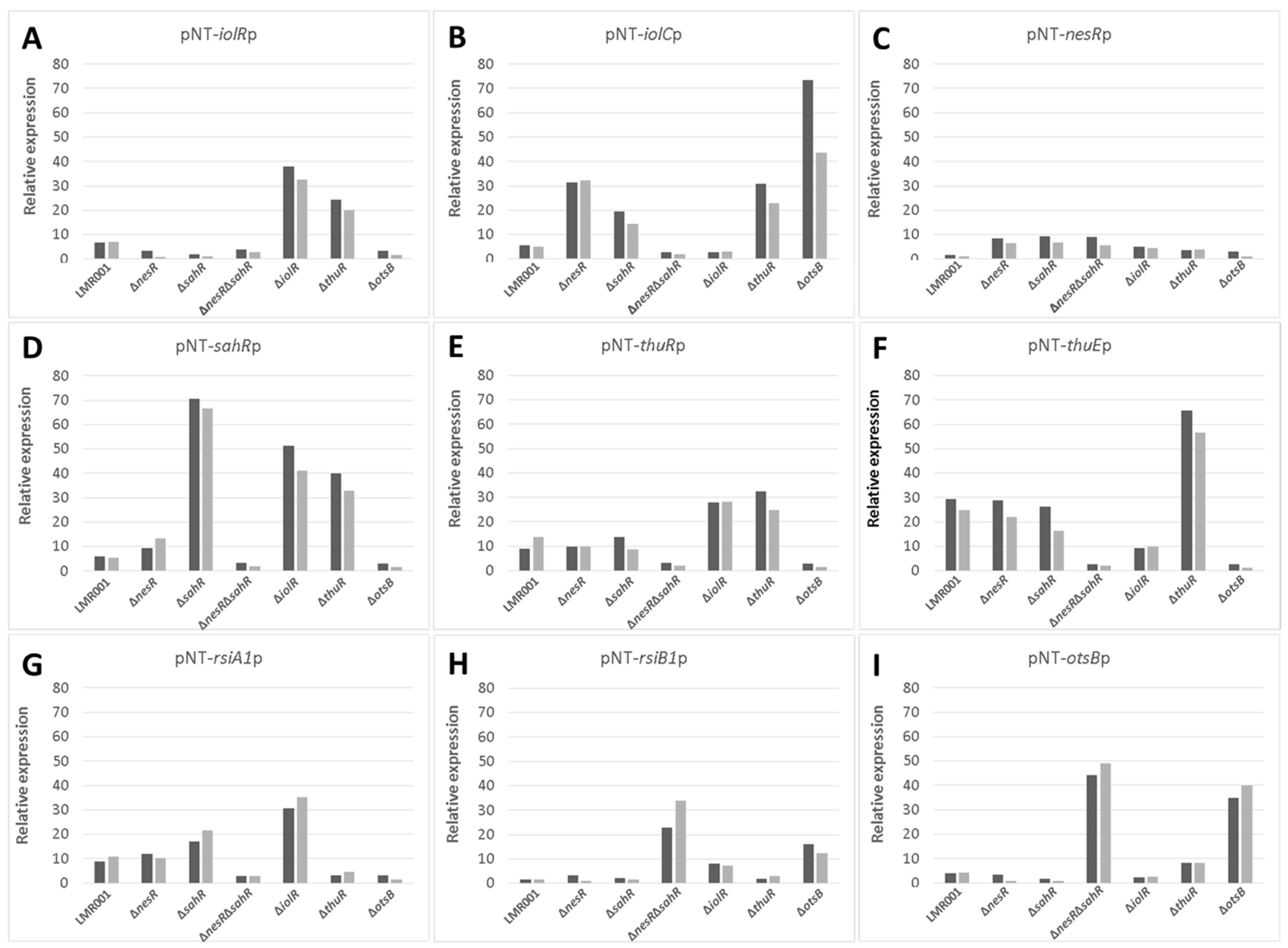
2.4. Transcirptional Fusion Assays

The promoters of selected genes were cloned previously in the broad-host-range mobilizable vector pROBE-NT and studied in E. aridi LMR001 and ΔrpoE2 strains [15]. These included the promoter regions of methionine cycle regulatory genes nesR (PEG5735) and sahR (PEG2239); GSR regulatory genes rsiA1 (PEG2540) and rsiB1 (PEG2541); genes involved in the trehalose import system and utilization thuEFGKAB (PEG6268-PEG6262) and its regulator thuR (PEG6269) or its endogenous biosynthesis otsBA (PEG4868, PEG4869) and treZ (PEG5323); and, finally, genes involved in inositol catabolism iolC (PEG373) and its regulation iolR (PEG374). Here, to explore the mutations’ effects on the transcriptional regulation of these genes, pPROBE derivatives were transferred by biparental conjugation into the new mutants using S17-1 E. coli as a donor. To estimate the relative expression of targeted genes’ transcriptions, the median relative expression of the 11 primary measures obtained every half hour was calculated for each mutant upon correction to controls and normalization by OD (600 nm) as previously described [15] and the data obtained from two kinetics were averaged.

3. Results and Discussion

To know whether the mutated genes were important for symbiosis development, the mutants were inoculated on two compatible hosts V. gummifera and V. tortilis, and their growth and nodulation were compared to those obtained with the LMR001 wild-type strain (Figure 1).
We found that deletions of genes involved in the regulation of the methionine cycle (nesR and sahR) significantly reduced the plant shoot weights for both Vachellia species as compared to the wild-type strain. Furthermore, the nodulation was also significantly lower than that of the LMR001 strain when the two regulatory genes were mutated in V. tortilis. In V. gummifera, all three mutants (ΔnesR, ΔsahR, and the double mutant ΔnesRΔsahR) significantly reduced the nodule number, suggesting that methionine cycling regulation generating the universal methyl donor SAM is important for proper symbiosis development and functioning. The regulation of the methionine cycle is complex and involves several transcription factors, such as NesR and SahR or RNA regulatory elements (SAM riboswitch) that remain to be investigated in Ensifer aridi. Nevertheless, given the strong homologies with the LuxR solo NesR of S. meliloti [31] and the ArsR family repressor SahR found in diverse proteobacteria, including rhizobiales [32], for which a conserved DNA-binding domain could be identified upstream LMR001 sahR, ahcY, metH, bhmT, and metK (Figure 2), our data suggest that a fine-tuning of the SAM-generating methionine cycle is essential for symbiosis establishment and functioning. Interestingly, it was recently shown in S. meliloti that the SAM synthetase MetK was also binding stress-inducible RNAs which previously showed to interact with the RNA chaperone Hfq and, more surprisingly, other mRNAs [33] which suggests that MetK-mediated post-transcriptional regulation may occur not only through epigenetics via the production of the universal methyl donor SAM but also by interacting with various RNAs and mRNAs.
The plant test also showed that a mutation in thuR or otsB, involved in the trehalose transport and/or metabolism, stimulated nodulation with both of the tested plants, while roots and shoots were also significantly increased in V. gummifera. Because ThuR is a LacI-GalR family regulator predicted to repress transport and catabolism genes (thuEFGKAB) through binding to upstream conserved motifs [34] also identified in E. aridi (Figure 3), the thuR deletion is expected to increase trehalose uptake capacity and utilization which may improve intracellular fitness and, thus, symbiotic efficiency; however, it remains to be functionally demonstrated. Strikingly, the otsB mutation significantly improved both plant weight and nodule number with the two plants. The trehalose-6P phosphatase OtsB is involved in the second step of the trehalose biosynthesis pathway which utilizes glucose-6P and UDP-glucose as primary substrates (Figure 3). A deletion of this gene should, therefore, result in an accumulation of trehalose-6P in the cell. Interestingly, an increased trehalose-6P synthase production by genetically manipulated rhizobial strains was reported to increase nodulation and plant biomasses in Phaseolus vulagris [35] or chickpea upon salt stress [36]. Even though much remains to be done to characterize factors involved in trehalose-6P-mediated plant growth and stress tolerance improvements, this phosphosugar is gaining increasing interest. It was recently shown to not only act as a compatible solute but also as signaling molecule in plant notably by modulating SnRK1 phosphorylation status, a central protein kinase involved in the coordination of the plant development that can lead to improved growth, notably under stress conditions [37,38]. Additional work is required to determine whether trehalose-6P accumulates in the ΔotsB strain during symbiosis development and functioning and whether the effects are linked to improved fitness and/or signaling.
The tolerance of the mutants to salt-induced osmotic upshift was compared to that of the LMR001 strain (Figure 4). The growth kinetics of tested strains in the TY medium, or when it was supplemented with 0.25 or 0.3 M of NaCl, were very similar. However, in the medium containing 0.4 M of salt, the growth of regulator mutants ΔnesR, ΔsahR, and the double mutant ΔnesRΔsahR were strongly impaired, suggesting that a fine-tuning regulation of the methionine cycle is important for salt tolerance in Ensifer aridi. The deletion of the trehalose uptake and utilization regulator thuR resulted in a continuous growth in the medium containing the highest salt concentration. Interestingly, while the otsB mutation resulted in reduced growth as compared to LMR001 in TY containing 0.4 M of salt after 2 days, the mutation enabled the bacterium to reach a higher OD at the stationary phase which almost reached the density observed in other media, suggesting that the strain impaired in trehalose phosphate phosphatase activity improves salt tolerance in LMR001. To estimate the viability of bacteria in tested media, five microliters of 2-week-old cultures were spotted onto the TY agar medium. While strains were all able to survive in media containing 0.3 M of NaCl or less, different growth phenotypes were found when bacteria were grown in the medium containing the highest salt concentration.
We found that after 2 weeks of growth in the medium supplemented with NaCl at 0.4 M, the LMR001 strain was still alive even though it showed a weak growth, as compared to cells grown in other media. All three mutants involved in methionine cycle regulation did not survive, showing that, at a higher osmotic strength, the proper regulation of the methionine cycling is essential. The ΔiolR strain that reached a lower OD at the stationary phase as compared to LMR001 strain did not survive prolonged incubation in TY supplemented with 0.4 M of NaCl. Interestingly, a deletion of thuR and more importantly otsB resulted in an improved long-term survival in the high salt concentrated medium as compared to the LMR001 strain. These results are globally in line with plant tests and further support the important role of stress-responsive genes in symbiosis functioning [39].
The motility of Ensifer aridi was previously shown to decrease in the presence of salt in the medium [15]. To test whether the mutated genes modified the swimming capacity of LMR001, their motility was compared to that of the LMR001 strain in soft agar plates that contained either TY medium or TY complemented with 0.1 or 0.25 M of NaCl. Figure 5 shows the results of the swimming assay for all strains. As shown for the LMR001 strain, the presence of salt in the medium reduced the motility in a dose-dependent manner. However, we found that after 5 days of growth in the TY medium, the ΔotsB showed continuous motility that exceeds that of the LMR001 strain. Whether this phenotype results from an increased trehalose-6P remains to be addressed. A similar phenotype was found for the double mutant ΔnesRΔsahR when, in contrast, single mutants of the methionine cycle regulators ΔnesR and ΔsahR presented a lower motility. These results are surprising as we would expect that the double mutation should result in a stronger effect. When salt was added in the medium, the motility was more strongly reduced for the double mutant as compared to the LMR001 strain and the single mutants and even totally impaired in the medium containing 0.25 M of salt, showing the importance for the bacterium to finely tune the methionine cycle for proper functioning. Given the involvement of methyl-accepting chemotaxis proteins in the regulation of diverse cellular activities including flagellum biosynthesis, it is not surprising that an alteration of the SAM-generating methionine salvage pathway regulation generates motility phenotypes. Nevertheless, additional work is required to characterize the effects of the mutations on SAM and whether these are responsible for the observed motility effects. Finally, after 5 days of growth, the ΔiolR and the ΔthuR mutants showed significantly lower motility in the medium containing 0.1 M of NaCl as compared to the LMR001 strain, suggesting that both trehalose and inositol affect swimming; however, mechanisms underlying these effects remain to be determined.
The tolerance to detergent (SDS), oxidative (H2O2), and acid (HCl) stresses of the studied strains was evaluated by comparing the growth inhibition zone resulting from their diffusion from a disc soaked with these chemicals (see Section 2). We found that only a double mutation of the two methionine cycle regulators resulted in a significant difference when compared to the LMR001 strain (Figure 6). The reduced inhibition zone found in the double mutant suggests that its membrane structure is altered, resulting in improved resistance to the three stresses which require additional work to be characterized.
To further describe the effects that mutations had on the transcriptional control of the inositol, the methionine, and the trehalose metabolisms, the relative expression levels of selected genes were assessed using the pPROBE derivatives previously screened in the ΔrpoE2 strain [15]. Figure 7 shows the mean relative expression obtained for all mutants. We found that in the ΔiolR and ΔthuR mutant backgrounds, iolR promoter activity was higher than in the wild-type strain, while in other mutants, the relative expression remained low and close to the background level (Figure 7A). The regulator IolR is a RpiR-like repressor shown to be involved in the regulation of the inositol catabolism in S. meliloti [40,41]. A regulatory model was proposed which involves IolR binding to promoter regions of target genes that are released when the intermediary product of the inositol catabolism 2-deoxy-5-keto-D-gluconic acid 6-phosphate (DKGP) is produced [41]. The promoters of genes involved in studied pathways of E. aridi were screened for putative regulator binding sites identified by Kohler and colleagues [41]. Interestingly, we found putative binding sites upstream of idhA, iolY, iolR, and iolC in LMR001 (Figure 8), suggesting a similar regulation in Ensifer aridi as all these genes were previously found to be induced upon salt stress [15] (Figure 8). In line with this model, we found that the mutation of iolR resulted in an induction of its own transcription, supporting a feedback regulatory loop (Figure 7A). However, we found that the iolC transcription was not induced in the iolR mutant background (Figure 7B), indicating that IolR is necessary for proper iolC transcription, thus suggesting a complex regulation of the inositol catabolism that may involve additional factors for proper regulation. Nevertheless, it is also possible that the iolR deletion resulted in an unexpected modification of the downstream genes involved in inositol catabolism that could impair the production of the product effector KDGP suggested to be required for antagonizing IolR repression. It should be noted that Kohler and colleagues [41] showed that even though IolR binding domains were found upstream of the iolY, iolR, and iolC genes, all the iolYRCDEB genes were transcribed as a single mRNA forming an operon. Recently, a iolR mutant in Aeromonas was shown to act both as a repressor and as an activator for a high number of genes [42]. Interestingly, a sequence close to the consensus IolR binding site was found in the intergenic region of the rsiA1-rsiB1 genes involved in the GSR (Figure S1). This putative IolR binding site overlaps with the predicted RpoE2-binding motif [20,43] located upstream of rsiB1 (Figure S1) which may suggest a possible implication of the IolR repressor in the modulation of the GSR. It should be noted that two motifs close to the consensus sequence of the ecfG sigma factor RpoE2-binding site were also predicted upstream of the iolR gene and the IolR putative binding site overlapping the primary putative RpoE2 binding site (Figure 8), which would support a possible cross regulation. By screening the genome of E. aridi for the presence of additional putative IolR binding sites, we found several consensus sequences. Among them, we identified the sequence motif upstream of a predicted ABC-type inositol transport system (PEG5307-5306-5305) that was also found induced by salt stress [15] (Figure 8).
Furthermore, another putative IolR binding site was also found upstream of PEG771, a LacI-type transcriptional repressor predicted to control inositol transport and utilization in R. etli [34]. Interestingly, a regulatory motif predicted as a binding motif of R. etli IolR (RHE_CH02362) by Regprecise was found twice in the intergenic region of PEG771–772, suggesting that inositol metabolism and transport regulation involves additional genes in E. aridi that may be modulated by IolR. Among putative IolR binding sites, we also found a putative IolR binding site upstream of the nesR start codon (Figure 2) which could suggest IolR-dependent methionine cycle regulation. However, this motif was located within the 5′ end of the pip gene, and nesR transcription using the GFP reporter system was close to background levels which make it difficult to assess. We also found that the iolC transcription was higher in the strains ΔnesR, ΔsahR, ΔthuR, and ΔotsB, while its transcription was close to background levels in the double mutant ΔnesRΔsahR (Figure 7B). Whether these increased iolC transcriptions are directly linked to the methionine and trehalose cycles remains to be determined, but these data strongly support a complex regulation of these metabolisms that appear interdependent and additional work is required to test the targets of IolR that may not be restricted to the regulation of inositol catabolism.
The relative expression of nesR encoding a LuxR solo showed to positively regulate the methionine salvage pathway generating SAM in S. meliloti [31], was higher in ΔnesR, ΔsahR and the double mutant ΔnesRΔsahR as compared to the other strains (Figure 7C). However, the expression remained low which makes it difficult to interpret. In contrast, the alphaproteobacterial ARS-type regulator SahR, which was predicted to act as a repressor [32], showed contrasting relative expression levels in the various strains (Figure 7D). Novichkov and colleagues showed that, in Desulfovibrio, SahR binds to operators of genes involved in SAM cycling depending on the cellular level of the effector S-adenosylhomocysteine (SAH) [32]. Here, the highest sahR relative expression was found in the ΔsahR strain supporting autoregulation. Accordingly, the promoter region of sahR in LMR001 possessed the predicted binding motif conserved among rhizobia that could also be identified upstream of the known target genes ahcY, metH, metK, and bhmT [32] (Figure 2). Given the fact that all these genes were found upregulated by salt previously [15], it is tempting to speculate on the role of SahR as a repressor of the SAM cycling in E. aridi. This raises the question regarding the regulatory mechanisms of this important pathway that, once again, involves several regulators, which is nevertheless not surprising given the importance of SAM as a universal methyl donor and the downstream activities it is involved in for cell functioning. Screening of the LMR001 genome for the presence of other putative SahR binding sites did not return any other hit, suggesting that SahR is primarily involved in SAM cycling regulation. However, the sahR promoter activity appeared also increased in the ΔiolR and the ΔthuR strains, as previously shown in ΔrpoE2 [15], which further show the pleiotropic effects alterations of the methionine cycling have on the other metabolisms.
Regarding the relative expression of thuR (Figure 7E) and thuE (Figure 7F), we previously found that thuR transcription was reduced in the ΔrpoE2 strain in contrast to that of thuE which supported a function of ThuR as a repressor of its own transcription and that of the thuEFGKAB operon that possesses 2 predicted ThuR binding motifs in their promoter regions (Figure 3) [15,34]. Here, we found that the thuR mutation resulted in inductions of both thuR and thuE, while the induction of thuR in the ΔiolR strain showed a concomitant repression of thuE when compared to LMR001. However, a strong repression of thuR in the ΔotsB and the double mutant ΔnesRΔsahR was found, which did not result in thuE inductions. Nevertheless, as shown in other LacI type regulators, such as the close regulator AglR, the DNA binding of such type of transcription factors depends on the presence of specific sugars as effectors whose concentrations and availability may be altered in the later strains. Indeed, given the inductility of thuEFGK by trehalose [44], it appears probable that the trehalose is involved in ThuR binding affinity to target promoters and a mutation of the trehalose-6P phosphatase may alter the availability of the effector to the repressor. Furthermore, as shown above, the double mutant ΔnesRΔsahR presented a stronger tolerance to detergent, oxidative, and acid stresses, suggesting a modification of the cell wall in this mutant that may modify cellular exchanges that could alter the availability of effectors to the regulator. However, additional work is required to further characterize the regulation of thu genes. It is interesting to note the presence of a motif (GGAAA-N17-AGAT) that resembles the RpoE2 sigma factor binding domains between these two ThuR recognition motifs upstream of the thuR start codon which would suggest that thuR transcription is RpoE2-mediated, in accordance with thuR transcriptional repression reported in the ΔrpoE2 strain [15]. The genome of the LMR001 strain was also screened for additional putative ThuR binding sites [34]. Among the conserved residues that matched LMR001 intergenic regions, putative binding motifs were found upstream of folD and metC involved in the methionine metabolism (Figure 2). Interestingly, in ΔthuR, sahR transcription was induced, which was shown to be activated through interaction of the SAH, i.e., the intermediary product of the methionine cycle acting as SahR effector [32], supporting the implication of ThuR in the methionine cycling. Nevertheless, whether ThuR is directly involved in the regulation of methionine precursors’ homeostasis remains to be determined experimentally. In addition, we also found the putative ThuR binding motif once upstream of the iolR gene and twice before the iolY start codon (Figure 8), which supports the involvement of ThuR in the regulation of the inositol catabolism. Again, iolR transcription was also found activated in ΔthuR, further supporting interplay between regulators of the methionine and trehalose metabolisms.
Among the putative ThuR binding sites identified, we also found the presence of two motifs upstream of the PEP carboxykinase-encoding gene pcKA (PEG3563) that was also strongly repressed by salt [15]. Interestingly, Di Cenzo and colleagues [45] showed that, in S. meliloti, pckA was regulated by PckR, another LacI-type transcriptional regulator that binds to a motif containing the following conserved residues (5′-TNNAANCGNTT-3′) that overlaps the predicted ThuR binding motif. Previous experimental data suggested a complex regulatory mechanism that relied on the presence of a single or a double PckR binding domain in the target promoter sequences which would result, respectively, in an induction or a repression of regulated genes in the presence of PEP, and inversely in the absence of the effector [45]. PckR was shown to regulate several genes of the glycolytic and gluconeogenic pathways, among which pckA and fbaB that possessed two PckR binding domains, and zwf, mgsA, and eda2, which possessed a single PckR binding domain, that were inversely regulated as compared to pckA and fbaB. Similarly, in E. aridi, both the presence and the number of PckR (PEG3052 in LMR001) binding domains could be identified in the promoter regions of these genes and similar expression profiles were found. Indeed, we found previously that a salt stress resulted in a strong repression of both pckA (PEG3563) and fbaB (PEG2954) and an induction of zwf (PEG714), mgsA (PEG118), and eda2 (PEG3069) [15]. This corroborates Di Cenzo and colleagues’ work on S. meliloti and suggests that, according to their regulatory model, under salt stress, E. aridi cells contain high levels of PEP. It should be noted that one of the two predicted ThuR binding sites upstream of thuR and aglE contained the PckR conserved motif, suggesting a possible involvement of PckR in ThuR-mediated regulation. di Cenzo and colleagues proposed several scenarios, including one that was based on a displacement of another negative regulator, thus enabling RNA polymerase to access the promoter. Given the fact that the predicted PckR binding motif includes conserved nucleotides of the ThuR binding motif, the ThuR regulator could be involved in modulating PckR-regulated genes; however, much work remains to be done to further characterize this complex regulatory mechanism and a possible interplay.
The transcriptional regulation of rsiA1 and rsiB1 was also studied in the various LMR001 derivatives (Figure 7G and Figure 7H, respectively). Regarding the rsiA1 relative expression, we found that, in comparison to the LMR001 strain, three mutants (ΔnesRΔsahR, ΔthuR, and ΔotsB) presented a lower expression. However, ΔsahR and, more importantly, ΔiolR presented a higher relative expression. Regarding the rsiB1 expression, three mutants (ΔnesRΔsahR, ΔiolR, and ΔotsB) presented an increased transcription level as compared with LMR001. It is interesting to note that both rsiA1 and rsiB1 relative expressions were increased in the ΔiolR strain as compared to the WT. Even if additional experimental data are required, the presence of a putative IolR binding site located in their promoter regions may be involved in modulating their transcriptional regulation and supports a role of IolR in GSR tuning. Surprisingly, both ΔnesRΔsahR and ΔotsB strains presented a reduced expression of rsiA1, while we observed stronger expressions of rsiB1 as compared to the WT strain. Because the anti-anti-sigma factor RsiB1 promotes RpoE2 recruitment to its operators through competitive binding to the anti-sigma factor RsiA1, our results suggest that GSR regulation may be altered in these mutants. Nevertheless, rpoE2 is co-transcribed with rsiA1 and the GSR is finely tuned and requires RsiB1 phosphorylation through a phosphorylation cascade that is not yet fully characterized, and studies are needed to functionally demonstrate the effects that these mutations may have on GSR. Interestingly, both strains presented a strong induction of the RpoE2-mediated otsBA operon (Figure 7I), suggesting that GSR was activated in these mutants. Surprisingly, with the exception of the iolC gene that was highly induced upon otsB deletion, the relative expression profiles obtained for ΔnesRΔsahR and ΔotsB strains were highly similar which contrasts with their responses to stress or their symbiotic performances. The fact that otsB deletion resulted in an induction of the otsBA operon suggests that, in this mutant, the cells accumulate the phosphor-sugar trehalose-6P as the phosphatase OtsB is nonfunctional. Whether tolerance to salt and symbiosis efficiency improvements are due to higher levels of trehalose-6P in the ΔotsB strain remains to be determined but further supports the idea that trehalose and its phosphorylated derivative are key metabolites for stress adaptation and plant growth promotion notably under abiotic stress [46]. Interestingly, Sengupta and colleagues previously showed in yeast that trehalose phosphate synthase methylation by a Cystein methyl transferase resulted in increased trehalose-6 phosphate biosynthesis [47], further supporting possible interplay between SAM-generating methionine cycling and trehalose metabolism. Additional research is, however, necessary to test whether such a type of post-translational regulation also occurs in bacteria.

4. Conclusions

In order to decipher the complex response of E. aridi to an osmotic upshift, a previous study based on RNA-seq enabled the identification of several metabolisms that were altered by salt. Despite activation of regulatory genes involved in the GSR, a deletion of the extra cytoplasmic function rpoE2 sigma factor-encoding gene did not result in phenotypic alterations, which suggested that alternative regulatory mechanisms could be recruited [15]. Here, a series of mutants in genes involved in the metabolisms of trehalose (thuR, otsB), inositol (iolR), and SAM-generating methionine (nesR and sahR) that appeared regulated upon salt stress were produced and phenotyped. To further describe the possible regulatory circuits between these metabolisms, the transcriptional state of selected genes and notable regulators involved in these metabolisms were assessed in these mutant backgrounds using a promoter—gfp fusion-based reporter system. The results show that mutations in genes involved in the methionine cycling regulation altered the symbiosis development and functioning with two compatible hosts, suggesting that a fine-tuned regulation of the methionine, and possibly SAM, is required. These mutants also showed a reduced growth in a medium containing 0.4 M of NaCl with a motility that was more strongly inhibited by salt than that of the wild-type LMR001 strain which may reduce plant colonization and in planta rhizobial fitness. Surprisingly, when the two genes nesR and sahR were deleted, the strain appeared more tolerant to detergent, oxidative, or acid stresses, suggesting an alteration of the cellular membranes. In contrast, mutants in the thuR or otsB genes resulted in improved symbiosis, showing that a modulation of the trehalose metabolism represents an interesting way to promote symbiosis and, thus, plant growth which was also correlated with an improved growth in a medium containing 0.4 M of NaCl. The transcription of genes encoding repressors IolR, ThuR, and SahR were induced when they were deleted, suggesting autoregulation which was shown in other bacteria for IolR and SahR and predicted for ThuR. We also found that IolR was required for proper iolC induction but was, in contrast, induced in ΔnesR, ΔsahR, ΔthuR, and ΔotsB, suggesting that the regulation of trehalose and methionine metabolisms can alter inositol catabolism while necessitating additional experimental evidences. Nevertheless, if nesR promoter activity remained low in all mutant backgrounds, we also found that the transcriptional state of the putative SahR repressor was also induced upon thuR or iolR mutations which further demonstrates an interplay between the studied metabolisms’ regulations. We also found that deletion of the iolR regulatory gene altered thuR and thuE transcriptions. When the transcriptional regulation of the rsiA1 and rsiB1 genes was assessed in the studied strains, different profiles were found depending on the mutant. Indeed, the thuR deletion resulted in a lower transcription of rsiA1 that was also found in the ΔotsB and ΔnesRΔsahR strains. In contrast, the two later strains presented a strong induction of the rsiB1 gene, suggesting that a deregulation of the methionine and trehalose metabolisms alter the GSR. Finally, GSR may also be influenced by inositol, as both rsiA1 and rsiB1 genes were induced in the ΔiolR strain. Whether these regulations are under the direct control of studied transcriptional regulators that possessed, in some cases, several putative binding motifs in their promoter regions, or indirect control through the involvement of yet unknown factors, may now be further explored. Nevertheless, the present study clearly shows that the stress-responsive genes are regulated by complex regulatory circuits, suggesting an interplay of genes involved in methionine, inositol, and trehalose regulatory pathways.

Supplementary Materials

The following are available online at https://0-www-mdpi-com.brum.beds.ac.uk/article/10.3390/microorganisms10020298/s1, Figure S1: Genetic map of the GSR regulatory genes rsiA1, rsiB1 and rpoE2.

Author Contributions

Conceptualization, A.F.-M., A.L.Q.; methodology, M.B., A.L.Q.; formal analysis, M.B., A.F.-M., A.L.Q.; writing—original M.B., A.L.Q.; writing—review and editing, M.B., A.F.-M., A.L.Q.; supervision A.F.-M., A.L.Q.; project administration, A.F.-M.; funding acquisition, A.F.-M., A.L.Q. All authors have read and agreed to the published version of the manuscript.

Funding

This research was funded by the French Institute of Research for the Development (IRD) through the LMI LBMV and an ARTS PhD grant for M.B.

Institutional Review Board Statement

Not applicable.

Informed Consent Statement

Not applicable.

Data Availability Statement

The genome and predicted gene sequences are accessible and can be downloaded from the NMPDR, SEED-based prokaryotic genome annotation service (https://rast.nmpdr.org/rast.cgi) using “guest” as login and password or upon request from the authors.

Acknowledgments

The authors would like to acknowledge Samir El Qaidi and Hassan Ait-Benhassou for methodological support for the construction of the mutants and Abderrahim Ferradous from the Centre Régional de Recherches Forestière in Marrakech for Vachellia seeds.

Conflicts of Interest

The authors declare no conflict of interest. The funders had no role in the design of the study; in the collection, analyses, or interpretation of data; in the writing of the manuscript, or in the decision to publish the results.

References

  1. Hakim, S.; Imran, A.; Mirza, M.S. Phylogenetic Diversity Analysis Reveals Bradyrhizobium Yuanmingense and Ensifer aridi as Major Symbionts of Mung Bean (Vigna radiata L.) in Pakistan. Braz. J. Microbiol. 2021, 52, 311–324. [Google Scholar] [CrossRef] [PubMed]
  2. Lamin, H.; Alami, S.; Bouhnik, O.; ElFaik, S.; Abdelmoumen, H.; Bedmar, E.J.; Missbah-El Idrissi, M. Nodulation of Retama monosperma by Ensifer aridi in an Abandonned Lead Mine Soils in Eastern Morocco. Front. Microbiol. 2019, 10, 1456. [Google Scholar] [CrossRef] [PubMed]
  3. Rocha, G.; Le Queré, A.; Medina, A.; Cuéllar, A.; Contreras, J.-L.; Carreño, R.; Bustillos, R.; Muñoz-Rojas, J.; del Carmen Villegas, M.; Chaintreuil, C.; et al. Diversity and Phenotypic Analyses of Salt- and Heat-Tolerant Wild Bean Phaseolus filiformis Rhizobia Native of a Sand Beach in Baja California and Description of Ensifer aridi sp. Nov. Arch. Microbiol. 2020, 202, 309–322. [Google Scholar] [CrossRef] [PubMed] [Green Version]
  4. Sakrouhi, I.; Belfquih, M.; Sbabou, L.; Moulin, P.; Bena, G.; Filali-Maltouf, A.; Le Quéré, A. Recovery of Symbiotic Nitrogen Fixing Acacia Rhizobia from Merzouga Desert Sand Dunes in South East Morocco—Identification of a Probable New Species of Ensifer Adapted to Stressed Environments. Syst. Appl. Microbiol. 2016, 39, 122–131. [Google Scholar] [CrossRef]
  5. Tak, N.; Awasthi, E.; Bissa, G.; Meghwal, R.R.; James, E.K.; Sprent, J.S.; Gehlot, H.S. Multi Locus Sequence Analysis and Symbiotic Characterization of Novel Ensifer Strains Nodulating Tephrosia Spp. in the Indian Thar Desert. Syst. Appl. Microbiol. 2016, 39, 534–545. [Google Scholar] [CrossRef]
  6. Le Quéré, A.; Tak, N.; Gehlot, H.S.; Lavire, C.; Meyer, T.; Chapulliot, D.; Rathi, S.; Sakrouhi, I.; Rocha, G.; Rohmer, M.; et al. Genomic Characterization of Ensifer aridi, a Proposed New Species of Nitrogen-Fixing Rhizobium Recovered from Asian, African and American Deserts. BMC Genom. 2017, 18, 85. [Google Scholar] [CrossRef]
  7. Cytryn, E.J.; Sangurdekar, D.P.; Streeter, J.G.; Franck, W.L.; Chang, W.; Stacey, G.; Emerich, D.W.; Joshi, T.; Xu, D.; Sadowsky, M.J. Transcriptional and Physiological Responses of Bradyrhizobium Japonicum to Desiccation-Induced Stress. J. Bacteriol. 2007, 189, 6751–6762. [Google Scholar] [CrossRef] [Green Version]
  8. López-Leal, G.; Tabche, M.; Castillo-Ramírez, S.; Mendoza-Vargas, A.; Ramírez-Romero, M.A.; Dávila, G. RNA-Seq Analysis of the Multipartite Genome of Rhizobium etli CE3 Shows Different Replicon Contributions under Heat and Saline Shock. BMC Genom. 2014, 15, 770. [Google Scholar] [CrossRef] [Green Version]
  9. Pérez-Montaño, F.; del Cerro, P.; Jiménez-Guerrero, I.; López-Baena, F.J.; Cubo, M.T.; Hungria, M.; Megías, M.; Ollero, F.J. RNA-Seq Analysis of the Rhizobium tropici CIAT 899 Transcriptome Shows Similarities in the Activation Patterns of Symbiotic Genes in the Presence of Apigenin and Salt. BMC Genom. 2016, 17, 198. [Google Scholar] [CrossRef]
  10. Alexandre, A.; Laranjo, M.; Oliveira, S. Global Transcriptional Response to Heat Shock of the Legume Symbiont Mesorhizobium loti MAFF303099 Comprises Extensive Gene Downregulation. DNA Res. 2014, 21, 195–206. [Google Scholar] [CrossRef] [Green Version]
  11. Laranjo, M.; Alexandre, A.; Oliveira, S. Genes Commonly Involved in Acid Tolerance Are Not Overexpressed in the Plant Microsymbiont Mesorhizobium loti MAFF303099 upon Acidic Shock. Appl. Microbiol. Biotechnol. 2014, 98, 7137–7147. [Google Scholar] [CrossRef] [PubMed]
  12. Laranjo, M.; Alexandre, A.; Oliveira, S. Global Transcriptional Response to Salt Shock of the Plant Microsymbiont Mesorhizobium loti MAFF303099. Res. Microbiol. 2017, 168, 55–63. [Google Scholar] [CrossRef] [PubMed]
  13. Liu, X.; Luo, Y.; Mohamed, O.A.; Liu, D.; Wei, G. Global Transcriptome Analysis of Mesorhizobium alhagi CCNWXJ12-2 under Salt Stress. BMC Microbiol. 2014, 14, 319. [Google Scholar] [CrossRef] [PubMed] [Green Version]
  14. Maynaud, G.; Brunel, B.; Mornico, D.; Durot, M.; Severac, D.; Dubois, E.; Navarro, E.; Cleyet-Marel, J.-C.; Le Quéré, A. Genome-Wide Transcriptional Responses of Two Metal-Tolerant Symbiotic Mesorhizobium Isolates to Zinc and Cadmium Exposure. BMC Genom. 2013, 14, 292. [Google Scholar] [CrossRef] [Green Version]
  15. Belfquih, M.; Sakrouhi, I.; Ait-Benhassou, H.; Dubois, E.; Severac, D.; Filali-Maltouf, A.; Le Quere, A. Ensifer aridi LMR001T Symbiosis and Tolerance to Stress Do Not Require the Alternative Sigma Factor RpoE2. Agronomy 2021, 11, 1787. [Google Scholar] [CrossRef]
  16. de Lucena, D.K.; Pühler, A.; Weidner, S. The Role of Sigma Factor RpoH1 in the PH Stress Response of Sinorhizobium meliloti. BMC Microbiol. 2010, 10, 265. [Google Scholar] [CrossRef] [Green Version]
  17. Domínguez-Ferreras, A.; Pérez-Arnedo, R.; Becker, A.; Olivares, J.; Soto, M.J.; Sanjuán, J. Transcriptome Profiling Reveals the Importance of Plasmid PSymB for Osmoadaptation of Sinorhizobium meliloti. J. Bacteriol. 2006, 188, 7617–7625. [Google Scholar] [CrossRef] [Green Version]
  18. Hellweg, C.; Pühler, A.; Weidner, S. The Time Course of the Transcriptomic Response of Sinorhizobium meliloti 1021 Following a Shift to Acidic PH. BMC Microbiol. 2009, 9, 37. [Google Scholar] [CrossRef] [Green Version]
  19. Lu, M.; Jiao, S.; Gao, E.; Song, X.; Li, Z.; Hao, X.; Rensing, C.; Wei, G. Transcriptome Response to Heavy Metals in Sinorhizobium meliloti CCNWSX0020 Reveals New Metal Resistance Determinants That Also Promote Bioremediation by Medicago lupulina in Metal-Contaminated Soil. Appl. Environ. Microbiol. 2017, 83, e01244-17. [Google Scholar] [CrossRef] [Green Version]
  20. Sauviac, L.; Philippe, H.; Phok, K.; Bruand, C. An Extracytoplasmic Function Sigma Factor Acts as a General Stress Response Regulator in Sinorhizobium meliloti. J. Bacteriol. 2007, 189, 4204–4216. [Google Scholar] [CrossRef] [Green Version]
  21. Vriezen, J.A.C.; de Bruijn, F.J.; Nüsslein, K. Responses of Rhizobia to Desiccation in Relation to Osmotic Stress, Oxygen, and Temperature. Appl. Environ. Microbiol. 2007, 73, 3451–3459. [Google Scholar] [CrossRef] [PubMed] [Green Version]
  22. Beringer, J.E. R Factor Transfer in Rhizobium leguminosarum. Microbiology 1974, 84, 188–198. [Google Scholar] [CrossRef] [PubMed] [Green Version]
  23. Sambrook, J.; Fritsch, E.; Maniatis, T. Molecular Cloning: A Laboratory Manual, 2nd ed.; Cold Spring Harbor: Long Island, NY, USA, 1989; Volume 2. [Google Scholar]
  24. Hanahan, D. Studies on Transformation of Escherichia coli with Plasmids. J. Mol. Biol. 1983, 166, 557–580. [Google Scholar] [CrossRef]
  25. Simon, R.; Priefer, U.; Pühler, A. A Broad Host Range Mobilization System for In Vivo Genetic Engineering: Transposon Mutagenesis in Gram Negative Bacteria. Bio/Technology 1983, 1, 784–791. [Google Scholar] [CrossRef]
  26. Quandt, J.; Hynes, M.F. Versatile Suicide Vectors Which Allow Direct Selection for Gene Replacement in Gram-Negative Bacteria. Gene 1993, 127, 15–21. [Google Scholar] [CrossRef]
  27. Figurski, D.H.; Helinski, D.R. Replication of an Origin-Containing Derivative of Plasmid RK2 Dependent on a Plasmid Function Provided in Trans. Proc. Natl. Acad. Sci. USA 1979, 76, 1648–1652. [Google Scholar] [CrossRef] [Green Version]
  28. Miller, W.G.; Leveau, J.H.J.; Lindow, S.E. Improved Gfp and InaZ Broad-Host-Range Promoter-Probe Vectors. Mol. Plant-Microbe Interact. 2000, 13, 1243–1250. [Google Scholar] [CrossRef] [Green Version]
  29. Chen, W.; Kuo, T. A Simple and Rapid Method for the Preparation of Gram-Negative Bacterial Genomic DNA. Nucleic Acids Res. 1993, 21, 2260. [Google Scholar] [CrossRef]
  30. Broughton, W.J.; Dilworth, M.J. Control of Leghaemoglobin Synthesis in Snake Beans. Biochem. J. 1971, 125, 1075–1080. [Google Scholar] [CrossRef] [Green Version]
  31. Patankar, A.V.; González, J.E. An Orphan LuxR Homolog of Sinorhizobium meliloti Affects Stress Adaptation and Competition for Nodulation. Appl. Environ. Microbiol. 2009, 75, 946–955. [Google Scholar] [CrossRef] [Green Version]
  32. Novichkov, P.S.; Li, X.; Kuehl, J.V.; Deutschbauer, A.M.; Arkin, A.P.; Price, M.N.; Rodionov, D.A. Control of Methionine Metabolism by the SahR Transcriptional Regulator in Proteobacteria: Control of Methionine Metabolism in Proteobacteria. Environ. Microbiol. 2014, 16, 1–8. [Google Scholar] [CrossRef] [PubMed] [Green Version]
  33. Robledo, M.; García-Tomsig, N.I.; Matia-González, A.M.; García-Rodríguez, F.M.; Jiménez-Zurdo, J.I. Synthetase of the Methyl Donor S-Adenosylmethionine from Nitrogen-Fixing α-Rhizobia Can Bind Functionally Diverse RNA Species. RNA Biol. 2021, 18, 1111–1123. [Google Scholar] [CrossRef] [PubMed]
  34. Ravcheev, D.A.; Khoroshkin, M.S.; Laikova, O.N.; Tsoy, O.V.; Sernova, N.V.; Petrova, S.A.; Rakhmaninova, A.B.; Novichkov, P.S.; Gelfand, M.S.; Rodionov, D.A. Comparative Genomics and Evolution of Regulons of the LacI-Family Transcription Factors. Front. Microbiol. 2014, 5, 294. [Google Scholar] [CrossRef] [PubMed] [Green Version]
  35. Suárez, R.; Wong, A.; Ramírez, M.; Barraza, A.; Orozco, M.D.C.; Cevallos, M.A.; Lara, M.; Hernández, G.; Iturriaga, G. Improvement of Drought Tolerance and Grain Yield in Common Bean by Overexpressing Trehalose-6-Phosphate Synthase in Rhizobia. Mol. Plant-Microbe Interact. 2008, 21, 958–966. [Google Scholar] [CrossRef] [Green Version]
  36. Moussaid, S.; Domínguez-Ferreras, A.; Muñoz, S.; Aurag, J.; Berraho, E.B.; Sanjuán, J. Increased Trehalose Biosynthesis Improves Mesorhizobium ciceri Growth and Symbiosis Establishment in Saline Conditions. Symbiosis 2015, 67, 103–111. [Google Scholar] [CrossRef]
  37. Paul, M.J.; Watson, A.; Griffiths, C.A. Trehalose 6-Phosphate Signalling and Impact on Crop Yield. Biochem. Soc. Trans. 2020, 48, 2127–2137. [Google Scholar] [CrossRef]
  38. Baena-González, E.; Lunn, J.E. SnRK1 and Trehalose 6-Phosphate—Two Ancient Pathways Converge to Regulate Plant Metabolism and Growth. Curr. Opin. Plant Biol. 2020, 55, 52–59. [Google Scholar] [CrossRef]
  39. da-Silva, J.R.; Alexandre, A.; Brígido, C.; Oliveira, S. Can stress response genes be used to improve the symbiotic performance of rhizobia? AIMS Microbiol. 2017, 3, 365–382. [Google Scholar] [CrossRef]
  40. Kohler, P.R.A.; Zheng, J.Y.; Schoffers, E.; Rossbach, S. Inositol Catabolism, a Key Pathway in Sinorhizobium meliloti for Competitive Host Nodulation. Appl. Environ. Microbiol. 2010, 76, 7972–7980. [Google Scholar] [CrossRef] [Green Version]
  41. Kohler, P.R.A.; Choong, E.-L.; Rossbach, S. The RpiR-Like Repressor IolR Regulates Inositol Catabolism in Sinorhizobium meliloti. J. Bacteriol. 2011, 193, 5155–5163. [Google Scholar] [CrossRef] [Green Version]
  42. Dong, Y.; Li, S.; Zhao, D.; Liu, J.; Ma, S.; Geng, J.; Lu, C.; Liu, Y. IolR, a Negative Regulator of the Myo-Inositol Metabolic Pathway, Inhibits Cell Autoaggregation and Biofilm Formation by Downregulating RpmA in Aeromonas hydrophila. NPJ Biofilms Microbiomes 2020, 6, 22. [Google Scholar] [CrossRef] [PubMed]
  43. Lang, C.; Barnett, M.J.; Fisher, R.F.; Smith, L.S.; Diodati, M.E.; Long, S.R. Most Sinorhizobium meliloti Extracytoplasmic Function Sigma Factors Control Accessory Functions. mSphere 2018, 3, e00454-18. [Google Scholar] [CrossRef] [PubMed] [Green Version]
  44. Jensen, J.B.; Peters, N.K.; Bhuvaneswari, T.V. Redundancy in Periplasmic Binding Protein-Dependent Transport Systems for Trehalose, Sucrose, and Maltose in Sinorhizobium meliloti. J. Bacteriol. 2002, 184, 2978–2986. [Google Scholar] [CrossRef] [PubMed] [Green Version]
  45. diCenzo, G.C.; Muhammed, Z.; Østerås, M.; O’Brien, S.A.P.; Finan, T.M. A Key Regulator of the Glycolytic and Gluconeogenic Central Metabolic Pathways in Sinorhizobium meliloti. Genetics 2017, 207, 961–974. [Google Scholar] [CrossRef] [PubMed] [Green Version]
  46. Onwe, R.O.; Onwosi, C.O.; Ezugworie, F.N.; Ekwealor, C.C.; Okonkwo, C.C. Microbial Trehalose Boosts the Ecological Fitness of Biocontrol Agents, the Viability of Probiotics during Long-Term Storage and Plants Tolerance to Environmental-Driven Abiotic Stress. Sci. Total Environ. 2022, 806, 150432. [Google Scholar] [CrossRef] [PubMed]
  47. Sengupta, S.; Banerjee, S.; Lahiri, S.; Dutta, T.; Dhar, T.K.; Ghosh, A.K. Purification, Characterization, Sequencing and Molecular Cloning of a Novel Cysteine Methyltransferase That Regulates Trehalose-6-Phosphate Synthase from Saccharomyces cerevisiae. Biochim. Biophys. Acta 2014, 1840, 1861–1871. [Google Scholar] [CrossRef]
Figure 1. Effects of the mutations on symbiosis development with Vachellia. The mean fresh shoot and root weights (shown, respectively, as FSW, dark grey and FRW, light grey; left axis) and nodule numbers (filled black circles; right axis) in V. tortilis (A) and V. gummifera (B) are shown for the mutants and non-inoculated control plants either watered using nitrogen complemented nutrient solution (+N) or without nitrogen (−N), as indicated at the bottom of the bar graphs. Error bars corresponding to standard deviations and significant differences with LMR001 data (p-value < 0.05, Tukey’s Test) are indicated by “*” preceded by the letter “S”, “R”, and/or “N” for mean shoot, root weights, and nodule numbers, respectively.
Microorganisms 10 00298 g001
Figure 2. Metabolism of methionine. Ensifer aridi predicted encoding genes (PEGs) involved in the metabolism of methionine are shown in the metabolic pathway. The presence of putative SahR, ThuR, and IolR binding sites upstream of genes (shown in the right boxes) are indicated, respectively, by “*”, “#”, and “$” on the left of genes when identified, and the position of conserved residues are bolded and colored accordingly in boxes. The salt response refers to fold change previously found by RNAseq [15]. Positive fold changes indicating upregulation (green) and negative ones showing repression (red) in salt containing medium are bolded when above 2.
Microorganisms 10 00298 g002
Figure 3. Metabolism of trehalose. Ensifer aridi predicted encoding genes (PEGs) involved in the metabolism and transport of trehalose are shown in the metabolic pathway. The presence of putative ThuR and RpoE2 binding sites upstream of genes (shown in the right boxes) are indicated by “#” and “σ”, respectively, on the left of genes when identified, and the position of conserved residues are bolded and colored accordingly in boxes. The salt response refers to fold change previously found by RNAseq [15]. Positive fold changes indicating upregulation (green) and negative ones showing repression (red) in salt containing medium are bolded when above 2.
Microorganisms 10 00298 g003
Figure 4. Effects of the mutations on salt tolerance. The growth kinetics are shown for the LMR001 (A), the strains ΔnesR (B), ΔsahR (C), ΔnesRΔsahR (D), ΔiolR (E), ΔthuR (F), and ΔotsB (G) in TY complemented with 0, 0.25, 0.3, or 0.4 M of NaCl (color code indicated at the bottom right of the figure). The OD (600 nm) corresponding to the mean of three replicates is shown using log 2 scale up to 9 days of growth (line graphs), and 5 µL of 2-week-old cultures was spotted onto TY to estimate long-term survival (pictures shown on the right of each line graph).
Microorganisms 10 00298 g004
Figure 5. Effects of the mutations and osmotic strength on bacterial swimming. The motility was estimated by measuring the growth diameter from triplicate TY soft agar plates supplemented with salt 0 M (A), 0.1 M (B) or 0.25 M (C). The mean motility after 2, 3, and 5 days are shown for the LMR001 and the 6 mutants (indicated on the right of the figure). Significant differences with LMR001 motility after 5 days (p-value < 0.05, Tukey’s Test) are indicated by “*” on the right of lines using the same color code.
Microorganisms 10 00298 g005
Figure 6. Effects of the mutations on detergent, oxidative and acids stresses. The mean diameters of the growth inhibition zone are shown for the 6 mutants (indicated on the right of the figure). The size of the inhibition zone is inversely correlated to the tolerance as the chemicals (indicted at the bottom of the graph) diffuse away from the disc into the agar (see Section 2). Error bars correspond to standard deviations and significant differences with LMR001 data (p-value < 0.05, Tukey’s Test) are indicated by “*” above the corresponding bar.
Microorganisms 10 00298 g006
Figure 7. Effects of the mutations on the relative expression level of selected genes using promoter–gfp fusion reporter system. The relative expression of iolR (A), iolC (B), nesR (C), sahR (D), thuR (E), thuE (F), rsiA1 (G), rsiB1 (H), and otsB (I) in the studied strains (indicated at the bottom of each bar graph) in TY or TY complemented with salt (250 mM of NaCl) are shown as dark and light grey bars, respectively. The mean relative expressions were calculated using the median fluorescence of 11 measures recorded every half an hour that were further corrected to controls and normalized to the OD (600 nm).
Microorganisms 10 00298 g007
Figure 8. Catabolism of inositol. Ensifer aridi-predicted encoding genes (PEGs) involved in the transport and catabolism of inositol are shown in the metabolic pathway. The presence of putative IolR, ThuR, and RpoE2 binding sites upstream of genes (shown on the right boxes) are indicated, respectively, by “$”, “#”, and “σ” on the left of genes when identified and the position of conserved residues are bolded and colored accordingly in boxes. The salt response refers to fold change previously found by RNAseq [15]. Positive fold changes indicating upregulation (green) in salt containing medium are bolded when ≥ 2.
Microorganisms 10 00298 g008
Table 1. List of bacteria and plasmid used in the study.
Strain/Plasmid NameDescriptionSource
Bacterial strains
LMR001TEnsifer aridi LMR001T isolated from Vachellia gummifera grown in Merzouga desert sand dune [4]
LMR001∆nesRnesR deletion mutant of LMR001This work
LMR001∆sahRsahR deletion mutant of LMR001This work
LMR001∆nesRsahRnesR deletion mutant of LMR001∆sahRThis work
LMR001∆iolRiolR deletion mutant of LMR001This work
LMR001∆thuRthuR deletion mutant of LMR001This work
LMR001∆otsBotsB deletion mutant of LMR001This work
E. coli DH5αhsdR17 endA1 thi-1 gyrA96 relA1 recA1 supE44 DlacU169 (f80lacZDM15)[24]
E. coli S17-1recA [SmR], thi, pro, RP4-2-Tc:Mu: aph::Tn7λpir. [25]
E. coli XL2 Blue Ultra-competentendA1 supE44 thi-1 hsdR17 recA1 gyrA96 relA1 lac [F’ proAB lacIqZΔM15 Tn10 (TetR) Amy CamR]Stratagene
Plasmids
pJQ200SKpACYC184-derived (p15A) suicide vector (GmR) [26]
pRK2013Helper plasmid containing the ColE1 replicon with RK2 tra genes (KmR) [27]
pGEM-T EasypUC origin, Multi Cloning Sites (MCS), lacZ gene fusion, β-lactamase coding region, lac operon sequences (ApR)Promega
pBluescript KS(+)ColEI, F1 Origin, MCS, lacZ gene fusion, β-lactamase coding region, lac operon sequences (ApR)Stratagene
TOPO vecteurpUC origin, région Plac promoteur, lacZ α-ccdB gene fuision. Topoisomerase enzyme (KmR, ApR) Invitrogen
pPROBE-NTpVS1-derived (p15a) vector, gfp (GmR) [28]
pNT-sahRppPROBE NT containing sahR (PEG2239) promoter fused to gfp (KmR)[15]
pNT-nesRppPROBE NT containing nesR (PEG5735) promoter fused to gfp (KmR)[15]
pNT-rsiA1ppPROBE NT containing rsiA1 (PEG2540) promoter fused to gfp (KmR)[15]
pNT-rsiB1ppPROBE NT containing rsiB1 (PEG2541) promoter fused to gfp (KmR)[15]
pNT-thuEppPROBE NT containing thuE (PEG6268) promoter fused to gfp (KmR)[15]
pNT-otsBppPROBE NT containing otsB (PEG4868) promoter fused to gfp (KmR)[15]
pNT-thuRppPROBE NT containing thuR (PEG6269) promoter fused to gfp (KmR)[15]
pNT-iolCppPROBE NT containing iolC (PEG373) promoter fused to gfp (KmR)[15]
pNT-iolRppPROBE NT containing iolR (PEG374) promoter fused to gfp (KmR)[15]
Table 2. Primers used in this study.
Target Gene (bp)Primer *Sequence (5′-3′) *5′ Region Size (bp)/Distance to Start (nt)3′ Region Size (bp)/Distance to Stop (nt)Deletion (bp)
iolR (861)IOLR-P374-A-XICCTCTAGACGAGATTTACGGCTCCAAGG457/−10 689
IOLR-P374-B-HIIITCAAAGACACCACAAGCTTCTCCAGCTTGCTCGTGTTC
IOLR-P374-C-HIIIAAGCTTGTGGTGTCTTTGACCGACTC 658/−183
IOLR-P374-D-XICCTCTAGAGCAATTGTCGCGATAGAAGA
otsB (750)OSTB-P4868-A-XIGGTCTAGATGCGCGATCTTCATGAACAA454/+75 503
OSTB-P4868-B-HIIITTCATCCGTCAGAAAGCTTGTCGGAGCAATGTCGAGAAG
OSTB-P4868-C-HIIIAAGCTTTCTGACGGATGAAGGAATGTT 500/−172
OSTB-P4868-D-XIGGTCTAGAATAGCCGGACATCTCATGCC
thuR (1020)THUR-P6269-A-XIGGTCTAGAGGTCCTCAGAAGCATTGTCA473/+87 865
THUR-P6269-B-HIIIGTTCGTGAATCTCAAGCTTCTCGCTGACCTCGGGATAG
THUR-P6269-C-HIIIAAGCTTGAGATTCACGAACTCTGGCC 453/−69
THUR-P6269-D-XIGGTCTAGATAGGTGCGATGAACATGACG
nesR (783)NESR-P5735-A-XICGCTCTAGAGGCTCGATATCACGCCAC186/+39 749
NESR-P5735-BTTAAGCCGCGGCGGTGAACTGGTTCCTGATC
NESR-P5735-CGCCGCGGCTTAAAACACC 160/+6
NESR-P5735-D-XIGCGTCTAGAACGACAAGCGGCAGCTTGC
sahR (1023)SAHR-P2239-A-AIGGGCCCGACAACATCTCGAAGG1062/+36 973
SAHR-P2239-B-PICTGCAGAGCATCCAAACCAAGCG
SAHR-P2239-C-PICTGCAGGGGGAGTTTGAGAATGA 1132/−14
SAHR-P2239-D-XITCTAGACCGAATTAGGGACTATAATTCCG
* Bold font indicate restriction sites (XI: XbaI, HIII: HindIII, AI: ApaI, PI: PstI). Red font shows complementary sequences used for the fusion of 5′ and 3′ regions.
Publisher’s Note: MDPI stays neutral with regard to jurisdictional claims in published maps and institutional affiliations.

