Brief Report

Modulation of Signal Regulatory Protein α (SIRPα) by Plasmodium Antigenic Extract: A Preliminary In Vitro Study on Peripheral Blood Mononuclear Cells

by
Priscilla da Costa Martins
,
Hugo Amorim dos Santos de Souza
,
Carolina Moreira Blanco
,
Luana Santos-de-Oliveira
,
Lilian Rose Pratt-Riccio
,
Cláudio Tadeu Daniel-Ribeiro
and
Paulo Renato Rivas Totino
*
Laboratório de Pesquisa em Malária, Instituto Oswaldo Cruz, Fundação Oswaldo Cruz, Rio de Janeiro 21040-900, Brazil
*
Author to whom correspondence should be addressed.
Microorganisms 2022, 10(5), 903; https://doi.org/10.3390/microorganisms10050903
Submission received: 12 January 2022 / Revised: 29 January 2022 / Accepted: 4 February 2022 / Published: 26 April 2022
(This article belongs to the Special Issue Parasitic Infection and Host Immunity)

Abstract

:
Signal regulatory protein α (SIRPα) is an immunoreceptor expressed in myeloid innate immune cells that signals for inhibition of both phagocytosis and inflammatory response. Malaria parasites have evolutionarily selected multiple mechanisms that allow them to evade host immune defenses, including the modulation of cells belonging to innate immunity. Notwithstanding, little attention has been given to SIRPα in the context of immunosuppressive states induced by malaria. The present study attempted to investigate if malaria parasites are endowed with the capacity of modulating the expression of SIRPα on cells of innate immune system. Human peripheral blood mononuclear cells (PBMC) from healthy individuals were incubated in the presence of lipopolysaccharide (LPS) or crude extracts of P. falciparum or P. vivax and then, the expression of SIRPα was evaluated by flow cytometry. As expected, LPS showed an inhibitory effect on the expression of SIRPα in the population of monocytes, characterized by cell morphology in flow cytometry analysis, while Plasmodium extracts induced a significant positive modulation. Additional phenotyping of cells revealed that the modulatory potential of Plasmodium antigens on SIRPα expression was restricted to the population of monocytes (CD14+CD11c+), as no effect on myeloid dendritic cells (CD14CD11c+) was observed. We hypothesize that malaria parasites explore inhibitory signaling of SIRPα to suppress antiparasitic immune responses contributing to the establishment of infection. Nevertheless, further studies are still required to better understand the role of SIRPα modulation in malaria immunity and pathogenesis.

1. Introduction

The innate immune system is the first line of defense against pathogens, acting basically through the recognition of pathogen-associated molecular patterns (PAMPs) found in different microorganism classes, including parasitic protozoa [1]. Upon sensing PAMPs by host pattern recognition receptors (PRRs), innate immune cells, such as macrophages, monocytes and dendritic cells (DCs), trigger intracellular signaling pathways leading to antimicrobial and inflammatory responses [2]. This event contributes to the direct elimination of pathogens, as well as the initiation of adaptive immune response mediated by lymphocytes. To ensure survival and propagation, pathogens have evolved a variety of strategies that facilitate their escape from innate immune responses, such as modulation of host cell activation [3,4,5]. Indeed, it has been demonstrated that malaria parasite antigens can inhibit maturation of lipopolysaccharide (LPS)-stimulated DCs both in vitro and in vivo, as well as fail to directly activate DCs in vitro, as shown by the down-regulation of the human leukocyte antigen (HLA-DR) and co-stimulatory molecules, and by reduced cytokine secretion, hampering T-cell responses [6,7,8]. In the same way, impairment of cell functions by malaria parasites has also been reported in monocytes and macrophages, in which suppression of important cellular effector events, such as phagocytosis, oxidative burst, cytokine release and HLA-DR expression, was observed [9,10,11,12,13].
In the context of suppressive immune responses, an immunoreceptor that has gained attention is the Signal Regulatory Protein alpha (SIRPα)—a transmembrane glycoprotein also designated as SHPS-1, CD172a and p84 that is mainly expressed in leukocytes of the myeloid lineage, including monocytes and DCs [14]. SIRPα belongs to the immunoglobulin (Ig) superfamily and presents as a key characteristic an immunoreceptor tyrosine-based inhibition motif (ITIM) in its cytoplasmic tail, which acts negatively in signaling pathways of cell activation [15]. It has been shown that by binding to its ligands, CD47, a ubiquitous cell membrane glycoprotein, and lung surfactant proteins A and D, SIRPα mediates inhibitory signaling of innate immunity functions, preventing, for instance, phagocytosis, proinflammatory cytokine production and DCs maturation [16,17,18,19]. Comprehensibly, it is believed that inhibitory signaling mediated by SIRPα can impact effective innate immune responses against infectious microbes—an issue that has not been widely addressed. To gain insight into SIRPα in malaria, in the present work, we investigate the modulatory effect of Plasmodium crude extracts on SIRPα expression in peripheral blood innate immune cells.

2. Material and Methods

2.1. P. falciparum and P. vivax Antigens

Asexual blood stages of P. falciparum (W2 strain) were maintained in continuous in vitro culture according to the method described by Trager and Jensen [20]. Parasites were cultured using O+ human red blood cells (RBCs) in RPMI-1640 medium (Sigma, St. Louis, MO, USA) supplemented with 25 mM Hepes (Sigma), 0.2% glucose (Sigma), 23 mM sodium bicarbonate (Sigma), 40 mg/mL gentamycin (Gibco Industries, Big Cabin, OK, USA) and 10% heat inactivated AB+ human serum (complete medium). Cultures were maintained at 5% hematocrit at 37 °C under an atmosphere of 5% O2, 5% CO2 and 90% N2 (White Martins Praxair Inc., Rio de Janeiro, Brazil). Parasites were synchronized by repeated sorbitol treatments and parasitized RBCs (pRBCs) were enriched by 60% Percoll density gradient using predominantly mature stage cultures, as described elsewhere [21]. P. vivax parasites were obtained from a peripheral blood sample of a patient presenting with non-complicated malaria and the enrichment of mature stages was performed using Percoll 45%, as described by Carvalho et al. [22]. Finally, the pRBCs were suspended in phosphate saline buffer (PBS), sonicated in an ice bath. Protein concentration was then determined using the Qubit Protein Assay Kit (Molecular Probes, Eugene, OR, USA).

2.2. PBMC Isolation and Antigenic Stimulation

Heparinized venous blood samples were collected from five clinically healthy individuals, as approved by the Human Research and Ethic Committee of the Oswaldo Cruz Foundation (CAAE 46084015.1.0000.5248). The same individuals were recruited for all experiments and peripheral blood mononuclear cells (PBMCs) were isolated through density gradient centrifugation using Histopaque-1077 (Sigma). Cells were washed twice in RPMI-1640 medium (Sigma) containing 2.05 mM l-glutamine, 25 mM Hepes, and 2.0 g/L sodium bicarbonate and then resuspended in RPMI medium supplemented with 200 U/mL penicillin (Gibco), 200 mg/mL streptomycin (Gibco), and 10% inactivated fetal calf serum (Gibco). Cells (2.5 × 10⁵) were incubated for 24 and 48 h in the absence or presence of Plasmodium extracts (0.1–10 μg/mL) or Escherichia coli lipopolysaccharides (LPS, 5 μg/mL, Sigma) in 96-well culture plates ( Corning Incorporated, Durham, NC, USA) at 37 °C in 5% CO2. PBMCs from the same individuals were used for all experiments.

2.3. Flow Cytometry Assay

Expression of SIRPα on PBMC was assayed by flow cytometry using an APC-conjugated anti-SIRPα monoclonal antibody (eBioscience, San Diego, CA, USA). Additionally, PerCP-Cy5.5-conjugated anti-CD11c (BD Pharmingen, San Diego, CA, USA) and PE-conjugated anti-CD14 (eBioscience) antibodies were used to identify monocyte and dendritic cell populations. Briefly, cells (2.5 × 10⁵) were washed in PBS and, subsequently, incubated at 4 °C for 30 min in PBS containing 10% fetal bovine serum (FBS) to block non-specific staining. After incubation, cells were stained with monoclonal antibodies for 40 min at 4 °C in 100 μL PBS containing 1% FBS. Cells were washed twice and finally analyzed by a FACSVerse flow cytometer (Becton Dickinson, Franklin Lakes, NJ, USA). Analysis of flow cytometry data was performed using BD FACSuite software (Becton Dickinson).

2.4. Statistical Analysis

Statistical analyses were performed using GraphPad Prism 5.0 software (San Diego, CA, USA) and differences were tested by one-way ANOVA and Tukey’s post-test. A p-value of <0.05 was considered statistically significant. Results presented (MFI: mean fluorescence intensity) were normalized to percentage of non-stimulated cells (Control—100%).

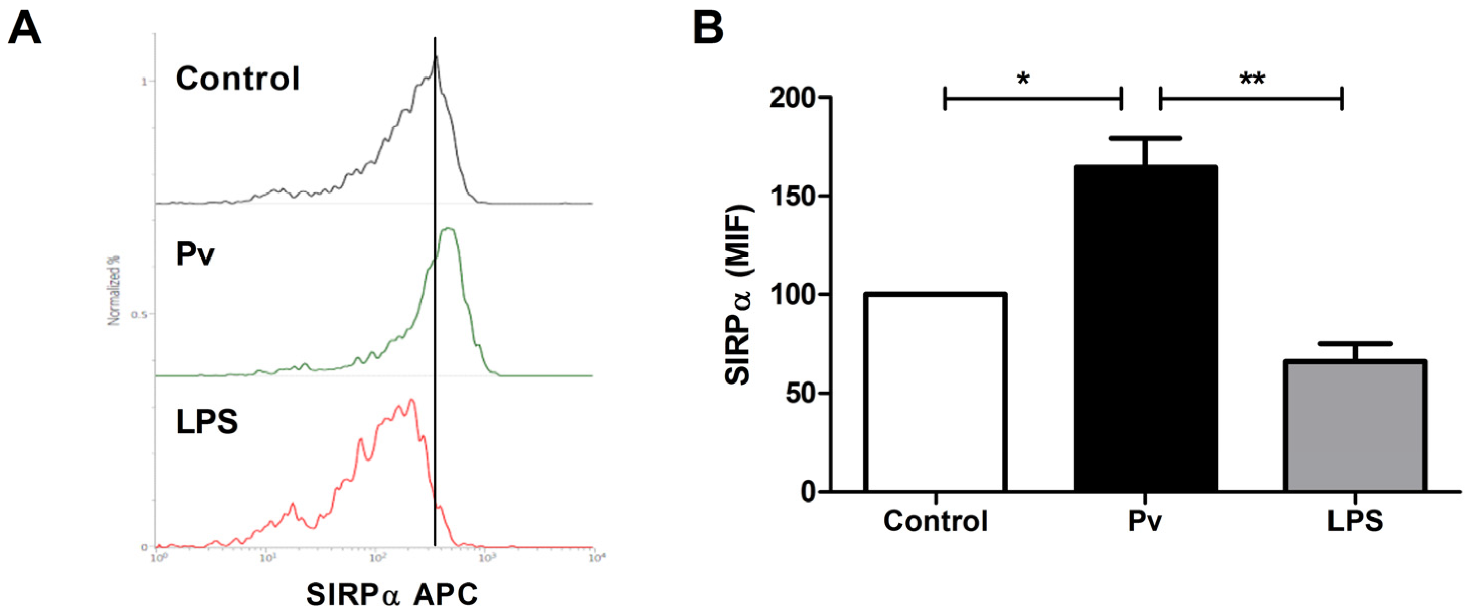
3. Results and Discussion

It is well known that Plasmodium infections suppress lymphoproliferative responses, which negatively impacts the development of an effective antimalarial immunity [23,24,25]. Some immunological mechanisms involved in such immune suppression states have been described and the modulation of innate immune cells by parasites plays a central role [7,26,27]. Thus, in order to study the immunomodulatory potential of malaria parasites on the expression of the innate immune receptor SIRPα, we incubated PBMC from healthy individuals in the presence of crude P. falciparum and P. vivax extracts, using LPS as a control stimulus.
As expected, stimulation of PBMCs with LPS by 24 and 48 h resulted in a decreased expression of SIRPα in the population of monocyte-like cells characterized by morphology criteria in flow cytometry analysis (Figure 1). This is in agreement with previous studies in which Saimiri monkey PBMCs [28], mouse macrophages [29] and DCs derived from human peripheral blood or mouse bone marrow [30] were stimulated with LPS. Similar downregulation of SIRPα has also been described after viral double-stranded RNA (polyI:C) stimulation of mouse macrophages, in which SIRPα signaling was shown to negatively regulate TLR3-dependent antiviral pathways [31]. Indeed, SIRPα is believed to be a negative modulator of TLR signaling in immune innate cells, as overexpression of SIRPα decreases pro-inflammatory cytokine response, while disruption of SIRPα signaling allows the activation of NF-κβ and an increase in pro-inflammatory cytokine production [29,32]. In this line, it has already been demonstrated, using human monocytic cell line THP-1, that LPS acts as a pro-inflammatory stimulus by triggering SIRPα proteolysis [33].
In marked contrast to LPS, P. falciparum crude extract modulated positively SIRPα expression in monocyte-like cells after 24 h incubation (Figure 1), an effect that was shown to be dose-dependent (Figure 2). Similar results were observed when P. vivax antigens were used to stimulate PBMC (Figure 3). Thus, although P. vivax and P. falciparum can cause different clinical and immunological outcomes during acute infection [34,35], the early induction of SIRPα expression might be a common feature avoiding initial parasite clearance and, consequently, allowing the establishment of blood stage. Indeed, besides promoting the engulfment of tumor cells by macrophages in vitro and in vivo [36], disruption of SIRPα signaling can attenuate parasite burden in the course of P. berghei ANKA infection in mice, as well as enhance in vitro phagocytosis of P. falciparum-pRBCs, which implicates SIRPα as an important component controlling malaria blood-stage [37]. Moreover, such involvement of SIRPα in the control of pathogen growth was also demonstrated in two other models, i.e., pneumonia and melioidosis. In the first model, the impaired phagocytic activity of alveolar macrophages against Escherichia coli and Staphylococcus aureus, imposed by primary pneumonia, was related to augmented SIRPα expression, which increased the susceptibility of mice to secondary pneumonia [38]. In contrast, the intracellular killing of the causative agent of melioidosis, Burkholderia pseudomallei, was associated with deflagration of TLR signaling and subsequent inducible nitric oxide synthase response, triggered as a result of downregulation of SIRPα expression in infected macrophages [32]. Nevertheless, both the stimuli and pathways regulating SIRPα expression remain largely unknown.
We finally investigated if Plasmodium antigens differentially modulate the innate immune cells by performing basic phenotyping for classical monocytes (CD14+CD11c+) and myeloid dendritic cells (CD14CD11c+), as classified previously [39]. As shown in Figure 4, P. falciparum extract positively modulated the expression of SIRPα in monocytes, but had no effect on dendritic cells population, whose susceptibility to negative modulation by LPS was similar to monocytes (Figure 4). It is not surprising, however, that subsets of innate immune cells, upon stimulation with certain pathogens, can respond through distinct signaling pathways, thus contributing in opposite ways to the immune response against the infection [40,41,42,43]. Under this premise, it is possible that the initial and transient upregulation of SIRPα induced by malaria parasites solely in monocytes (Figure 1) could favor the establishment of blood infection at the very early phase, in which the parasite load is still limited, while preserving the role of dendritic cells in priming T cell response. Consistent with this possibility, it was already demonstrated that the activity of dendritic cells and macrophages suffers temporal alterations in the course of experimental malaria [44,45] and that, for instance, the impairment of dendritic cells to stimulate T cells was restricted to late, but not early, phases of blood-stage infection with P. chabaudi or P. yoelii [46,47].
In conclusion, our work shows that Plasmodium crude extracts can positively stimulate the expression of SIRPα in cells of the innate immune system, representing a putative mechanism of parasite evasion contributing to the early establishment of blood-stage infection. Nevertheless, further studies, exploring the dynamic of SIRPα expression in the course of infection and its relation to both immune responses and parasite elimination, are still required for a better understanding of the role of SIRPα in malaria. In this regard, a study with malaria patients from the Brazilian Amazon, where P. falciparum and P. vivax are endemic, is currently in progress.

Author Contributions

P.d.C.M.: conduction of experiments, data analysis and writing of the manuscript. H.A.d.S.d.S., C.M.B. and L.S.-d.-O.: conduction of experiments. L.R.P.-R. and C.T.D.-R.: critical review of the manuscript. P.R.R.T.: study design, conduction of experiments, data analysis, and writing and revision of the manuscript. All authors have read and agreed to the published version of the manuscript.

Funding

The study was supported by Instituto Oswaldo Cruz (IOC-Fiocruz); Conselho Nacional de Desenvolvimento Científico e Tecnológico (CNPq) and; Fundação de Amparo à Pesquisa do Estado do Rio de Janeiro (FAPERJ). CTDR is recipient of a Research Productivity Fellowship from the CNPq and, CTDR and PRRT received grants from FAPERJ as “Cientista do Nosso Estado” and “Jovem Cientista do Nosso Estado”, respectively. The Laboratório de Pesquisa em Malária (LPM, IOC, Fiocruz) is a National Institute of Science & Technology (INCT, CNPq, Brazil) Associated Laboratory on Neuroimmunomodulation and also an Associated Laboratory of the Neuroinflammation Network (Faperj, RJ, Brazil).

Institutional Review Board Statement

The study was conducted according to the guidelines of the Declaration of Helsinki, and approved by the Fundação Oswaldo Cruz Research Ethics Committee, CEP-Fiocruz (CAAE 46084015.1.0000.5248/2016).

Informed Consent Statement

Informed consent was obtained from all subjects involved in the study.

Data Availability Statement

The data of the study are contained within the article.

Acknowledgments

We would like to thank the Multi-User Research Facility of Flow Cytometry/Multiparametric Analysis of the Instituto Oswaldo Cruz, Fiocruz-RJ, for sample acquisition.

Conflicts of Interest

The authors declare no conflict of interest.

References

  1. Olive, C. Pattern recognition receptors: Sentinels in innate immunity and targets of new vaccine adjuvants. Expert Rev. Vaccines 2012, 11, 237–256. [Google Scholar] [CrossRef] [PubMed]
  2. Beutler, B. Innate immunity: An overview. Mol. Immunol. 2004, 40, 845–859. [Google Scholar] [CrossRef]
  3. Lang, C.; Gross, U.; Lüder, C.G.K. Subversion of innate and adaptive immune responses by Toxoplasma gondii. Parasitol. Res. 2007, 100, 191–203. [Google Scholar] [CrossRef] [PubMed]
  4. Rénia, L.; Goh, Y.S. Malaria parasites: The great escape. Front. Immunol. 2016, 7, 463. [Google Scholar] [CrossRef] [PubMed] [Green Version]
  5. Rikihisa, Y. Ehrlichia subversion of host innate responses. Curr. Opin. Microbiol. 2006, 9, 95–101. [Google Scholar] [CrossRef]
  6. Elliott, S.R.; Spurck, T.P.; Dodin, J.M.; Maier, A.G.; Voss, T.S.; Yosaatmadja, F.; Payne, P.D.; McFadden, G.I.; Cowman, A.F.; Rogerson, S.J.; et al. Inhibition of dendritic cell maturation by malaria is dose dependent and does not require Plasmodium falciparum erythrocyte membrane protein 1. Infect. Immun. 2007, 75, 3621–3632. [Google Scholar] [CrossRef] [Green Version]
  7. Millington, O.R.; Di Lorenzo, C.; Phillips, R.S.; Garside, P.; Brewer, J.M. Suppression of adaptive immunity to heterologous antigens during Plasmodium infection through hemozoin-induced failure of dendritic cell function. J. Biol. 2006, 5, 5. [Google Scholar] [CrossRef] [Green Version]
  8. Urban, B.; Ferguson, D.; Pain, A.; Willcox, N.; Plebanski, M.; Austyn, J.M.; Roberts, D.J. Plasmodium falciparum-infected erythrocytes modulate the maturation of dendritic cells. Nature 1999, 400, 73–77. [Google Scholar] [CrossRef]
  9. Leitner, W.W.; Krzych, U. Plasmodium falciparum malaria blood stage parasites preferentially inhibit macrophages with high phagocytic activity. Parasite Immunol. 1997, 19, 103–110. [Google Scholar] [CrossRef]
  10. Sampaio, N.G.; Eriksson, E.M.; Schofield, L. Plasmodium falciparum PfEMP1 modulates monocyte/macrophage transcription factor activation and cytokine and chemokine responses. Infect. Immun. 2017, 86, e00447-17. [Google Scholar] [CrossRef] [Green Version]
  11. Schwarzer, E.; Alessio, M.; Ulliers, D.; Arese, P. Phagocytosis of the malarial pigment, hemozoin, impairs expression of major histocompatibility complex class II antigen, CD54, and CD11c in human monocytes. Infect. Immun. 1998, 66, 1601–1606. [Google Scholar] [CrossRef] [PubMed] [Green Version]
  12. Schwarzer, E.; Arese, P. Phagocytosis of malarial pigment hemozoin inhibits NADPH-oxidase activity in human monocyte-derived macrophages. Biochim. Biophys. Acta 1996, 1316, 169–175. [Google Scholar] [CrossRef] [Green Version]
  13. Urban, B.C.; Hien, T.T.; Day, N.P.; Phu, N.H.; Roberts, R.; Pongponratn, E.; Jones, M.; Mai, N.T.H.; Bethell, D.; Turner, G.D.H.; et al. Fatal Plasmodium falciparum malaria causes specific patterns of splenic architectural disorganization. Infect. Immun. 2005, 73, 1986–1994. [Google Scholar] [CrossRef] [Green Version]
  14. Van den Berg, T.K.; van Beek, E.M.; Bühring, H.J.; Colonna, M.; Hamaguchi, M.; Howard, C.J.; Kasuga, M.; Liu, Y.; Matozaki, T.; Neel, B.G.; et al. A nomenclature for signal regulatory protein family members. J. Immunol. 2005, 175, 7788–7789. [Google Scholar] [CrossRef] [Green Version]
  15. Daëron, M.; Jaeger, S.; Du Pasquier, L.; Vivier, E. Immunoreceptor tyrosine-based inhibition motifs: A quest in the past and future. Immunol. Rev. 2008, 224, 11–43. [Google Scholar] [CrossRef] [PubMed]
  16. Gardai, S.J.; Xiao, Y.-Q.; Dickinson, M.; Nick, J.A.; Voelker, D.R.; Greene, K.E.; Henson, P.M. By binding SIRPalpha or calreticulin/CD91, lung collectins act as dual function surveillance molecules to suppress or enhance inflammation. Cell 2003, 115, 13–23. [Google Scholar] [CrossRef] [Green Version]
  17. Janssen, W.J.; McPhillips, K.A.; Dickinson, M.G.; Linderman, D.J.; Morimoto, K.; Xiao, Y.Q.; Oldham, K.M.; Vandivier, R.W.; Henson, P.M.; Gardai, S.J. Surfactant proteins A and D suppress alveolar macrophage phagocytosis via interaction with SIRP alpha. Am. J. Respir. Crit. Care Med. 2008, 178, 158–167. [Google Scholar]
  18. Latour, S.; Tanaka, H.; Demeure, C.; Mateo, V.; Rubio, M.; Brown, E.J.; Maliszewski, C.; Lindberg, F.P.; Oldenborg, A.; Ullrich, A.; et al. Bidirectional negative regulation of human T and dendritic cells by CD47 and its cognate receptor signal-regulator protein-alpha: Down-regulation of IL-12 responsiveness and inhibition of dendritic cell activation. J. Immunol. 2001, 167, 2547–2554. [Google Scholar] [CrossRef] [PubMed] [Green Version]
  19. Smith, R.E.; Patel, V.; Seatter, S.D.; Deehan, M.R.; Brown, M.H.; Brooke, G.P.; Goodridge, H.S.; Howard, C.J.; Rigley, K.P.; Harnett, W.; et al. A novel MyD-1 (SIRP-1alpha) signaling pathway that inhibits LPS-induced TNFalpha production by monocytes. Blood 2003, 102, 2532–2540. [Google Scholar] [CrossRef] [PubMed] [Green Version]
  20. Trager, W.; Jensen, J.B. Human malaria parasites in continuous culture. Science 1976, 193, 673–675. [Google Scholar] [CrossRef]
  21. Moll, K.; Kaneko, A.; Scherf, A.; Wahlgren, M. Methods in Malaria Research; MR4/ATCC: Manassas, VA, USA, 2013. [Google Scholar]
  22. Carvalho, B.O.; Lopes, S.; Nogueira, P.A.; Orlandi, P.P.; Bargieri, D.; Blanco, Y.C.; Mamoni, R.; Leite, J.A.; Rodrigues, M.M.; Soares, I.D.S.; et al. On the cytoadhesion of Plasmodium vivax-infected erythrocytes. J. Infect. Dis. 2010, 202, 638–647. [Google Scholar]
  23. Goonewardene, R.; Carter, R.; Gamage, C.P.; Del Giudice, G.; David, P.H.; Howie, S.; Mendis, K.N. Human T cell proliferative responses to Plasmodium vivax antigens: Evidence of immunosuppression following prolonged exposure to endemic malaria. Eur. J. Immunol. 1990, 20, 1387–1391. [Google Scholar] [CrossRef] [PubMed]
  24. Riley, E.M.; Andersson, G.; Otoo, L.N.; Jepsen, S.; Greenwood, B.M. Cellular immune responses to Plasmodium falciparum antigens in Gambian children during and after an acute attack of falciparum malaria. Clin. Exp. Immunol. 1988, 73, 17–22. [Google Scholar]
  25. Wangoo, A.; Ganguly, N.K.; Mahajan, R.C. Immunosuppression in murine malaria: Suppressor role of macrophages and their products during acute and chronic Plasmodium berghei infection. APMIS 1990, 98, 407–414. [Google Scholar] [CrossRef] [PubMed]
  26. Bobade, D.; Khandare, A.V.; Deval, M.; Shastry, P.; Deshpande, P. Hemozoin-induced activation of human monocytes toward M2-like phenotype is partially reversed by antimalarial drugs-chloroquine and artemisinin. MicrobiologyOpen 2019, 8, e00651. [Google Scholar] [CrossRef] [PubMed]
  27. Vallejo, A.F.; Read, R.C.; Arevalo-Herrera, M.; Herrera, S.; Elliott, T.; Polak, M.E. Malaria systems immunology: Plasmodium vivax induces tolerance during primary infection through dysregulation of neutrophils and dendritic cells. J. Infect. 2018, 77, 440–447. [Google Scholar] [CrossRef] [PubMed] [Green Version]
  28. Souza, H.A.D.S.D.; Costa-Correa, E.H.; Bianco-Junior, C.; Andrade, M.C.R.; Lima-Junior, J.D.C.; Pratt-Riccio, L.R.; Daniel-Ribeiro, C.T.; Totino, P.R.R. Detection of signal regulatory protein α in Saimiri sciureus (Squirrel Monkey) by anti-human monoclonal antibody. Front. Immunol. 2017, 8, 1814. [Google Scholar] [CrossRef]
  29. Kong, X.-N.; Yan, H.-X.; Chen, L.; Dong, L.-W.; Yang, W.; Liu, Q.; Yu, L.-X.; Huang, D.-D.; Liu, S.-Q.; Liu, H.; et al. LPS-induced down-regulation of signal regulatory protein α contributes to innate immune activation in macrophages. J. Exp. Med. 2007, 204, 2719–2731. [Google Scholar] [CrossRef]
  30. Liu, Q.; Wen, W.; Tang, L.; Qin, C.-J.; Lin, Y.; Zhang, H.-L.; Wu, H.; Ashton, C.; Wu, H.-P.; Ding, J.; et al. Inhibition of SIRPα in dendritic cells potentiates potent antitumor immunity. Oncoimmunology 2016, 5, e1183850. [Google Scholar] [CrossRef] [Green Version]
  31. Dong, L.-W.; Kong, X.-N.; Yan, H.-X.; Yu, L.-X.; Chen, L.; Yang, W.; Liu, Q.; Huang, D.-D.; Wu, M.-C.; Wang, H.-Y. Signal regulatory protein alpha negatively regulates both TLR3 and cytoplasmic pathways in type I interferon induction. Mol. Immunol. 2008, 45, 3025–3035. [Google Scholar] [CrossRef]
  32. Baral, P.; Utaisincharoen, P. Involvement of signal regulatory protein α, a negative regulator of Toll-like receptor signaling, in impairing the MyD88-independent pathway and intracellular killing of Burkholderia pseudomallei-infected mouse macrophages. Infect. Immun. 2012, 80, 4223–4231. [Google Scholar] [CrossRef] [Green Version]
  33. Londino, J.D.; Gulick, D.; Isenberg, J.S.; Mallampalli, R.K. Cleavage of signal regulatory protein α (SIRPα) enhances inflammatory signaling. J. Biol. Chem. 2015, 290, 31113–31125. [Google Scholar] [CrossRef] [PubMed] [Green Version]
  34. Burel, J.G.; Apte, S.H.; McCarthy, J.S.; Doolan, D.L. Plasmodium vivax but not Plasmodiumfalciparum blood-stage infection in humans is associated with the expansion of a CD8+ T cell population with cytotoxic potential. PLoS Negl. Trop. Dis. 2016, 10, e0005031. [Google Scholar]
  35. Goncalves, R.M.; Scopel, K.K.G.; Bastos, M.S.; Ferreira, M.U. Cytokine balance in human malaria: Does Plasmodium vivax elicit more inflammatory responses than Plasmodium falciparum? PLoS ONE 2012, 7, e44394. [Google Scholar] [CrossRef] [PubMed]
  36. Veillette, A.; Chen, J. SIRPα-CD47 immune checkpoint blockade in anticancer therapy. Trends Immunol. 2018, 39, 173–184. [Google Scholar] [CrossRef] [PubMed]
  37. Ayi, K.; Lu, Z.; Serghides, L.; Ho, J.M.; Finney, C.; Wang, J.; Liles, W.C.; Kain, K.C. CD47-SIRPα interactions regulate macrophage uptake of Plasmodium falciparum-infected erythrocytes and clearance of malaria in vivo. Infect. Immun. 2016, 84, 2002–2011. [Google Scholar] [CrossRef] [PubMed] [Green Version]
  38. Roquilly, A.; Jacqueline, C.; Davieau, M.; Mollé, A.; Sadek, A.; Fourgeux, C.; Rooze, P.; Broquet, A.; Misme-Aucouturier, B.; Chaumette, T.; et al. Alveolar macrophages are epigenetically altered after inflammation, leading to long-term lung immunoparalysis. Nat. Immunol. 2020, 21, 636–648. [Google Scholar] [CrossRef]
  39. Haniffa, M.; Bigley, V.; Collin, M. Human mononuclear phagocyte system reunited. Semin. Cell Dev. Biol. 2015, 41, 59–69. [Google Scholar] [CrossRef]
  40. Galgani, M.; Busiello, I.; Censini, S.; Zappacosta, S.; Racioppi, L.; Zarrilli, R. Helicobacter pylori induces apoptosis of human monocytes but not monocyte-derived dendritic cells: Role of the cag pathogenicity island. Infect. Immun. 2004, 72, 4480–4485. [Google Scholar] [CrossRef] [Green Version]
  41. Khan, N.; Vidyarthi, A.; Pahari, S.; Agrewala, J.N. Distinct strategies employed by dendritic cells and macrophages in restricting Mycobacterium tuberculosis infection: Different philosophies but same desire. Int. Rev. Immunol. 2016, 35, 386–398. [Google Scholar] [CrossRef]
  42. Manuzak, J.; Dillon, S.; Wilson, C. Differential interleukin-10 (IL-10) and IL-23 production by human blood monocytes and dendritic cells in response to commensal enteric bacteria. Clin. Vaccine Immunol. 2012, 19, 1207–1217. [Google Scholar] [CrossRef] [PubMed] [Green Version]
  43. Norimatsu, M.; Harris, J.; Chance, V.; Dougan, G.; Howard, C.J.; Villarreal-Ramos, B. Differential response of bovine monocyte-derived macrophages and dendritic cells to infection with Salmonella typhimurium in a low-dose model in vitro. Immunology 2003, 108, 55–61. [Google Scholar] [CrossRef]
  44. Ahvazi, B.C.; Jacobs, P.; Stevenson, M.M. Role of macrophage-derived nitric oxide in suppression of lymphocyte proliferation during blood-stage malaria. J. Leukoc. Biol. 1995, 58, 23–31. [Google Scholar] [CrossRef] [PubMed]
  45. Fu, Y.; Ding, Y.; Zhou, T.; Fu, X.; Xu, W. Plasmodium yoelii blood-stage primes macrophage-mediated innate immune response through modulation of toll-like receptor signaling. Malar. J. 2012, 11, 104. [Google Scholar] [CrossRef] [Green Version]
  46. Ocaña-Morgner, C.; A Wong, K.; Rodriguez, A. Interactions between dendritic cells and CD4+ T cells during Plasmodium infection. Malar. J. 2008, 7, 88. [Google Scholar] [CrossRef] [Green Version]
  47. Sponaas, A.-M.; Belyaev, N.; Falck-Hansen, M.; Potocnik, A.; Langhorne, J. Transient deficiency of dendritic cells results in lack of a merozoite surface protein 1-specific CD4 T cell response during peak Plasmodium chabaudi blood-stage infection. Infect. Immun. 2012, 80, 4248–4256. [Google Scholar] [CrossRef] [PubMed] [Green Version]
Figure 1. Modulation of SIRPα expression by P. falciparum crude extract. PBMCs from healthy individuals were stimulated for 24 and 48 h with P. falciparum extract (10 µg/mL) or LPS (5 µg/mL) and SIRPα expression on cell surface of monocyte-like cells was evaluated by flow cytometry using APC-conjugated anti-SIRPα monoclonal antibody. (A,B) Representative cytometric analysis of SIRPα expression (B) in gated monocyte-like cells (A; P1) after stimulation with P. falciparum antigens (Pf) or LPS. (C) Levels of SIRPα expression on monocyte-like cells population (P1), as measured by mean fluorescence intensity (MFI). Non-stimulated PBMCs were used as control (Control). Data are shown as mean ± standard error (SEM) and represent one of two independent experiments performed with PBMC samples from five individuals. **: p < 0.01; ***: p < 0.001.
Microorganisms 10 00903 g001
Figure 2. Dose-dependent effect of P. falciparum extract on SIRPα expression. PBMCs from healthy individuals were stimulated for 24 h with crescent concentrations of P. falciparum extract (Pf; 0.1, 1.0 and 10 µg/mL) or 5 µg/mL LPS and, the levels of SIRPα expression (MFI) on cell surface of monocyte-like cells was evaluated by flow cytometry, as in Figure 1. Data are shown as mean ± standard error (SEM) and represent one of two independent experiments performed with PBMC samples from five individuals. **: p < 0.01; ***: p < 0.001, as compared with non-stimulated cells (Control).
Microorganisms 10 00903 g002
Figure 3. Modulation of SIRPα expression by P. vivax crude extract. PBMCs from healthy individuals were stimulated for 24 h with P. vivax extract (10 µg/mL) or LPS (5 µg/mL) and SIRPα expression on cell surface of monocyte-like cells was evaluated by flow cytometry using APC-conjugated anti-SIRPα monoclonal antibody. (A) Representative histogram analysis of SIRPα expression in gated monocyte-like cells after stimulation with P. vivax antigens or LPS. (B) Levels of SIRPα expression on monocyte-like cells population, as measured by mean fluorescence intensity (MFI). Non-stimulated PBMCs were used as control (Control). Data are shown as mean ± standard error (SEM) and represent one of two independent experiments performed with PBMC samples from five individuals. *: p < 0.05; **: p < 0.01.
Microorganisms 10 00903 g003
Figure 4. Cell type-specific modulation of SIRPα expression by P. falciparum crude extract. PBMCs from healthy individuals were stimulated for 24 h with P. falciparum extract (10 µg/mL) or LPS (5 µg/mL) and SIRPα expression on cell surface of monocytes and dencritic cells was evaluated by flow cytometry. (A) Representative phenotyping analysis of classical monocytes (P2; CD14+CD11+) and myeloid dendritic cells (P3; CD14CD11+) populations. (B,C) Levels of SIRPα expression in gated monocytes (B) and dendritic cells (C), as measured by mean fluorescence intensity (MFI). Non-stimulated PBMCs were used as control (Control). Data are shown as mean ± standard error (SEM) and represent one of two independent experiments performed with PBMC samples from five individuals. *: p < 0.05; **: p < 0.01; ***: p < 0.001.
Microorganisms 10 00903 g004
Publisher’s Note: MDPI stays neutral with regard to jurisdictional claims in published maps and institutional affiliations.

