Communication

Exploring Mitogenomes Diversity of Fusarium musae from Banana Fruits and Human Patients

1
Department of Food, Environmental and Nutritional Science (DeFENS), University of Milan, Via Celoria 2, 20133 Milan, Italy
2
Department of Biomedical Sciences for Health, University of Milan, Via Pascal 36, 20133 Milan, Italy
*
Author to whom correspondence should be addressed.
These authors contributed equally to this work.
Submission received: 1 May 2022 / Revised: 21 May 2022 / Accepted: 23 May 2022 / Published: 28 May 2022
(This article belongs to the Special Issue Human Pathogens in Primary Production Systems)

Abstract

:
Fusarium musae has recently been described as a cross-kingdom pathogen causing post-harvest disease in bananas and systemic and superficial infection in humans. The taxonomic identity of fungal cross-kingdom pathogens is essential for confirming the identification of the species on distant infected hosts. Understanding the level of variability within the species is essential to decipher the population homogeneity infecting human and plant hosts. In order to verify that F. musae strains isolated from fruits and patients are part of a common population and to estimate their overall diversity, we assembled, annotated and explored the diversity of the mitogenomes of 18 F. musae strains obtained from banana fruits and human patients. The mitogenomes showed a high level of similarity among strains with different hosts’ origins, with sizes ranging from 56,493 to 59,256 bp. All contained 27 tRNA genes and 14 protein-coding genes, rps3 protein, and small and large ribosomal subunits (rns and rnl). Variations in the number of endonucleases were detected. A comparison of mitochondrial endonucleases distribution with a diverse set of Fusarium mitogenomes allowed us to specifically discriminate F. musae from its sister species F. verticillioides and the other Fusarium species. Despite the diversity in F. musae mitochondria, strains from bananas and strains from human patients group together, indirectly confirming F. musae as a cross-kingdom pathogen.

1. Introduction

Different fungal species are able to cross hosts, causing diseases not only in plants but also in humans and animals. In general, cross-kingdom fungi are weak pathogens for both plants and humans; they can also be asymptomatic in plants but can have clinical significance, especially in people with impaired immunity or those who have sustained penetrating trauma [1]. The role of food and agriculture in the transmissibility of cross-kingdom pathogens deserves to be studied accurately.
Fusarium musae VanHove is a pathogenic species belonging to the F. fujikuroi species complex [2]. It causes crown rot in bananas, a post-harvest disease [3,4], and it also causes keratitis and skin infections as well as systemic infections in immunocompromised patients [5,6].
Based on multilocus sequence typing, F. musae can be distinguished from its sister species F. verticillioides [2]. The two species show a diverse susceptibility to azoles, with F. musae having a higher tolerance to some fungicides compared to F. verticillioides [7].
F. musae has been identified on banana fruits in Central American regions, in the Philippines and Canary Islands and in European and Japanese markets of banana fruits. In patients, it has been identified in the US and Europe [5,8]. Understanding the level of diversity of strains infecting humans and banana fruits may help address the question about the transmissibility of F. musae in different hosts [9].
Mitochondrial genomes evolve independently of and faster than the nuclear genome [10]. Mitogenomes have also been proposed as a useful tool for diagnostic purposes in Fusarium species [11]. The concept of using mitogenome diversity for the identification of species mainly derives from their higher DNA copy number compared with nuclear DNA, and hence the high recovery and amplification success in eukaryotes where insufficient phylogenetic signals have accumulated in nuclear genes [12].
Fungal mitochondrial genomes are typically small, circular and double-stranded DNA molecules, with a typical set of mitochondrial genes with identical gene order. Extensive mitochondrial genome comparisons within the fungal kingdom have shown that any gene has a mitochondrial localization in all fungal species [13], suggesting that the use of mitochondrial gene diversity cannot be applied as a universal marker for fungi. Nonetheless, the analysis of mitochondrial diversity can be successfully applied to differentiate species within orders [14] or species [15,16]. For example, a unique feature apparently common in all Fusarium species is the presence of a large open reading frame with an unknown function (LV-uORF) firstly described in mitogenomes of F. graminearum, F. verticillioides and F. solani [17] and probably acquired prior to the divergence of Fusarium species.
In addition, fungal mitogenomes harbour a variable number of mobile genetic elements (MGEs) such as intron-associated homing endonucleases (HEGs), which have invaded mitogenomes throughout their evolution. Most of the MGEs insertion sites are highly conserved [18] and occur in mitochondrial protein-coding genes but can display remarkably different MGE densities. The same MGEs can be irregularly distributed in evolutionarily distant species and mosaicism in MGE patterns can be found between different populations or even strains of the same species, driving large genome size differences among them. Exploring MGE distribution can help discriminate species and subgroups, tracking the spread of fungal populations [19].
We, therefore, explored the mitogenomes of a collection of 18 F. musae strains in order to describe their mitochondrial diversity. The other objective of this study was to discover whether the pattern of MGE diversity might facilitate the identification of F. musae species within the Fusarium genus. Moreover, we explored whether F. musae from bananas and human patients shared the same mitochondrial sequence in order to see if genetic subgroups associated with the host origin could be identified.

2. Materials and Methods

2.1. DNA Extraction and Sequencing

DNA used for sequencing was obtained from fresh mycelia of 16 strains according to a modified CTAB method [20], followed by Genomic tips column purification (Qiagen, Germantown, MD, USA). Sequencing was carried out using Illumina Hiseq 2000 (151 bp x2) by Novogene (Cambridge, UK). Mitogenomes of NRRL25059 [21] and F31 [22] strains were obtained from the NCBI database.

2.2. Other Fungal Mitochondrial Genomes

Mitogenomes from fungal strains belonging to different Fusarium species (see supplementary Table S1) were retrieved from NCBI and partially annotated if the annotation was missing (protein genes) using MFannot online software (University of Montreal, Montreal, QC, Canada), (https://megasun.bch.umontreal.ca/cgi-bin/mfannot/mfannotInterface.pl (accessed on 4 March 2022)).

2.3. Assembly and Annotation of Mitogenome

Assembly for all F. musae mitochondrial DNAs was carried out de novo with NOVOplasty 4.2 [23] using F. musae F31 strain as a reference and the first 715 bp from cox1 gene as seed sequence. The obtained mitogenomes were then annotated by integrating MFannot and RNAWeasel [24]. In order to improve the final annotation, the BlastX tool was used to define the nature of the different annotated ORFs and endonucleases.

2.4. Alignment of Protein Genes

Mitogenomes comparison included a total of 14 concatenated sequences, which represent the 14 coding protein nucleotide sequences corresponding to the set of conserved mitochondrial genes, using the “concatenated sequences” tool on Geneious Prime Software (Biomatters Ltd., Auckland, Australia). Sequences were then aligned using the MAFFT alignment tool with default parameters and visually checked.

2.5. MGE Analysis (Minimap2)

Mobile genetic element analysis was performed using the distribution pattern of F. musae IUM_11-0508 as a reference. The goal was to identify the distribution of the same MGEs across Fusarium genus diversity. After MGE sequences collection, the Minimap2 tool on Geneoius Prime Software (setting a threshold of identity at 90% and minimum coverage of 75%) against all other Fusarium mtDNA and BlastX (coverage and identity threshold: 75 and 90% respectively) analyses were used to identify the presence of those sequences in other Fusarium species and other fungal species.

2.6. Analysis of Nad1 Intron

To confirm the sequence of the intron in nad1 in order to describe the possible path of inactivation of the nad1 endonuclease present in F. verticillioides, primers musaeendoF1- (5′-TGGAAAATCAGCAGGTTGACC-3′) and musaeendoR1- (5′-ACTGCTGCGTGTTCTGTCAT-3′) were designed on the coding region of the nad1 gene using primer 3 software online (https://primer3.ut.ee/, accessed on 4 November 2021). PCR amplification was carried out using Q5 master mix (NEB, Ipswich, MA, UK) in a total of 25 microliters of reaction using a PCR program that included 3 min at 95 °C, 35 cycles including 20 s at 95 °C, 20 s at 60 °C and 1 min at 72 °C, followed by a last step at 72 °C for 5 min, which was carried out in a VeritiPRO Thermal Cycler (Applied Byosystem, Waltham, MA, USA). Sequences were obtained by PCR purification and Sanger sequencing (Eurofins genomics, GER, Ebersberg, Germany). The obtained sequences were then manually checked and assembled and then aligned using the MAFFT tool in Geneious Prime Software to verify the correctness of the assembly. Moreover, BlastN, BlastX and BlastP were used to characterize the mutations occurring in the region and to identify the inactivation of endonuclease functional domains.

2.7. Haplotype Analysis of Mitogenomes

Whole MAFFT aligned mitogenomes using Geneious prime software were manually checked and analysed using Median Joining Network in Popart [25].

3. Results

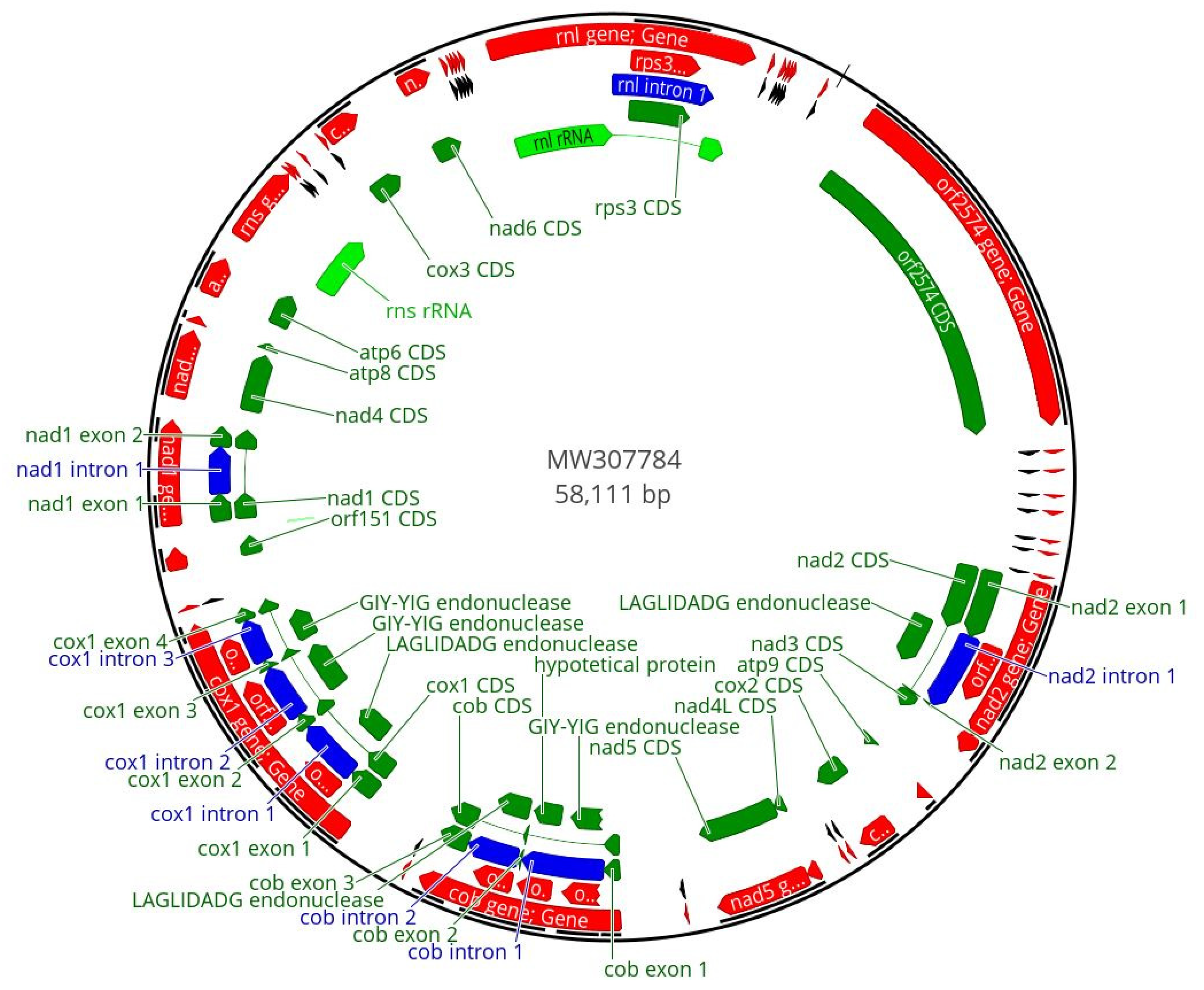
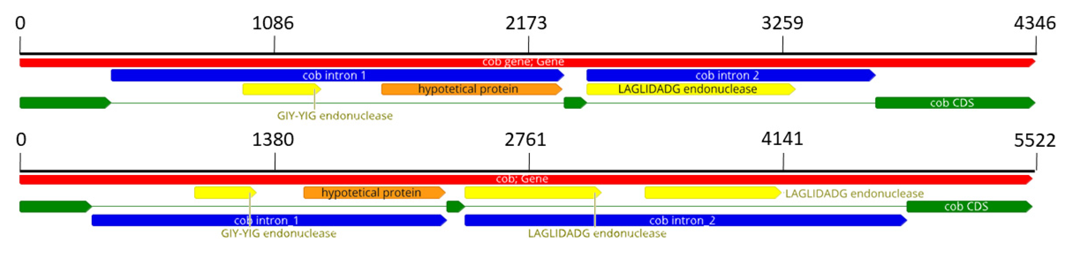
3.1. F. musae Mitogenomes

Mitogenomes of F. musae strains ranged from 56,439 to 59,256 bp (Table 1).
All mitogenomes had identical gene and tRNA distribution. Analysis carried out on the nine annotated ORFs, using the BlastP tool, showed that six of these ORFs represent mobile genetic elements (MGEs) of LAGLIDADG and GIY-YIG family, one represents a hypothetical protein, while two have to be considered ORFs with unassigned functions.
Further investigation of nad1_intron revealed the presence of an inactivated endonuclease with very high similarity to the F. verticillioides GIY-YIG endonuclease positioned within the nad1 gene.
The final annotation of the mitogenomes of F. musae strains includes all the 14 protein-coding genes (atp6, atp8, atp9, cob, cox1, cox2, cox3, nad1, nad2, nad3, nad4, nad4 L, nad5, nad6); the ribosomal protein rps3 and two ribosomal rRNA (rns and rnl); 27 tRNA genes and 2 ORFs; 6 MGEs (3 LAGLIDADG and 3 GIY-YIG positioned in nad2 intron (n = 1), in cob introns (n = 2) and in cox1 introns (n = 3)); 1 hypothetical protein; and 1 inactivated GIY-YIG endonuclease present in nad1 intron (not annotated). Figure 1 shows the example of the IUM_11-0508 strain mitogenome.
Size difference among mitogenomes was mostly due to variations in two regions: for strain ITEM_1149, a deletion in the ORF located between rps3 and nad2 reduces the mitogenome to 56,493, while for strains IHEM_20180, MUCL_51371, NRRL_28893 and NRRL_28897, the increased size above 59 kb is due to changes in the cob region. The cob gene in F. musae includes three exons and two introns (Figure 2). The first intron is composed of an endonuclease with GIY-YIG domain and a hypothetical protein, common in all the F. musae strains. The second intron shows differences within the species: in four strains (IHEM_20180, NRRL_28893, NRRL_28897, MUCL_51371), it shows a duplication of a 247 AA LAGLIDADG endonuclease, while in all the other strains, a single LAGLIDAGD of 296 AA is present (Figure 2).
BlastX analysis on these two endonucleases showed different LAGLIDADG domains for the two endonucleases (LAGLIDADG_1 and LAGLIDADG_2). Investigation for a similar pattern in other species showed that a similar duplication could be observed in F. bactridioides, F. begoniae and F. pseudograminearum as well as in Cladobotryum mycophilum (Supplementary Table S2).
F. verticillioides has an endonuclease within the nad1 gene. Interestingly, F. musae strains also have an intron in nad1. Intron annotation using alignment and BlastX tool showed that F.musae introns have highly similar sequences to the endonuclease of F. verticillioides, but the presence of different mutations leading to stop codons and frame shifts suggest the inactivation of the endonuclease and the loss of functional domains.
Analysing endonad1 (the most variable endonuclease within F. musae species) using F. verticillioides as a reference, we could identify three different ways of silencing the endonuclease in our F. musae population that are caused by insertion and nonsynonymous substitution to add stop codons in the sequence of the endonuclease (Figure 3 and Supplementary Figure S1).

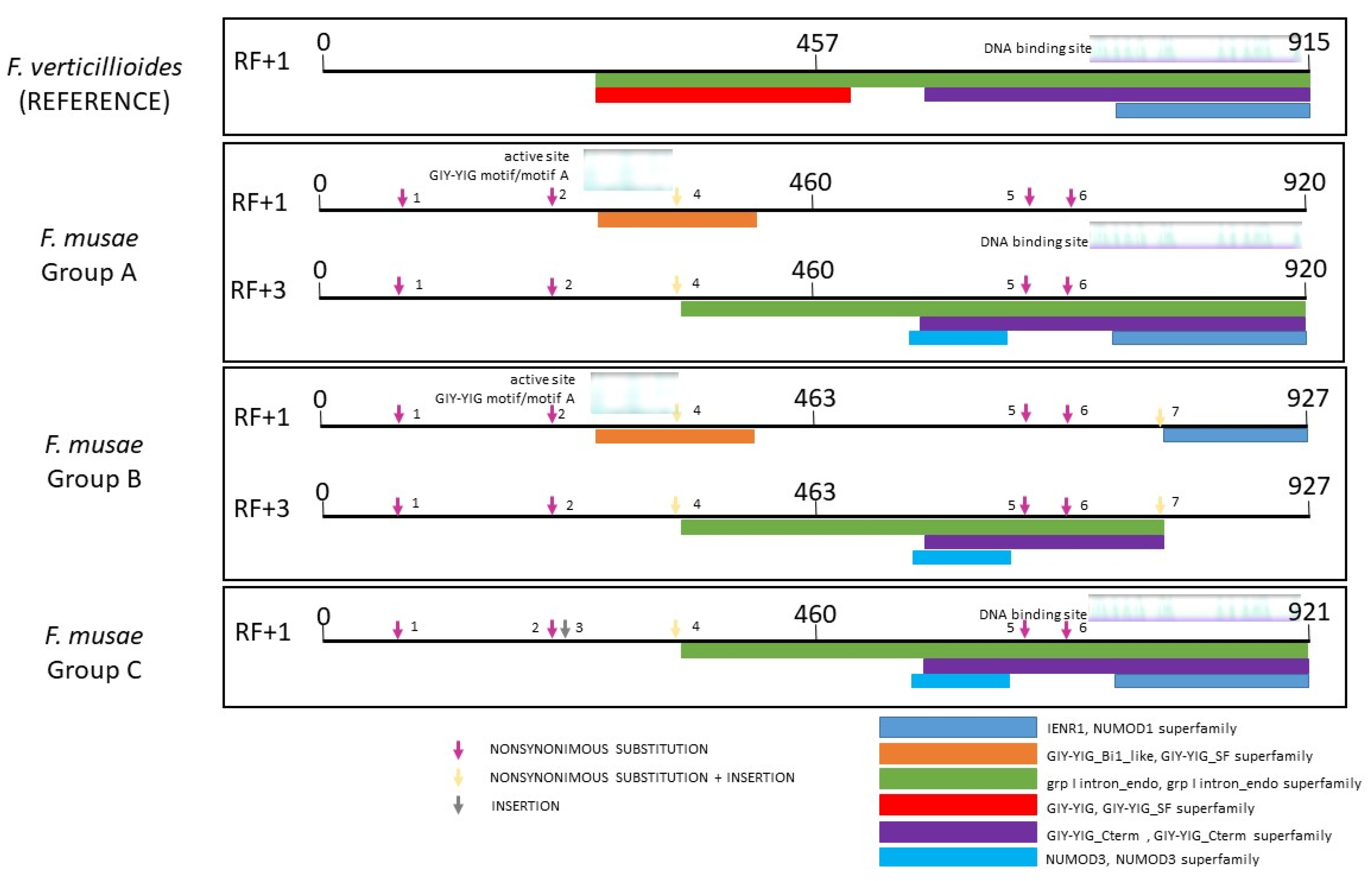
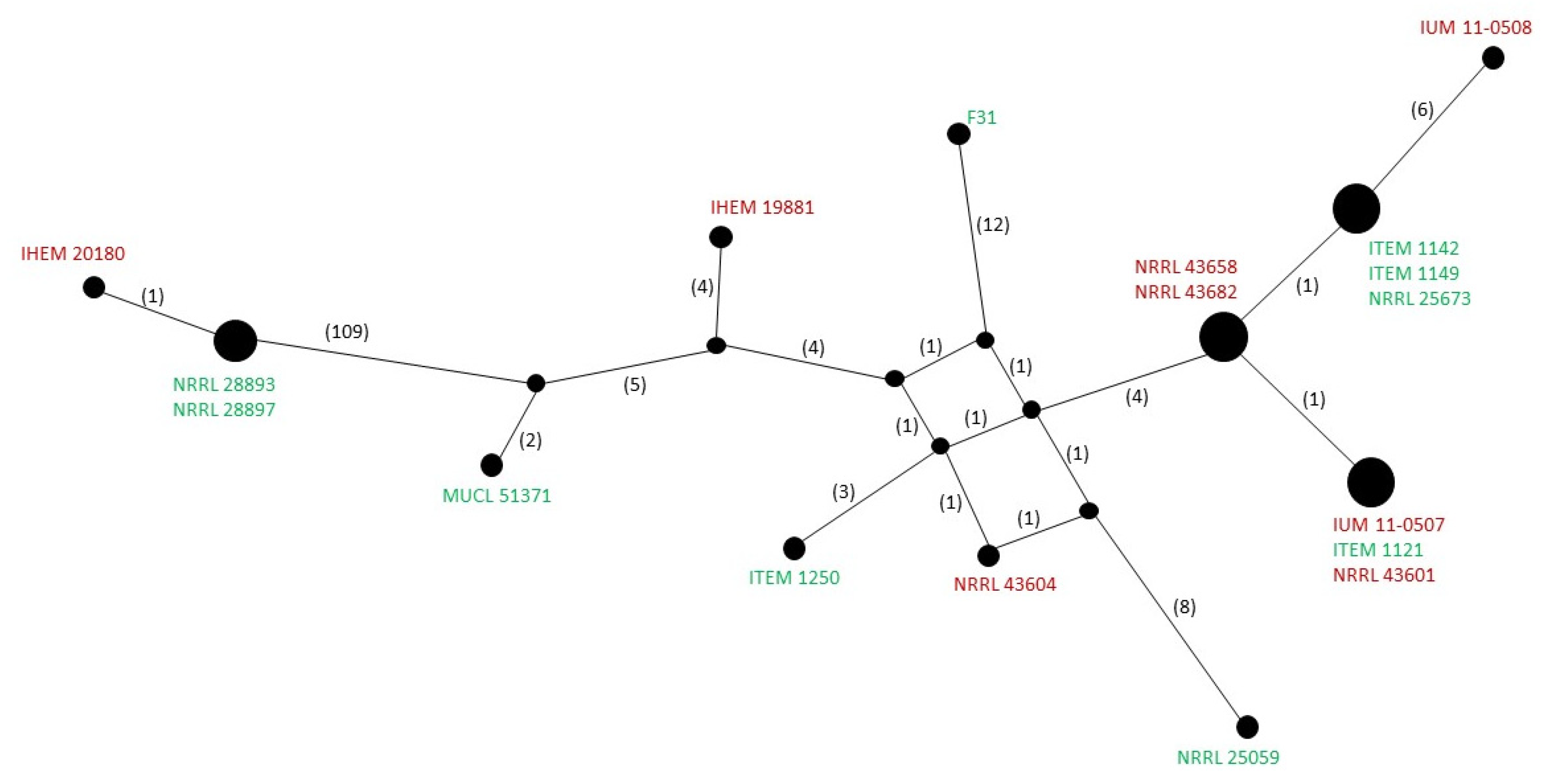
3.2. Diversity within F. musae

Given the limited exploitation of mitogenomes for population studies, the other goal of our work was to assess whether mitochondrial diversity observed within the species could be explored to differentiate species subgroups.
Triest and Hendrickx [9] hypothesized that F. musae may have been transmitted from bananas to patients. To confute this hypothesis, we would expect that the hosts’ origin can be associated with different subgroups within the species, suggesting a host specialization driven by evolutionary constraints [26]. We therefore tested whether F. musae mitochondria diversity could be used to clearly separate strains obtained from humans and bananas. The overall diversity of mitogenome haplotypes (Figure 4) suggests that different subgroups of F. musae strains exist within the analysed population.
This is consistent with the existence of different nuclear gene haplotypes [2]. Interestingly, at least one set of strains belonging to the same mitochondrial haplotype includes both human and banana derived strains from different geographic regions (ITEM_1121 from a banana fruit from Panama, IUM_11-0507 from a patient in Greece and NRRL_43601 from a patient in Maryland, USA), supporting the hypothesis that the infection of banana fruits and human patients occurs from strains with similar genetic profile.

3.3. The Diagnostic Power of Mitochondrial Genomes

Another objective of the paper was to explore the use of mitochondrial diversity as a tool for the detection of species. The distribution of MEGs in a mitogenome may differentiate species. The hypothesis has been validated for other Fusarium species [27]. One of the major diagnostic challenges in F. musae is to discriminate it from F. verticillioides, as morphologically, the species are often misclassified [5,8].
We therefore compared the F. musae pattern of MGE (Supplementary Table S3) with all the other available Fusarium mtDNAs (Supplementary Table S1). The 6 + 1 MGEs were extracted and searched in all other mtDNAs. MGE distribution within the Fusarium genus showed discontinuity. In particular, F. musae species showed a specific pattern of MGE distribution that is unique within the Fusarium genus. It is possible to identify F. musae species based on the unique presence of two MGEs (nad1 intron that is showing polymorphic inactivations and endo1cox), offering potentially a very powerful methodology to identify F. musae.

4. Discussion

This study, analysing the mitochondrial genomes of 18 F. musae strains, confirms previous observations based on nuclear genes (TEF and RPB2) [5,7], which showed that F. musae strains from banana and human patients are interspersed in the species tree. This indirectly confirms the ability of F. musae to cross-infect distant hosts. One strain, F31, showed a nonsynonymous substitution that may indicate some divergence within the species. Further analyses to appropriately characterize the strain are warranted. Within F. musae species, nad5 proved to be the gene with higher recombinations, as observed previously in other species within the F. fujikuroi species complex [28]. Our mitogenome study of the F. musae population confirms that intergenic regions and endonucleases may be exploited to identify subgroups within a species [15]. As observed in other fungal species [29,30], we also observed that major contributors to mitogenome size diversity within a species are intron rearrangements. In our population, two regions showed variability in size: the ORF located between rps3 and nad2 and the intron between exon2_cob and exon3_cob. Future studies may focus on the diversity of these regions in a larger F. musae population.
We observed different haplotype groups comparing whole mitochondrial diversity in F. musae. Previous analysis of nuclear gene haplotype diversity carried out on F. musae strains [2] showed the existence of nine haplotypes based on RPB2, B-tub and TEF diversity. Our panel of strains included representatives of six haplotypes. We could verify that nuclear gene haplotypes were all distinguished in different mitogenome groups. A larger dataset of mitogenomes needs to be analysed to verify the correspondence of nuclear gene haplotypes and mitochondrial whole diversity haplotypes. This preliminary observation seems to confirm that in F. musae, mitochondrial diversity is concordant with nuclear gene variations, as observed for other fungal pathogens [30], and it is therefore a valuable tool for strain typing.
The use of mitochondrial genomes for diagnostic purposes has been often proposed and explored [19,31,32,33]. We could verify that all F. musae strains with different geographic origins (Europe, Asia, America) and different years of isolation (from 1991 to 2013) share the same specific co-occurrence of two endonucleases that allow us to differentiate them from all other Fusarium species whose mitogenome is available. With the increased number of available genomes within single species, the possibility to test the hypothesis of using MGE distribution for species discrimination will be tested thoroughly.
Our study demonstrates that the variable landscape of MGEs is the most prominent type of variation among mitogenomes, including those of closely related F. verticillioides and F. musae, as shown here for the intron of nad1, contributing to expanding the knowledge on inter- and intraspecies mitochondrial diversity in the F. fujikuroi species complex [34]. Moreover, it shows that intraspecies diversity in mitochondrial size can be due to the acquisition or loss of mobile elements. The presence of strains with a human and a banana origin in the same mitochondrial haplotype group is an indirect confirmation that F. musae is able to cause diseases in both humans and plants. Therefore, studying the disease mechanisms of F. musae on hosts belonging to the two kingdoms is warranted to shed light on the infection arsenal that this cross-kingdom pathogen employs.

Supplementary Materials

The following supporting information can be downloaded at: https://0-www-mdpi-com.brum.beds.ac.uk/article/10.3390/microorganisms10061115/s1, Table S1: Presence of F. musae endonucleases in other Fusarium mitogenomes; Table S2: Coverage and Identity of best hits using Blastx with endonucleases of cob gene of the four F. musae strains with longer mitochondrial DNA.; Table S3: Endonuclease identity in all F. musae strains; Figure S1: Whole nucleotide alignment of intron of nad1 gene in F. verticillioides and in the 18 F. musae strains.

Author Contributions

Conceptualization, M.P.; methodology, V.T. and L.D.; software, L.D.; validation, A.P., M.C.E., A.M.T., A.K., M.S. and P.C.; formal analysis, M.P.; investigation, L.D., V.T., M.P., A.K., A.P. and M.C.E.; resources, A.M.T., M.S., P.C. and M.P.; data curation, L.D. and V.T.; writing—original draft preparation, M.P., L.D. and V.T.; writing—review and editing, A.K., P.C., M.S., A.M.T., A.P. and M.C.E.; visualization, L.D. and V.T.; supervision, M.P., A.M.T. and P.C.; funding acquisition, M.P. All authors have read and agreed to the published version of the manuscript.

Funding

The authors acknowledge support from the University of Milan through the APC initiative. V.T. is funded by the School of Food Systems of the University of Milan.

Institutional Review Board Statement

Not applicable.

Informed Consent Statement

Not applicable.

Data Availability Statement

The genome sequence data that support the findings of this study are openly available in GenBank of NCBI at [https://www.ncbi.nlm.nih.gov] (accessed on 22 May 2022)) under the accession numbers listed in Table 1. We acknowledge the fundamental contribution of mycological banks: strains used in this study are available at IHEM/MUCL (Belgium), NRRL (USA) and ITEM (Italy) and DSMZ (Germany) mycological repositories.

Acknowledgments

We thank Cristina Pizzatti for her technical support.

Conflicts of Interest

The authors declare no conflict of interest.

References

  1. Gauthier, G.M.; Keller, N.P. Crossover Fungal Pathogens: The Biology and Pathogenesis of Fungi Capable of Crossing Kingdoms to Infect Plants and Humans. Fungal Genet. Biol. 2013, 61, 146–157. [Google Scholar] [CrossRef] [PubMed]
  2. Hove, F.V.; Waalwijk, C.; Logrieco, A.; Munaut, F.; Moretti, A. Gibberella musae (Fusarium musae) Sp. Nov., a Recently Discovered Species from Banana Is Sister to F. verticillioides. Mycologia 2011, 103, 570–585. [Google Scholar] [CrossRef] [PubMed] [Green Version]
  3. Kamel, M.A.M.; Cortesi, P.; Saracchi, M. Etiological Agents of Crown Rot of Organic Bananas in Dominican Republic. Postharvest Biol. Technol. 2016, 120, 112–120. [Google Scholar] [CrossRef]
  4. Molnár, O.; Bartók, T.; Szécsi, Á. Occurrence of Fusarium verticillioides and Fusarium musae on Banana Fruits Marketed in Hungary. Acta Microbiol. Immunol. Hung. 2015, 62, 109–119. [Google Scholar] [CrossRef] [Green Version]
  5. Triest, D.; Stubbe, D.; De Cremer, K.; Piérard, D.; Detandt, M.; Hendrickx, M. Banana Infecting Fungus, Fusarium musae, Is Also an Opportunistic Human Pathogen: Are Bananas Potential Carriers and Source of Fusariosis? Mycologia 2015, 107, 46–53. [Google Scholar] [CrossRef]
  6. Verbeke, V.; Bourgeois, T.; Lodewyck, T.; Van Praet, J.; Lagrou, K.; Reynders, M.; Nulens, E. Successful Outcome of Disseminated Fusarium musae Fungemia with Skin Localization Treated with Liposomal Amphotericin B and Voriconazole in a Patient with Acute Myeloid Leukemia. Mycopathologia 2020, 185, 1085–1089. [Google Scholar] [CrossRef]
  7. Tava, V.; Prigitano, A.; Cortesi, P.; Esposto, M.C.; Pasquali, M. Fusarium musae from Diseased Bananas and Human Patients: Susceptibility to Fungicides Used in Clinical and Agricultural Settings. J. Fungi 2021, 7, 784. [Google Scholar] [CrossRef]
  8. Esposto, M.C.; Prigitano, A.; Tortorano, A.M. Fusarium musae as Cause of Superficial and Deep-Seated Human Infections. J. Mycol. Med. 2016, 26, 403–405. [Google Scholar] [CrossRef]
  9. Triest, D.; Hendrickx, M. Postharvest Disease of Banana Caused by Fusarium musae: A Public Health Concern? PLoS Pathog. 2016, 12, e1005940. [Google Scholar] [CrossRef] [Green Version]
  10. De Almeida, J.R.; Riaño Pachón, D.M.; Franceschini, L.M.; Dos Santos, I.B.; Ferrarezi, J.A.; de Andrade, P.A.M.; Monteiro-Vitorello, C.B.; Labate, C.A.; Quecine, M.C. Revealing the High Variability on Nonconserved Core and Mobile Elements of Austropuccinia psidii and Other Rust Mitochondrial Genomes. PLoS ONE 2021, 16, e0248054. [Google Scholar] [CrossRef]
  11. Wyrębek, J.; Molcan, T.; Myszczyński, K.; van Diepeningen, A.D.; Stakheev, A.A.; Żelechowski, M.; Bilska, K.; Kulik, T. Uncovering Diagnostic Value of Mitogenome for Identification of Cryptic Species Fusarium graminearum Sensu Stricto. Front. Microbiol. 2021, 12, 714651. [Google Scholar] [CrossRef] [PubMed]
  12. Kulik, T.; Bilska, K.; Żelechowski, M. Promising Perspectives for Detection, Identification, and Quantification of Plant Pathogenic Fungi and Oomycetes through Targeting Mitochondrial DNA. Int. J. Mol. Sci. 2020, 21, 2645. [Google Scholar] [CrossRef] [PubMed] [Green Version]
  13. Fonseca, P.L.C.; De-Paula, R.B.; Araújo, D.S.; Tomé, L.M.R.; Mendes-Pereira, T.; Rodrigues, W.F.C.; Del-Bem, L.-E.; Aguiar, E.R.G.R.; Góes-Neto, A. Global Characterization of Fungal Mitogenomes: New Insights on Genomic Diversity and Dynamism of Coding Genes and Accessory Elements. Front. Microbiol. 2021, 12, 787283. [Google Scholar] [CrossRef] [PubMed]
  14. Valenti, I.; Degradi, L.; Kunova, A.; Cortesi, P.; Pasquali, M.; Saracchi, M. The First Mitochondrial Genome of Ciborinia camelliae and Its Position in the Sclerotiniaceae Family. Front. Fungal Biol. 2022, 2, 802511. [Google Scholar] [CrossRef]
  15. Theelen, B.; Christinaki, A.C.; Dawson, T.L.; Boekhout, T.; Kouvelis, V.N. Comparative Analysis of Malassezia furfur Mitogenomes and the Development of a Mitochondria-Based Typing Approach. FEMS Yeast Res. 2021, 21, foab051. [Google Scholar] [CrossRef] [PubMed]
  16. Ma, Q.; Wu, H.; Geng, Y.; Li, Q.; Zang, R.; Guo, Y.; Xu, C.; Zhang, M. Mitogenome-Wide Comparison and Phylogeny Reveal Group I Intron Dynamics and Intraspecific Diversification within the Phytopathogen Corynespora cassiicola. Comput. Struct. Biotechnol. J. 2021, 19, 5987–5999. [Google Scholar] [CrossRef]
  17. Al-Reedy, R.M.; Malireddy, R.; Dillman, C.B.; Kennell, J.C. Comparative Analysis of Fusarium Mitochondrial Genomes Reveals a Highly Variable Region That Encodes an Exceptionally Large Open Reading Frame. Fungal Genet. Biol. 2012, 49, 2–14. [Google Scholar] [CrossRef]
  18. Hamari, Z.; Juhász, A.; Kevei, F. Role of Mobile Introns in Mitochondrial Genome Diversity of Fungi (a Mini Review). Acta Microbiol. Immunol. Hung. 2002, 49, 331–335. [Google Scholar] [CrossRef]
  19. Yang, M.; Zhang, H.; van der Lee, T.A.J.; Waalwijk, C.; van Diepeningen, A.D.; Feng, J.; Brankovics, B.; Chen, W. Population Genomic Analysis Reveals a Highly Conserved Mitochondrial Genome in Fusarium asiaticum. Front. Microbiol. 2020, 11, 839. [Google Scholar] [CrossRef]
  20. Pasquali, M.; Acquadro, A.; Balmas, V.; Migheli, Q.; Gullino, M.L.; Garibaldi, A. Development of PCR Primers for a New Fusarium oxysporum Pathogenic on Paris Daisy (Argyranthemum frutescens L.). Eur. J. Plant Pathol. 2004, 110, 7–11. [Google Scholar] [CrossRef]
  21. Brankovics, B.; van Diepeningen, A.D.; de Hoog, G.S.; van der Lee, T.A.J.; Waalwijk, C. Detecting Introgression Between Members of the Fusarium fujikuroi and F. oxysporum Species Complexes by Comparative Mitogenomics. Front. Microbiol. 2020, 11, 1092. [Google Scholar] [CrossRef] [PubMed]
  22. Degradi, L.; Tava, V.; Kunova, A.; Cortesi, P.; Saracchi, M.; Pasquali, M. Telomere to Telomere Genome Assembly of Fusarium musae F31, Causal Agent of Crown Rot Disease of Banana. Mol. Plant Microbe Interact. 2021, 34, 1455–1457. [Google Scholar] [CrossRef] [PubMed]
  23. Dierckxsens, N.; Mardulyn, P.; Smits, G. Unraveling Heteroplasmy Patterns with NOVOPlasty. NAR Genom. Bioinform. 2020, 2, lqz011. [Google Scholar] [CrossRef] [PubMed] [Green Version]
  24. Lowe, T.M.; Chan, P.P. TRNAscan-SE On-Line: Integrating Search and Context for Analysis of Transfer RNA Genes. Nucleic Acids Res. 2016, 44, W54–W57. [Google Scholar] [CrossRef]
  25. Bandelt, H.J.; Forster, P.; Röhl, A. Median-Joining Networks for Inferring Intraspecific Phylogenies. Mol. Biol. Evol. 1999, 16, 37–48. [Google Scholar] [CrossRef]
  26. Hartmann, F.E.; Snirc, A.; Cornille, A.; Godé, C.; Touzet, P.; Van Rossum, F.; Fournier, E.; Le Prieur, S.; Shykoff, J.; Giraud, T. Congruent Population Genetic Structures and Divergence Histories in Anther-Smut Fungi and Their Host Plants Silene italica and the Silene nutans Species Complex. Mol. Ecol. 2020, 29, 1154–1172. [Google Scholar] [CrossRef]
  27. Kulik, T.; Brankovics, B.; van Diepeningen, A.D.; Bilska, K.; Żelechowski, M.; Myszczyński, K.; Molcan, T.; Stakheev, A.; Stenglein, S.; Beyer, M.; et al. Diversity of Mobile Genetic Elements in the Mitogenomes of Closely Related Fusarium culmorum and F. graminearum Sensu Stricto Strains and Its Implication for Diagnostic Purposes. Front. Microbiol. 2020, 11, 1002. [Google Scholar] [CrossRef]
  28. Fourie, G.; Van der Merwe, N.A.; Wingfield, B.D.; Bogale, M.; Wingfield, M.J.; Steenkamp, E.T. Mitochondrial Introgression and Interspecies Recombination in the Fusarium fujikuroi Species Complex. IMA Fungus 2018, 9, 37–48. [Google Scholar] [CrossRef] [Green Version]
  29. Xiao, S.; Nguyen, D.T.; Wu, B.; Hao, W. Genetic Drift and Indel Mutation in the Evolution of Yeast Mitochondrial Genome Size. Genome Biol. Evol. 2017, 9, 3088–3099. [Google Scholar] [CrossRef] [Green Version]
  30. Sommerhalder, R.J.; McDonald, B.A.; Zhan, J. Concordant Evolution of Mitochondrial and Nuclear Genomes in the Wheat Pathogen Phaeosphaeria nodorum. Fungal Genet. Biol. 2007, 44, 764–772. [Google Scholar] [CrossRef]
  31. Misas, E.; Chow, N.A.; Gómez, O.M.; Muñoz, J.F.; McEwen, J.G.; Litvintseva, A.P.; Clay, O.K. Mitochondrial Genome Sequences of the Emerging Fungal Pathogen Candida auris. Front. Microbiol. 2020, 11, 560332. [Google Scholar] [CrossRef] [PubMed]
  32. Jelen, V.; de Jonge, R.; Van de Peer, Y.; Javornik, B.; Jakše, J. Complete Mitochondrial Genome of the Verticillium-Wilt Causing Plant Pathogen Verticillium nonalfalfae. PLoS ONE 2016, 11, e0148525. [Google Scholar] [CrossRef] [PubMed] [Green Version]
  33. Santamaria, M.; Vicario, S.; Pappadà, G.; Scioscia, G.; Scazzocchio, C.; Saccone, C. Towards Barcode Markers in Fungi: An Intron Map of Ascomycota Mitochondria. BMC Bioinform. 2009, 10, S15. [Google Scholar] [CrossRef] [PubMed] [Green Version]
  34. Fourie, G.; van der Merwe, N.A.; Wingfield, B.D.; Bogale, M.; Tudzynski, B.; Wingfield, M.J.; Steenkamp, E.T. Evidence for Inter-Specific Recombination among the Mitochondrial Genomes of Fusarium Species in the Gibberella fujikuroi Complex. BMC Genom. 2013, 14, 605. [Google Scholar] [CrossRef] [PubMed] [Green Version]
Figure 1. Graphical representation of circular mtDNA of Fusarium musae IUM_11-0508. The red colour indicates genes, the green colour refers to the coding sequences, light green refers to ribosomal subunits and the blue colour represents the introns. tRNA are represented in black.The analysis of codon usage in coding sequences did not reveal any significant difference among the whole set of analysed strains (not shown). Indeed, all the F. musae strains showed a high similarity for protein-coding regions. Two differences were present in strain F31: a triplet change for coding the same amino acid (Leu) and a single SNP, which causes a transversion. Strains FM_IHEM20180, FM_NRRL28993 and NRRL28997 showed one SNP with no effect on the amino acid sequence. All changes were localized in the nad5 gene.
Microorganisms 10 01115 g001
Figure 2. The two different patterns of the cob gene. On the top, the most common configuration with only one endonuclease inside the intron_2; on the bottom, the other configuration of the 4 F. musae strains with 2 endonucleases in intron_2. Red colour represents the gene, yellow and orange are respectively endonucleases and hypothetical protein while introns are in blue.
Microorganisms 10 01115 g002
Figure 3. BlastX results of the intron of nad1 gene in F. verticillioides and in F. musae strains. Types of endonuclease functional domains are visible. Numbers near arrows indicate different types of variations of the intron gene in F. musae strains compared to functional endonuclease in F. verticillioides (details on the alignment can be observed in Supplementary Figure S1). Group A includes: IHEM19981, ITEM1121, ITEM1142, ITEM1149, ITEM1250, IUM_11-0508, IUM_11-0507, MUCL51371, NRRL25673, NRRL43601, NRRL43604, NRRL43658, NRRL43682; group B: IHEM20180, NRRL25059, NRRL28893, NRRL28897; group C: F31.
Microorganisms 10 01115 g003
Figure 4. Representation of mitogenomes network haplotypes using Popart software. The red colour indicates strains isolated from human patients; the green colour indicates strains isolated from banana fruits.
Microorganisms 10 01115 g004
Table 1. List of F. musae strains used in this study with GeneBank accession numbers and mitogenome size and composition.
F. musae Strain/FeatureIUM 110508F31NRRL 43682NRRL 43658NRRL 43604NRRL 43601NRRL 28897NRRL 28893NRRL 25673MUCL 51371IUM 11-0507ITEM 1250ITEM 1149ITEM 1142ITEM 1121IHEM 20180IHEM 19881NRRL 25059
mtDNAMW 307784MW 296866ON 240679ON 240982ON 240983ON 240980ON 240981ON 240987ON 240984ON 240985ON 240992ON 240991ON 240990ON 240989ON 240988ON 240986ON 012582MT 010916
Length (bp)58,11158,07258,07658,07858,08958,06359,25259,25258,08059,25658,10558,07556,49358,10858,07759,24958,09358,099
Identity (%)REF99.899.999.999.999.999.599.599.999.999.9799.999.999.9899.999.599.999.8
Protein genes *
Number151515151515151515151515151515151515
Length (bp)14,14814,14814,14814,14814,14814,14814,14814,14814,14814,14814,14814,14814,14814,14814,14814,14814,14814,148
Identity (%)REF99.9610010010010099.9999.9910010010010010010010099.99100100
DifferencesREF20000110000000100
TransversionREF10000000000000000
MGEs
Number **6 + 16 + 16 + 16 + 16 + 16 + 17 + 17 + 16 + 17 + 16 + 16 + 16 + 16 + 16 + 17 + 16 + 16 + 1
Length (bp)700970107009700970097009746474647009744870097009700970097009746470097017
Identity (%)REF100%100%100%100%100%-- ***-- ***100%-- ***100%100%100%100%100%-- ***100%99.80%
ORF
Number333333333333333333
Length (bp)895289528952895289528952894989498952895289528955737789528952894989528952
Identitcal sitesREF89488945894589458944893189318946894389448945736989468944893189438945
rns + tRNA
Length (bp)1668 + 20051668 + 20051668 + 20051668 + 20051668 + 20051668 + 20051668 + 20051668 + 20051668 + 20051668 + 20051668 + 20051668 + 20051668 + 20051668 + 20051668 + 20051668 + 20051668 + 20051668 + 2005
Identity (%)REF100100100100100100100100100100100100100100100100100
* Protein list: atp6, atp8, atp9, cob, cox1, cox2, cox3, nad1, nad2, nad3, nad4, nad4L, nad5, nad6, rps3; ** +1 represent the inactivated endonuclease; *** identity not calculated due to differences in number of endonucleases.
Publisher’s Note: MDPI stays neutral with regard to jurisdictional claims in published maps and institutional affiliations.

