Article

Characterization of Phytopythium Species Involved in the Establishment and Development of Kiwifruit Vine Decline Syndrome

1
Department of Agricultural, Forestry and Food Sciences (DiSAFA), University of Torino, Via Paolo Braccini 2, 10095 Grugliasco, Italy
2
Centre of Competence for the Innovation in the Agro-Environmental Sector—AGROINNOVA, University of Turin, Via Paolo Braccini 2, 10095 Grugliasco, Italy
3
Fondazione Agrion, Via Falicetto, 24, 12030 Manta, Italy
4
Dipartimento di AGRARIA, Università Mediterranea di Reggio Calabria, Feo di Vito, 89122 Reggio Calabria, Italy
*
Author to whom correspondence should be addressed.
Submission received: 29 November 2022 / Revised: 11 January 2023 / Accepted: 12 January 2023 / Published: 15 January 2023

Abstract

:
Since 2012, the kiwifruit vine decline syndrome (KVDS) has progressively compromised Italian kiwifruit orchards. Different abiotic and biotic factors have been associated with the establishment and development of KVDS. During monitoring of orchards affected by KVDS in north-western Italy during 2016–2019, 71 Phytopythium spp. were isolated. Based on maximum likelihood concatenated phylogeny on the ITS1-5.8S-ITS2 region of the rDNA, large subunit rDNA, and cytochrome oxidase I, isolates were identified as P. vexans (52), P. litorale (10), P. chamaehyphon (7) and P. helicoides (2). Phytopythium litorale and P. helicoides are reported for the first time as agents of KVDS in Italy. To demonstrate pathogenicity and fulfil Koch’s postulates, representative isolates of P. vexans, P. litorale, P. chamaehyphon and P. helicoides were inoculated in potted plants. In these trials, waterlogging was applied to stress plant with a temporary anoxia and to favour the production of infective zoospores by the oomycetes. In experiments in vitro, the four species showed the highest growth at 25–30 °C, depending on the media used. P. helicoides was able to grow also at 40 °C. The four species were able to grow in vitro at a pH ranging from 5.0 to 8.0, showing that pH had less effect on growth than temperature. The present study suggests a strong role of different species of Phytopythium in the establishment and development of KVDS. Phytopythium spp. could be favoured by the average increase in soil temperatures during summer, associated with global warming.

1. Introduction

Kiwifruit production is around 3.5 million tons worldwide [1], and Italy is the second kiwifruit producer after China, with 316,443 tons in 2019, whose 250,000 tons are exported [2]. Since 2012, kiwifruit vine decline syndrome (KVDS) has affected over 10% (almost 2900 ha) of Italian kiwifruit orchards, in various regions of northern (Veneto, Piedmont, Friuli Venezia Giulia), central (Lazio, Emilia Romagna) and southern Italy (Calabria) [1]. However, it is estimated that over 25% of the Italian kiwifruit orchards are compromised [3]. In the areas where KVDS is observed, the most common irrigation system consists of temporary flooding [4,5].
Up to now, the syndrome has been reported only in Italy. Similar vine decline disorders were reported also in other countries but were attributed to a specific pathogen [6,7], to waterlogging [8], to root rotting, or to root asphyxia [9].
Typical KVDS symptoms are a reduction in plant vigour, progressive leaf desiccation starting from the margin to the centre of the leaves, leaf curling with a progression from the basal to the upper leaves [3,10]. Canopy symptoms are generally related to damage of structural roots with brown rotting areas and absence of feeder roots, even though vines with compromised roots could still have asymptomatic canopy [11]. The symptomatology is more evident during summer, when high temperatures and transpiration are recorded. Once the syndrome becomes visible, the plant dies within one year [12].
Different research has aimed to understand the aetiology of KVDS, suggesting that different abiotic and biotic factors may be involved. Sorrenti et al. [1] reported a high disease frequency in silty-sandy soil, where temporary waterlogging occurs. Soil drainage seems to represent one important factor involved in the syndrome. Waterlogging could promote KVDS, even though the disease is reported also on sandy or well-drained soils [3,5,13,14]. Soil structure and its management are also involved in KVDS development [15], as well as the interaction between these factors and weather conditions [12]. As reported by Tacconi et al. [16], a delay in the development of KVDS was obtained using soil convexing and compost in order to improve water drainage and soil texture.
The involvement of biotic factors in KVDS was demonstrated by Savian et al. [3], where symptom development was obtained using soil from infected KVDS orchards, whereas no symptoms were observed using sterilised soil. Different pathogens have been associated with KVDS. Among fungal species isolated from symptomatic tissues, the pathogenicity on kiwifruits plants was demonstrated for Phytophthora cryptogea, P. citrophthora, Phytopythium vexans, P. chamaehyphon, and Desarmillaria tabescens [5,10,11,17]. Among bacteria, the genus Clostridium was associated with the disease [18] even though Donati et al. [11] did not isolate bacterial pathogens neither from affected KVDS orchards nor from healthy ones.
The aim of the present work was to investigate the presence of microorganisms associated with KVDS and their role in the syndrome development, with a focus on oomycetes. The strains were identified by morphological and molecular approaches. Biological characterisation of the isolates included pathogenicity tests performed to reproduce typical symptoms of KVDS and the evaluation of the effect of temperature and pH on their growth.

2. Materials and Methods

2.1. Oomycete Isolation

Phytopythium spp. isolates were collected from 18 A. chinensis orchards during the period August–October of 2016, 2018 and 2019, in Piedmont, north-western Italy (Table 1 and Table S1). The strains were isolated from kiwifruit plants showing typical KVDS symptoms, i.e., reduction in plant vigour, leaf curling, or complete decline, focusing on microorganisms associated with infected tissues. Isolation was carried out from symptomatic rotten roots at the margin between infected and healthy tissue to fulfil the first postulate of Koch, as previously described by Prencipe et al. [10]. Briefly, roots were first surface-disinfected with 1% sodium hypochlorite, washed in sterile deionised water and air-dried. Five fragments of each root were cut at the symptom edges and plated onto Potato Dextrose Agar (PDA, Merck, Germany) and semi-selective oomycete PARP medium (17 g corn meal agar, 0.01 g Pimaricin, 0.01 g Ampicillin 0.01 g, Rifampicin and 0.07 g Pentachloronitrobenzene,) Petri dishes. After 4 days of incubation at 25 ± 1 °C, 71 representative isolates were selected (Table 1), based on colony morphology, and they were maintained in tubes of PARP medium.

2.2. Molecular Identification

Genomic DNA was extracted from isolates grown in Potato dextrose broth (PDB, Merck, Darmstadt, Germany) at 25 ± 1 °C on rotary shaker for 8 days, using an Omega E.Z.N.A. Fungal DNA Mini Kit (VWR, Radnor, USA), according to manufacturer’s instructions. The ITS1-5.8S-ITS2 region of the rDNA was amplified using primers ITS1 and ITS4 or ITS4 and ITS6 and protocols reported in White et al. [19] and Cooke and Duncan [20]. The large subunit (LSU) rDNA, was amplified using primers (NL1/NL4) and protocol reported in Baten et al. [21] Finally the cytochrome oxidase I gene (COI) was amplified using primes (FM85mod/OomCOILevup) and protocol reported in Robideau et al. [22]. PCR were carried in a volume of 25 µL using: 2.5 µL of Qiagen PCR Buffer, 0.5 µL of MgCl2, 0.75 µL of dNTPs (10 mM), 1 µL of each primer (10 µM), 0.2 µL of Taq DNA polymerase and 1 µL (20 ng) of template DNA. The amplicons were checked by electrophoresis at 110 V/cm for 40 min in 1% agarose gel stained with 1 µL of GelRed™ (VWR). Single PCR fragments were purified using QIAquick© PCR purification Kit (Qiagen, Hilden, Germany), and sequenced in both directions by Macrogen, Inc. (Amsterdam, The Netherlands). The DNA Baser program (Heracle Biosoft S.R.L., Arges, Romania) was used to obtain the consensus sequences and alignment was performed using CLUSTALW through Molecular Evolutionary Genetics Analysis (MEGA6) software, version 6.0. After cutting the trimmed regions and manual correction, a dataset of 761 bp, 723 bp and 658 bp for ITS, LSU and COI, respectively, was obtained. The best-fit nucleotide model for the concatenated dataset was obtained using MEGA version 6, as well as to perform the phylogenetic analysis with the Maximum likelihood (ML) algorithm. Reference sequences used for phylogeny, according to the last revision of the genus [23] and the latest species descriptions, are reported in Table S2. All sequences were deposited in GenBank (Table 1).

2.3. Morphological Observation

For the macro-morphology, two representative isolates per species (Table 1) were grown onto PDA, Corn Meal Agar (CMA) and Potato Carrot Agar (PCA) Petri dishes [24], as described by de Cock & Lévesque [25]. Each plate was inoculated with a 6 mm mycelial plug in the centre of the plate and observed for colony characteristics (growth, colour, margin shape and texture), after 5 days of incubation at 25 ± 1 °C in the dark.
For the micro-morphology, sporangia and zoospore productions were induced for two isolates per species (Table 1), as described by de Cock & Lévesque [25]. Briefly, pieces of sterilised grass blades were placed onto CMA Petri dishes colonised by actively growing mycelium of the oomycetes. After 24 h, grass blades were transferred into Petri dishes filled with 10 mL of sterile soil broth (100 g sandy soil, 1 L deionised water), incubated at 20 ± 1 °C and exposed to 16 h daylight. Observations were carried out using a Nikon (Eclipse 55t) microscope (Tokyo, Japan) at 40× magnification after 7–14 days, depending on the strain. Twenty measurements were made for each isolate.

2.4. Effect of Temperature and pH on In Vitro Growth

For the assessment of the growth cardinal temperatures, the same isolates used for morphological observation (Table 1) were inoculated onto PDA, PCA and CMA, and incubated at 10, 15, 20, 25, 30, 35, 40 and 45 ± 1 °C for 5 days according to de Cock & Lévesque [25] and de Cock et al. [23]. Radial growth was measured daily, along two lines intersecting the centre of the plate, where the inoculum plug (6 mm) was positioned, and data were expressed as growth rate (mm/day).
To assess the effect of pH on the growth of colonies, a mycelial plug (6 mm) of each isolate was inoculated on three plates containing PCA adjusted at specific pH and incubated at 25 ± 1 °C for 5 days. To obtain PCA at specific pH (5.0, 5.5, 6.0, 6.5, 7.0, 7.5, and 8.0), NaoH 1M and HCl 1M solutions were used. The pH was measured using FiveEasy pH meter (Mettler Toledo, Milano, Italy). Radial growth was measured daily along two lines intersecting the centre of the mycelial plug, and data were expressed as growth rate (mm/day).

2.5. Pathogenicity Test

The pathogenicity test was carried out on 1-year-old plants of A. chinensis var. deliciosa ‘Hayward’ grown in 3 L pots containing a sterilised agriperlite-peat mixture. One representative isolate strain per species was used for the inoculation of five kiwifruit plants: P. vexans strain PP1, P. chamaehyphon strain PH6, P. helicoides strain CA2 and P. litorale strain R3A. The inoculum was prepared according to Prencipe et al. [10]. Briefly, each strain was grown for 7 days on a wheat and hemp mixture (100 g wheat, 50 g hemp and 170 mL of water), and used to inoculate the soil at a rate of 6 g/L per pot. Similarly, negative control plants were inoculated with a sterile seed mixture, whereas positive control plants were potted onto diseased-infected soil taken from an orchard of A. chinensis var. deliciosa ‘Hayward’ located in Saluzzo (Piedmont, north-western Italy), showing typical KVDS symptoms. Plants were kept in greenhouse at 28 ± 5 °C. In order to simulate the field capacity, 2 days post inoculation (dpi), all of the plants were submerged up to the crown level for 72 h. Based on symptoms observed, the disease severity (DS) on Actinidia plants was scored at 12 and 26 dpi using a scale of 0 to 4 (Figure 1), corresponding to: 0 = healthy plant; 1 = plant starting to wither at the basal level; 2 = plant showing withering to the upper level and basal leaves showing curling; 3 = basal leaves dried and upper leaves curling; 4 = dead plant.
To fulfil Koch’s postulates, re-isolations were performed from the roots of all the plants and the isolates were identified by sequencing the COI gene, as described above. Furthermore, soil pH and temperature were measured at the beginning of the trial and at 26 dpi with a pH-meter and a temperature probe. After 26 dpi, the dry weight of the roots was also measured.

2.6. Statistical Analysis

Statistical analyses on the in vitro test and the pathogenicity test were carried out by using IBM SPSS (Chicago, IL, USA) statistics version 25 for the normal distribution analysis using Shapiro-Wilk normality test, the homogeneity of variance using Levene’s test and one-way analysis of variance using Tukey’s test (p ≤ 0.05).

3. Results

3.1. Oomycete Isolation and Identification

During the summer-autumn period of three years (2016, 2018 and 2019), several isolates of Phytopythium spp. were collected from plants of A. chinensis showing symptoms of KVDS. Based on colony morphology 26, 12, and 33 representative isolates were selected in 2016, 2018, and 2019, respectively, to obtain a total of 71 isolates (Table 1). The isolates collected in 2016 were all from ‘Hayward’ kiwifruit, except for one isolate from the cultivar ‘Soreli’. The isolates collected in 2018 were from ‘Hayward’ plants, while those collected in 2019 were from different cultivars: twenty-three from ‘Hayward’, a green pulp variety, eight from ‘Soreli’, a yellow pulp variety, and two from ‘Dong Hong’, a red pulp variety.
Sequencing of the ITS1-5.8S-ITS2 region, the large subunit (LSU) rDNA, and cytochrome oxidase I gene (COI) were used for species assignation. The best-fit model used for the concatenated dataset was Tamura Nei + Gamma distribution. Based on concatenated phylogeny, 52 isolates were identified as P. vexans, 10 as P. litorale, seven as P. chamaehyphon, and two as P. helicoides (Figure 2).
A great intraspecific variability was observed. P. vexans strains were divided into three main groups: one with 39 strains, isolated in the three years of sampling, the second group with three strains, two isolated in 2016 and one in 2018, and 10 strains in the last group, all isolated in 2016 (Figure 2, Table 1). P. chamaehyphon strains clustered into two groups: one with four strains isolated during 2016 and the second group with three strains isolated in 2019 (Figure 2, Table 1). P. litorale strains were also divided into two groups: the first comprises three strains isolated in 2016 and one in 2019, and the second group has one strain isolated in 2016 and five strains isolated during 2019 (Figure 2, Table 1).
In 2016, 10 orchards were sampled (Table S1) and enabled the isolation of 26 strains: 18 identified as P. vexans, four as P. litorale and four as P. chamaehyphon. P. vexans was isolated in all the orchards while P. chamaehyphon and P. litorale were isolated only in orchards 1 and 2, respectively. In 2018, all isolates (12) were identified as P. vexans, whereas in 2019 the sampling in eight orchards (Table S1) yielded 22 strains: 11 identified as P. vexans, six as P. litorale, three as P. chamaehyphon, and 2 as P. helicoides. Only P. vexans was isolated from six orchards sampled in 2019. In orchard 12 both P. chamaehyphon and P. helicoides were isolated, whereas in orchard 16 both P. vexans and P. litorale were found.

3.2. Morphological Observations

For the macro-morphology, two representative isolates per species were grown onto PDA, CMA and PCA Petri dishes and observed after 5 days. The same two strains were also grown on soil broth containing grass blades and their micro-morphology was observed under microscope after 7–14 days.
Phytopythium vexans colonies showed aerial mycelium and radiate chrysanthemum mycelial pattern on CMA (Figure 3A; mean diameter: 78 mm), a submerged mycelium without visible pattern on PDA (Figure 3A; mean diameter: 46 mm), and a submerged mycelium and a slight radiate chrysanthemum pattern on PCA (Figure 3A; mean diameter: 79 mm). Hyphae were hyaline, 7 to 15.19 μm wide. Sporangia were subglobose (20.17 ± 3.56 μm × 19.59 ± 3.26 μm) non-papillate. Oogonia were not produced. No differences between the two strains were observed.
Phytopythium litorale colonies showed aerial mycelium and rosette mycelial pattern on CMA and PDA (Figure 3B; mean diameter of 79 and 63 mm, respectively), whereas submerged mycelium and radiate chrysanthemum pattern on PCA (Figure 3B; mean diameter: 79 mm). Hyphae were hyaline, 6 to 15.95 μm wide. Sporangia and oogonia were not produced. No differences between the two strains were observed.
Phytopythium chamaehyphon colonies showed aerial mycelium and a radiate chrysanthemum pattern on CMA and PDA (Figure 3C; mean diameter: 79 mm), whereas submerged mycelium and radiate chrysanthemum pattern on PCA (Figure 3C; mean diameter: 79 mm). Hyphae were hyaline, 2.29 to 5.13 µm wide. Sporangia were subglobose (25.80 ± 2.60 μm × 25.73 ± 3.69 µm) non-papillate. Oogonia were not produced.
Phytopythium helicoides colonies showed aerial mycelium and no specific pattern on CMA, PDA and PCA (Figure 3D; mean diameter: 79 mm). Hyphae were hyaline, 8.49 to 13.42 μm wide. Sporangia were globose or ovoid (36.93 ± 6.78 μm × 39.04 ± 7.96 μm) mainly without papilla, and oogonia were smooth and spherical (27.78 ± 4.28 × 26.06 μm ± 2.96 μm). No differences between the two strains were observed.

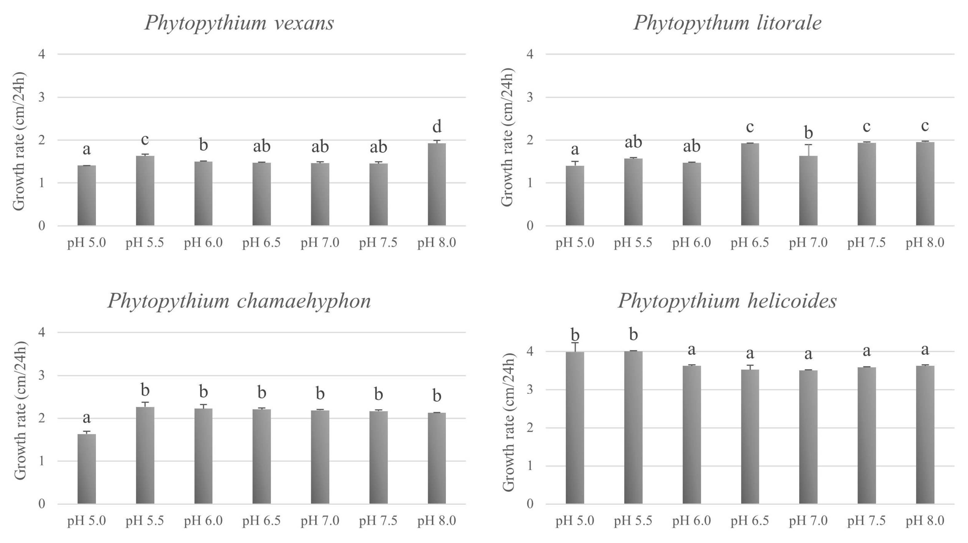
3.3. Effect of Temperature and pH on In Vitro Growth

The optimal growth for P. vexans strains occurred at 25 °C on CMA and PCA, with an average radial growth of 21 and 29 mm/24 h, respectively (Figure 4a). Onto PDA, it was 25 °C for the strain R1A, with an average radial growth of 13 mm/24 h, whereas it was at 30 °C for the strain PPA, with an average radial growth of 17 mm/24 h (Figure 4a). The minimum and maximum growth temperature were 10 °C and 30 °C, respectively, on all media.
The optimal growth for P. litorale strains occurred at 30 °C on CMA, with an average radial growth of 20 mm/24 h (Figure 4b). Onto PDA, it was at 25 °C for the strain R3A, with an average radial growth of 12 mm/24 h, and at 30 °C for the strain P8G, with an average radial growth of 12 mm/24 h. Onto PCA, for both strains, it was at 25 °C with an average radial growth of 21 mm/24 h. The minimum and maximum growth temperature were 10 °C and 35 °C, respectively, on all media.
The optimal growth for P. chamaehyphon strains occurred at 25 °C on CMA, PDA and PCA, with an average radial growth of 25, 23, and 23 mm/24 h, respectively (Figure 4c). The minimum and maximum growth temperature were 10 °C and 30 °C, respectively, on all media.
The optimal growth for P. helicoides strains occurred at 25 °C on CMA and PCA media with an average radial growth of 40 mm/24 h and 42 mm/24 h, respectively (Figure 4d). Onto PDA media was 30 °C, with an average radial growth of 23 mm/24 h. The minimum temperature for growth was 10 °C in all the media, whereas the maximum growth temperatures was 35 °C on PDA and 40 °C on CMA and PCA.
The four species tested were able to grow on PCA at pH from 5.0 to 8.0, with different growth rate (Figure 5). Since no statistical different growth rate (p ≥ 0.05) was found for both strains of the same species, the values shown are the mean of the two strains. P. vexans showed the highest growth rate at pH 8.0 and pH 5.5 (p ≤ 0.05), whereas P. litorale at pH from pH 6.5 to 8.0 (p ≤ 0.05). For P. chamaehyphon, there was no statistical different growth rate from pH 5.5 to 8.0, whereas a lower growth rate was shown at pH 5.0 (p ≤ 0.05). The optimal pH was between pH 5.0 and 5.5 (p ≤ 0.05) for P. helicoides.

3.4. Pathogenicity Test

All Phytopythium species under investigation were able to induce leaf curling, root rot, and decline of inoculated Actinidia plants. The first symptoms occurred after 12 days post-inoculation (Table 2) in all of the inoculated plants, whereas negative controls remained symptomless. P. helicoides showed the highest disease index (3.67 ± 0.58) and no statistical differences were observed when compared to the positive control (infected soil). The other species showed a slow progression of symptoms (Table 2). After 26 dpi, the species with the highest virulence remained P. helicoides (4.00 ± 0.00) compared to the other species tested, and no statistical differences were observed when compared to the positive control (Table 2). P. vexans and P. chamaehyphon showed similar virulence, with a mean disease index of 2.17 ± 0.58 and 2.67 ± 0.29, respectively. These species showed a lower disease index and the disease progressed more slowly during the test. P. litorale showed to be more virulent (3.17 ± 0.76) compared to P. vexans, but no statistical differences were observed compared to the positive control (Table 2).
To fulfil Koch’s postulates, re-isolations were performed from the roots of all the plants and the isolates were identified as P. vexans, P. helicoides, P. chamaehyphon and P. litorale.
The soil pH at the beginning of the trial was 4.35 ± 0.22, whereas after 26 dpi it was 5.33 ± 0.22. The soil temperature was 23.74 ± 0.76 °C when the trial started and it was 22.06 ± 0.81 at the end of the trial. The highest dry weight was recorded from roots sampled from healthy plants (40.05 ± 8.32 g) compared to the other roots (Table 3). No statistical differences were observed between roots inoculated with P. vexans, P. litorale and P. chamaehyphon (p ≤ 0.05), whereas a statistically lower dry weight was observed for the roots of plants inoculated with P. helicoides. No statistical differences were observed when P. helicoides was compared to the positive control.

4. Discussion

Field surveys carried out in 18 A. chinensis orchards of north-western Italy during 2016–2019 permitted us to collect 71 isolates of Phytopythium spp. from kiwifruit plants, showing typical symptoms of KVDS, such as reduction in plant vigour, leaf curling, or complete decline. Isolation from roots of plants affected by KVDS is a difficult process, as it involves root surface disinfection, washing, air-drying, tissue sample taking from the affected area, plating on PDA and oomycete PARP media and, after 4 days, transplant into tubes with PARP medium. All of the isolates showed to belong to the oomycete genus Phytopythium.
The role of the microbial community in KVDS development was previously demonstrated. Donati and colleagues [11] underlined the role of the rhizosphere microbial community, since a high incidence of Phytophthora spp. and Phytopythium spp. was associated with plants affected with KVDS. The role of biotic components in KVDS development was also described by Savian et al. [3], where in greenhouse experiments the symptoms were reproduced using unsterilised soil from a KVDS affected orchard, whereas no symptoms were observed using the same soil sterilised. The most frequently isolated species from kiwifruit affected by KVDS were Phytophthora citrophthora, P. cryptogea, P. infestans, P. megasperma, and Cylindrocarpon spp. [5,11]. Furthermore, Phytopythium spp. were isolated from symptomatic kiwifruits plants in several investigations [5,10,11,17].
In the present study, based on ML concatenated phylogeny on the ITS1-5.8S-ITS2 region of the rDNA, the large subunit (LSU) rDNA, and cytochrome oxidase I gene (COI), 52 strains were identified as P. vexans, 10 strains as P. litorale, seven strains as P. chamaehyphon and two strains as P. helicoides. Both, P. litorale and P. helicoides are reported for the first time as agents of KVDS on kiwifruit plants in Italy. Most of the isolates showed to belong to the species P. vexans, which is known to cause root and crown root in different crops, including kiwifruit [23,26,27]. Most orchards sampled showed the presence of only one Phytopythium species, except for some exceptions where two species were found.
A great intraspecific variability was observed among the strains of each of the four species, which were divided into clusters. This variability was previously observed for Phytopythium species, such as P. vexans [21,28,29], P. helicoides [21,28,30,31], P. litorale, P. kandeliae [32], P. mercuriale and P. oedochilum [33]. Based on micro- and macro-morphology analyses, the selected strains confirmed the typical characteristics of species [25,33,34,35,36].
In the pathogenicity tests, the virulence of all of the isolated species was demonstrated. Previously, P. vexans and P. chamaehyphon resulted pathogenic on 1-year-old or 6-month-old Actinidia chinensis var. deliciosa ‘Hayward’ plants [10,17]. Furthermore, the symptoms reproduction was also demonstrated for Desarmillaria tabescens and one isolate of Phytopythium spp. [11]. In this work, we demonstrated for the first time the pathogenicity of P. litorale on kiwifruit plants and P. helicoides was reported for the first time on kiwifruit plants in Italy. P. litorale, P. helicoides and P. vexans were previously reported as pathogenic on other hosts, such as on Platanus orientalis [37], Rhododendron pulchrum [38], on citrus, apple, and pear [39], and on almond [40]. Moreover, P. helicoides was already reported as agent of root and collar rot on kiwifruit in China [41].
In the pathogenicity test, flooding was used to reproduce soil water content proximal to field capacity. Waterlogging was previously investigated and, when applied alone, it was unable to reproduce KVDS symptoms. However, waterlogging is able to promote the progression of KVDS symptoms [3] and oomycetes were mainly reisolated from plants subjected to high irrigations volumes [11]. It should be noted that Phytopythium spp. live in water and soil and need a high humidity to produce sporangia and zoospores that are important infective propagules [42]. Therefore, the presence of water seems an important factor to promote Phytopythium spp. propagules and the onset of the disease.
In experiments in vitro, the four species showed an optimal growth at a temperature between 25 and 30 °C, depending on the media used. The maximum growth temperature was 30 °C for P. vexans, P. chamaehyphon, and P. litorale. Only P. helicoides was able to grow at 35 °C and even at 40 °C has a slow growth, confirming that the species is tolerant to high temperatures [43]. The data confirmed what reported in literature for P. litorale [33,44]. In other papers, where different strains were tested, 35 °C was reported as the maximum growth limit for P. vexans and 38 °C was reported as the optimal temperature for P. helicoides [31,45]. All of the tested strains showed an optimal growth at temperatures, which are in accordance with what has been observed in the soil of the orchards. In a monitoring performed in kiwifruit orchard during 2019, the average soil temperature measured during summer, when the KVDS symptoms occurred, was 23.2 ± 1.3 °C, whereas the average air temperature was 23.4 ± 4.5 °C.
The test to evaluate the tolerance of the species to different pH in vitro, showed their ability to grow at all the tested pH, ranging from 5.0 to 8.0. The maximum growth (p ≤ 0.05) was observed at pH 8.0 for P. vexans, at pH 6.5, 7.5 and 8.0 for P. litorale and at 5.0 and 5.5 for P. helicoides, whereas P. chamaehyphon seems the least influenced species by pH, where the highest growth was recorded in the range from 5.5 to 8.0. The average radial growth rate was similar for the species P. vexans, P. litorale and P. chamaehyphon, whereas P. helicoides showed the highest radial growth. For all the species under investigation, results showed that pH had less effect on growth than temperature, as previously shown by Cantrell and Dowler [45] for P. vexans. It should be noted that the effects of pH on fungal growth are complex, as an initial pH may affect growth, and subsequently growth can affect pH through release of metabolites into the growing medium. The values of pH and temperatures of the substrates recorded during pathogenicity tests were around the optimal values for the growth of the species of Phytopythium tested.

5. Conclusions

The present study demonstrates the strong role of oomycetes in the establishment and development of KVDS. The presence of different species of oomycetes suggests that the oomycete component of the soil microbiota present in the soil is involved in the development of KVDS, and not only a single species is involved in this complex syndrome. The isolated species have a relatively high optimal and maximum temperature for growth in soil, and they could be favoured by the average increase in soil temperatures during summer, associated with global warming. Waterlogging could exert a double effect, both on stressing the plant with a temporary anoxia and on favouring the release of infective zoospores by oomycetes. Further studies should investigate the complex interactions between kiwifruit, the oomycete species, the soil environment, and the effect of different management strategies in the field. Moreover, a study of the soil and rhizosphere microbiome could help to clarify the changes occurring in the soil microbiota of kiwifruit orchards.

Supplementary Materials

The following supporting information can be downloaded at: https://0-www-mdpi-com.brum.beds.ac.uk/article/10.3390/microorganisms11010216/s1. Table S1: Strain name and information about orchards from where the strains were isolated (geographical location, geographical coordinates and orchard number); Table S2: List of species, strain designation and accession numbers for ITS and LSU regions and COI gene used for the phylogeny of Phytopythium spp. isolated from kiwifruit roots in this study.

Author Contributions

Conceptualization, S.P. and D.S.; methodology, S.P. and D.S.; formal analysis, S.P., G.S. and M.R.; investigation, S.P. and L.N.; data curation, S.P. and D.S.; writing—original draft preparation, S.P.; writing—review and editing, S.P., D.S. and L.S.; funding acquisition, D.S. All authors have read and agreed to the published version of the manuscript.

Funding

This research was partially funded by Piedmont Region (Italy) with the project “KIRIS—Kiwifruit Vine Decline Syndrome: deepening the aetiology and development of prevention and control tool”.

Data Availability Statement

Conflicts of Interest

The authors declare no conflict of interest. The funders had no role in the design of the study; in the collection, analyses, or interpretation of data; in the writing of the manuscript; or in the decision to publish the results.

References

  1. Sorrenti, G.; Tacconi, G.; Tosi, L.; Vittone, G.; Nari, L.; Savian, F.; Saro, S.; Ermacora, P.; Graziani, S.; Toselli, M. Avanza la “moria del kiwi”: Evoluzione e primi riscontri della ricerca. Riv. Fruttic. 2019, 2, 34–42. [Google Scholar]
  2. Balestra, G.M.; Costa, G. La situazione in Italia: Evoluzione varietale sì, ma con tante avversità. Riv. Fruttic. 2020, 82, 10–16. [Google Scholar]
  3. Savian, F.; Ginaldi, F.; Musetti, R.; Sandrin, N.; Tarquini, G.; Pagliari, L.; Firrao, G.; Martini, M.; Ermacora, P. Studies on the aetiology of kiwifruit decline: Interaction between soil-borne pathogens and waterlogging. Plant Soil 2020, 456, 113–128. [Google Scholar] [CrossRef]
  4. Xiloyannis, C.; Baroni, G.; Bovo, M.; Angelini, P.; Pezzarossa, B.; Pagliai, M. L’irrigazione per scorrimento nel Veronese: Spreco di acqua e di concimi; effetti sulla porosità del terreno. Riv. Fruttic. 1992, 6, 55–62. [Google Scholar]
  5. Tacconi, G.; Paltrinieri, S.; Mejia, J.F.; Fuentealba, S.P.; Bertaccini, A.; Tosi, L.; Giacopini, A.; Mazzucchi, U.; Favaron, F.; Sella, L.; et al. Vine decline in kiwifruit: Climate change and effect on waterlogging and Phytophthora in north Italy. Acta Hortic. 2015, 1096, 93–97. [Google Scholar] [CrossRef]
  6. Akilli, S.; Serçe, Ç.U.; Zekaİ Katircioğlu, Y.; Karakaya, A.; Maden, S. Involvement of Phytophthora citrophthora in kiwifruit decline in Turkey. J. Phytopathol. 2011, 159, 579–581. [Google Scholar] [CrossRef]
  7. Kurbetli, İ.; Ozan, S. Occurrence of Phytophthora root and stem rot of kiwifruit in Turkey. J. Phytopathol. 2013, 161, 887–889. [Google Scholar] [CrossRef]
  8. Reid, J.B.; Tate, K.G.; Brown, N.S. Effects of flooding and alluvium deposition on kiwifruit (Actinidia deliciosa). N. Z. J. Crop Hortic. Sci. 1991, 19, 247–257. [Google Scholar] [CrossRef]
  9. McAneney, K.J.; Clough, A.; Green, A.; Harris, B.; Richardson, A. Waterlogging and vine death at Kerikeri. N. Z. Kiwifruit 1989, 56, 15. [Google Scholar]
  10. Prencipe, S.; Savian, F.; Nari, L.; Ermacora, P.; Spadaro, D.; Martini, M. First report of Phytopythium vexans causing decline syndrome of Actinidia deliciosa ‘Hayward’ in Italy. Plant Dis. 2020, 104, 2032. [Google Scholar] [CrossRef] [Green Version]
  11. Donati, I.; Cellini, A.; Sangiorgio, D.; Caldera, E.; Sorrenti, G.; Spinelli, F. Pathogens associated to kiwifruit vine decline in Italy. Agriculture 2020, 10, 119. [Google Scholar] [CrossRef] [Green Version]
  12. Bardi, L. Early kiwifruit decline: A soil-borne disease syndrome or a climate change effect on plant–soil relations? Front. Agron. 2020, 2, 3. [Google Scholar] [CrossRef]
  13. Tosi, L.; Tacconi, G.; Giacopini, A. La moria del kiwi, situazione e prospettive. L’Informatore Agrar. 2015, 44, 67–70. [Google Scholar]
  14. Sorrenti, G.; Toselli, M.; Reggidori, G.; Spinelli, F.; Tosi, L.; Giacopini, A.; Tacconi, G. Implicazioni della gestione idrica nella “moria del kiwi” del veronese. Riv. Fruttic. 2016, 3, 45–51. [Google Scholar]
  15. Bardi, L.; Nari, L.; Morone, C.; Faga, M.G.; Malusà, E. Possible Role of High Temperature and Soil Biological Fertility on Kiwifruit Early Decline Syndrome. Front. Agron. 2020, 2, 580659. [Google Scholar] [CrossRef]
  16. Tacconi, G.; Giacopini, A.; Vittone, G.; Nari, L.; Spadaro, D.; Savian, F.; Ermacora, P.; Saro, S.; Morone, C.; Bardi, L.; et al. “Moria del kiwi”: Situazione disastrosa al nord, preoccupante nel resto d’Italia. Kiwi Inf. 2019, 2, 32–37. [Google Scholar]
  17. Savian, F.; Prencipe, S.; Filippini, N.; Nari, L.; Martini, M.; Ermacora, P.; Spadaro, D. Pathogenicity of Phytopythium chamaehyphon: A New Player in Kiwifruit Vine Decline Syndrome of Actinidia chinensis var. deliciosa ‘Hayward’ in Italy. Plant Dis. 2021, 105, 2781–2784. [Google Scholar] [CrossRef]
  18. Spigaglia, P.; Barbanti, F.; Marocchi, F.; Mastroleo, M. Clostridium bifermentans and C. subterminale are associated with kiwifruit vine decline, known as moria, in Italy. Plant Pathol. 2020, 69, 765–774. [Google Scholar] [CrossRef]
  19. White, T.J.; Bruns, T.D.; Lee, S.B.; Taylor, J.W. Amplification and Direct Sequencing of Fungal Ribosomal RNA Genes for Phylogenetics. In PCR Protocols: A Guide to Methods and Applications; Innis, M.A., Gelfand, D.H., Sninsky, J.J., White, T.J., Eds.; Academic Press: San Diego, CA, USA, 1990; pp. 315–322. [Google Scholar]
  20. Cooke, D.E.L.; Duncan, J.M. Phylogenetic analysis of Phytophthora species based on ITS1and ITS2 sequences of the ribosomal RNA gene repeat. Mycol. Res. 1997, 101, 667–677. [Google Scholar] [CrossRef]
  21. Baten, M.A.; Asano, T.; Motohashi, K.; Ishiguro, Y.; Rahman, M.Z.; Inaba, S.; Suga, H.; Kageyama, K. Phylogenetic relationships among Phytopythium species, and re-evaluation of Phytopythium fagopyri comb. nov., recovered from damped-off buckwheat seedlings in Japan. Mycol. Prog. 2014, 13, 1003. [Google Scholar] [CrossRef]
  22. Robideau, G.P.; de Cock, A.W.A.M.; Coffey, M.D.; Voglmayr, H.; Brouwer, H.; Bala, K.; Chitty, D.W.; Désaulniers, N.; Eggertson, Q.A.; Gachon, C.M.M.; et al. DNA barcoding of oomycetes with cytochrome c oxidase subunit I and internal transcribed spacer. Mol. Ecol. Resour. 2011, 11, 1002–1011. [Google Scholar] [CrossRef] [PubMed]
  23. de Cock, A.W.A.M.; Lodhi, A.M.; Rintoul, T.L.; Bala, K.L.; Robideau, G.P.; Gloria Abad, Z.; Coffey, M.D.; Shahzad, S.; Lévesque, C.A. Phytopythium: Molecular phylogeny and systematics. Peersonia 2015, 34, 25–39. [Google Scholar] [CrossRef] [PubMed] [Green Version]
  24. Crous, P.W.; Verkley, G.J.M.; Groenewald, J.Z.; Samson, R.A. Fungal Biodiversity. In CBS Laboratory Manual Series 1; CBS-KNAW Fungal Biodiversity Centre: Utrecht, The Netherlands, 2009; p. 269. [Google Scholar]
  25. de Cock, A.W.A.M.; Lévesque, C.A. New species of Pythium and Phytophthora. Stud. Mycol. 2004, 50, 481–487. [Google Scholar]
  26. Adhikari, B.N.; Hamilton, J.P.; Zerillo, M.M.; Tisserat, N.; Lévesque, C.A.; Buell, C.R. Comparative genomics reveals insight into virulence strategies of plant pathogenic oomycetes. PLoS ONE 2013, 8, e75072. [Google Scholar] [CrossRef]
  27. Polat, Z.; Awan, Q.N.; Hussain, M.; Akgul, D.S. First Report of Phytopythium vexans causing root and collar rot of kiwifruit in Turkey. Plant Dis. 2017, 101, 1058. [Google Scholar] [CrossRef]
  28. Baten, M.A.; Li, M.; Motohashi, K.; Ishiguro, Y.; Rahman, M.Z.; Suga, H.; Kageyama, K. Two new species, Phytopythium iriomotense sp. nov. and P. aichiense sp. nov., isolated from river water and water purification sludge in Japan. Mycol. Prog. 2015, 14, 2. [Google Scholar] [CrossRef]
  29. Rodríguez Padrón, C.; Siverio, F.; Pérez-Sierra, A.; Rodríguez, A. Isolation and pathogenicity of Phytophthora species and Phytopythium vexans recovered from avocado orchards in the Canary Islands, including Phytophthora niederhauserii as a new pathogen of avocado. Phytopathol. Mediterr. 2018, 57, 89–106. [Google Scholar]
  30. Kageyama, K.; Senda, M.; Asano, T.; Suga, H.K.; Ishiguro, K. Intro isolate heterogeneity of the ITS region of rDNA in Pythium helicoides. Mycol. Res. 2007, 111, 416–423. [Google Scholar] [CrossRef]
  31. Chen, X.R.; Liu, B.B.; Xing, Y.P.; Cheng, B.P.; Liu, M.L.; Tong, Y.H.; Xu, J.Y. Identification and characterization of Phytopythium helicoides causing stem rot of Shatangju mandarin seedlings in China. Eur. J. Plant Pathol. 2016, 146, 715–727. [Google Scholar] [CrossRef]
  32. Marano, A.V.; Jesus, A.L.; De Souza, J.I.; Leãno, E.M.; James, T.Y.; Jerônimo, G.H.; de Cock, A.W.A.M.; Pires-Zottarelli, C.L.A. A new combination in Phytopythium: P. kandeliae (Oomycetes, Straminipila). Mycosphere 2014, 5, 510–522. [Google Scholar] [CrossRef]
  33. Bouket, C.A.; Babai-Ahari, A.; Arzanlou, M.; Tojo, M. Morphological and molecular characterization of Phytopythium litorale and Pp. oedochilum from Iran. Nova Hedwig. 2016, 102, 257–270. [Google Scholar] [CrossRef]
  34. Sideris, C.P. Taxonomic studies in the family Pythiaceae II. Pythium. Mycologia 1932, 24, 14–61. [Google Scholar] [CrossRef]
  35. Bennett, R.M.; Bora, N.; Dedeles, G.R.; Thines, M. Phytopythium leanoi sp. nov. and Phytopythium dogmae sp. nov., Phytopythium species associated with mangrove leaf litter from the Philippines. Acta Mycol. 2017, 52, 1103. [Google Scholar] [CrossRef] [Green Version]
  36. Nam, B.; Choi, Y.J. Phytopythium and Pythium Species (Oomycota) isolated from freshwater environments of Korea. Mycobiology 2019, 47, 261–272. [Google Scholar] [CrossRef] [PubMed] [Green Version]
  37. Derviş, S.; Türkölmez, S.; Çiftçi, O.; Özer, G.; Ulubaş Serçe, Ç.; Dikilitas, M. Phytopythium litorale: A novel killer pathogen of plane (Platanus orientalis) causing canker stain and root and collar rot. Plant Dis. 2020, 104, 2642–2648. [Google Scholar] [CrossRef]
  38. Li, Y.; Feng, Y.; Wu, C.; Xue, J.; Jiao, B.; Li, B.; Dai, T. First Report of Phytopythium litorale Causing Crown and Root Rot on Rhododendron pulchrum in China. Plant Dis. 2021, 105, 4173. [Google Scholar] [CrossRef]
  39. Jabiri, S.; Bahra, C.; MacLean, D.; Radouane, N.; Barka, E.A.; Bendriss Amraoui, M.; Lahlali, R. Phytopythium vexans Associated with Apple and Pear Decline in the Saïss Plain of Morocco. Microorganisms 2021, 9, 1916. [Google Scholar] [CrossRef] [PubMed]
  40. Beluzán, F.; Miarnau, X.; Torguet, L.; Armengol, J.; Abad-Campos, P. Survey of Oomycetes Associated with Root and Crown Rot of Almond in Spain and Pathogenicity of Phytophthora niederhauserii and Phytopythium vexans to ‘Garnem’ Rootstock. Agriculture 2022, 12, 294. [Google Scholar] [CrossRef]
  41. Wang, K.X.; Xie, Y.L.; Yuan, G.Q.; Li, Q.Q.; Lin, W. First report of root and collar rot caused by Phytopythium helicoides on kiwifruit (Actinidia chinensis). Plant Dis. 2015, 99, 725–726. [Google Scholar] [CrossRef]
  42. Tao, Y.H.; Zeng, F.; Ho, H.H.; Wei, J.; Wu, Y.; Yang, L.; He, Y. Pythium vexans causing stem rot of Dendrobium in Yunnan Province, China. J. Phytopathol. 2011, 159, 255–259. [Google Scholar] [CrossRef]
  43. Afandi, A.; Murayama, E.; Hieno, A.; Suga, H.; Kageyama, K. Population structures of the waterborne plant pathogen Phytopythium helicoides reveal its possible origins and transmission modes in Japan. PLoS ONE 2018, 13, e0209667. [Google Scholar] [CrossRef] [PubMed] [Green Version]
  44. Nechwatal, J.; Mendgen, K. Pythium litorale sp. nov., a new species from the littoral of Lake Constance, Germany. FEMS Microbiol. Lett. 2006, 255, 96–101. [Google Scholar] [CrossRef] [PubMed] [Green Version]
  45. Cantrell, H.F.; Dowler, W.M. Effects of temperature and pH on growth and composition of Pythium irregulare and Pythium vexans. Mycologia 1971, 63, 31–37. [Google Scholar] [CrossRef] [PubMed]
Figure 1. Disease severity index used to evaluate in greenhouse the pathogenicity of the Phytopythium species inoculated on 1-year-old A. chinensis var. deliciosa ‘Hayward’ plants.
Microorganisms 11 00216 g001
Figure 2. Best scoring Maximum Likelihood tree based on the concatenated ITS, LSU, and COI sequence datasets. The numbers at the major nodes indicate the bootstrap value from 1000 bootstrapped datasets. Branches with lower bootstrap values than 70% are not shown. Phylogeny was rooted by Pythium ultimum. Evolutionary analyses were conducted using MEGA, version 6. The highlighted strains are the isolates used for pathogenicity test.
Microorganisms 11 00216 g002
Figure 3. Colony morphology of Phytopythium species isolated from kiwifruits on Corn Meal Agar (CMA), Potato Dextrose Agar (PDA) and Potato Carrot Agar (PCA) after 6 days at 25 ± 1 °C. (A) Phytopythium vexans; (B) Phytopythium litorale; (C) Phytopythium chamaehyphon; (D) Phytopythium helicoides.
Microorganisms 11 00216 g003
Figure 4. Average radial growth rate (mm/24 h) of Phytopythium species grown onto CMA, PDA and PCA media at different temperatures for 5 days. (a) P. vexans strains R1A and PPA; (b) P. litorale strains R3A and P8G; (c) P. chamaehyphon strains CA3 and PH6; (d) P. helicoides strains CA1 and CA2.
Microorganisms 11 00216 g004
Figure 5. Average radial growth rate (cm/24 h) of two strains per Phytopythium species grown onto PCA media adjusted at specific pH at 25 °C for 5 days. Values are expressed as mean values ± SD (n = 6). Values followed by the same letter are not statistically different by Tukey’s test (p ≤ 0.05).
Microorganisms 11 00216 g005
Table 1. Strain name, molecular identification, cultivar, year of isolation and GenBank accession numbers of the Phytopythium spp. strains isolated from kiwifruit roots in this study.
Strain *SpeciesCultivarYear of IsolationGenBank Accession Numbers **
ITSLSUCOI
PH1Phytopythium vexansHayward2016OL891590OL957094ON228590
PH6Phytopythium chamaehyphonHayward2016OL891528OL957144ON228528
PH3Phytopythium vexansHayward2016MN510425 *MN510427MN510423
PH2Phytopythium vexansHayward2016OL891591OL957095ON228591
GBIPhytopythium vexansHayward2016OL891578OL957096ON228578
4SRE1Phytopythium vexansHayward2016OL891579OL957097ON228579
4SRE2Phytopythium vexansHayward2016OL891580OL957098ON228580
4SBE_2C1Phytopythium vexansHayward2016OL891581OL957099ON228581
4SBE_C2APhytopythium litoraleHayward2016OL891534OL957153ON228534
4SBE_C2BPhytopythium litoraleHayward2016OL891536OL957154ON228536
4SBE_C2DPhytopythium litoraleHayward2016OL891533OL957155ON228533
4SBE_1C1Phytopythium vexansHayward2016OL891587OL957100ON228587
4SBE_C1APhytopythium vexansHayward2016OL891586OL957101ON228586
4SBE_C2CPhytopythium litoraleHayward2016OL891535OL957162ON228535
4SBE_4C2Phytopythium vexansHayward2016OL891588OL957102ON228588
4SBE_3C2Phytopythium vexansHayward2016OL891589OL957103ON228589
4/16_DREPhytopythium chamaehyphonHayward2016OL891529OL957145ON228529
5/16_CBEPhytopythium vexansHayward2016OL891582OL957104ON228582
6/16_BREPhytopythium vexansSoreli2016OL891583OL957105ON228583
11/16_AB1Phytopythium chamaehyphonHayward2016OL891531OL957146ON228531
12/16_DR2Phytopythium vexansHayward2016MN510426MN510428MN510424
12/16_CR1Phytopythium vexansHayward2016OL891584OL957106ON228584
12/16_AB1 Phytopythium vexansHayward2016OL891585OL957107ON228585
13/16_DR2Phytopythium vexansHayward2016OL891564OL957108ON228564
13/16_DR3Phytopythium vexansHayward2016OL891565OL957109ON228565
10/16_BRPhytopythium chamaehyphonHayward2016OL891530OL957147ON228530
PP1Phytopythium vexansHayward2018OL891571OL957110ON228571
PP2Phytopythium vexansHayward2018OL891572OL957111ON228572
PP3Phytopythium vexansHayward2018OL891573OL957112ON228573
PP4Phytopythium vexansHayward2018OL891574OL957113ON228574
PP5Phytopythium vexansHayward2018OL891575OL957114ON228575
PP6Phytopythium vexansHayward2018OL891576OL957115ON228576
PP8Phytopythium vexansHayward2018OL891577OL957116ON228577
PPAPhytopythium vexansHayward2018OL891566OL957117ON228566
PPCPhytopythium vexansHayward2018OL891567OL957118ON228567
PPDPhytopythium vexansHayward2018OL891568OL957119ON228568
PPEPhytopythium vexansHayward2018OL891569OL957120ON228569
PPFPhytopythium vexansHayward2018OL891570OL957121ON228570
CA1Phytopythium helicoidesHayward2019OL891523OL957151ON228523
CA2Phytopythium helicoidesHayward2019OL891524OL957152ON228524
CA3Phytopythium chamaehyphonHayward2019OL891525OL957148ON228525
CA4Phytopythium chamaehyphonHayward2019OL891526OL957149ON228526
CA5Phytopythium chamaehyphonHayward2019OL891527OL957150ON228527
R1APhytopythium vexansHayward2019OL891543OL957122ON228543
R1BPhytopythium vexansHayward2019OL891544OL957123ON228544
R1CPhytopythium vexansHayward2019OL891545OL957124ON228545
R1DPhytopythium vexansHayward2019OL891546OL957125ON228546
R1EPhytopythium vexansHayward2019OL891547OL957126ON228547
R1FPhytopythium vexansHayward2019OL891548OL957127ON228548
R1GPhytopythium vexansHayward2019OL891549OL957128ON228549
R1HPhytopythium vexansHayward2019OL891550OL957129ON228550
R3APhytopythium litoraleHayward2019OL891532OL957156ON228532
T4APhytopythium vexansHayward2019OL891562OL957130ON228562
T4BPhytopythium vexansHayward2019OL891563OL957131ON228563
R7APhytopythium vexansHayward2019OL891551OL957132ON228551
R7BPhytopythium vexansHayward2019OL891552OL957133ON228552
R7CPhytopythium vexansHayward2019OL891553OL957134ON228553
R7DPhytopythium vexansHayward2019OL891554OL957135ON228554
R7EPhytopythium vexansHayward2019OL891555OL957136ON228555
R7FPhytopythium vexansHayward2019OL891556OL957137ON228556
P8APhytopythium vexansSoreli 2019OL891557OL957138ON228557
P8BPhytopythium vexansSoreli 2019OL891558OL957139ON228558
P8DPhytopythium vexansSoreli 2019OL891559OL957140ON228559
P8EPhytopythium vexansSoreli 2019OL891560OL957141ON228560
P8FPhytopythium vexansSoreli 2019OL891561OL957142ON228561
P8GPhytopythium litoraleSoreli 2019OL891537OL957157ON228537
P8HPhytopythium litoraleSoreli 2019OL891538OL957158ON228538
P8IPhytopythium litoraleSoreli 2019OL891539OL957159ON228539
R10APhytopythium litoraleDong Hong2019OL891540OL957160ON228540
R10BPhytopythium litoraleDong Hong 2019OL891541OL957161ON228541
RE3Phytopythium vexansHayward2019OL891542OL957143ON228542
* Strains in bold are used for morphological observations and in vitro tests. ** Sequences in bold are from Prencipe et al. [10].
Table 2. Results of disease severity (DS) on 1-year-old plants of A. chinensis var. deliciosa ‘Hayward’ plants at 12 and 26 dpi. Values in the same column followed by the same letter are not statistically different by Tukey test (p ≤ 0.05).
DS 12 dpi DS 26 dpi
TreatmentMean ± SDTukey TestMean ± SDTukey Test
P. vexans strain PP11.50 ± 0.50bc2.17 ± 0.58b
P. chamaehyphon strain PH61.00 ± 0.50ab2.67 ± 0.29bc
P. helicoides strain CA23.67 ± 0.58d4.00 ± 0.00e
P. litorale strain R3A2.17 ± 0.76c3.17 ± 0.76cd
Positive control (infected soil)3.50 ± 0.87d3.83 ± 0.29de
Negative control (healthy soil)0.00-a0.00-a
Table 3. Dry weight (g) of roots sampled from 1-year-old plants of A. chinensis var. deliciosa ‘Hayward’ plants after 26 dpi. Values in the same column followed by the same letter are not statistically different by Tukey test (p ≤ 0.05).
TreatmentMean ± SDTukey Test
P. vexans strain PP123.94 ± 9.16b
P. chamaehyphon strain PH626.06 ± 2.77b
P. helicoides strain CA27.35 ± 0.76a
P. litorale strain R3A17.13 ± 0.90ab
Positive control (infected soil)13.43 ± 2.91a
Negative control (healthy soil)40.05 ± 8.32c
Disclaimer/Publisher’s Note: The statements, opinions and data contained in all publications are solely those of the individual author(s) and contributor(s) and not of MDPI and/or the editor(s). MDPI and/or the editor(s) disclaim responsibility for any injury to people or property resulting from any ideas, methods, instructions or products referred to in the content.

