Article

Stable Isotope and Metagenomic Profiling of a Methanogenic Naphthalene-Degrading Enrichment Culture

by
Courtney R. A. Toth
,
Carolina Berdugo-Clavijo
,
Corynne M. O’Farrell
,
Gareth M. Jones
,
Andriy Sheremet
,
Peter F. Dunfield
and
Lisa M. Gieg
*
Department of Biological Sciences, University of Calgary, 2500 University Drive NW, Calgary, AB T2N 1N4, Canada
*
Author to whom correspondence should be addressed.
Current Address: Department of Chemical Engineering and Applied Chemistry, University of Toronto, 200 College Street, Toronto, ON M5S 3E5, Canada
Submission received: 1 June 2018 / Revised: 2 July 2018 / Accepted: 8 July 2018 / Published: 10 July 2018
(This article belongs to the Special Issue Metabolic Diversity of Anaerobic Microbial Communities)

Abstract

:
Polycyclic aromatic hydrocarbons (PAH) such as naphthalene are widespread, recalcitrant pollutants in anoxic and methanogenic environments. A mechanism catalyzing PAH activation under methanogenic conditions has yet to be discovered, and the microbial communities coordinating their metabolism are largely unknown. This is primarily due to the difficulty of cultivating PAH degraders, requiring lengthy incubations to yield sufficient biomass for biochemical analysis. Here, we sought to characterize a new methanogenic naphthalene-degrading enrichment culture using DNA-based stable isotope probing (SIP) and metagenomic analyses. 16S rRNA gene sequencing of fractionated DNA pinpointed an unclassified Clostridiaceae species as a putative naphthalene degrader after two months of SIP incubation. This finding was supported by metabolite and metagenomic evidence of genes predicted to encode for enzymes facilitating naphthalene carboxylic acid CoA-thioesterification and degradation of an unknown arylcarboxyl-CoA structure. Our findings also suggest a possible but unknown role for Desulfuromonadales in naphthalene degradation. This is the first reported functional evidence of PAH biodegradation by a methanogenic consortium, and we envision that this approach could be used to assess carbon flow through other slow growing enrichment cultures and environmental samples.

1. Introduction

Polycyclic aromatic hydrocarbons (PAH) are ubiquitous environmental pollutants generated during the incomplete combustion of organic materials (e.g., fossil fuels, refined fuel products, and wood), but also from natural seepage of petroleum or coal deposits, accidental fuel releases, and volcanic activities [1]. Due to their high hydrophobicity, PAHs tend to partition and deposit in sedimentary environments, but water-soluble components (e.g., 2-to 3-ringed PAHs) are also capable of migrating through anoxic groundwater systems [2]. Given that PAHs exhibit toxigenic, mutagenic, and/or carcinogenic properties [1,3], PAH-contaminated aquifers pose a significant threat to ecological and human health. This issue has prompted more than 40 years of field- and microcosm-based research documenting the extent of PAH mineralization. As a result, it is firmly established that two- and three- ringed PAHs are susceptible to microbial mineralization under ferric iron-reducing, sulfate-reducing, and methanogenic conditions [4,5]. Reproducible evidence of nitrate-reducing PAH mineralization has yet to be reported [5]. Similarly, there is no evidence to date demonstrating that ≥ 4-ringed PAHs can be metabolized anaerobically as a sole carbon source, though co-metabolic PAH degradation is possible [6,7].
Recent efforts have shifted towards profiling the molecular landscape of anaerobic PAH degradation by conducting genomic and proteomic surveys of PAH-degrading cultures. Most of our biochemical understanding of anaerobic PAH biodegradation have been gleaned from the marine isolate NaphS2 and the freshwater enrichment culture N47 [8,9,10,11]. NaphS2 and the dominant organism of N47 are unclassified sulfate-reducing Desulfobacteraceae belonging to the Deltaproteobacteria, sharing organizational and sequence homology across two gene clusters differentially expressed during naphthalene and 2-methylnaphthalene degradation [12,13,14]. One gene cluster harbors a UbiD-like (de)carboxylase putatively responsible for the carboxylation of naphthalene, while the second gene cluster encodes for the fumarate adding catalytic subunit of naphthyl-2-methylsuccinate synthase (NmsA). Several enzymes predicted to facilitate downstream degradation steps of naphthalene and 2-methylnaphthalene have also been tentatively identified or experimentally verified [10,12,13,14,15,16,17], including the novel de-aromatization enzyme 2-naphthoyl-CoA reductase and the predicted 5, 6-dihydro-2-naphthoyl CoA reductase [18,19,20]. The partial elucidation of these pathways has also helped to identify signature metabolites and functional genes that can serve as diagnostic markers of anaerobic PAH biodegradation in the environment [5,21]. Nevertheless, these model strains only represent a small fraction of the metabolic potential and physiology of organisms likely involved in anaerobic PAH degradation.
Methanogenic PAH degradation has been investigated for over a decade, providing analytical evidence of methyl-substituted naphthalene mineralization and/or microbial community data [22,23,24,25,26,27,28,29,30,31]. However, the identification of catabolic genes, mechanisms, and key microbes involved in methanogenic PAH metabolism has remained largely elusive. A major challenge associated with studying anaerobic PAH degradation is the relatively slow growth rates of PAH degraders, complicating cultivation-dependent biochemical assays [4,5]. This is especially problematic for methanogenic incubations, wherein thermodynamic constraints restrict energy yields for growth and metabolism [5,32]. Instead, cultivation-independent molecular methods such as nucleic acid-based stable isotope probing (SIP) may offer provisional solutions for pinpointing PAH-degrading microorganisms and help to elucidate (novel) anaerobic PAH activation mechanisms. For example, DNA-SIP was recently conducted for an iron-reducing enrichment, revealing that Thermoanaerobacteraceae and Desulfobacterales phylotypes are involved in 1-methylnaphthalene degradation [33]. However, identifying primary PAH degraders by SIP in complex consortia is difficult as long incubation times generally increase the risk of labeling secondary degraders [34,35]. To our knowledge, only one study has previously used DNA-SIP to monitor PAH degradation under methanogenic conditions, wherein the authors identified three unexpected Proteobacteria (Methylibium, Legionella, Rhizobiales) isotopically enriched during methanogenic 13C6-anthracene degradation [30].
In this study, we aimed to identify key microorganisms and reactions responsible for the initial degradation steps of the PAH naphthalene by a new methanogenic naphthalene-degrading enrichment culture (designated NDC) using a variety of molecular ecology toolbox approaches including DNA-SIP and metagenomic analyses. The culture described in this study originated from a crude oil-degrading enrichment [36,37] found to metabolize methyl-substituted naphthalenes to near stoichiometric amounts of methane [22]. Naphthalene was not initially reported to be susceptible to methanogenic degradation by this culture, but enhanced methane production and naphthalene loss was ultimately verified by gas chromatography more than a year after the original publication date [38]. The results of this present study revealed that members of the Clostridiaceae, and possibly Desulfuromonadales, are involved in naphthalene degradation by NDC under methanogenic conditions, as these phylotypes became labeled during incubations with isotopically heavy naphthalene and/or harbored genes known to be involved in anaerobic naphthalene metabolism.

2. Materials and Methods

2.1. Culture Establishment

Preliminary attempts to establish the present methanogenic naphthalene-degrading enrichment culture were first described by Berdugo-Clavijo et al. [22], but new details are presented here. Parent cultures were prepared by adding 3 mL of resuspended pelleted cells from a crude oil-degrading culture [37] to 60 mL of sterile, anoxically prepared bicarbonate-buffered minimal salts medium containing resazurin as a redox indicator and reduced with 0.05% w/v cysteine sulfide. Triplicate microcosms were amended with 8 mg naphthalene pre-adsorbed in 0.3 g of resin (Amberlite XAD-7 [39]), and incubated at room temperature (~22 °C) under dark and static conditions. Substrate-free controls containing resin only and cell-free sterile controls were also established in triplicate, for a total of nine microcosms. To monitor methanogenic activity, microcosm headspaces were routinely measured for methane production by gas chromatography (GC) [22]. After 140 days of incubation, naphthalene-amended enrichments had only produced equal or lesser amounts of methane relative to substrate-free microcosms (Figure S1).
In order to further verify whether naphthalene could be used as a substrate by this methanogenic culture, the enrichment (10% v/v) was transferred into 70 mL of fresh medium. Two microcosms were amended with 58.5 mM naphthalene, and dissolved in 1 mL of the inert carrier 2,2,4,4,6,8,8-heptamethylnonane (HMN). HMN, rather than resin, was used in order to enable the measurement of naphthalene loss over time and to assess stoichiometric yields of methane over time. Two unamended microcosms (containing HMN only) and a sterile control were also prepared, for a total of five microcosms. We verified that no naphthalene carry-over had occurred in the unamended controls (see below). Incubation time and temperature were increased to 320 days and 30 °C respectively, to help stimulate methanogenic activity.
Naphthalene loss was measured by injecting 1 µL of the HMN overlay into an Agilent 7890A GC equipped with an HP-5 capillary column (30 m × 320 µm × 0.25 µm; Agilent, Santa Clara, CA, USA) and a flame ionization detector (GC-FID). The oven was held at 50 °C for 2 min, then increased at a rate of 7 °C min to 140 °C, and held at this temperature for 3 min. The injector, operated in split mode (50:1), was held at 275 °C and the detector was held at 300 °C. Calibration curves were prepared from naphthalene standards of known concentrations dissolved in HMN (2–80 mM).

2.2. Metabolite Analysis and 13C-bicarbonate Tracer Experiments

Subsamples of active NDC enrichments (≤50 mL) were periodically extracted for putative naphthalene metabolites and analyzed by gas chromatography–mass spectrometry (GC-MS) [22]. We also prepared a series of 13C-bicarbonate tracer incubations to search for evidence of a carboxylation activation mechanism. To prepare the inoculum, pelleted cells from 75 mL of NDC were washed and resuspended in an equal volume of anoxic medium buffered with 25 mM HEPES solution. Incubations containing 70 mL fresh medium were inoculated with 3 mL of HEPES-buffered inoculum and amended with 19.5 mM naphthalene in HMN and 23 mM of 13C-sodium bicarbonate (99%; Cambridge Isotope Laboratories, Inc., Tewksbury, MA, USA) as a co-substrate in a N2 headspace. Cultures were incubated at room temperature for over a year before they were extracted and analyzed for putative 13C-labeled metabolites by GC-MS.

2.3. Microbial Community Analysis and Targeted Functional Gene Analysis

An actively-degrading methanogenic naphthalene enrichment culture was subsampled (4 mL) and extracted for genomic DNA using a commercial kit (FastDNA SPIN Kit for Soils, MP Biomedicals, Solon, OH, USA) according to the manufacturer’s procedure. PCR-based targeted gene surveys assays of the extracted DNA were performed to search for putative anaerobic naphthalene degradation genes, summarized in Table S1. PCR reactions (50 µL) were prepared using the Taq DNA Polymerase kit (Qiagen, Toronto, ON, Canada) and contained 2 µL of template DNA (0.2 ng/µL). 16S rRNA gene sequencing (by 454 pyrotag sequencing) of extracted genomic DNA was carried out using an established two-step PCR method targeting the V6-V8 regions of the 16S rRNA gene [22]. Purified amplicons of expected size were sequenced using a GS FLX Titanium Series Kit XLR70 (Roche Diagnostics Corporation, Indianapolis, IN, USA), and data analysis was conducted in Phoenix 2 as previously described [22]. Pyrosequencing reads are available within NCBI’s Short Read Archive (SRA) under BioProject PRJNA478574.

2.4. 13C-Naphthalene Stable Isotope Probing

DNA-based SIP was conducted to help pinpoint key naphthalene degraders in NDC. This methodology was selected in particular because it offers higher taxonomic information than protein-SIP, and 13C-labeled DNA is less challenging to isolate in sufficient quantities than 13C-labeled mRNA [35,40]. Due to limited volumes of available culture and slow growth rates, SIP microcosms were prepared using 10 mL of active, undiluted culture (100% transfer v/v) and amended (in triplicate) with 4 mg unlabeled [12C10] or fully labeled [13C10] naphthalene (99 atom %; Sigma Aldrich, St. Louis, MO, USA) dissolved in 2 mL of HMN. Substrate-unamended (containing HMN only) and sterile control replicates (fresh medium containing 12C-naphthalene) were also established, for a total of eight experimental microcosms.
Once a month, for a total of three months, a 12C and 13C replicate were sacrificed for SIP analysis following the procedures outlined by Neufeld et al. [40]; unamended and sterile replicates were harvested after three months of incubation. DNA was extracted as before and quantified using Qubit fluorometry (Invitrogen, Carlsbad, CA, USA). Up to 227 ng of DNA were retrieved per 10 mL replicate and combined with cesium chloride (density = 1.88 g/mL) and gradient buffer in polyallomer Quick-Seal centrifuge tubes (Beckman Coulter, Brea, CA, USA). Samples were centrifuged at 50,000 rpm (∼300,000 × gav) in a VTi 90 rotor (Beckman Coulter, Brea, CA, USA) at 20 °C with vacuum for 65 hours. DNA was retrieved by density fractionation resulting in an average of 12 fractions (425 µL each), where fraction 1 was the heaviest and fraction 12 was the lightest. The density of each fraction was measured with a refractometer (AR200, Reichert, Depew, NY, USA) to confirm proper gradient formation. DNA was precipitated from the CsCl with polyethylene glycol and glycogen, washed with 70% ethanol, and eluted in 30 µL Tris-EDTA buffer.
Following density gradient fractionation, most DNA concentrations could no longer be determined directly by fluorometry. A preliminary attempt to quantify the distribution of total 16S rRNA genes in SIP fractions was performed using a previously described qPCR assay [41], but most samples failed to amplify above threshold levels (102 copies/mL) even after 40 cycles (data not shown). Instead, we sought to identify SIP fractions containing any detectable amounts of DNA by amplifying the V6-V8 regions of 16S rRNA gene fragments using a PCR assay adapted for low concentrations of DNA [42]. Here, PCR reactions (25 µL) contained 12.5 µL 2× PCR Master Mix (Fermentas, Waltham, MA, USA), 9 µL RNase-free water, 2.5 µL gDNA template, and 0.5 µL each (10 µM) of universal primers 926Fi5 (TCGTCGGCAGCGTCAGATGTGTATAAGAGACAGAAACTYAAKGAATTGACGG) and 1392Ri7 (GTCTCGTGGGCTCGGAGATGTGTATAAGAGACAGACGGGCGGTGTGTRC). PCR assays were performed using a three-step thermoprofile: an initial denaturation at 95 °C for 5 min; 25 cycles of 95 °C (40 s), 55 °C (2 min), 72 °C (1 min); and a final extension at 72 °C for 7 min. Amplification of the target region was confirmed on a 1% agarose gel, purified using the Agencourt AMPure XP magnetic bead system (Beckman Coulter, Brea, CA, USA) according to the manufacturer’s protocol, and quantified as before. The relative abundance of reportable 16S rRNA gene amplicons was then plotted against each fraction’s CsCl buoyant densities. 13C ‘light’ fractions were identified based on overlapping densities with either 12C-naphthalene replicates or unamended fractions. In contrast, ‘heavy’ fractions exclusively detected in 13C-naphthalene enrichments were considered as candidates for having incorporated the isotopic label into their genetic material. 16S rRNA amplicons of the selected light and heavy fractions were pooled into separate volumes and processed for subsequent amplification and Illumina sequencing and analysis as previously described [43]. Trimmed 13C-labeled OTU sequences were queried against the NCBI non-redundant nucleotide database using BLASTn to identify homology to anaerobic benzene/PAH degraders and other representative organisms, and used to build bootstrapped maximum likelihood trees (500 replicates) in MEGA7 [44]. 13C-labeled OTU sequences have been submitted to NCBI under the accession numbers MH548302–MH548312, and raw reads retrieved from all SIP microcosms are available under BioProject PRJNA478574.

2.5. Metagenomic Sequencing, Binning, and Genomic Annotation

Three 13C ‘heavy’ fractions were identified after two months of incubation (relative to control replicates) and were further processed for metagenomic sequencing. Briefly, 1 ng of 13C-labeled DNA (pooled from all three ‘heavy’ fractions) was used to prepare a metagenomic sequencing library using the Nextera XT DNA Library Prep Kit (Illumina, San Diego, CA, USA). Library quality was assessed using a Bioanalyzer High Sensitivity Chip (Agilent, Santa Clara, CA, USA). The library was then sequenced using MiSeq technology (Illumina), obtaining a total of 12.85 million reads. Raw reads were processed with BBmap (http://jgi.doe.gov/data-and-tools/bbtools/): phiX contaminants, residual adapters and low-quality ends were trimmed and removed with bbduk.sh set to the following parameters: k = 23, ktrim = r, kmin = 11, trimq = 20. Bulk assembly of processed reads was performed using SPAdes v3.9.1 [45]. The resulting scaffolds were analyzed and binned using MetaBAT v0.32.4 under default parameters [46]. Each bin was queried by BLASTN against the NCBI non-redundant database to validate functional identity and to provide taxonomic information. The BLASTN outputs were processed with MEtaGenome ANalyzer (MEGAN v.5.11.3) for taxonomic assignment [47], and matched to most of the dominant phylogenies appearing in the respective 16S rRNA microbial community analysis. Annotation of the complete metagenome and draft genomes was conducted using the Integrated Microbial Genomes & Metagenomics (IMG/M) system of the Joint Genome Institute (JGI) website and submitted to the IMG database under GOLD study Gs0127588, “Stable isotope and metagenomic profiling of a methanogenic naphthalene-degrading enrichment culture”.

3. Results and Discussion

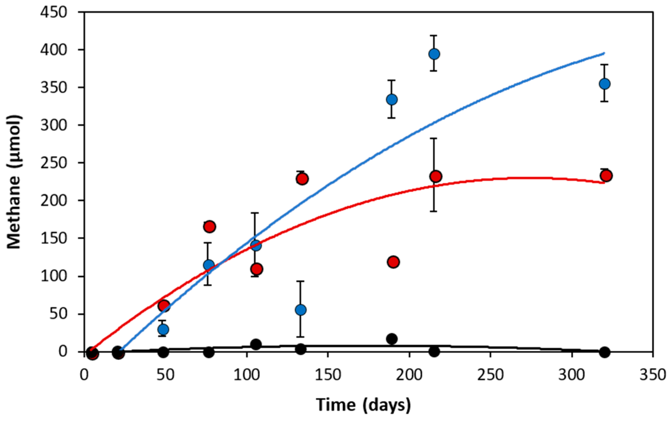
3.1. Analytical Evidence of Methanogenic Naphthalene Degradation

Initial anaerobic PAH degradation investigations conducted by Berdugo-Clavijo et al. [22] suggested that naphthalene could not be metabolized as a methanogenic substrate as similar amounts of methane were produced in substrate-unamended controls compared to naphthalene-amended incubations. However, after transferring the culture into fresh medium and adjusting its incubation conditions, enhanced methane production (up to 162 µmol) was observed in duplicate naphthalene-amended incubations relative to unamended controls; negligible amounts of methane were detected in the naphthalene-amended sterile replicate (Figure 1).
Methane production in the unamended controls (235 µmol) was likely due to metabolism of the cysteine-containing reducing agent. Based on the theoretical stoichiometric conversion of cysteine to methane (4 C3H7NO2S + 6 H2O + 4 H+ → 4 H2S + 4 NH4+ + 5 CH4 +7 CO2), the amount of cysteine sulfide added as a reductant may result in the production of up to 222 µmol methane. The balance of carbon could have originated from other trace organics (e.g. dead biomass, trace residual naphthalene) in the growth medium. Accounting for these background methane levels, naphthalene-amended NDC cultures yielded near-stoichiometric amounts of methane (101%) for their reported 20.5 µmol substrate loss (C10H8 + 8 H2O → 6 CH4 + 4 HCO3- + 4 H+), which is comparable to methanogenic methylnaphthalene-degrading enrichment cultures (2-MN and 2-6-dimethylnaphthalene; 2, 6-diMN) maintained by our laboratory [22].

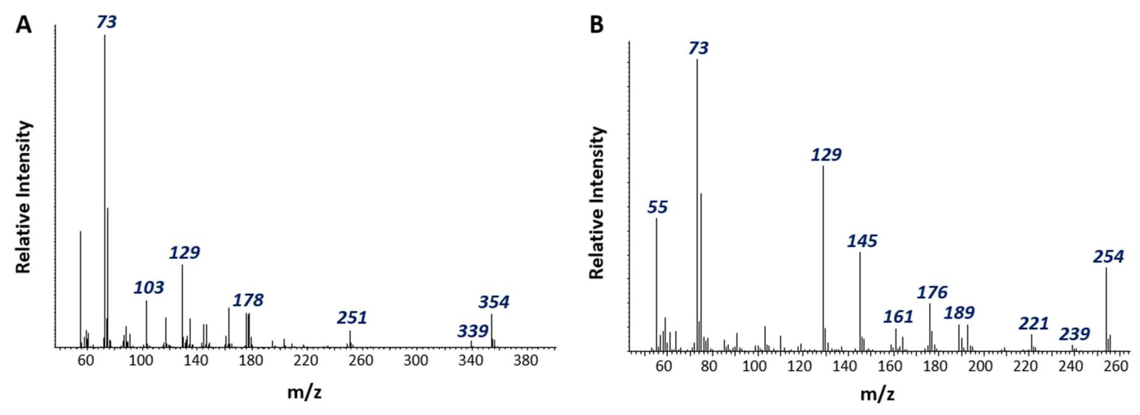
3.2. Detection of Putative Naphthalene Metabolites

Based on our GC-MS detection limits (≥10 nM), no metabolites indicative of carboxylation (naphthoic acid), fumarate addition ((methyl)naphthylsuccinic acid), or methylation (methyl naphthalene) were detected during active naphthalene degradation or in 13C-bicarbonate tracer experiments. This was supported by the results of our targeted functional gene assays of NDC, wherein no PCR amplification products were obtained using functional primers targeting fumarate addition (nmsA) [48] or using an in-house designed primer set targeting aromatic ring carboxylation (abcA) [49] (Table S1). We were also unable to detect evidence of the dearomatization gene 2-naphthoyl-CoA reductase using Ncr primers [50]. As these PCR-based assays were designed to target highly conserved regions within sulfate-reducing Deltaproteobacteria isolates and enrichment cultures, they may be unable to detect a broader diversity of related functional markers in other taxonomic lineages.
A silylated organic acid tentatively identified as a cyclohexene dicarboxylic acid (M = 354, M − 15 = 339) was detected exclusively in actively degrading enrichment cultures (Figure 2A). Notably, the free acid form of the putative metabolite was 2 mass units less than a cyclohexane dicarboxylic acid (C11H16O4) previously detected during naphthalene degradation by culture N47 [51]. We also detected a peak tentatively identified as decalin carboxylic acid (M = 254, M – 15 = 239) in PAH-amended NDC cultures (Figure 2B). The mass spectrum and GC retention time of this compound differed from those of an authentic standard of decahydro-2-naphthoic acid but could be an isomer. Although decahydro-2-naphthoic acid and its isomeric structures are thought to be dead-end products of anaerobic naphthalene metabolism [5], both putative metabolites suggest that naphthalene degradation by methanogenic consortium NDC undergoes a series of hydrogenation and ring cleavage reactions not unlike those catalyzed by NaphS2 and strain N47.

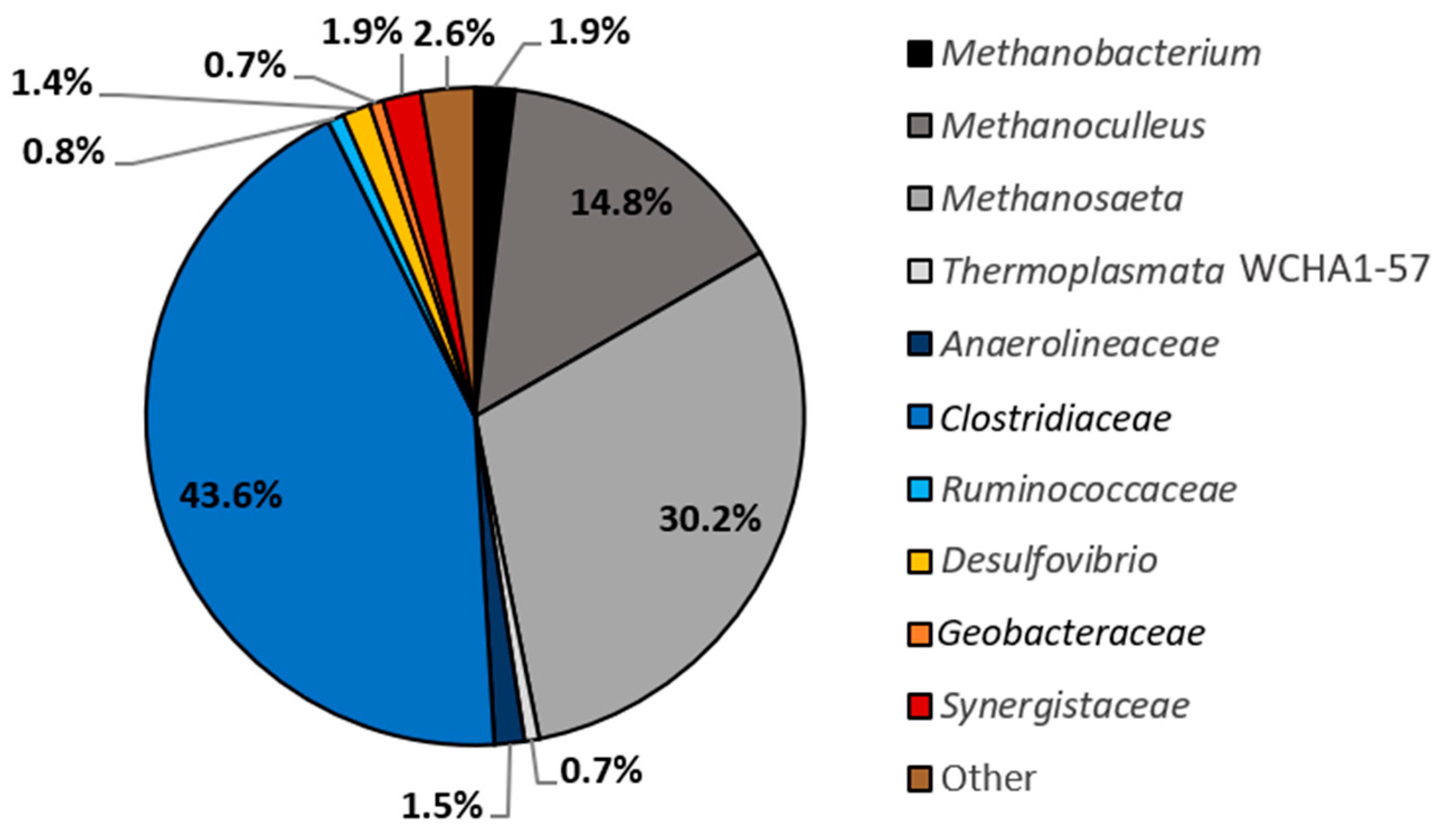
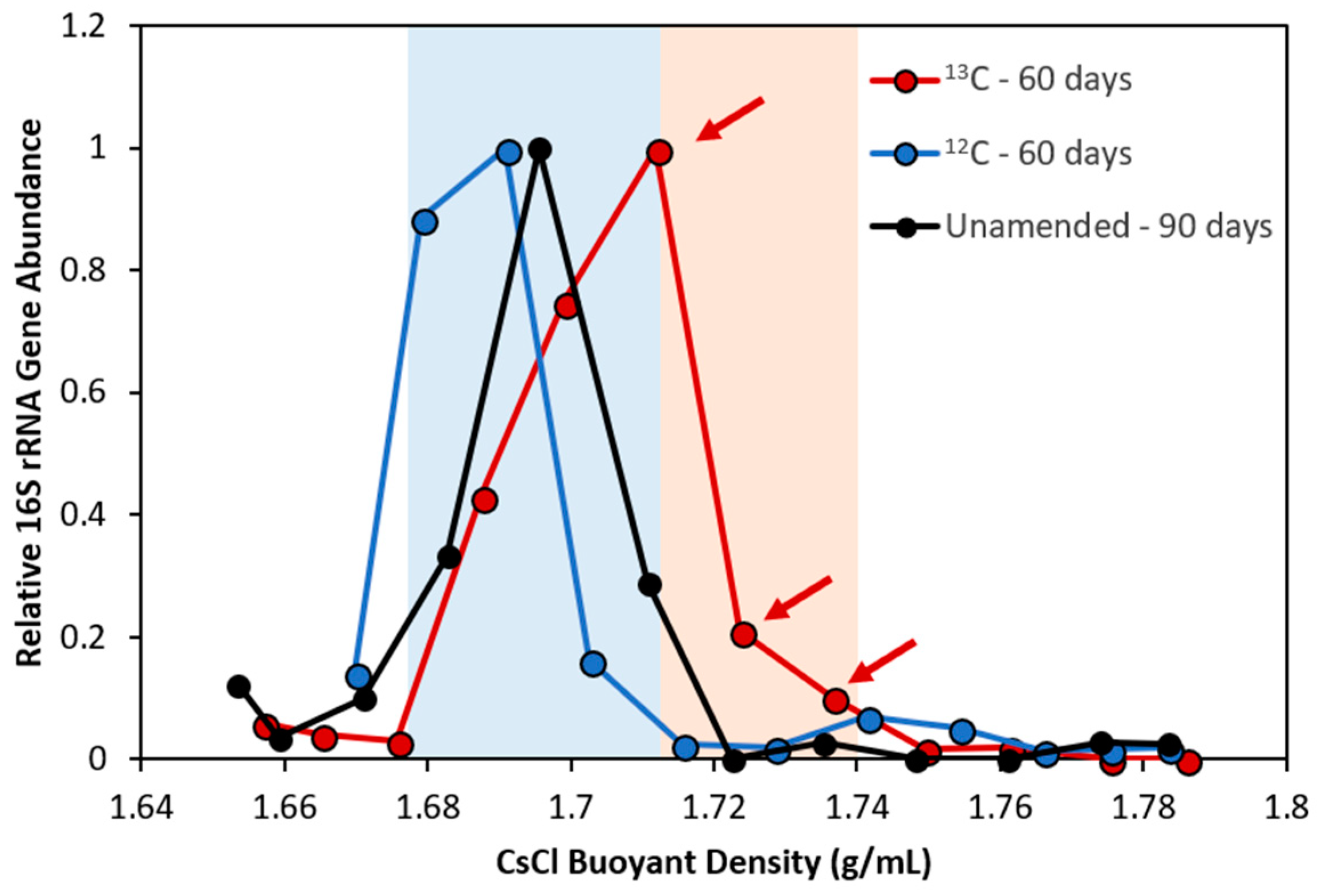
3.3. Pinpointing Putative Naphthalene Degraders Using Pyrosequencing and DNA-SIP

Pyrotag sequencing of 16S rRNA genes amplified from NDC revealed the predominance of bacterial OTUs belonging to Clostridiaceae (43.6% of reads) (Figure 3). This was not surprising, as members of this family were also heavily enriched in 2-MN and 2, 6-diMN-degrading methanogenic cultures (24.0% and 32.2% of reads, respectively) [22]. Methanogenic archaeal sequences belonging to Methanosaeta (30.2% of reads) and Methanoculleus (14.8% of reads) were also present in high abundance. All other OTUs recovered from NDC were only minor constituents (<2% read abundance) and collectively comprised less than 12% of reads.
Stable isotope probing with 13C-naphthalene and 12C-naphthalene as a control was performed to help pinpoint key organisms in NDC that actively participate in methanogenic naphthalene degradation. Triplicate microcosms were sacrificed and harvested for DNA in one month intervals to search for the earliest possible labeling of naphthalene degraders and to minimize risk of secondary cross-feeding. Up to 227 ng of gDNA was extracted from each microcosm, or ~80% less nucleic acid material than was successfully fractionated by Abu Laban et al. [52] during DNA-SIP surveys of a methanogenic toluene-degrading culture originating from an oil sands tailings pond. Note that this is still considerably less than the 5–10 µg of genetic material recommended for density gradient fractionation to ensure proper banding efficiency of labeled and unlabeled nucleic acid species [40]. While low DNA concentrations had no apparent effect on density gradient formation or fractionation, DNA could not be accurately quantified using conventional qPCR techniques after being separated across a dozen fractions (<102 16S rRNA gene copies/mL). We therefore chose to assess fractions for the presence of amplifiable 16S rRNA gene fragments using PCR as a semi-qualitative substitute for qPCR analysis.
After two months of incubation, three ‘heavy’ fractions were recovered (1.712–1.736 g/mL) from gDNA extracted from a [13C10] naphthalene microcosm, harboring OTUs belonging primarily to Desulfuromonadales (68.5% of reads) and Clostridiaceae (14.5% of reads) (Figure 4, Table 1). A similar microbial community composition was reported in the unlabeled [12C10] naphthalene control (Figure 4), supporting that these organisms were indeed the most abundant (and presumably most active) members in NDC at this time point. Recovered [13C10] naphthalene ‘light’ fractions (1.681–1.704 g/mL) contained a 9.52% lower abundance of Clostridiaceae than in corresponding ‘heavy’ fractions, showing that this taxon became enriched with the isotopic material, likely due to its involvement in naphthalene metabolism. Though the recovered [13C10] naphthalene ‘light’ fractions showed a 12.5% greater abundance of Desulfuromonadales compared to the ‘heavy’ fractions but its high relative abundance in NDC in general suggests it may also play a role in naphthalene metabolism.
Three minor constituents of NDC (Methanoculleus, 0.72%; Desulfovibrio, 0.43%; Clostridium, 0.27%) may have also been labeled with heavy isotope, though their low abundances make this observation speculative at best (Table 1). A problem arises, particularly for tracking nucleic acids, when substrate incorporation and incubation time are insufficient, generating poorly labeled biomarker molecules that are near-indistinguishable from a background of (relatively abundant) unlabeled molecules [34]. That said, no new 13C-labeling was observed after three months of SIP incubation. Determination of isotopic labeling via buoyant densities has a detection limit for 13C stable isotope incorporation of ~20 atom% [35], so we may have been very close to this threshold.
It was unclear why Desulfuromonadales became enriched in both [13C10] and [12C10] naphthalene-amended microcosms (Table 1), having only comprised a small fraction (0.7%) of previous pyrosequencing reads (shown as Geobacteraceae, Figure 3). Active members of the Desulfuromonadales are not typically detected in methanogenic enrichment cultures. Though this family has occasionally been identified in methane-producing coal-bed materials [53,54], their community role(s) have yet to be determined. Marozava et al. [33] identified a member of this family (Desulfobulbaceae) to incorporate a 13C-carbon label from SIP of an iron-reducing, 1-methylnaphthalene-degrading consortium, predicted to catalyze Fe (III) reduction rather than degrade PAHs. The dominant Desulfuromonadales OTU in NDC (NDC OTU_1) appears to more phylogenetically related to acetate-oxidizing Geobacter lovleyi strain SZ and Geobacter sp. AOG2 (87.0% sequence identity to each) than to known hydrocarbon-degrading isolates (84.6–84.8% sequence identity) (Figure 5). Overall, despite its high relative abundance in NDC, the possible role of Desulfuromonadales in naphthalene degradation is ambiguous given our pyrosequencing and DNA-SIP results (and metagenome analysis, see below).
Recovered 13C-labeled SIP and pyrosequencing OTUs belonging to Clostridiaceae shared 86–99% nucleotide sequence identity to cultured representatives of the Youngiibacter (formerly known as Acetivibrio), a recently described genus capable of fermenting a broad range of carbohydrates and aromatic acids [55,56,57] (Figure 5). The OTUs also share 84–99% sequence identity to the dominant Firmicutes detected in 2-MN and 2, 6-diMN [22]. This clade appears to be phylogenetically distinct from Clostridium and does not cluster with any other known or predicted PAH degraders, including the three anthracene-degrading SIP clones recovered by Zhang et al. [30] and the Thermoanerobacteraceae identified by Marozova et al. [33].

3.4. Metagenomic Evidence Predicts Clostridiaceae to Be a Key Naphthalene Degrader in NDC

Whole metagenomic shotgun sequencing and assembly of pooled 13C-labeled fractions was performed to generate a primary culture metagenome (IMG Genome ID 3300013290) and 10 supporting draft genomes (Table 2). The final metagenome consisted of an estimated 51212 scaffolds encoding 103278 protein-coding genes, of which roughly one-third were not orthologous to known proteins. A single Clostridiaceae sp. bin was assembled from the metagenomic sequences, predicted to be NDC_OTU2 (Figure 5), and shared >87% protein sequence similarity to Youngiibacter fragilis across 54% of its predicted 5424 protein-encoding genes (Table S2). Y. fragilis is a strictly anaerobic member of the Clostridiaceae family isolated from a natural gas-producing coal bed system [55]. Previous genomic sequencing by Wawrik et al. [56] revealed no evidence of known anaerobic hydrocarbon activation pathways in the pure culture. However, the remaining half of the genomic material in our Clostridiaceae bin appears to be weakly similar or unrelated to the type strain.
When comparing annotated protein-encoding genes to connected hydrocarbon metabolism KEGG routes, most draft genomes yielded only single hits and were not considered to be significant. We were also unable to detect any evidence of sequences orthologous to previously proposed anaerobic naphthalene carboxylation genes (abc, ubiX, ubiD), naphthoyl-2-coenzyme A reductase, or fumarate addition mechanisms. Thus, we further sought evidence of downstream anaerobic naphthalene degradation pathways using a curated database of all publicly available predicted anaerobic benzene/PAH degradation genes in NBCI and IMG (as of March 2017). A total of 804 genes of interest (with a minimum e-value cutoff = 10−5) were detected across 453 scaffolds in the prepared draft genomes, of which 85% contained less than three naphthalene degradation gene orthologues and were thus removed from further analysis. The remaining 73 candidate scaffolds were manually assessed for functional and structural consistency to predicted/known PAH-degrading genes in strains NaphS2, N47, and in other reference genomes. Most scaffolds were found to encode for predicted Fe-containing hydrogenases (including in the Desulfuromonadales bins) and have yet to be investigated further, as their broad functional diversity makes it difficult to discern an exclusive role in anaerobic naphthalene degradation.
In the Clostridiaceae bin, we identified a gene cluster of key interest (Scaffold 1013) wherein 9 of the 12 predicted protein-encoding genes shared 44–65% protein sequence similarity to Deltaproteobacterium strain NaphS2 (Figure 6). The enzymes encoded in Scaffold 1013 are also shown in Table 3 together with their predicted putative function according to sequence homologs in NaphS2 and other organisms using BLASTP. We manually confirmed that Scaffold 1013 contained no orthologous regions to the genome of Y. fragilis.
Two genes predicted to encode CoA-transferase Family III proteins were identified in Scaffold 1013 shared 44–61% similarity across a handful of NaphS2 loci and may serve to catalyze CoA-thioesterification in a reaction that is similar but not identical to naphthoyl-CoA ligase (Figure 6, Table 3). Family III CoA-transferases occur in prokaryotes and eukaryotes, and catalyze reversible, ATP-independent CoA-transfer reactions in a highly substrate- and stereo-specific manner [58]. These enzymes operate via a ternary complex mechanism, allowing for recycling of the CoA donor within the enzyme complex and increased energy conservation during substrate catabolism. Scaffold 1013 also encodes for a predicted acetyl-CoA C-acetyltransferase and a predicted H+/gluconate symporter (GntP) that could help to accumulate the CoA derivative compounds within the cell [5,58]. Several anaerobic catabolic pathways rely on Family III CoA-transferases to recycle CoA intermediates [55], including succinyl-CoA during anaerobic toluene degradation (succinyl CoA:(R)-benzylsuccinate CoA-transferase) [59,60], and 2-naphthoyl-CoA during 2-methylnaphthalene metabolism by NaphS2 and strain N47 (succinyl-CoA: naphthyl-2-methyl-succinate CoA-transferase) [12,61]. Though fumarate addition does not appear to be the mechanism of naphthalene activation by NDC, a similar unknown intermediate may be being used instead. Notably, if the Clostridiaceae phylotype is indeed catalyzing the addition of CoA to an unknown naphthalene activation product, then it is plausible that the organism is also chiefly responsible for initiating methanogenic attack of the PAH in NDC.
Scaffold 1013 harbored two predicted flavin-containing enzymes with 48–60% similarity to two NaphS2 orthologs annotated as 2-nitropropane dioxygenases [12] (Figure 6, Table 3). As NDC was maintained under strict anoxic conditions and 2-nitropropane is not a proposed intermediate of anaerobic naphthalene degradation, these annotations are more than likely erroneous. We hypothesize that these protein sequences may instead encode for dearomatizing arylcarboxyl-CoA reductases similar to those of 2-naphthoyl-CoA reductase and 5, 6-dihydro-2-naphthoyl CoA reductase in NaphS2 and N47. However, because the protein-encoding genes are not sequential to one another in Scaffold 1013 (Figure 6) as they are in strain N47 (N47_G38220 and N47_G38210, respectively) and in NaphS2 (NPH_5475 and NPH_5476, respectively), the pathway for reduction of the unknown arylcarboxyl-CoA structure appears to be unique. Enzyme assays conducted by Estelmann et al. [20] showed that predicted 5, 6-dihydro-2-naphthoyl CoA reductases contained flavin adenine dinucleotide (FAD) and flavin mononucleotide (FMN), and that complete reduction of both flavin cofactors (and a [4Fe-4S] cluster) was required to catalyze highly negative (E°′= −493 mV) two-electron reduction of 2-naphthoyl-CoA to 5,6-dihydro-2-naphthoyl-CoA. Therefore, a similar reaction may be required to reduce naphthalene under methanogenic conditions.
Four predicted ß-oxidation-like degradation genes detected in Scaffold 1013 were orthologous to sequences encoded within the proposed tetralin (thn) operon in NaphS2 (Figure 6; Table 3). Enzymes encoded by this operon are predicted to catalyze tetrahydronaphthoyl-CoA ring cleavage in NaphS2 and N47 [12,13]. Meckenstock et al. [5] proposed that enoyl-CoA hydratases/isomerases/hydrolases (ThnKLM) and acyl-CoA dehydrogenases (ThnO) facilitated ß-oxidation-like reactions leading towards ring opening, while dehydrogenation reactions following ring cleavage were catalyzed by predicted 3-hydroxyacyl-CoA dehydrogenases (ThnK and ThnY). Given that we also detected tentative metabolite evidence of dearomatization and ring cleavage reactions (Figure 2), it is plausible that Clostridiaceae members of NDC may coordinate at least some similar naphthalene degradation reactions under methanogenic conditions. However, another possible function of these enzymes is to catalyze ß-oxidation-like degradation steps of the unknown arylcarboxyl-CoA structure, similar to benzyl- and naphthyl-2-methyl-succinyl-CoA [12,13,61,62]. This would result in the formation of an intermediate aryl-acid CoA-ester similar to 2-napthoyl-CoA that could then be further degraded by Clostridiaceae.

4. Conclusions

PAH are important components that can persist in anoxic fuel-contaminated environments, thus understanding their natural fate processes, such as their microbial degradation, can aid in environmental clean-up efforts. The results of this present study add to the small body of knowledge describing methanogenic PAH biodegradation. Through the use of 16S rRNA amplicon sequencing, metabolite analysis, DNA-SIP using 13C-naphthalene, and assembly-based metagenomics of heavy fractions, our data suggest that a member of the Clostridiaceae is a key player in naphthalene metabolism in the culture. Enrichment of a Desulfuromonadales phylotype in NDC suggest a possible role for this organism as well, though this remains ambiguous as our metagenomic data did not reveal naphthalene-specific genes in the bins associated with the organism. While metabolite and genomic approaches did not allow us to identify an initial postulated mode of attack of naphthalene (e.g., via carboxylation), we did identify metabolites (tentatively) and genes predicted to encode for enzymes facilitating metabolic processes for downstream catabolic reactions including naphthalene carboxylic acid CoA-thioesterification and ring reduction steps only partially similar to NaphS2 and strain N47. These data suggest that more than one transformation pathway may be used to drive anoxic naphthalene degradation, demanding greater in-depth genomic investigations of diverse anaerobic PAH degrading taxa. Ongoing scale-up efforts of NDC and other naphthalene-degrading methanogenic enrichments that yield increased cell material will allow for additional studies to determine whether initial steps are also similar or different.

Supplementary Materials

The following are available online at https://0-www-mdpi-com.brum.beds.ac.uk/2076-2607/6/3/65/s1, Figure S1: Methane production from triplicate parent NDC microcosms amended with naphthalene (blue) relative to an unamended control (red), Table S1: Primer sets screened for the targeted detection of anaerobic functional genes in a methanogenic naphthalene-degrading enrichment culture, Table S2: Summary of draft genomes assembled from pooled 13C-labeled NDC SIP fractions.

Author Contributions

C.R.A.T., C.B.-C, and L.M.G. contributed to the conception of this research project and manuscript preparation. C.B.-C. established and maintained the methanogenic naphthalene-degrading enrichment culture, as well as conducted GC-MS, targeted functional gene PCR assays, and pyrosequencing surveys of NDC. C.R.A.T. was the lead researcher conducting DNA-SIP and metagenomic analyses of NDC with the assistance of C.M.O., G.M.J., A.S., and P.F.D.

Funding

This research was supported by an NSERC Discovery grant (no. 05214–2015) and a Deep Life Community (DLC) Pilot Project Award (Deep Carbon Observatory subcontract no. 50895) awarded to L.M.G.

Acknowledgments

The authors gratefully acknowledge Ilona Ruhl for assistance with Illumina MiSeq preparations and Oscar Montoya for assistance with 16S rRNA pipeline execution.

Conflicts of Interest

The authors declare no conflict of interest.

References

  1. Abdel-Shafy, H.I.; Mansour, M.S.M. A review on polycyclic aromatic hydrocarbons: Source, environmental impact, effect on human health and remediation. Egypt. J. Petrol. 2016, 25, 107–123. [Google Scholar] [CrossRef]
  2. Jobelius, C.; Ruth, B.; Griebler, C.; Meckenstock, R.U.; Hollender, J.; Reineke, A.; Frimmel, F.H.; Zwiener, C. Metabolites indicate hot spots of biodegradation and biogeochemical gradients in a high-resolution monitoring well. Environ. Sci. Technol. 2011, 45, 474–481. [Google Scholar] [CrossRef] [PubMed]
  3. Sverdrup, L.E.; Nielsen, T.; Henning Krogh, P. Soil ecotoxicity of polycyclic aromatic hydrocarbons in relation to soil sorption, lipophilicity, and water solubility. Environ. Sci. Technol. 2002, 36, 2429–2435. [Google Scholar] [CrossRef] [PubMed]
  4. Meckenstock, R.U.; Mouttaki, H. Anaerobic degradation of non-substituted aromatic hydrocarbons. Curr. Opin. Biotechnol. 2011, 22, 406–414. [Google Scholar] [CrossRef] [PubMed]
  5. Meckenstock, R.U.; Boll, M.; Mouttaki, H.; Koelschbach, J.S.; Cunha Tarouco, P.; Weyrauch, P.; Dong, X.; Himmelberg, A.M. Anaerobic degradation of benzene and polycyclic aromatic hydrocarbons. J. Mol. Microbiol. Biotechnol. 2016, 26, 92–118. [Google Scholar] [CrossRef] [PubMed]
  6. Chang, B.V.; Shiung, L.C.; Yuan, S.Y. Anaerobic biodegradation of polycyclic aromatic hydrocarbon in soil. Chemosphere 2002, 48, 717–724. [Google Scholar] [CrossRef]
  7. Safinowski, M.; Griebler, C.; Meckenstock, R.U. Anaerobic cometabolic transformation of polycyclic and heterocyclic aromatic hydrocarbons: Evidence from laboratory and field studies. Environ. Sci. Technol. 2006, 40, 4165–4173. [Google Scholar] [CrossRef] [PubMed]
  8. Musat, F.; Galushko, A.; Jacob, J.; Widdel, F.; Kube, M.; Reinhardt, R.; Wilkes, H.; Schink, B.; Rabus, R. Anaerobic degradation of naphthalene and 2-methylnaphthalene by strains of marine sulfate-reducing bacteria. Environ. Microbiol. 2009, 11, 209–219. [Google Scholar] [CrossRef] [PubMed] [Green Version]
  9. Meckenstock, R.U.; Annweiler, E.; Michaelis, W.; Richnow, H.H.; Schink, B. Anaerobic naphthalene degradation by a sulfate-reducing enrichment culture. Appl. Environ. Microbiol. 2000, 66, 2743–2747. [Google Scholar] [CrossRef] [PubMed]
  10. Selesi, D.; Jehmlich, N.; von Bergen, M.; Schmidt, F.; Rattei, T.; Tischler, P.; Lueders, T.; Meckenstock, R.U. Combined genomic and proteomic approaches identify gene clusters involved in anaerobic 2-methylnaphthalene degradation in the sulfate-reducing enrichment culture N47. J. Bacteriol. 2010, 192, 295–306. [Google Scholar] [CrossRef] [PubMed]
  11. Galushko, A.; Minz, D.; Schink, B.; Widdel, F. Anaerobic degradation of naphthalene by a pure culture of a novel type of marine sulphate-reducing bacterium. Environ. Microbiol. 1999, 1, 415–420. [Google Scholar] [CrossRef] [PubMed]
  12. DiDonato, R.J., Jr.; Young, N.D.; Butler, J.E.; Chin, K.J.; Hixson, K.K.; Mouser, P.; Lipton, M.S.; DeBoy, R.; Methe, B.A. Genome sequence of the Deltaproteobacterial strain NaphS2 and analysis of differential gene expression during anaerobic growth on naphthalene. PLoS ONE 2010, 5, e14072. [Google Scholar] [CrossRef] [PubMed]
  13. Bergmann, F.; Selesi, D.; Weinmaier, T.; Tischler, P.; Rattei, T.; Meckenstock, R.U. Genomic insights into the metabolic potential of the polycyclic aromatic hydrocarbon degrading sulfate-reducing Deltaproteobacterium N47. Environ. Microbiol. 2011, 13, 1125–1137. [Google Scholar] [CrossRef] [PubMed]
  14. Bergmann, F.D.; Selesi, D.; Meckenstock, R.U. Identification of new enzymes potentially involved in anaerobic naphthalene degradation by the sulfate-reducing enrichment culture N47. Arch. Microbiol. 2011, 193, 241–250. [Google Scholar] [CrossRef] [PubMed]
  15. Buckel, W.; Kung, J.W.; Boll, M. The benzoyl-coenzyme A reductase and 2-hydroxyacyl-coenzyme A dehydratase radical enzyme family. Chembiochem 2014, 15, 2188–2194. [Google Scholar] [CrossRef] [PubMed]
  16. Mouttaki, H.; Johannes, J.; Meckenstock, R.U. Identification of naphthalene carboxylase as a prototype for the anaerobic activation of non-substituted aromatic hydrocarbons. Environ. Microbiol. 2012, 14, 2770–2774. [Google Scholar] [CrossRef] [PubMed]
  17. Weyrauch, P.; Zaytsev, A.V.; Stephan, S.; Kocks, L.; Schmitz, O.J.; Golding, B.T.; Meckenstock, R.U. Conversion of cis-2-carboxycyclohexylacetyl-CoA in the downstream pathway of anaerobic naphthalene degradation. Environ. Microbiol. 2017, 19, 2819–2830. [Google Scholar] [CrossRef] [PubMed]
  18. Eberlein, C.; Estelmann, S.; Seifert, J.; von Bergen, M.; Muller, M.; Meckenstock, R.U.; Boll, M. Identification and characterization of 2-naphthoyl-coenzyme a reductase, the prototype of a novel class of dearomatizing reductases. Mol. Microbiol. 2013, 88, 1032–1039. [Google Scholar] [CrossRef] [PubMed]
  19. Eberlein, C.; Johannes, J.; Mouttaki, H.; Sadeghi, M.; Golding, B.T.; Boll, M.; Meckenstock, R.U. ATP-dependent/-independent enzymatic ring reductions involved in the anaerobic catabolism of naphthalene. Environ. Microbiol. 2013, 15, 1832–1841. [Google Scholar] [CrossRef] [PubMed]
  20. Estelmann, S.; Blank, I.; Feldmann, A.; Boll, M. Two distinct old yellow enzymes are involved in naphthyl ring reduction during anaerobic naphthalene degradation. Mol. Microbiol. 2015, 95, 162–172. [Google Scholar] [CrossRef] [PubMed]
  21. Gieg, L.M.; Toth, C.R.A. Signature metabolite analysis to determine in situ anaerobic hydrocarbon biodegradation. In Anaerobic Utilization of Hydrocarbons, Oils, and Lipids; Springer: Cham, Switzerland, 2017; pp. 1–30. [Google Scholar]
  22. Berdugo-Clavijo, C.; Dong, X.; Soh, J.; Sensen, C.W.; Gieg, L.M. Methanogenic biodegradation of two-ringed polycyclic aromatic hydrocarbons. FEMS Microbiol. Ecol. 2012, 81, 124–133. [Google Scholar] [CrossRef] [PubMed] [Green Version]
  23. Berdugo-Clavijo, C.; Gieg, L.M. Conversion of crude oil to methane by a microbial consortium enriched from oil reservoir production waters. Front. Microbiol. 2014, 5, 197. [Google Scholar] [CrossRef] [PubMed]
  24. Chang, W.; Um, Y.; Holoman, T.R. Polycyclic aromatic hydrocarbon (PAH) degradation coupled to methanogenesis. Biotechnol. Lett. 2006, 28, 425–430. [Google Scholar] [CrossRef] [PubMed]
  25. Christensen, N.; Batstone, D.J.; He, Z.; Angelidaki, I.; Schmidt, J.E. Removal of polycyclic aromatic hydrocarbons (PAHs) from sewage sludge by anaerobic degradation. Water Sci. Technol. 2004, 9, 237–244. [Google Scholar] [CrossRef]
  26. Chang, W.; Um, Y.; Hoffman, B.; Pulliam Holoman, T.R. Molecular characterization of polycyclic aromatic hydrocarbon (PAH)-degrading methanogenic communities. Biotechnol. Prog. 2005, 21, 682–688. [Google Scholar] [CrossRef] [PubMed]
  27. Maillacheruvu, K.Y.; Pathan, I.A. Biodegradation of naphthalene, phenanthrene, and pyrene under anaerobic conditions. J. Environ. Sci. Health A 2009, 44, 1315–1326. [Google Scholar] [CrossRef] [PubMed]
  28. Siegert, M.; Cichocka, D.; Herrmann, S.; Grundger, F.; Feisthauer, S.; Richnow, H.H.; Springael, D.; Krüger, M. Accelerated methanogenesis from aliphatic and aromatic hydrocarbons under iron- and sulfate-reducing conditions. FEMS Microbiol. Lett. 2011, 315, 6–16. [Google Scholar] [CrossRef] [PubMed]
  29. Wan, R.; Zhang, S.; Xie, S. Microbial community changes in aquifer sediment microcosm for anaerobic anthracene biodegradation under methanogenic condition. J. Environ. Sci. 2012, 24, 1498–1503. [Google Scholar] [CrossRef]
  30. Zhang, S.; Wang, Q.; Xie, S. Stable isotope probing identifies anthracene degraders under methanogenic conditions. Biodegradation 2012, 23, 221–230. [Google Scholar] [CrossRef] [PubMed]
  31. Zhang, S.Y.; Wang, Q.F.; Xie, S.G. Molecular characterization of phenanthrene-degrading methanogenic communities in leachate-contaminated aquifer sediment. Int. J. Environ. Sci. Technol. 2012, 9, 705–712. [Google Scholar] [CrossRef] [Green Version]
  32. Dolfing, J.; Xu, A.; Gray, N.D.; Larter, S.R.; Head, I.M. The thermodynamic landscape of methanogenic PAH degradation. Microb. Biotechnol. 2009, 2, 566–574. [Google Scholar] [CrossRef] [PubMed] [Green Version]
  33. Marozava, S.; Mouttaki, H.; Muller, H.; Laban, N.A.; Probst, A.J.; Meckenstock, R.U. Anaerobic degradation of 1-methylnaphthalene by a member of the Thermoanaerobacteraceae contained in an iron-reducing enrichment culture. Biodegradation 2018, 29, 23–39. [Google Scholar] [CrossRef] [PubMed]
  34. Neufeld, J.D.; Dumont, M.G.; Vohra, J.; Murrell, J.C. Methodological considerations for the use of stable isotope probing in microbial ecology. Microb. Ecol. 2007, 53, 435–442. [Google Scholar] [CrossRef] [PubMed]
  35. Vogt, C.; Lueders, T.; Richnow, H.H.; Krüger, M.; von Bergen, M.; Seifert, J. Stable isotope probing approaches to study anaerobic hydrocarbon degradation and degraders. J. Mol. Microbiol. Biotechnol. 2016, 26, 195–210. [Google Scholar] [CrossRef] [PubMed]
  36. Townsend, G.T.; Prince, R.C.; Suflita, J.M. Anaerobic oxidation of crude oil hydrocarbons by the resident microorganisms of a contaminated anoxic aquifer. Environ. Sci. Technol. 2003, 37, 5213–5218. [Google Scholar] [CrossRef] [PubMed]
  37. Gieg, L.M.; Duncan, K.E.; Suflita, J.M. Bioenergy production via microbial conversion of residual oil to natural gas. Appl. Environ. Microbiol. 2008, 74, 3022–3029. [Google Scholar] [CrossRef] [PubMed]
  38. Berdugo-Clavijo, C. Methanogenic Biodegradation of Crude Oil and Polycyclic Aromatic Hydrocarbons. Ph.D. Thesis, University of Calgary, Calgary, AB, Canada, 2015. [Google Scholar]
  39. Morasch, B.; Annweiler, E.; Warthmann, R.J.; Meckenstock, R.U. The use of a solid adsorber resin for enrichment of bacteria with toxic substrates and to identify metabolites: Degradation of naphthalene, o-, and m-xylene by sulfate-reducing bacteria. J. Microbiol. Methods 2001, 44, 183–191. [Google Scholar] [CrossRef]
  40. Neufeld, J.D.; Vohra, J.; Dumont, M.G.; Lueders, T.; Manefield, M.; Friedrich, M.W.; Murrell, J.C. DNA stable-isotope probing. Nat. Protoc. 2007, 2, 860–866. [Google Scholar] [CrossRef] [PubMed]
  41. Cheng, L.; Rui, J.; Li, Q.; Zhang, H.; Lu, Y. Enrichment and dynamics of novel syntrophs in a methanogenic hexadecane-degrading culture from a Chinese oilfield. FEMS Microbiol. Ecol. 2013, 83, 757–766. [Google Scholar] [CrossRef] [PubMed]
  42. Klindworth, A.; Pruesse, E.; Schweer, T.; Peplies, J.; Quast, C.; Horn, M.; Glockner, F.O. Evaluation of general 16S ribosomal RNA gene PCR primers for classical and next-generation sequencing-based diversity studies. Nucleic Acids Res. 2013, 41, e1. [Google Scholar] [CrossRef] [PubMed]
  43. Toth, C.R.A.; Gieg, L.M. Time course-dependent methanogenic crude oil biodegradation: Dynamics of fumarate addition metabolites, biodegradative genes, and microbial community composition. Front. Microbiol. 2018, 8, 2610. [Google Scholar] [CrossRef] [PubMed]
  44. Kumar, S.; Stecher, G.; Tamura, K. MEGA7: Molecular evolutionary genetics analysis version 7.0 for bigger datasets. Mol. Biol. Evol. 2016, 33, 1870–1874. [Google Scholar] [CrossRef] [PubMed]
  45. Bankevich, A.; Nurk, S.; Antipov, D.; Gurevich, A.A.; Dvorkin, M.; Kulikov, A.S.; Lesin, V.M.; Nikolenko, S.I.; Pham, S.; Prjibelski, A.D.; et al. SPAdes: A new genome assembly algorithm and its applications to single-cell sequencing. J. Comput. Biol. 2012, 19, 455–477. [Google Scholar] [CrossRef] [PubMed]
  46. Kang, D.D.; Froula, J.; Egan, R.; Wang, Z. MetaBAT, an efficient tool for accurately reconstructing single genomes from complex microbial communities. PeerJ 2015, 3, e1165. [Google Scholar] [CrossRef] [PubMed]
  47. Huson, D.H.; Auch, A.F.; Qi, J.; Schuster, S.C. MEGAN analysis of metagenomic data. Genome Res. 2007, 17, 377–386. [Google Scholar] [CrossRef] [PubMed] [Green Version]
  48. Von Netzer, F.; Pilloni, G.; Kleindienst, S.; Kruger, M.; Knittel, K.; Grundger, F.; Lueders, T. Enhanced gene detection assays for fumarate-adding enzymes allow uncovering of anaerobic hydrocarbon degraders in terrestrial and marine systems. Appl. Environ. Microbiol. 2013, 79, 543–552. [Google Scholar] [CrossRef] [PubMed]
  49. Fowler, S.J. Syntrophic Hydrocarbon Metabolism under Methanogenic Conditions. Ph.D. Thesis, University of Calgary, Calgary, AB, Canada, 2014. [Google Scholar]
  50. Morris, B.E.; Gissibl, A.; Kümmel, S.; Richnow, H.H.; Boll, M. A PCR-based assay for the detection of anaerobic naphthalene degradation. FEMS Microbiol. Lett. 2014, 354, 55–59. [Google Scholar] [CrossRef] [PubMed] [Green Version]
  51. Annweiler, E.; Michaelis, W.; Meckenstock, R.U. Identical ring cleavage products during anaerobic degradation of naphthalene, 2-methylnaphthalene, and tetralin indicate a new metabolic pathway. Appl. Environ. Microbiol. 2002, 68, 852–858. [Google Scholar] [CrossRef] [PubMed]
  52. Abu Laban, N.; Dao, A.; Foght, J. DNA stable-isotope probing of oil sands tailings pond enrichment cultures reveals different key players for toluene degradation under methanogenic and sulfidogenic conditions. FEMS Microbiol. Ecol. 2015, 91. [Google Scholar] [CrossRef] [PubMed] [Green Version]
  53. Krüger, M.; Beckmann, S.; Engelen, B.; Thielemann, T.; Cramer, B.; Schippers, A.; Cypionka, H. Microbial methane formation from hard coal and timber in an abandoned coal mine. Geomicrobiol. J. 2008, 25, 315–321. [Google Scholar] [CrossRef]
  54. Beckmann, S.; Lueders, T.; Kruger, M.; von Netzer, F.; Engelen, B.; Cypionka, H. Acetogens and acetoclastic Methanosarcinales govern methane formation in abandoned coal mines. Appl. Environ. Microbiol. 2011, 77, 3749–3756. [Google Scholar] [CrossRef] [PubMed]
  55. Lawson, P.A.; Wawrik, B.; Allen, T.D.; Johnson, C.N.; Marks, C.R.; Tanner, R.S.; Harriman, B.H.; Strapoc, D.; Callaghan, A.V. Youngiibacter fragilis gen. nov., sp. nov., isolated from natural gas production-water and reclassification of Acetivibrio multivorans as Youngiibacter multivorans comb. nov. Int. J. Syst. Evol. Microbiol. 2014, 64, 198–205. [Google Scholar] [CrossRef] [PubMed]
  56. Wawrik, C.B.; Callaghan, A.V.; Stamps, B.W.; Wawrik, B. Genome sequence of Youngiibacter fragilis, the type strain of the genus Youngiibacter. Genome Announc. 2014, 2, e01183-13. [Google Scholar] [CrossRef] [PubMed]
  57. Tanaka, K.; Nakamura, K.; Mikami, E. Fermentation of cinnamate by a mesophilic strict anaerobe, Acetivibrio multivorans sp. nov. Arch. Microbiol. 1991, 155, 120–124. [Google Scholar] [CrossRef]
  58. Heider, J. A new family of CoA-transferases. FEBS Lett. 2001, 509, 345–349. [Google Scholar] [CrossRef] [Green Version]
  59. Leuthner, B.; Heider, J. Anaerobic toluene catabolism of Thauera aromatica: The bbs operon codes for enzymes of beta oxidation of the intermediate benzylsuccinate. J. Bacteriol. 2000, 182, 272–277. [Google Scholar] [CrossRef] [PubMed]
  60. Leutwein, C.; Heider, J. Succinyl-CoA:(R)-benzylsuccinate CoA-transferase: An enzyme of the anaerobic toluene catabolic pathway in denitrifying bacteria. J. Bacteriol. 2001, 183, 4288–4295. [Google Scholar] [CrossRef] [PubMed]
  61. Safinowski, M.; Meckenstock, R.U. Enzymatic reactions in anaerobic 2-methylnaphthalene degradation by the sulphate-reducing enrichment culture N47. FEMS Microbiol. Lett. 2004, 240, 99–104. [Google Scholar] [CrossRef] [PubMed]
  62. Leutwein, C.; Heider, J. (R)-Benzylsuccinyl-CoA dehydrogenase of Thauera aromatica, an enzyme of the anaerobic toluene catabolic pathway. Arch. Microbiol. 2002, 178, 517–524. [Google Scholar] [CrossRef] [PubMed]
Figure 1. Methane production from transferred NDC incubations amended with naphthalene (blue) relative to substrate-unamended (red) and sterile (black) controls. Bars indicate ± average of duplicate microcosms.
Microorganisms 06 00065 g001
Figure 2. Mass spectra of silylated GC-MS peaks tentatively identified as (A) cyclohexane dicarboxylic acid and (B) an isomer of deahydro-2-naphthoic acid detected in NDC.
Microorganisms 06 00065 g002
Figure 3. Distribution of bacterial and archaeal 16S rRNA gene pyrotag sequences in an active naphthalene-degrading replicate of NDC. Values are shown as the percentage of sequences per taxon based on 2034 bacterial sequences and 1881 archaeal sequences. OTUs included in ‘Other’ make up less than 0.4% of prokaryotic sequences each and include Spirochaeta (0.26%), Proteiniphilum (0.13%), and Candidatus Methanoregula (0.36%).
Microorganisms 06 00065 g003
Figure 4. Relative distribution of total 16S rRNA gene fragments in NDC SIP fractions containing 13C- and 12C-DNA extracted after 60 days of incubation. The red shaded area indicates fractions considered to be enriched in 13C-DNA compared to background 12C-DNA microorganisms (shaded area in blue) and were predicted to harbor naphthalene degraders. Fractions chosen and pooled for metagenomic sequencing analysis are indicated with arrows.
Microorganisms 06 00065 g004
Figure 5. Maximum likelihood consensus tree showing the affiliation of putative 13C-labeled sequencing OTUs comprising at least 0.5% abundance (bold, shaded) to methanogenic (methyl)naphthalene pyrosequencing clones (red), known anaerobic benzene/PAH degraders and other published reference strains. Evolutionary analyses of aligned nucleotide sequences were conducted in MEGA7; the consensus tree was constructed using the Tamura–Nei model at all nucleotide positions (528 in total) and performing 500 bootstrap replicates (values below 60% are not shown). Archaeal 16S rRNA gene sequences retrieved from NCBI were used to root the tree (collapsed). Asterisks indicate sequences where more than one clone was obtained and a single representative sequence (accession number) was selected for tree construction.
Microorganisms 06 00065 g005
Figure 6. Organization of putative methanogenic naphthalene degradation gene cluster of binned Clostridiaceae scaffold 1013. Protein functions were predicted based on sequence orthology to NaphS2 loci and BLASTP searches. Protein annotation: 1. enoyl CoA hydratase; 2. Class III CoA transferase; 3. FAD-dependent arylcarboxyl-CoA reductase; 4. enoyl-CoA hydratase/isomerase/hydrolase; 5. 3-hydroxybutyryl-CoA dehydrogenase; 6. FAD-dependent arylcarboxyl-CoA reductase; 7. H+/gluconate symporter; 8. 3-hydroxybutyryl-CoA dehydrogenase; 9. acetyl-CoA C-acetyltransferase; 10. Class III CoA transferase; 11. hypothetical protein.
Microorganisms 06 00065 g006
Table 1. Distribution of the most abundant classified taxa (%) across SIP fractions collected after 60 days of incubation, as determined by 16S rRNA gene sequencing.
Taxon13C Heavy12C Control13C Light
Archaea
Methanosaeta1.495.184.83
Methanobacterium0.140.642.19
Methanoculleus0.720.170.04
Thermoplasmata WCHA-570.000.040.35
Bacteroidetes
Proteiniphilum0.070.530.30
Rikenellaceae vadinBC270.000.170.70
Chloroflexi
Anaerolineaceae0.141.480.73
Firmicutes
Clostridiaceae14.517.84.98
Sedimentibacter0.071.590.47
Clostridium0.270.070.03
Peptococcaceae0.160.130.01
Proteobacteria
Desulfuromonadales68.561.681.0
Geobacter1.881.702.06
Desulfuromonas0.451.680.36
Desulfobulbus0.320.420.54
Desulfovibrio0.430.440.11
Deltaproteobacteria0.410.150.21
12C Control: indicates cultures amended with unlabeled naphthalene that were incubated alongside those amended with isotopically labeled naphthalene.
Table 2. Genome sequence similarity of prepared draft genomes to their closest matching cultured representative or enriched strain. Pairwise average nucleotide identity (ANI) calculations were used to assess the bidirectional best hits (BBH) of genes having 70% or more identity to reference strains and at least 70% coverage of the shorter gene. Alignment fraction (AF) calculations were prepared to determine the % coverage of the draft genomes to reference strains.
Genome1 IMG IDGenome1 NameGenome Size, bp (Assembled)Gene Count (Assembled)Genome2 IMG IDGenome2 NameANI 1→2ANI 2→1AF 1→2AF 2→1Total BBH
2724679725Anaerolinea sp. Bin 123337882249649633005Anaerolinea thermophila UNI-167.667.60.080.05221
2724679726Anaerolinea sp. Bin 222716252153649633005Anaerolinea thermophila UNI-167.767.70.130.08318
2724679696Unclassified Clostridiaceae sp. Bin 1543813254242582580929Youngiibacter fragilis87.587.50.540.752904
2724679721Desulfovibrio sp. Bin 1308824229592561511137Desulfovibrio alcoholivorans DSM 543372.672.70.400.251337
2724679697Unclassified Desulfuromonadales sp. Bin 143900794104640427115Geobacter uraniireducens Rf472.472.40.290.251225
2724679723Unclassified Desulfuromonadales sp. Bin 238766843546640427115Geobacter uraniireducens Rf472.272.20.350.261230
2724679724Unclassified Desulfuromonadales sp. Bin 325265382519642555130Geobacter lovleyi SZ77.077.00.690.431737
2724679722Methanosaeta sp. Bin 126833222798650716054Methanosaeta concilii GP-696.696.60.810.762209
2724679727Sphaerochaeta sp. Bin 117509541956650377973Sphaerochaeta globosa Buddy80.580.50.700.371321
2724679720Unclassified Rhodospirillales Bin 1347316836512651870079Unclassified Rhodospirillaceae Bin 3569.569.50.150.14546
Table 3. Overview of binned Clostridiaceae Scaffold 1013. Percent sequence identity/similarity of protein-encoding encoding genes are compared to orthologous loci detected in the NaphS2 genome and within NCBI protein sequence database.
Gene IDIMG Predicted Protein AnnotationLength (aa)NaphS2 OrthologLocus% Identity
(% Similarity)
Best BLASTP OrthologAccession No.% Identity
(% Similarity)
2727804033Hypothetical protein60N/A--hypothetical protein [Clostridiales bacterium VE202-03]WP_02472363443 (71)
2727804034Crotonobetainyl-CoA:carnitine CoA-transferase CaiB412CoA-transferase family III proteinNPH_4605
NPH_6726
NPH_5274
31 (50)
24 (46)
27 (44)
Formyl-coenzyme A transferase [uncultured Clostridium sp.]SCJ8019555 (74)
2727804035acetyl-CoA C-acetyltransferase387Acetyl-CoA C-acyltransferaseNPH_3581
NPH_6994
NPH_6993
NPH_5273
43 (61)
43 (62)
38 (62)
38 (55)
MULTISPECIES: acetyl-CoA acetyltransferase [Geobacillus]WP_07404374451 (69)
27278040363-hydroxybutyryl-CoA dehydrogenase2883-hydroxybutyryl-CoA dehydrogenaseNPH_5812
NPH_5906
NPH_5896
NPH_7219
38 (58)
40 (56)
36 (53)
34 (53)
3-hydroxyacyl-CoA dehydrogenase [Clostridiales bacterium PH28_bin88]KKM1008953 (71)
2727804037H+/gluconate symporter444N/A--MULTISPECIES: hypothetical protein [Clostridiales]WP_00786230843 (65)
2727804038enoyl-[acyl-carrier protein] reductase II3152-nitropropane dioxygenaseNPH_5508
NPH_6957
35 (52)
33 (53)
2-nitropropane dioxygenase [Anaerosporomusa subterranea]WP_06623762452 (70)
2727804039Enoyl-CoA hydratase/isomerase853-hydroxybutyryl-CoA dehydrataseNPH_5887
NPH_5907
47 (61)
40 (56)
MULTISPECIES: 3-hydroxybutyryl-CoA dehydratase [Clostridiales]WP_00786230052 (74)
2727804040Enoyl-CoA hydratase/isomerase783-hydroxybutyryl-CoA dehydrataseNPH_6695
NPH_5897
NPH_5898
39 (62)
38 (63)
45 (65)
enoyl-CoA hydratase [Desulfotomaculum thermosubterraneum]WP_07286985555 (77)
27278040412-nitropropane dioxygenase precursor3212-nitropropane dioxygenaseNPH_6957
NPH_5508
38 (60)
31 (48)
nitronate monooxygenase [Clostridium citroniae]WP_08342401871 (84)
2727804042Crotonobetainyl-CoA:carnitine CoA-transferase CaiB409CoA-transferase family III proteinNPH_4605
NPH_1868
34 (52)
26 (44)
MULTISPECIES: CoA transferase [Clostridiales]WP_00786230548 (68)
27278040432-(1,2-epoxy-1,2-dihydrophenyl)acetyl-CoA isomerase262putative enoyl-CoA hydrataseNPH_0885
NPH_5898
NPH_5897
31 (50)
32 (54)
32 (52)
enoyl-CoA hydratase [Enterococcus silesiacus]WP_07187930647 (71)
2727804044Hypothetical protein25N/A--ATP-dependent endonuclease [Bacillus cereus]OPA1154972 (77)

