Article

Bacteria-Derived Hemolysis-Related Genes Widely Exist in Scuticociliates

1
Shenzhen Institute of Guangdong Ocean University, Shenzhen 518120, China
2
Key Laboratory of Aquatic Biodiversity and Conservation, Institute of Hydrobiology, Chinese Academy of Sciences, Wuhan 430072, China
3
Guangdong Provincial Engineering Research Center for Aquatic Animal Health Assessment, Shenzhen 518120, China
4
Shenzhen Dapeng New District Science and Technology Innovation Service Center, Shenzhen 518119, China
5
University of Chinese Academy of Sciences, Beijing 100049, China
6
State Key Laboratory of Freshwater Ecology and Biotechnology of China, Wuhan 430072, China
7
CAS Center for Excellence in Animal Evolution and Genetics, Kunming 650223, China
*
Author to whom correspondence should be addressed.
These authors contributed equally to this work.
Submission received: 30 September 2020 / Revised: 20 November 2020 / Accepted: 20 November 2020 / Published: 22 November 2020
(This article belongs to the Section Molecular Microbiology and Immunology)

Abstract

:
Scuticociliatosis is an invasive external or systemic infection caused by ciliated protozoa, mainly those within the subclass Scuticociliatia (scuticociliates). Many scuticociliates are fish pathogens, including Miamiensis avidus, Philasterides dicentrarchi, Pseudocohnilembus persalinus, and Uronema marinum. Our previous study showed that hemolysis-related genes derived from bacteria through horizontal gene transfer (HGT) may contribute to virulence in P. persalinus. Hemorrhagic lesions are a common feature of scuticociliatosis, but it is not known whether other scuticociliates also have bacteria-derived hemolysis-related genes. In this study, we constructed a high-quality macronuclear genome of another typical pathogenic scuticociliate, U. marinum. A total of 105 HGT genes were identified in this species, of which 35 were homologs of hemolysis-related genes (including hemolysin-like genes) that had previously been identified in P. persalinus. Sequencing of an additional five species from four scuticociliate families showed that bacteria-derived hemolysis-related genes (especially hemolysin-like genes) are widely distributed in scuticociliates. Based on these findings, we suggest that hemolysin-like genes may have originated before the divergence of scuticociliates.

1. Introduction

Scuticociliatosis is one of the most serious parasitic diseases in fish worldwide, with a high mortality rate in cultured marine fish, e.g., olive flounder and turbot [1,2]. Associated outbreaks of fatal infection have led to serious economic losses. Scuticociliates, which belong to the ciliate subclass Scuticociliatia, have been identified as causative agents of scuticociliatosis. A salient clinical manifestation of scuticociliatosis is hemorrhagic lesions [3]. More than 20 species are facultative parasites that can destroy host tissues, including Miamiensis avidus, Philasterides dicentrarchi, Pseudocohnilembus persalinus, and Uronema marinum [4], suggesting a common molecular basis for virulence in scuticociliates. However, many studies on scuticociliates have focused on species identification, morphology, and phylogenetic analysis, and few have examined the molecular basis or mechanism of virulence. One study on P. dicentrarchi found that cysteine proteases are activated and secreted into the extracellular medium and probably participate in host tissue invasion and degradation, as well as in the degradation of hemoglobin within erythrocytes [5]. Metalloprotease, phosphatidylcholine (PC)-specific phospholipase D (PC-PLD), and PC-specific phospholipase C (PC-PLC) activities have been detected in U. marinum-infected tissues, and may play an important role in host cell penetration [6].
In 2015, we sequenced the first macronuclear (MAC) genome of a scuticociliate, P. persalinus. Comparative genomics analysis showed that the MAC genome is relatively small compared with other free-living ciliates and that the protein domain composition resembles that of an obligate fish parasite, Ichthyophthirius multifiliis. More importantly, the P. persalinus MAC genome harbors many bacteria-derived genes that encode proteins which may be involved in cell adhesion, hemolysis, and heme utilization processes [7].
Similarly to P. persalinus, U. marinum is a facultative parasite [8] that can be cultured at a high density in a variety of media and cryopreserved in liquid nitrogen using glycerol and fetal bovine serum [9]. To explore the molecular basis of the virulence of scuticociliates, we sequenced the MAC genome of U. marinum using Oxford Nanopore Technologies long-read sequencing and acquired a high-quality genome assembly. Horizontal gene transfer (HGT) analysis showed that U. marinum has similar hemolysis-related genes to P. persalinus. Sequencing of five other scuticociliates (members of four families) using Illumina short-read sequencing showed that hemolysis-related genes are widely distributed in scuticociliates. These findings suggest that these genes originated in an ancestor of the scuticociliates through HGT.

2. Methods

2.1. Ciliate Culture and Identification

U. marinum (a gift from Dr Xuming Pan, Harbin Normal University) was isolated from seawater in Qingdao (QD), China (36°08′ N, 120°43′ E; salinity 3.5%) [10]. The body shape was elongated with a rounded posterior and bluntly pointed anterior end. The length and width of cells averaged 24.09 μm and 14.95 μm, respectively. For 4′,6-diamidino-2-phenylindole (DAPI) staining, the cells were fixed by 37% formaldehyde and mounted with DAPI, then incubated for 5 min at 4 °C. A total of 10 μL cell suspension was applied to the slides. Cell staining was observed by means of fluorescence microscopy using an Olympus BX51 (Olympus Corp., Tokyo, Japan). The staining showed two typical types of nucleus: MAC and micronucleus (MIC), of 8.13 μm and 1.61 μm in diameter, respectively. For silver staining, the method is briefly described as follows [11]. The enriched cells were fixed with 50% formalin solution for 5 min at room temperature (RT), washed several times, then added an equal-volume mixture of 10% formalin and Fernandez–Galiano solution. The cells were heated in a 60 °C water bath under continual observation until the color of the cell body became brownish black, and were then added to the same volume of 5% Na2S2O3 solution. Finally, the cells were tipped onto the slides with glass pipette and observed using light microscopy with an Olympus BX51 (Figure 1A).
The U. marinum 18S small subunit (SSU) ribosomal DNA (rDNA) gene was PCR-amplified using the universal ciliate primer pairs 18S-F (AACCTGGTTGATCCTGCCAGT) and 18S-R (TGATCCTTCTGCAGGTTCACCTAC) as follows: Denaturation at 94 °C for 5 min; 33 cycles of denaturation at 94 °C for 30 s, annealing at 55 °C for 30 s, and extension at 72 °C for 2 min; and a final extension step at 72 °C for 5 min [12]. The 18S rDNA gene shared 99.11% sequence identity with the previously reported U. marinum (GenBank accession: GQ259745.1) [13].
The U. marinum cells were cultured in PGY medium (proteose peptone 1.5%, glucose 0.5%, yeast exact 0.5%, pH 8.0) containing sterilized seawater (filtered through a 200 μm pore membrane) at 25 °C. Bacterial contaminations were reduced by adding 200 μg/mL normocin (Invivogen, San Diego, CA, USA) to the culture medium.
Five other scuticociliates were also isolated. One was isolated from seawater in Qingdao (QD), China; two were isolated from a fish farm (Larimichthys polyactis) in Ningde (ND), China; and two were isolated from seawater in Shenzhen (SZ) Bay Port, China. All five scuticociliates were cultured in the medium described above supplemented with wheat (one grain per 10 mL sterilized water). Bacterial contaminations were reduced using the method above. DNA was extracted using the phenol/chloroform method. PCR amplification for 18S rDNA was conducted using the primers as before. Species identification was based on Basic Local Alignment Search Tool (BLAST) search results against the GenBank database (Table S1).

2.2. DNA and RNA Sample Preparation and Sequencing

U. marinum cells were lysed in TEN buffer (100 mM Tris-HCl at pH 8.0, 50 mM EDTANa2 at pH 8.0, 1 M NaCl) containing 0.1% β-mercaptoethanol, 30 mg proteinase K, and 2.5% SDS. DNA was then extracted from a chloroform:isoamyl alcohol (24:1) mixture and precipitated in isopropanol. The DNA pellet was washed in 70% ethanol and resuspended in sterile water. The contimated RNA was removed by incubation at 37 °C with 100 µg/mL RNase A for 45 min. A Blood and Cell Culture DNA Midi Kit (Qiagen Sciences, Germantown, MD, USA) was used to purify DNA according to the manufacturer’s protocols. DNA was then purified using an equal volume of AMPure XP beads (Beckman Coulter, Indianapolis, IN, USA). DNA quality and concentration were checked using a Qubit 3.0 Fluorometer and 0.8% agarose gel electrophoresis. Total DNA from U. marinum was sequenced using Oxford Nanopore sequencing (for long reads) and MGI sequencing (for short reads). For Nanopore sequencing, a library was prepared using 1D Genomic DNA sequencing (SQK-LSK109 Ligation Sequencing Kit, Oxford Nanopore Technologies) according to the manufacturer’s protocols. An average fragment length of 8 Kb was selected with a high-throughput automated DNA (fragment) recovery system (Sage Science, Beverly, MA, USA) and was used to construct the library. The final library was loaded onto a PromethION flow cell and monitored using MinKNOW software (version 1.15.1) over a 24-h sequencing period. For MGI sequencing, a library was prepared using a DNA Sample Preparation Kit, as recommended by the manufacturer. The library was used for 300 bp paired-end sequencing using a MGISEQ-2000 sequencer (MGI Tech Co., Ltd., Shenzhen, China).
Total RNA was extracted from U. marinum cells in the growth phase using an RNeasy Protect Cell Mini Kit (Qiagen, Valencia, CA, USA), as described in the Tetrahymena Functional Genomics Database (TetraFGD) [14]. Pair-end (150 bp) Illumina sequencing libraries were constructed according to the manufacturer’s protocols and analyzed with a HiSeq 4000 sequencer (Illumina, San Diego, CA, USA). Raw read adaptors were trimmed with Trim-Galore version 0.4.0 [15] and mapped to the U. marinum genome using TopHat version 2.0.9 [16].
Total DNA was extracted from the other five scuticociliates using a method previously described for Tetrahymena [17]. Briefly, cells were harvested by centrifugation at 5000 g for 3 min after filtering with a 200 µm pore membrane (excluding the bacterial aggregates), and then lysed with urea buffer (20 mM Tris-HCl pH 7.4, 50 mM NaCl, 12.5 mM EDTANa2 pH 8.0, 2% SDS, 42% urea). DNA was extracted using a phenol/chloroform/isoamyl alcohol (25:24:1) mixture and precipitated in isopropanol. The DNA pellet was washed in 70% ethanol and resuspended in sterile water. DNA samples were sequenced using the NovaSeq 6000 platform (Illumina, San Diego, CA, USA) in 150 bp × 2 mode according to the manufacturer’s protocols. The quality of raw sequence reads was assessed using FastQC version 0.11.8 and Trim-Galore [15,17]. After adaptor trimming and filtering low-quality reads, the clean sequence data were used for further bioinformatics analysis.

2.3. U. marinum Genome Assembly and Gene Prediction

Error-prone nanopore long reads were used for de novo assembly of the U. marinum genome using Canu version 1.2 [18]. The higher-accuracy MGIseq short reads were used to correct errors in long read assembly (i.e., polishing). In detail, MGIseq paired-end reads were mapped to the long-read assembly using BWA-MEM and the assembly was polished using Pilon version 1.2.3 [19,20]. A total of three rounds of polishing were performed, in accordance with Canu recommendations [18].
For gene prediction, RNA-Seq data were de novo assembled using Trinity and reference-guided assembled using the TopHat and Cufflinks pipeline [16,21]. A combination of de novo and reference-guided assembled transcripts were validated by aligning putative transcripts to the assembled genome using PASA [22]. The full-length transcripts determined by PASA were used to train the Augustus and GlimmerHMM gene prediction software [23]. The training parameters were then used by the two programs for de novo prediction of gene models. As Augustus software can accept cDNA or protein evidence, assembled transcripts were also used as cDNA evidence for Augustus. Finally, an integrated set of gene models was created using Evidence Modeler by merging all of the predicted gene models [24]. Protein domains encoded by the genes were annotated using InterProScan [25]. Genes from U. marinum, P. persalinus, and I. multifliis were taken to perform all-by-all comparisons among ciliates using BLASTP with an E-value less than 1 × 10⁻⁵. Ortholog groups (clusters) were then annotated using OrthoMCL, which provided the best overall balance of sensitivity and specificity for multiple species ortholog clustering [26]. The important parameter inflation index of OrthoMCL was set at 1.5 to balance sensitivity and selectivity, as used in OrthoMCL-DB construction.

2.4. Identification of HGT Genes in U. marinum

HGT genes were identified using a published pipeline [7]. In detail, all predicted genes were BLASTP searched against the NCBI non-redundant protein database using an E-value threshold of 1 × 10⁻⁵, and any prokaryote genes that had a best hit were regarded as candidate HGT genes.
Second, phylogenetic approaches were used to validate the HGT genes identified in U. marinum. Briefly, we divided the NCBI non-redundant database into a eukaryote database and a prokaryote database using blastdbcmd [27]. Next, all candidate genes retrieved from the first step were independently BLASTP searched against the two databases in order to retrieve both eukaryotic and prokaryotic homologs. U. marinum proteins with more than five homologs with an E-value of less than 1 × 10⁻⁵ in prokaryotes but no homologs with an E-value of less than 1 × 10⁻⁵ in eukaryotes were not included in the phylogenetic trees because of their low similarity to eukaryotic proteins. Such proteins were defined as HGT proteins if the E-value of the best prokaryote hit divided by the E-value of best eukaryote hit was less than 1 × 10⁻⁵. For homologs with an E-value of less than 1 × 10⁻⁵ in both prokaryotes and eukaryotes, a phylogenetic tree was used to determine whether the gene originated from prokaryotes or eukaryotes. Two programs (FASTTREE and PHYML) were used to construct the phylogenetic tree [28,29]. All homologs with an E-value of less than 1 × 10⁻⁵ were used to construct the phylogenetic tree in FASTTREE due to its fast computation speed. In PHYML, only the top 10 homologs (if present) were used to construct the phylogenetic tree. Sequences were aligned in MUSCLE before constructing the phylogenetic tree. A gene was accepted as being encoded by an HGT gene originating in prokaryotes if it clustered within a prokaryotic clade that had a eukaryotic outgroup.

2.5. Homologs of U. marinum HGT Genes in Other Scuticociliates

In addition to U. marinum, we sequenced the genome/transcriptome of the other five scuticociliates. For four species (Uronemita sp. ND, Uronema sp. ND, Parauronema sp. SZ, and Uronema sp. SZ), we generated only short reads (Illumina) to assemble the draft genomes using Megahit [30]. For one species (Paralembus sp. QD), the transcriptome was sequenced and assembled using Trinity [31]. The removal of contaminating bacterial genes was based on a BLASTX search against the NCBI non-redundant protein database and the guanine-cytosine (GC) distribution (as for U. marinum). Regions homologous to U. marinum and P. persalinus HGT genes in the five scuticociliates were identified by TBLASTN searches, and gene models in these homologous regions were predicted using Augustus and GeneWise [32,33,34]. All the raw sequenced data were deposited in the Genome Sequence Archive (GSA) database under the BioProject number PRJCA003485, and all the assembled sequences and annotation were deposited in the Genome Warehouse (GWH) under the submission number WGS012693.

3. Results and Discussion

3.1. The U. marinum MAC Genome

Polyploidy is a common feature of ciliate MACs. For example, the MAC ploidy is ~45 C in Tetrahymena, ~800 C in Paramecium, and more than 1000 C in Oxytricha [35,36,37]. Therefore, as with those species, bulk DNA from U. marinum should also predominantly comprise MAC DNA. A preliminary MAC genome of 91.7 Mb was assembled using long reads generated by nanopore sequencing. The GC content distribution of the preliminary assembly had two peaks at 0.18 and 0.64 (Figure S1). Sequences with a high GC content were found to be a result of bacterial contamination. Based on the GC distribution and a BLAST search (the same procedure applied for P. persalinus), bacterial contaminations were successfully removed, and 86.8 Mb MAC genome assembly with 403 scaffolds was obtained for U. marinum. In all, 54% (218 out of 403) of scaffolds were chromosome-level assemblies with telomere sequence repeats ([C4A2]n) at both ends, indicating that the U. marinum MAC genome assembly was of high quality compared with other ciliates (Table 1) [7,38,39,40]. The U. marinum genome was 60% larger than that of a previously sequenced facultative scuticociliate parasite, P. persalinus, and 80% larger than that of the obligate fish parasite I. multifiliis, but 20% smaller than that of the free-living ciliate Tetrahymena thermophila.
Using both de novo and homology-based gene prediction pipelines, a total of 24,582 genes were identified in U. marinum (Table 1). This number was about 1.86-fold greater than that of P. persalinus and comparable to that of T. thermophila. Segmental DNA duplications were found to be the main contributors to the high gene number in U. marinum (Table S2). U. marinum shared many orthologs with P. persalinus (n = 3235) that were absent in I. multifiliis (Figure S2). Global protein domain analysis showed that the domain composition of U. marinum predicted proteins was similar to that of P. persalinus and I. multifiliis, but different from that of T. thermophila (Figure 1B). More importantly, we found that U. marinum, P. persalinus, and I. multifiliis had similar proportions of parasitic lifestyle-related gene families—they all contained a higher percentage of proteases compared with free-living ciliates (Figure 1C). Both U. marinum and P. persalinus had more proteases than I. multifiliis, especially in the cysteine and serine catalytic classes (Figure 1C), which may contribute to virulence in scuticociliates.

3.2. U. marinum Acquired Hemolysis-Related Genes through HGT

MAC genome sequencing showed that the facultative parasite P. persalinus acquired many genes from bacteria through HGT, especially hemolysis-related genes [7]. The histophagous ciliate U. marinum is closely related to P. persalinus and can also induce systemic scuticociliatosis in marine fish [41,42]. To determine whether U. marinum also acquires bacterial genes through a similar mechanism, we identified all HGT genes in U. marinum. A total of 105 putative HGT genes were identified in U. marinum. The GC content of these genes was found to be similar to the other U. marinum genes (Figure 2A), and 87.62% were predicted to have introns, a typical feature of eukaryotic genes (Figure 2B). These results suggest that the 105 genes were not derived from bacterial contamination.
Comparison of HGT genes between U. marinum (105 genes; Table S3) and P. persalinus (54 genes) showed that 33.3% (n = 35) of U. marinum HGT genes are homologs of P. persalinus genes (Figure 2C), including hemolysis-related genes, such as those encoding the cell adhesion proteins hemolysin III-like (Hly-III-like), lysophospholipase L1 (LYPLA1), and PC-PLC (Figure 2D).
Cell adhesion is an initial process in the infection response involving the recognition of signals between the pathogen and the host [43]. Different sets of cell adhesion genes were identified in the HGT genes of both U. marinum and P. persalinus. In P. persalinus, two Ig family genes encode an He_PIG domain (PF05345) that contains a conserved core region of about 90 residue repeats found in several haemagglutinins and other cell-surface proteins, indicating that these proteins may contribute to cell adhesion [7]. In U. marinum, cell adhesion gene (UMARIN_00095740 and UMARIN_00112310)-encoded proteins include a surface layer (S-layer) family protein. S-layers are the outermost proteinaceous cell envelope structures, found on members of nearly all taxonomic groups of bacteria and archaea [44]. The function of cell adhesion is very different between species, which may be related to species-specific cell recognition during infection.
LYPLA1, PC-PLC, and Hly-III-like proteins are reported to contribute to hemolysis. Two lypla1 genes (UMARIN_00121000 and UMARIN_00165080) and one pc-plc gene (UMARIN_00092030) were identified as HGT genes in U. marinum. LYPLA1 and PC-PLC are phospholipases that can destroy the cell membrane and function as virulence factors in the bacterial invasion of host cells [45]. Destruction of the host surface cells is necessary for pathogens to gain access to nutrients such as hemocytes [46,47].
Hly-III is hemolytic toxin found in Bacillus cereus that plays an important role in destroying blood cells [48]. The toxin acts by forming an oligomeric pore in three steps: The protein first binds to the erythrocyte surface, and then monomers assemble to form the transmembrane pore, leading to cell lysis. A Hly-III-like protein encoded by the HGT gene (UMARIN_00191820) identified in U. marinum showed high-sequence similarity to the Hly-III-like protein previously identified in P. persalinus, and shared a conserved domain (Pfam accession: PF03006) and several transmembrane helices with B. cereus Hly-III (Figure 3A,B). These results indicate that the Hly-III-like protein is a key protein involved in forming hemorrhagic lesions in scuticociliatosis.

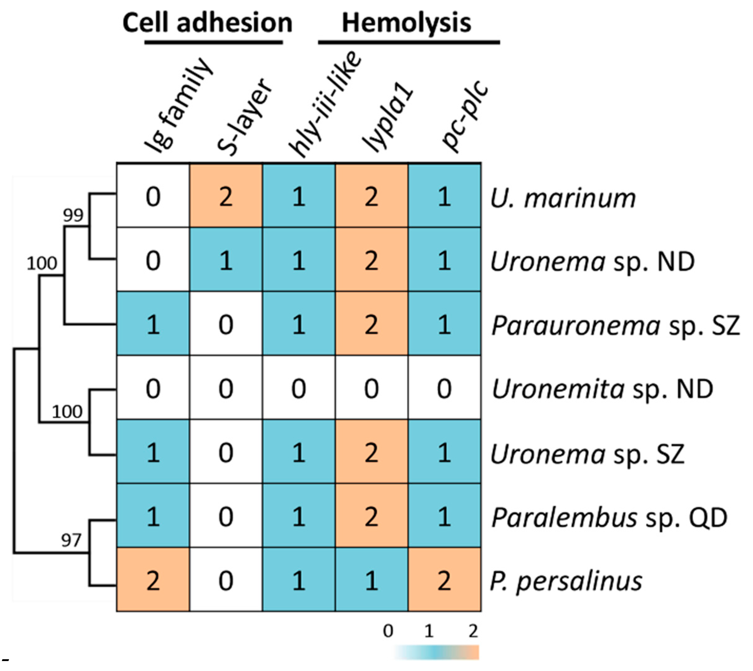
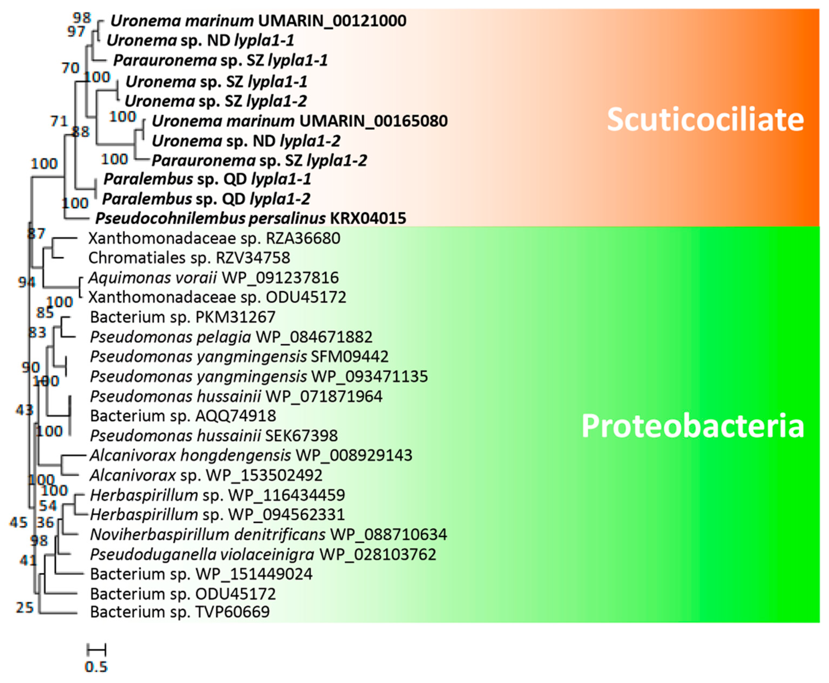
3.3. Hemolysis-Related Genes Are Widely Distributed in Scuticociliates

U. marinum and P. persalinus belong to two different families of scuticociliates—Uronematidae and Pseudocohnilembidae. The presence of hemolysis-related HGT genes led us to speculate that these genes originated very early in the evolution of scuticociliates and may be widely distributed in this subclass. To address this possibility, we checked the homologs of hemolysis-related HGT genes in five additional scuticociliates belonging to four families in the class Scuticociliatia—a Paralembus sp. isolated in Jiaozhou Bay, a Uronemita sp. and a Uronema sp. isolated in an L. polyactis farm in ND (Fujian province, China), and a Parauronema sp. and a Uronema sp. isolated in SZ Bay Port, China. Comparative genomics analysis showed that all seven scuticociliates contain homologs of different cell adhesion genes, either Ig family genes or S-layer genes (Figure 4). These results indicate that different scuticociliates have acquired different cell adhesion genes during their evolution. Interestingly, homologs of genes encoding hly-iii-like, lypla1, and pc-plc were found in six of the seven scuticociliates (Figure 4), suggesting a wide distribution of hemolysis genes in scuticociliates. Phylogenetic analysis of hly-iii-like, lypla1, and pc-plc genes showed that these scuticociliate genes cluster into a monophyletic group (Figure 5, Figure 6 and Figure 7) that is separate from bacterial genes. This result suggests that ancestral hly-iii-like, lypla1, and pc-plc genes originated through HGT before the divergence of scuticociliates. Phylogenetic analysis showed that the closest bacterial relatives of hly-iii-like are in the Terrabacteria group (Figure 5), whereas the closest bacterial relatives of lypla1 and pc-plc genes are proteobacteria (Figure 6 and Figure 7). These results indicate hly-iii-like, lypla1, and pc-plc genes probably originated from different HGT events involving different bacteria, but more evidence is needed.

4. Conclusions

We sequenced and assembled a high-quality MAC genome of U. marinum, a typical scuticociliatosis pathogen. As in P. persalinus, bacteria-derived hemolysis genes have also been acquired by U. marinum. These genes were found to be widely distributed in scuticociliates, suggesting that they have important roles in scuticociliatosis.

Supplementary Materials

The following are available online at https://0-www-mdpi-com.brum.beds.ac.uk/2076-2607/8/11/1838/s1, Figure S1. GC distribution of preliminarily assembled genome. Figure S2. Venn diagram showing specific and orthologous genes in U. marinum, P. persalinus, and I. multifiliis. Table S1. Sampling information and species identification for the five scuticociliates. Table S2. Number and type of segmental duplications in U. marinum and P. persalinus. WGD, whole-genome duplication. Table S3. Description of the 105 HGT genes in the U. marinum MAC genome.

Author Contributions

Conceptualization, W.M., J.X., Y.L., and J.Z.; methodology, J.Z., K.C., C.J., W.Y., S.G., and G.W.; formal analysis, K.C. and J.Z.; writing–original draft preparation, J.Z. and K.C.; writing–review and editing, J.X., W.M., and Y.L.; supervision, J.X., W.M., and Y.L.; funding acquisition, W.M., J.X., Y.L., J.Z., and K.C. All authors have read and agreed to the published version of the manuscript.

Funding

This project was supported by grants from the National Natural Science Foundation of China to W.M. (31525021 and 91631303) and J.X. (31672281 and 31872221), from the Natural Science Foundation of Hubei Province to J.X. (2019CFB808), from the Shenzhen Science and Technology Project to Y.L. (JCYJ20180507183240459) and from the China Postdoctoral Science Foundation to J.Z. (2019M652735) and K.C. (2020M682528).

Conflicts of Interest

The authors declare no conflict of interest.

References

  1. Iglesias:, R.; Parama, A.; Alvarez, M.F.; Leiro, J.; Fernandez, J.; Sammartin, M.L. Philasterides dicentrarchi (Ciliophora, Scuticociliatida) as the causative agent of scuticociliatosis in farmed turbot Scophthalmus maximus in galicia (nw spain). Dis. Aquat. Organ. 2001, 46, 47–55. [Google Scholar] [CrossRef] [PubMed] [Green Version]
  2. Kim, S.M.; Cho, J.B.; Kim, S.K.; Nam, Y.K.; Kim, K.H. Occurrence of scuticociliatosis in olive flounder Paralichthys olivaceus by Phiasterides dicentrarchi (Ciliophora: Scuticociliatida). Dis. Aquat. Organ. 2004, 62, 233–238. [Google Scholar] [CrossRef] [PubMed] [Green Version]
  3. Azad, I.S.; Al-Marzouk, A.; James, C.M.; Almatar, S.; Al-Gharabally, H. Scuticociliatosis-associated mortalities and histopathology of natural infection in cultured silver pomfret (pampus argenteus euphrasen) in kuwait. Aquaculture 2007, 262, 202–210. [Google Scholar] [CrossRef]
  4. Harikrishnan, R.; Balasundaram, C.; Heo, M.S. Scuticociliatosis and its recent prophylactic measures in aquaculture with special reference to South Korea taxonomy, diversity and diagnosis of scuticociliatosis: Part i control strategies of scuticociliatosis: Part ii. Fish Shellfish Immun. 2010, 29, 15–31. [Google Scholar] [CrossRef] [PubMed]
  5. Parama, A.; Iglesias, R.; Alvarez, M.F.; Leiro, J.; Ubeira, F.M.; Sanmartin, M.L. Cysteine proteinase activities in the fish pathogen Philasterides dicentrarchi (Ciliophora: Scuticociliatida). Parasitology 2004, 128, 541–548. [Google Scholar] [CrossRef]
  6. Lee, E.H.; Kim, C.S.; Cho, J.B.; Ahn, K.J.; Kim, K.H. Measurement of protease activity of live Uronema marinum (Ciliata: Scuticociliatida) by fluorescence polarization. Dis. Aquat. Organ. 2003, 54, 85–88. [Google Scholar] [CrossRef] [Green Version]
  7. Xiong, J.; Wang, G.Y.; Cheng, J.; Tian, M.; Pan, X.M.; Warren, A.; Jiang, C.; Yuan, D.; Miao, W. Genome of the facultative scuticociliatosis pathogen Pseudocohnilembus persalinus provides insight into its virulence through horizontal gene transfer. Sci. Rep. 2015, 5, 15470. [Google Scholar] [CrossRef] [Green Version]
  8. Cheung, P.J.; Nigrelli, R.F.; Ruggieri, G.D. Studies on the morphology of Uronema-marinum Dujardin (Ciliatea, Uronematidae) with a description of the histopathology of the infection in marine fishes. J. Fish Dis. 1980, 3, 295–303. [Google Scholar] [CrossRef]
  9. Zheng, W.B.; Gao, F.; Warren, A. High-density cultivation of the marine ciliate Uronema marinum (Ciliophora, Oligohymenophorea) in axenic medium. Acta Protozool. 2015, 54, 325–330. [Google Scholar]
  10. Wang, C.N.; Bourland, W.A.; Mu, W.J.; Pan, X.M. Transcriptome analysis on chlorpyrifos detoxification in Uronema marinum (Ciliophora, Oligohymenophorea). Environ. Sci. Pollut. Res. 2018, 25, 33402–33414. [Google Scholar] [CrossRef]
  11. Fernandezgaliano, D. The ammoniacal silver carbonate method as a general procedure in the study of protozoa from sewage (and other) waters. Water Res. 1994, 28, 495–496. [Google Scholar] [CrossRef]
  12. Medlin, L.; Elwood, H.J.; Stickel, S.; Sogin, M.L. The characterization of enzymatically amplified eukaryotic 16s-like rRNA-coding regions. Gene 1988, 71, 491–499. [Google Scholar] [CrossRef] [Green Version]
  13. Dykova, I.; Tyml, T.; Kostka, M.; Peckova, H. Strains of Uronema marinum (Scuticociliatia) co-isolated with amoebae of the genus Neoparamoeba. Dis. Aquat. Organ. 2010, 89, 71–77. [Google Scholar] [CrossRef] [PubMed]
  14. Xiong, J.; Lu, Y.M.; Feng, J.M.; Yuan, D.X.; Tian, M.; Chang, Y.; Fu, C.J.; Wang, G.Y.; Zeng, H.H.; Miao, W. Tetrahymena functional genomics database (TetraFGD): An integrated resource for Tetrahymena functional genomics. Database-Oxf. 2013, 2013, bat008. [Google Scholar] [CrossRef]
  15. Auwera, G.A.; Carneiro, M.O.; Hartl, C.; Poplin, R.; Del Angel, G.; Levy-Moonshine, A.; Jordan, T.; Shakir, K.; Roazen, D.; Thibault, J.; et al. From fastq data to high confidence variant calls: The genome analysis toolkit best practices pipeline. Curr. Protoc. Bioinform. 2013, 43, 11.10.1–11.10.33. [Google Scholar] [CrossRef]
  16. Kim, D.; Pertea, G.; Trapnell, C.; Pimentel, H.; Kelley, R.; Salzberg, S.L. Tophat2: Accurate alignment of transcriptomes in the presence of insertions, deletions and gene fusions. Genome Biol. 2013, 14, R36. [Google Scholar] [CrossRef] [Green Version]
  17. Chen, K.; Wang, G.Y.; Xiong, J.; Jiang, C.Q.; Miao, W. Exploration of genetic variations through single-cell whole-genome sequencing in the model ciliate Tetrahymena thermophila. J. Eukaryot. Microbiol. 2019, 66, 954–965. [Google Scholar] [CrossRef]
  18. Koren, S.; Walenz, B.P.; Berlin, K.; Miller, J.R.; Bergman, N.H.; Phillippy, A.M. Canu: Scalable and accurate long-read assembly via adaptive k-mer weighting and repeat separation. Genome Res. 2017, 27, 722–736. [Google Scholar] [CrossRef] [Green Version]
  19. Li, H.; Durbin, R. Fast and accurate long-read alignment with burrows-wheeler transform. Bioinformatics 2010, 26, 589–595. [Google Scholar] [CrossRef] [Green Version]
  20. Walker, B.J.; Abeel, T.; Shea, T.; Priest, M.; Abouelliel, A.; Sakthikumar, S.; Cuomo, C.A.; Zeng, Q.D.; Wortman, J.; Young, S.K.; et al. Pilon: An integrated tool for comprehensive microbial variant detection and genome assembly improvement. PLoS ONE 2014, 9, e112963. [Google Scholar] [CrossRef]
  21. Trapnell, C.; Roberts, A.; Goff, L.; Pertea, G.; Kim, D.; Kelley, D.R.; Pimentel, H.; Salzberg, S.L.; Rinn, J.L.; Pachter, L. Differential gene and transcript expression analysis of RNA-seq experiments with Tophat and Cufflinks. Nat. Protoc. 2012, 7, 562–578. [Google Scholar] [CrossRef] [PubMed] [Green Version]
  22. Haas, B.J.; Delcher, A.L.; Mount, S.M.; Wortman, J.R.; Smith, R.K.; Hannick, L.I.; Maiti, R.; Ronning, C.M.; Rusch, D.B.; Town, C.D.; et al. Improving the Arabidopsis genome annotation using maximal transcript alignment assemblies. Nucleic Acids Res. 2003, 31, 5654–5666. [Google Scholar] [CrossRef] [PubMed] [Green Version]
  23. Majoros, W.H.; Pertea, M.; Salzberg, S.L. Tigrscan and Glimmerhmm: Two open source ab initio eukaryotic gene-finders. Bioinformatics 2004, 20, 2878–2879. [Google Scholar] [CrossRef] [PubMed]
  24. Haas, B.J.; Salzberg, S.L.; Zhu, W.; Pertea, M.; Allen, J.E.; Orvis, J.; White, O.; Buell, C.R.; Wortman, J.R. Automated eukaryotic gene structure annotation using evidencemodeler and the program to assemble spliced alignments. Genome Biol. 2008, 9, R7. [Google Scholar] [CrossRef] [PubMed] [Green Version]
  25. Jones, P.; Binns, D.; Chang, H.Y.; Fraser, M.; Li, W.; McAnulla, C.; McWilliam, H.; Maslen, J.; Mitchell, A.; Nuka, G.; et al. Interproscan 5: Genome-scale protein function classification. Bioinformatics 2014, 30, 1236–1240. [Google Scholar] [CrossRef] [PubMed] [Green Version]
  26. Li, L.; Stoeckert, C.J.; Roos, D.S. Orthomcl: Identification of ortholog groups for eukaryotic genomes. Genome Res. 2003, 13, 2178–2189. [Google Scholar] [CrossRef] [Green Version]
  27. Camacho, C.; Coulouris, G.; Avagyan, V.; Ma, N.; Papadopoulos, J.; Bealer, K.; Madden, T.L. Blast plus: Architecture and applications. BMC Bioinform. 2009, 10, 1–9. [Google Scholar] [CrossRef] [Green Version]
  28. Price, M.N.; Dehal, P.S.; Arkin, A.P. Fasttree: Computing large minimum evolution trees with profiles instead of a distance matrix. Mol. Biol. Evol. 2009, 26, 1641–1650. [Google Scholar] [CrossRef]
  29. Guindon, S.; Dufayard, J.F.; Lefort, V.; Anisimova, M.; Hordijk, W.; Gascuel, O. New algorithms and methods to estimate maximum-likelihood phylogenies: Assessing the performance of PhyML 3.0. Syst. Biol. 2010, 59, 307–321. [Google Scholar] [CrossRef] [Green Version]
  30. Li, D.H.; Liu, C.M.; Luo, R.B.; Sadakane, K.; Lam, T.W. Megahit: An ultra-fast single-node solution for large and complex metagenomics assembly via succinct de bruijn graph. Bioinformatics 2015, 31, 1674–1676. [Google Scholar] [CrossRef] [Green Version]
  31. Haas, B.J.; Papanicolaou, A.; Yassour, M.; Grabherr, M.; Blood, P.D.; Bowden, J.; Couger, M.B.; Eccles, D.; Li, B.; Lieber, M.; et al. De novo transcript sequence reconstruction from RNA-seq using the Trinity platform for reference generation and analysis. Nat. Protoc. 2013, 8, 1494–1512. [Google Scholar] [CrossRef] [PubMed]
  32. Stanke, M.; Diekhans, M.; Baertsch, R.; Haussler, D. Using native and syntenically mapped cDNA alignments to improve de novo gene finding. Bioinformatics 2008, 24, 637–644. [Google Scholar] [CrossRef] [PubMed] [Green Version]
  33. Birney, E.; Clamp, M.; Durbin, R. Genewise and genomewise. Genome Res. 2004, 14, 988–995. [Google Scholar] [CrossRef] [Green Version]
  34. Stanke, M.; Morgenstern, B. Augustus: A web server for gene prediction in eukaryotes that allows user-defined constraints. Nucleic Acids Res. 2005, 33, W465–W467. [Google Scholar] [CrossRef] [PubMed] [Green Version]
  35. Aury, J.M.; Jaillon, O.; Duret, L.; Noel, B.; Jubin, C.; Porcel, B.M.; Segurens, B.; Daubin, V.; Anthouard, V.; Aiach, N.; et al. Global trends of whole-genome duplications revealed by the ciliate Paramecium tetraurelia. Nature 2006, 444, 171–178. [Google Scholar] [CrossRef] [PubMed]
  36. Doerder, F.P.; Deak, J.C.; Lief, J.H. Rate of phenotypic assortment in Tetrahymena thermophila. Dev. Genet. 1992, 13, 126–132. [Google Scholar] [CrossRef] [PubMed]
  37. Xu, K.; Doak, T.G.; Lipps, H.J.; Wang, J.M.; Swart, E.C.; Chang, W.J. Copy number variations of 11 macronuclear chromosomes and their gene expression in Oxytricha trifallax. Gene 2012, 505, 75–80. [Google Scholar] [CrossRef] [PubMed]
  38. Eisen, J.A.; Coyne, R.S.; Wu, M.; Wu, D.; Thiagarajan, M.; Wortman, J.R.; Badger, J.H.; Ren, Q.; Amedeo, P.; Jones, K.M.; et al. Macronuclear genome sequence of the ciliate Tetrahymena thermophila, a model eukaryote. PLoS Biol. 2006, 4, e286. [Google Scholar] [CrossRef]
  39. Coyne, R.; Hannick, L.; Shanmugam, D.; Hostetler, J.; Brami, D.; Joardar, V.; Johnson, J.; Radune, D.; Singh, I.; Badger, J.; et al. Comparative genomics of the pathogenic ciliate Ichthyophthirius multifiliis, its free-living relatives and a host species provide insights into adoption of a parasitic lifestyle and prospects for disease control. Genome Biol. 2011, 12, R100. [Google Scholar] [CrossRef] [Green Version]
  40. McGrath, C.L.; Gout, J.F.; Doak, T.G.; Yanagi, A.; Lynch, M. Insights into three whole-genome duplications gleaned from the Paramecium caudatum genome sequence. Genetics 2014, 197, 1417–1482. [Google Scholar] [CrossRef] [Green Version]
  41. Gao, F.; Katz, L.A.; Song, W.B. Insights into the phylogenetic and taxonomy of philasterid ciliates (Protozoa, Ciliophora, Scuticociliatia) based on analyses of multiple molecular markers. Mol. Phylogenet. Evol. 2012, 64, 308–317. [Google Scholar] [CrossRef] [PubMed]
  42. Feng, J.M.; Jiang, C.Q.; Warren, A.; Tian, M.; Cheng, J.; Liu, G.L.; Xiong, J.; Miao, W. Phylogenomic analyses reveal subclass Scuticociliatia as the sister group of subclass Hymenostomatia within class Oligohymenophorea. Mol. Phylogenet. Evol. 2015, 90, 104–111. [Google Scholar] [CrossRef]
  43. Stow, J.L.; Condon, N.D. The cell surface environment for pathogen recognition and entry. Clin. Transl. Immunol. 2016, 5, e71. [Google Scholar] [CrossRef] [PubMed]
  44. Hynonen, U.; Palva, A. Lactobacillus surface layer proteins: Structure, function and applications. Appl. Microbiol. Biot. 2013, 97, 5225–5243. [Google Scholar] [CrossRef] [PubMed] [Green Version]
  45. White, M.J.; Boyd, J.M.; Horswill, A.R.; Nauseef, W.M. Phosphatidylinositol-specific phospholipase C contributes to survival of Staphylococcus aureus USA300 in human blood and neutrophils. Infect. Immun. 2014, 82, 1559–1571. [Google Scholar] [CrossRef] [PubMed] [Green Version]
  46. Baida, G.E.; Kuzmin, N.P. Mechanism of action of hemolysin III from Bacillus cereus. BBA-Biomembranes 1996, 1284, 122–124. [Google Scholar] [CrossRef] [Green Version]
  47. Kalliomaki, M.A.; Walker, W.A. Physiologic and pathologic interactions of bacteria with gastrointestinal epithelium. Gastroenterol. Clin. N. 2005, 34, 383–399. [Google Scholar] [CrossRef]
  48. Ramarao, N.; Sanchis, V. The pore-forming haemolysins of Bacillus cereus: A review. Toxins 2013, 5, 1119–1139. [Google Scholar] [CrossRef] [Green Version]
Figure 1. Genomic evidence for parasitism in U. marinum. (A) U. marinum morphology under (left to right) light microscopy, 4′,6-diamidino-2-phenylindole (DAPI) staining, and silver staining, with the latter showing the dorsal and ventral surfaces. White arrows indicate micronucleus (MIC) and macronucleus (MAC); black arrows indicate the oral cilium and the caudal cilium. (B) Correlations of protein domain composition between U. marinum and four other ciliates. Both the horizontal and vertical axes represent the number of protein domains. (C) Number of protease genes in five different ciliates.
Microorganisms 08 01838 g001
Figure 2. Horizontal gene transfer (HGT) genes in U. marinum. (A) Guanine-cytosine (GC) content of the 105 HGT genes. (B) Distribution of intron number in the 105 HGT genes. (C) Venn diagram showing the numbers of specific and homologous HGT genes in U. marinum and P. persalinus. (D) Potential hemolysis-related HGT genes identified in U. marinum.
Microorganisms 08 01838 g002
Figure 3. Conservation of hly-iii genes. (A) Sequence alignment of predicted Hly-III-like proteins in U. marinum, P. persalinus, and B. cereus. Color scheme was based on the default settings on the website http://ekhidna.biocenter.helsinki.fi/pfamz/clustal_colours. (B) Conserved domain predicated through Pfam database and transmembrane predicated by TMHMM (http://w3ww.cbs.dtu.dk/services/TMHMM/).
Microorganisms 08 01838 g003
Figure 4. Distribution of hemolysis-related genes in seven scuticociliates. Numbers indicate the number of homologs identified in each species. The functional category was assigned as described in our previous study [7].
Microorganisms 08 01838 g004
Figure 5. Phylogenetic tree of hly-iii-like genes in scuticociliates and bacteria. hly-iii-like genes were identified in all scuticociliates. The top 20 prokaryotic BLASTP hits (using the encoded U. marinum Hly-III-like proteins as the seed sequence) in the NCBI non-redundant protein database were used to construct the phylogenetic tree.
Microorganisms 08 01838 g005
Figure 6. Phylogenetic tree of lypla1 genes in scuticociliates and bacteria. lypla1 genes were identified in all scuticociliates. The top 20 prokaryotic BLASTP hits (using the encoded U. marinum LYPLA1 proteins as the seed sequence) in the NCBI non-redundant protein database were used to construct the phylogenetic tree.
Microorganisms 08 01838 g006
Figure 7. Phylogenetic tree of pc-plc genes in scuticociliates and bacteria. pc-plc genes were identified in all scuticociliates. The top 20 prokaryotic BLASTP hits (using the encoded U. marinum PC-PLC proteins as the seed sequence) in the NCBI non-redundant protein database were used to construct the phylogenetic tree.
Microorganisms 08 01838 g007
Table 1. Statistics of the assembled macronuclear (MAC) genomes for five ciliates.
SpeciesU. marinumP. persalinusT. thermophilaI. multifiliisP. tetraurelia
SubclassScuticociliatiaScuticociliatiaOligohymenophoreaOligohymenophoreaOligohymenophorea
Genome size (Mb)86.855.5103.047.872.1
N50 (Kb)47036852166413
Scaffold number40328811481375697
Longest scaffold (Mb)2.482.02.20.41.0
Sequencing method/platformNanopore/MGIIlluminaSangerSanger/454Sanger
Average guanine-cytosine (GC) content18%19%22%16%28%
Assembled chromosome number218 (54%)0 (0%)129 (11.2%)0 (0%)8(1%)
Completeness84.8%79.5%85.1%67.0%85.5%
Gene number24,58213,18626,460806239,642
Publisher’s Note: MDPI stays neutral with regard to jurisdictional claims in published maps and institutional affiliations.

