Review

In Vitro Zika Virus Infection of Human Neural Progenitor Cells: Meta-Analysis of RNA-Seq Assays

by
Rossella Gratton
1,2,*,
Paola Maura Tricarico
1,
Almerinda Agrelli
3,4,
Heverton Valentim Colaço da Silva
3,4,
Lucas Coêlho Bernardo
3,4,
Sergio Crovella
1,5,
Antonio Victor Campos Coelho
6,
Ronald Rodrigues de Moura
4,5 and
Lucas André Cavalcanti Brandão
3,4
1
Department of Advanced Translational Microbiology, Institute for Maternal and Child Health IRCCS Burlo Garofolo, Via dell’Istria 65/1, 34137 Trieste, Italy
2
Department of Medical, Surgical and Health Sciences, University of Trieste, Strada di Fiume 447, 34129 Trieste, Italy
3
Laboratory of Immunopathology Keizo Asami (LIKA), Federal University of Pernambuco (UFPE), Av. Prof. Moraes Rego, 1235 Cidade Universitária, 50670-901 Recife, Brazil
4
Department of Pathology-Federal University of Pernambuco (UFPE), Av. Prof. Moraes Rego, 1235-Cidade Universitária, 50670-901 Recife, Brazil
5
Department of Genetics-Federal University of Pernambuco (UFPE), Av. Prof. Moraes Rego, 1235-Cidade Universitária, 50670-901 Recife, Brazil
6
Department of Molecular Biology-Federal University of Paraíba (UFPB), Campus I-Lot.-Cidade Universitária, 58051-900 João Pessoa, Brazil
*
Author to whom correspondence should be addressed.
Submission received: 7 January 2020 / Revised: 12 February 2020 / Accepted: 14 February 2020 / Published: 17 February 2020
(This article belongs to the Special Issue Virus-Host Interaction: From Physiology to Pathology)

Abstract

:
The Zika virus (ZIKV) is an emergent arthropod-borne virus (arbovirus) responsible for congenital Zika syndrome (CZS) and a range of other congenital malformations. Evidence shows that ZIKV infects human neural progenitor cells (hNPCs) in the fetal brain, prompting inflammation and tissue damage/loss. Despite recent advances, little is known about the pathways involved in CZS pathogenesis. We performed a meta-analysis, gene ontology (GO), and pathway analysis of whole transcriptome studies with the aim of clarifying the genes and pathways potentially altered during hNPCs infection with ZIKV. We selected three studies (17 samples of infected hPNCs compared to hPNCs uninfected controls) through a systematic search of the Gene Expression Omnibus (GEO) database. The raw reads were trimmed, counted, and normalized. Next, we performed a rank product meta-analysis to detect consistently differentially expressed genes (DEGs) in these independent experiments. We detected 13 statistically significant DEGs. GO ontology and reactome analysis showed an enrichment of interferon, pro-inflammatory, and chemokines signaling and apoptosis pathways in ZIKV-infected cells. Moreover, we detected three possible new candidate genes involved in hNPCs infection: APOL6, XAF1, and TNFRSF1. Our results confirm that interferon (IFN) signaling dominates the ZIKV response, and that a crucial contribution is given by apoptotic pathways, which might elicit the CZS phenotype.

Graphical Abstract

1. Introduction

The Zika virus (ZIKV) is an arthropod-borne virus (arbovirus), member of the Flaviviridae family and of the Flavivirus genus, transmitted by Aedes genus mosquitoes [1]. In 2015, during the outbreak in Brazil, ZIKV was correlated for the first time to neonatal microcephaly [2] and to a variety of other congenital malformations, especially of neurological origin, collectively known as congenital Zika syndrome (CZS) [3].
CZS is characterized by a spectrum of congenital malformations associated with ZIKV infection during embryonic development [4]. The most commonly reported neurological feature of CZS is microcephaly, a condition characterized by a head circumference ≥2 standard deviations below the mean for sex and gestational age at birth, although other neurological abnormalities, including brainstem dysfunction, absence of swallowing reflex, and polymalformative syndromes, may also be present [5]. General features (redundant scalp skin, anasarca, low birth weight, polyhydramnios, and arthrogryposis) and ophthalmological defects (intraocular calcifications, cataract, asymmetrical eye sizes, macular atrophy, optic nerve hypoplasia, iris coloboma, and lens subluxation) have also been reported [6,7,8].
The human central nervous system (CNS) development begins during the third week of embryogenesis [9]. The embryonic brain is basically composed of human neural progenitor cells (hNPCs), progenitor cells that give rise to all of the glial and neuronal cell types that populate the CNS; therefore, the onset of pathogenic processes might cause neuroinflammation and the secretion of immunoregulatory molecules [10]. As a result, these events may trigger cell death mechanisms, leading to an impairment of hNPCs proliferation, growth, and differentiation, and consequently to a defective brain development [11].
Several studies demonstrated that ZIKV infects hNPCs in the fetal brain, prompting inflammation and tissue damage and loss [12,13,14,15]. Despite recent advances in the characterization of the impact of ZIKV infection on embryonic CNS development, it is still necessary to identify which pathways in hNPCs are involved during these pathogenic mechanisms. This gap of knowledge is clearly restrictive for the development of therapeutic approaches that could prevent the severe clinical consequences of the infection.
Transcriptional profiling has provided remarkable opportunities for understanding the relationship between cellular function and metabolic pathways, as well as to define the possible implications of genetic variability and environmental conditions in many tissues and organisms [16]. RNA-sequencing (RNA-Seq) has been widely used over the last decade and has become the main option for these studies [17,18].
In this work, we performed a meta-analysis of whole transcriptome studies, aiming to clarify which genes and cellular networks were up- or downregulated during ZIKV infection in hNPCs. Next, we assessed a comprehensive pathway analysis to predict how the modulation of these genes could affect the outcome of the disease.

2. Materials and Methods

2.1. Study Search

We used the SRAdb package [19] for R software version 3.6.1 [20] to search for RNA-Seq experiments deposited in the Gene Expression Omnibus (GEO) database related to ZIKV infection in hNPCs that matched the following criteria: only whole transcriptome studies; experiments carried out in patients’ cells (ex vivo) or human cell lines (in vitro); and availability of the raw data (.fastq files) for each sample. The following search terms were used: “ZIKV RNA-Seq” and “ZIKV transcriptome” including studies from 19 July 2015 to 19 July 2019.

2.2. RNA-Seq Data Collection, Processing, and Analysis

For all analyzed samples, Raw .fastq files were downloaded and re-processed using the same pipeline analysis. For this purpose, Trimmomatic v0.39 [21] was used to trim adapters and to exclude reads counting less than 25 bases. Then, the remaining reads were mapped on the National Center for Biotechnology (NCBI) human GRCh38 reference genome and sorted by coordinates using STAR aligner [22]. Aligned reads were imported into R software version 3.6.1 [20], together with a .gtf annotation file from the reference genome, using packages Rsamtools [23], GenomicFeatures, and GenomicAlignments [24].
The gene counts were normalized and filtered in order to remove low-expressed genes (i.e., genes expressed in less than three samples and less than two copies). Differentially expressed genes (DEGs) for each study were re-calculated using a Wald test with correction for multiple tests implemented in the DESeq2 package [25]. Genes with |log2(fold change)| > 1 and false discovery rate (FDR)-adjusted p-values < 0.05 were considered to be statistically significant.

2.3. Meta-Analysis

The normalized and filtered gene expression dataset, together with the samples’ information relative to the type of treatment (ZIKV-infected or control) and the team that performed the study (Zhang et al., McGrath et al., or Caires-Júnior et al.) were included in the meta-analysis using the RankProd package for R software version 3.6.1 [20]. Briefly, the package performs the rank product (RP) and rank sum tests, non-parametric tests that detect consistently differentially expressed genes in independent and replicated experiments. The test ranks expression fold-changes calculated in a pairwise manner among several experimental replicates. Under the null hypothesis (no differentially expressed genes), it “is extremely unlikely to find the same gene at the top of each list [of ranked fold changes among experiments] just by chance” [26,27]. We adopted the meta-analysis methodology derived from a previously published study, in which a gene ontology enrichment analysis was also included (see the next section) [28].

2.4. Gene Ontology Enrichment Analysis

In parallel with the identification of differentially expressed genes via meta-analysis, we performed a gene ontology enrichment analysis by employing the GOexpress package [29], also suitable for R software version 3.6.1 [20]. Briefly, the package scores each gene feature (through a random forest statistical framework) on “its ability to classify samples from different treatments separately, before summarizing this information at the ontology level” [29]. Then, the package queries the ranked genes in the Ensembl gene ontology database and configures the package to assess the statistical significance of the gene ontology ranking via permutation-based calculation of p-values (100,000 permutations), representing the probability of seeing at least five genes out of the total number of genes in the list attributed to a particular gene ontology term [30,31]. Then, the genes were ranked from the lowest to highest p-value below the limit of p < 0.05.

2.5. Reactome Pathway Analysis

For each independent study and for the pooled dataset in the meta-analysis, we conducted a pathway analysis, based on the REACTOME database, of the statistically significant DEGs using the ReactomePA package [20]. Also, in this case, only the results with false discovery rate (FDR)-adjusted p-values < 0.05 were considered significant.

3. Results

The search strategy retrieved 30 studies. Three people (R.R.M., H.V.C.S., and L.C.B.) independently reviewed the search hits, from which 10 studies were excluded because only viral genomes were sequenced; four were excluded since they were studies performed in mice; three experiments were eliminated since they involved other animals (mice and Aedes aegypti mosquitoes); one study was removed since it was relative to the sequencing of the Chikungunya virus genome; six studies involving other human cell types and three studies based on CNS organoids were also excluded as they contemplated the usage of other cell types beside hNPCs. Finally, the three reviewers agreed that three studies matched the fixed criteria (Table 1) [32,33,34].
The first study, from Zhang et al. (SRAdb id: SRP073493, GSE id: GSE80434) [32], analyzed the differences between ZIKV and Dengue virus (DENV) infection in hNPCs. In the selected research, hNPCs were infected with both African (ZIKVM) and Asian (ZIKVC) lineages to then compare the levels of transcriptional changes, gene function, and protein interactions among the DEGs. We focused our attention only on the comparisons between ZIKVM versus mock treatment and ZIKVC versus mock treatment. Their study highlighted 1345 DEGs between ZIKVM and its mock group and 601 DEGs for the ZIKVC versus mock group comparison.
The work of McGrath et al. [33] (SRAdb id: SRP096367, GSE id: GSE93385) was the second study to be included. It comprised the analysis of hNPC samples derived from three deceased children. Whole transcriptome profiles were compared between infected and non-infected cells of each patient separately. As a result, they detected eight upregulated and four downregulated genes, found to be shared between the samples.
The third study, conducted by Caires-Júnior et al. [34] (SRAdb id: SRP114529, GSE id: GSE102128) described the performance of an RNA-Seq experiment on cells derived from three pairs of discordant phenotypes of CZS dizygotic twins. According to their results, authors identified 64 DEGs, and specifically the DDIT4L gene, which plays a crucial role in the mammalian target of rapamycin (mTOR) signaling pathway and emerged as the most relevant one.
Since each study possesses its own approach in terms of handling and statistical procedures of RNA-Seq data, we reprocessed the reads using the same protocols for all samples and performed a meta-analysis. The normalization of expression data resulted in 29,318 features present in all 17 samples. Among those, we identified 13 upregulated genes in virus-infected cells through the rank product method with a percentage of false prediction, pfp < 0.05 and |log2(fold change)| > 1 (Table 2).
The gene ontology analysis identified 847 enriched terms. Due to the stem cell nature of the cells used in the examined experiments, as expected, most of the top-ranked terms included events representative of cell cycle progression (“mitotic spindle midzone assembly”, rank #1, p < 0.00001) and cell differentiation (“nervous system development”, #12, p < 0.00001, “multicellular organism development”, #15, p < 0.00001; “cell differentiation”; #39, p = 0.00021). We then selected 20 terms possibly related to ZIKV-infection and response (Table 3), including for example: “response to virus” (#49, p = 0.00033), “apoptotic process” (#57, p = 0.00042), “viral process” (#64, p = 0.0005), “positive regulation of neuron death” (#132, p = 0.00239), and “regulation of inflammatory response” (#142, p = 0.00266). The complete list of enriched GO terms can be found in the Supplementary Table S1.
From the 13 DEGs that reached our |log2(fold change)| threshold, reactome analysis returned 12 statistically significant pathways in which these genes are involved (Table 4). Briefly, these pathways are related to interferon signaling and response, as well as to interleukin-10, chemokines, and other receptor signaling.

4. Discussion

We searched for studies involving hNPCs transcriptome analysis in response to ZIKV infection. We included three available studies and analyzed the gene expression patterns using a meta-analysis with the rank product method. Using a different approach, we detected 13 statistically significant DEGs found to be upregulated in hNPCs infected by ZIKV. No downregulated gene was observed as statistically associated with hNPCs infected by ZIKV. Our goal was to identify the expression pattern during ZIKV infection in hNPCs, primarily in order to highlight potential molecules that could be used as an antiviral barrier, namely restriction factors, and identify which molecules are released during the antiviral response.
Differences amongst the number and the identity of differentially expressed genes found in our meta-analysis, and in the selected studies, individually rely on three main factors. The first aspect to be considered is that each study has its own peculiarities in terms of the analyzed samples and conditions of infection, as highlighted in Table 1. The second aspect depends on the fact that the studies did not apply the same method for the quantitative evaluation of each mRNA. Specifically, the study of Zhang et al. [32] used fragments per kilobase million (FPKM) measurements for mRNA counting and then applied gamma-Poison normalization, while we opted for the usage of a simple count followed by the application of the gamma-Poison normalization. Third, the experimental design among the studies was clearly different. For instance, in the work conducted by McGrath et al. [33], they assessed a pair-wise comparison between infected and non-infected cells from each evaluated brain sample followed by the application of a Venn diagram to verify the differences (or resemblances) between infected and non-infected samples as a whole. Furthermore, the statistical procedures carried out by Caires-Junior et al. [34] were very similar to the ones we employed in our study, although it is important to note that the contexts of application were different since we were assessing them in a meta-analysis context, whereas they made a single study.
Being that the sampled cells were hNPCs, between the identified DEGs, three are known to be involved in tissue development: the NK3 homeobox 1 (NKX3-1) and homeobox A2 (HOXA2), which encode for homeobox-containing transcription factors and promote animal morphogenesis and tissue differentiation [35,36], and chitinase-3-like protein 1 (CHI3L1), which codes for a chitinase-like protein that lost the capability for chitin cleavage, but retained the carbohydrate-binding affinity, and is thought to regulate tissue remodeling and angiogenesis [37].
2′-5′-Oligoadenylate synthetase 1 and 3 (OAS1 and OAS3) and 2′-5′-oligoadenylate synthetase like (OASL) are related to antiviral responses and were detected as being upregulated in their ZIKV-infected counterparts. They are interferon-inducible genes that bind to double-stranded RNA (dsRNA) and single-stranded RNA (ssRNA) viral genomes and activate the RNase L degradation pathway of viral and cellular RNA, therefore arresting viral production [38]. A recent study demonstrated that while ZIKV ssRNA genome is susceptible to RNase L activity, the virus otherwise efficiently evades the enzymatic activity due to its unique replication factories in the endoplasmic reticulum, which confers a higher resistance to host viral sensors when compared to other Flaviviruses, such as DENV, that employ similar assembly strategies [39].
However, the most intriguing evidence is represented by the upregulation of interferon-inducible genes, such as interferon induced with helicase C domain 1 (IFIH1) and apolipoprotein L6 (APOL6) genes. IFIH1, also known as melanoma differentiation-associated protein 5 (MDA5), encodes for a pattern recognition receptor that, along with the DDX58 (RIG1) protein, is able to bind dsRNA and ssRNA derived from other Flaviviruses, such as DENV, and elicits innate immune responses through type I interferon (IFN-1) production [40]. Moreover, this gene has already been seen to be upregulated in skin cells during ZIKV infection [41].
APOL6 has been reported to not only promote antiviral responses against some viruses, such as picornaviruses, enteroviruses, and respiratory syncytial virus [42,43], but also to possess pro-apoptotic properties [44]. We could not find any other independent evidence relating the expression of this gene in the context of ZIKV infection.
It is important to note that in our analysis, we identified two other DEGs not yet independently associated with ZIKV infection, to our knowledge: X- linked inhibitor of apoptosis protein associated factor 1 (XAF1) and tumor necrosis factor receptor superfamily member 1A (TNFRSF1). XAF1 is involved in the apoptotic route and it encodes for a protein acting as an antagonist of inhibitors of apoptosis proteins (IAPs), therefore also exerting pro-apoptotic properties. Its expression is also induced by interferon (IFN) signaling and is thought to function as a tumor suppressor [45]. We could not ascertain other studies showing XAF1 association with ZIKV infection. TNFRSF1 encodes for a widely expressed protein, which is the main receptor involved in mediating soluble TNFα-induced signaling, therefore exerting a pivotal role in regulating pro-inflammatory and immune responses [46]. TNFRSF1 has not been reported to be involved with ZIKV infection; nevertheless, other studies highlight its role in the immune defense in the gut mucosa [47].
As previously described in the text, the majority of the identified DEGs mediate antiviral intracellular pathways. However, besides TNFRSF1, some other genes involved in inflammatory and immune responses were also upregulated in ZIKV-infected hNPCs: C-C motif chemokine ligand 5 (CCL5) and C-X-C motif chemokine ligand 10 and 11 (CXCL10 and CXCL11) [48]. CCL5, CXCL10, and CXCL11 have already been shown to be expressed in ZIKV-infected skin cells [41].
Integrating our results in the context of neurogenesis, we suppose that the presence of ZIKV in hNPCs is sensed by interferon induced with helicase C domain 1 (IFIH1), which acts as a restriction viral factor, enabling the cell to activate type 1 interferon apoptosis and inflammatory pathways, as suggested by reactome analysis. Furthermore, the activation of inflammatory processes might increase the production of cytokines and chemokines (CXCL10 and CXCL11) that may play a key role during neurogenesis by regulating the expression of developmental genes. Thus, some neurological CZS symptoms may be due to the development of an interfering inflammatory response during neurogenesis [49].

5. Conclusions

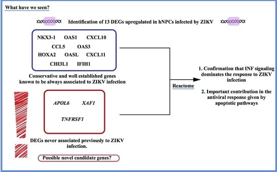
RNA-Seq has contributed to increase the knowledge relative to the biological and cellular processes involved during infections, especially in the context of an emergent pathogen, such as ZIKV. Meta-analysis of RNA-Seq assays increases the statistical power of samples and we applied this method for re-examining past evidence to better understand the pathogenesis of CZS. The results of our GO enrichment and reactome analysis seem to support the assumption that the cellular antiviral pathways activated in response to ZIKV infection could be responsible for an impaired cellular neurogenesis. As mentioned above, other studies investigating ZIKV infection also support the biological significance of our meta-analysis findings. Moreover, we detected three possible novel candidate genes involved during this antiviral response: APOL6, XAF1, and TNFRSF1. Finally, we confirmed that IFN signaling dominates the cellular response against ZIKV infection, and that under these conditions, an important contribution is given by apoptotic pathways that might elicit the CZS phenotype (Figure 1).

Supplementary Materials

The following are available online at https://0-www-mdpi-com.brum.beds.ac.uk/2076-2607/8/2/270/s1, Table S1: A complete list of all 847 GO terms that were enriched in the meta-analysis of three RNA-Seq assays involving human neural stem cells.

Author Contributions

Conceptualization, A.A., R.R.d.M., and L.A.C.B.; methodology, R.R.d.M, H.V.C.d.S., and L.C.B.; formal analysis, R.R.d.M. and A.V.C.C.; writing—original draft preparation, A.A., R.R.d.M., and A.V.C.C.; writing—review and editing, R.G. and P.M.T.; supervision, L.A.C.B; funding acquisition, R.G., P.M.T., and S.C. All authors have read and agreed to the published version of the manuscript.

Funding

This research was funded by Comissão de Aperfeiçoamento de Pessoal do Nível Superior (CAPES), grant number 88881.130808/2016.1, and by Conselho Nacional de Desenvolvimento Científico e Tecnológico (CNPq), from the Brazilian public notice Chamada 14/2016–Prevenção e Combate ao vírus Zika, grant number 440371/2016-3, both granted to S.C and 308540/2017-4 granted to L.A.C.B.

Conflicts of Interest

The authors declare no conflict of interest.

References

  1. Azar, S.R.; Weaver, S.C. Vector Competence: What Has Zika Virus Taught Us? Viruses 2019, 11, 867. [Google Scholar] [CrossRef] [PubMed] [Green Version]
  2. Schuler-Faccini, L.; Ribeiro, E.M.; Feitosa, I.M.; Horovitz, D.D.; Cavalcanti, D.P.; Pessoa, A.; Doriqui, M.J.; Neri, J.I.; Neto, J.M.; Wanderley, H.Y.; et al. Possible Association Between Zika Virus Infection and Microcephaly—Brazil, 2015. Morb. Mortal. Wkly. Rep. 2016, 65, 59–62. [Google Scholar] [CrossRef] [PubMed]
  3. Miranda-Filho Dde, B.; Martelli, C.M.; Ximenes, R.A.; Araujo, T.V.; Rocha, M.A.; Ramos, R.C.; Dhalia, R.; Franca, R.F.; Marques Junior, E.T.; Rodrigues, L.C. Initial Description of the Presumed Congenital Zika Syndrome. Am. J. Public Health 2016, 106, 598–600. [Google Scholar] [CrossRef] [PubMed]
  4. Moore, C.A.; Staples, J.E.; Dobyns, W.B.; Pessoa, A.; Ventura, C.V.; Fonseca, E.B.; Ribeiro, E.M.; Ventura, L.O.; Neto, N.N.; Arena, J.F.; et al. Characterizing the Pattern of Anomalies in Congenital Zika Syndrome for Pediatric Clinicians. JAMA Pediatr. 2017, 171, 288–295. [Google Scholar] [CrossRef] [Green Version]
  5. Chan, J.F.; Choi, G.K.; Yip, C.C.; Cheng, V.C.; Yuen, K.Y. Zika fever and congenital Zika syndrome: An unexpected emerging arboviral disease. J. Infect. 2016, 72, 507–524. [Google Scholar] [CrossRef] [Green Version]
  6. Ventura, C.V.; Maia, M.; Ventura, B.V.; Linden, V.V.; Araujo, E.B.; Ramos, R.C.; Rocha, M.A.; Carvalho, M.D.; Belfort, R., Jr.; Ventura, L.O. Ophthalmological findings in infants with microcephaly and presumable intra-uterus Zika virus infection. Arq. Bras. Oftalmol. 2016, 79, 1–3. [Google Scholar] [CrossRef]
  7. De Paula Freitas, B.; de Oliveira Dias, J.R.; Prazeres, J.; Sacramento, G.A.; Ko, A.I.; Maia, M.; Belfort, R., Jr. Ocular Findings in Infants With Microcephaly Associated With Presumed Zika Virus Congenital Infection in Salvador, Brazil. JAMA Ophthalmol. 2016, 134, 529–535. [Google Scholar] [CrossRef] [Green Version]
  8. Ventura, C.V.; Maia, M.; Bravo-Filho, V.; Gois, A.L.; Belfort, R., Jr. Zika virus in Brazil and macular atrophy in a child with microcephaly. Lancet 2016, 387, 228. [Google Scholar] [CrossRef] [Green Version]
  9. O’Rahilly, R.; Muller, F. Developmental stages in human embryos: Revised and new measurements. Cells Tissues Org. 2010, 192, 73–84. [Google Scholar] [CrossRef]
  10. Martinez-Cerdeno, V.; Noctor, S.C. Neural Progenitor Cell Terminology. Front. Neuroanat. 2018, 12, 104. [Google Scholar] [CrossRef]
  11. Hammack, C.; Ogden, S.C.; Madden, J.C., Jr.; Medina, A.; Xu, C.; Phillips, E.; Son, Y.; Cone, A.; Giovinazzi, S.; Didier, R.A.; et al. Zika Virus Infection Induces DNA Damage Response in Human Neural Progenitors That Enhances Viral Replication. J. Virol. 2019, 93, e00638-19. [Google Scholar] [CrossRef] [PubMed] [Green Version]
  12. Tang, H.; Hammack, C.; Ogden, S.C.; Wen, Z.; Qian, X.; Li, Y.; Yao, B.; Shin, J.; Zhang, F.; Lee, E.M.; et al. Zika Virus Infects Human Cortical Neural Progenitors and Attenuates Their Growth. Cell Stem Cell 2016, 18, 587–590. [Google Scholar] [CrossRef] [PubMed] [Green Version]
  13. Siddique, R.; Liu, Y.; Nabi, G.; Sajjad, W.; Xue, M.; Khan, S. Zika Virus Potentiates the Development of Neurological Defects and Microcephaly: Challenges and Control Strategies. Front. Neurol. 2019, 10, 319. [Google Scholar] [CrossRef] [PubMed] [Green Version]
  14. Rosa-Fernandes, L.; Cugola, F.R.; Russo, F.B.; Kawahara, R.; de Melo Freire, C.C.; Leite, P.E.C.; Bassi Stern, A.C.; Angeli, C.B.; de Oliveira, D.B.L.; Melo, S.R.; et al. Zika Virus Impairs Neurogenesis and Synaptogenesis Pathways in Human Neural Stem Cells and Neurons. Front. Cell Neurosci. 2019, 13, 64. [Google Scholar] [CrossRef]
  15. Lima, M.C.; de Mendonca, L.R.; Rezende, A.M.; Carrera, R.M.; Anibal-Silva, C.E.; Demers, M.; D’Aiuto, L.; Wood, J.; Chowdari, K.V.; Griffiths, M.; et al. The Transcriptional and Protein Profile From Human Infected Neuroprogenitor Cells Is Strongly Correlated to Zika Virus Microcephaly Cytokines Phenotype Evidencing a Persistent Inflammation in the CNS. Front. Immunol. 2019, 10, 1928. [Google Scholar] [CrossRef] [Green Version]
  16. Wang, Z.; Gerstein, M.; Snyder, M. RNA-Seq: A revolutionary tool for transcriptomics. Nat. Rev. Genet. 2009, 10, 57–63. [Google Scholar] [CrossRef]
  17. Costa-Silva, J.; Domingues, D.; Lopes, F.M. RNA-Seq differential expression analysis: An extended review and a software tool. PLoS ONE 2017, 12, e0190152. [Google Scholar] [CrossRef] [Green Version]
  18. Wang, B.; Kumar, V.; Olson, A.; Ware, D. Reviving the Transcriptome Studies: An Insight Into the Emergence of Single-Molecule Transcriptome Sequencing. Front. Genet. 2019, 10, 384. [Google Scholar] [CrossRef] [Green Version]
  19. Zhu, Y.; Stephens, R.M.; Meltzer, P.S.; Davis, S.R. SRAdb: Query and use public next-generation sequencing data from within R. BMC Bioinformat. 2013, 14, 19. [Google Scholar] [CrossRef] [Green Version]
  20. R Core Team. R: A Language and Environment for Statistical Computing. Available online: http://www.r-project.org/ (accessed on 3 July 2019).
  21. Bolger, A.M.; Lohse, M.; Usadel, B. Trimmomatic: A flexible trimmer for Illumina sequence data. Bioinformatics 2014, 30, 2114–2120. [Google Scholar] [CrossRef] [Green Version]
  22. Dobin, A.; Davis, C.A.; Schlesinger, F.; Drenkow, J.; Zaleski, C.; Jha, S.; Batut, P.; Chaisson, M.; Gingeras, T.R. STAR: Ultrafast universal RNA-seq aligner. Bioinformatics 2013, 29, 15–21. [Google Scholar] [CrossRef] [PubMed]
  23. Morgan, M.; Pagès, H.; Obenchain, V.; Hayden, N. Rsamtools: Binary alignment (BAM), FASTA, variant call (BCF), and tabix file import. R Package Vers. 2016, 1, 677–689. [Google Scholar]
  24. Lawrence, M.; Huber, W.; Pages, H.; Aboyoun, P.; Carlson, M.; Gentleman, R.; Morgan, M.T.; Carey, V.J. Software for computing and annotating genomic ranges. PLoS Comput. Biol. 2013, 9, e1003118. [Google Scholar] [CrossRef] [PubMed]
  25. Love, M.I.; Huber, W.; Anders, S. Moderated estimation of fold change and dispersion for RNA-seq data with DESeq2. Genome Biol. 2014, 15, 550. [Google Scholar] [CrossRef] [Green Version]
  26. Hong, F.; Breitling, R.; McEntee, C.W.; Wittner, B.S.; Nemhauser, J.L.; Chory, J. RankProd: A bioconductor package for detecting differentially expressed genes in meta-analysis. Bioinformatics 2006, 22, 2825–2827. [Google Scholar] [CrossRef] [Green Version]
  27. Del Carratore, F.; Jankevics, A.; Eisinga, R.; Heskes, T.; Hong, F.; Breitling, R. RankProd 2.0: A refactored bioconductor package for detecting differentially expressed features in molecular profiling datasets. Bioinformatics 2017, 33, 2774–2775. [Google Scholar] [CrossRef] [Green Version]
  28. Lee, S.Y.; Park, Y.K.; Yoon, C.H.; Kim, K.; Kim, K.C. Meta-analysis of gene expression profiles in long-term non-progressors infected with HIV-1. BMC Med. Genomics 2019, 12, 3. [Google Scholar] [CrossRef]
  29. Rue-Albrecht, K. GOexpress: Visualise Microarray and RNAseq Data Using Gene Ontology Annotations. R Package Version 1.18.0. Available online: https://github.com/kevinrue/GOexpress (accessed on 7 January 2020).
  30. Ashburner, M.; Ball, C.A.; Blake, J.A.; Botstein, D.; Butler, H.; Cherry, J.M.; Davis, A.P.; Dolinski, K.; Dwight, S.S.; Eppig, J.T.; et al. Gene ontology: Tool for the unification of biology. The Gene Ontology Consortium. Nat. Genet. 2000, 25, 25–29. [Google Scholar] [CrossRef] [Green Version]
  31. The Gene Ontology. The Gene Ontology Resource: 20 years and still GOing strong. Nucleic Acids Res. 2019, 47, D330–D338. [Google Scholar] [CrossRef] [Green Version]
  32. Zhang, F.; Hammack, C.; Ogden, S.C.; Cheng, Y.; Lee, E.M.; Wen, Z.; Qian, X.; Nguyen, H.N.; Li, Y.; Yao, B.; et al. Molecular signatures associated with ZIKV exposure in human cortical neural progenitors. Nucleic Acids Res. 2016, 44, 8610–8620. [Google Scholar] [CrossRef]
  33. McGrath, E.L.; Rossi, S.L.; Gao, J.; Widen, S.G.; Grant, A.C.; Dunn, T.J.; Azar, S.R.; Roundy, C.M.; Xiong, Y.; Prusak, D.J.; et al. Differential Responses of Human Fetal Brain Neural Stem Cells to Zika Virus Infection. Stem Cell Rep. 2017, 8, 715–727. [Google Scholar] [CrossRef] [PubMed]
  34. Caires-Junior, L.C.; Goulart, E.; Melo, U.S.; Araujo, B.H.S.; Alvizi, L.; Soares-Schanoski, A.; de Oliveira, D.F.; Kobayashi, G.S.; Griesi-Oliveira, K.; Musso, C.M.; et al. Discordant congenital Zika syndrome twins show differential in vitro viral susceptibility of neural progenitor cells. Nat. Commun. 2018, 9, 475. [Google Scholar] [CrossRef] [PubMed] [Green Version]
  35. Shen, M.M.; Abate-Shen, C. Roles of the Nkx3.1 homeobox gene in prostate organogenesis and carcinogenesis. Dev. Dyn. 2003, 228, 767–778. [Google Scholar] [CrossRef] [PubMed]
  36. Mallo, M.; Alonso, C.R. The regulation of Hox gene expression during animal development. Development 2013, 140, 3951–3963. [Google Scholar] [CrossRef] [Green Version]
  37. Coffman, F.D. Chitinase 3-Like-1 (CHI3L1): A putative disease marker at the interface of proteomics and glycomics. Crit. Rev. Clin. Lab. Sci. 2008, 45, 531–562. [Google Scholar] [CrossRef] [PubMed]
  38. Kristiansen, H.; Gad, H.H.; Eskildsen-Larsen, S.; Despres, P.; Hartmann, R. The oligoadenylate synthetase family: An ancient protein family with multiple antiviral activities. J. Interferon Cytokine Res. 2011, 31, 41–47. [Google Scholar] [CrossRef]
  39. Whelan, J.N.; Li, Y.; Silverman, R.H.; Weiss, S.R. Zika Virus Production Is Resistant to RNase L Antiviral Activity. J. Virol. 2019, 93, e00313-19. [Google Scholar] [CrossRef] [Green Version]
  40. Urcuqui-Inchima, S.; Cabrera, J.; Haenni, A.L. Interplay between dengue virus and Toll-like receptors, RIG-I/MDA5 and microRNAs: Implications for pathogenesis. Antivir. Res. 2017, 147, 47–57. [Google Scholar] [CrossRef]
  41. Hamel, R.; Dejarnac, O.; Wichit, S.; Ekchariyawat, P.; Neyret, A.; Luplertlop, N.; Perera-Lecoin, M.; Surasombatpattana, P.; Talignani, L.; Thomas, F.; et al. Biology of Zika Virus Infection in Human Skin Cells. J. Virol. 2015, 89, 8880–8896. [Google Scholar] [CrossRef] [Green Version]
  42. Schoggins, J.W.; Wilson, S.J.; Panis, M.; Murphy, M.Y.; Jones, C.T.; Bieniasz, P.; Rice, C.M. A diverse range of gene products are effectors of the type I interferon antiviral response. Nature 2011, 472, 481–485. [Google Scholar] [CrossRef]
  43. Schoggins, J.W.; MacDuff, D.A.; Imanaka, N.; Gainey, M.D.; Shrestha, B.; Eitson, J.L.; Mar, K.B.; Richardson, R.B.; Ratushny, A.V.; Litvak, V.; et al. Pan-viral specificity of IFN-induced genes reveals new roles for cGAS in innate immunity. Nature 2014, 505, 691–695. [Google Scholar] [CrossRef] [PubMed]
  44. Liu, Z.; Lu, H.; Jiang, Z.; Pastuszyn, A.; Hu, C.A. Apolipoprotein l6, a novel proapoptotic Bcl-2 homology 3-only protein, induces mitochondria-mediated apoptosis in cancer cells. Mol. Cancer Res. 2005, 3, 21–31. [Google Scholar] [PubMed]
  45. Plenchette, S.; Cheung, H.H.; Fong, W.G.; LaCasse, E.C.; Korneluk, R.G. The role of XAF1 in cancer. Curr. Opin. Investig. Drugs 2007, 8, 469–476. [Google Scholar] [PubMed]
  46. Siebert, S.; Fielding, C.A.; Williams, B.D.; Brennan, P. Mutation of the extracellular domain of tumor necrosis factor receptor 1 causes reduce NF-kappaB activation due to decreased surface expression. FEBS Lett. 2005, 579, 5193–5198. [Google Scholar] [CrossRef] [Green Version]
  47. Shui, J.W.; Kronenberg, M. HVEM is a TNF Receptor with Multiple Regulatory Roles in the Mucosal Immune System. Immune Netw. 2014, 14, 67–72. [Google Scholar] [CrossRef] [Green Version]
  48. Commins, S.P.; Borish, L.; Steinke, J.W. Immunologic messenger molecules: Cytokines, interferons, and chemokines. J. Allergy Clin. Immunol. 2010, 125, S53–S72. [Google Scholar] [CrossRef]
  49. Morimoto, K.; Nakajima, K. Role of the Immune System in the Development of the Central Nervous System. Front. Neurosci. 2019, 13, 916. [Google Scholar] [CrossRef] [Green Version]
Figure 1. Meta-analysis of RNA-Seq assays for the characterization of congenital Zika syndrome (CZS). ZIKV can infect hNPCs in the fetal brain, possibly leading to congenital malformations. Despite recent advances, the characterization of the main cellular pathways involved in the anti-ZIKV response are yet not fully understood and clearly constitute a limitation for the development of therapeutic approaches that could prevent the severe clinical consequences of the infection. By performing a meta-analysis, gene ontology, and reactome pathway analysis of whole transcriptome studies, we aimed at clarifying the genes and pathways that are potentially altered during hNPCs infection with ZIKV. Our results led to the identification of 13 DEGs found to be upregulated in hNPCs infected by ZIKV. Specifically, we detected three possible new candidate genes, never previously associated to ZIKV-infection, expressed during the antiviral response in infected hPNCs: APOL6, XAF1, and TNFRSF1. Finally, we have confirmed that INF signaling dominates the response against ZIKV infection and that an important contribution is given by apoptotic pathways that might elicit the CZS phenotype.
Microorganisms 08 00270 g001
Table 1. Detailed information regarding the three selected studies that matched the study criteria. DEGs: differentially expressed genes, hiPSCs (human-induced pluripotent stem cells), MOI (multiplicity of infection), NPCs (neural progenitor cells), RNA-Seq (RNA sequencing), ZIKV (Zika virus).
SRATitleSamplesReplicatesMain Results
SRP073493Molecular Signatures Associated with ZIKV Exposure in Human Cortical Neural Progenitors [32]Three infected (two with African and one with Asian lineage); two non-infectedTwo per sampleThe RNA-Seq extraction was gone in 56 hpi for African lineage and 64 hpi for Asian lineage. MOI of 0.2 and 0.4. DEGs include TP53.
SRP096367Differential Responses of Human Fetal Brain Neural Stem Cells to Zika Virus Infection [33]Three infected with Asian or African lineage; three non-infectedThree per sampleUsage of isolates from Mexico (Asian lineage), Cambodia (Asian lineage), and Senegal strains (African lineage). Following 120 hpi to RNA-Seq extraction. MOI of 0.1 and 1. The DEGs found were FAS, SOX1, and TUBB3.
SRP114529RNA-seq of hiPSCs-Derived NPCs from Three Pairs of Dizygotic Discordant Twins for Congenital Zika Syndrome [34]Three infected with Asian lineage; three non-infectedOne per sampleBrazilian strain (Asian lineage) used at a MOI of 0.01 and 0.1. RNA-Seq extracted 96 hpi. Indentified DEGs included DEPDC5, GPR108, MICAL3, OR12D2, OR4K5, PHF2, SLC6A18, and TTC16.
Table 2. Meta-analysis results of three RNA-Seq assays involving human neural stem cells experimentally infected in vitro with Zika virus when compared to control cells, ranked by lowest p-values corrected by the percentage of false prediction, pfp (rank product test).
GeneRank ProductFold Changelog2 (Fold Change)p-Valuepfp
(Control/ZIKV-Infected)
OAS1334.50.228−2.13291.874 × 10−90.0001
CXCL10350.90.3021−1.72692.664 × 10−90.00004
OASL840.80.3739−1.41930.0000010.0088
CCL5880.80.2667−1.90670.0000020.0095
CXCL11997.50.1634−2.61350.0000040.0153
TNFRSF110010.2218−2.17270.0000040.0137
IFIH110850.2048−2.28770.0000060.0205
OAS311500.3442−1.53870.0000090.0266
CHI3L111580.3843−1.37970.000010.0254
NKX3-111640.3466−1.52870.000010.0241
APOL612030.1532−2.70650.000010.0273
XAF112510.283−1.82130.000020.0323
HOXA213140.4441−1.17100.000020.0383
Table 3. List of 20 gene ontology (GO) terms that were enriched in the meta-analysis of three RNA-Seq assays involving human neural stem cells that were experimentally infected in vitro with Zika virus when compared to control cells (false discovery rate (FDR)-adjusted p-values). The complete list of all 847 terms can be found in Supplementary Table S1.
RankGO TermGenes in GO TermGenes of GO Term Present in DataAdjusted p-ValueGO Term Description
23GO:00430665415260.00071Negative regulation of apoptotic process
49GO:00096151091090.00570Response to virus
57GO:00069156936660.00617Apoptotic process
64GO:00160324764600.00662Viral process
69GO:00332091181170.00749Tumor necrosis factor-mediated signaling pathway
125GO:190223711110.01385Positive regulation of endoplasmic reticulum stress-induced intrinsic apoptotic signaling pathway
132GO:190121641410.01523Positive regulation of neuron death
142GO:005072781810.01576Regulation of inflammatory response
152GO:00069543863720.01612Inflammatory response
257GO:000695956560.02419Humoral immune response
282GO:00430653753600.02500Positive regulation of apoptotic process
328GO:00516072011950.02596Defense response to virus
546GO:004277129290.03671Intrinsic apoptotic signaling pathway in response to DNA damage by p53 class mediator
570GO:003461232320.03800Response to tumor necrosis factor
576GO:0002523990.03854Leukocyte migration involved in inflammatory response
639GO:009719418180.04256Execution phase of apoptosis
641GO:004508925250.04259Positive regulation of innate immune response
683GO:0002741880.04391Positive regulation of cytokine secretion involved in immune response
704GO:000243715150.04585Inflammatory response to antigenic stimulus
805GO:0002827990.04832Positive regulation of T-helper 1 type immune response
Table 4. A list of “reactome” pathways that were enriched in the meta-analysis of three RNA-Seq assays involving human neural stem cells experimentally infected in vitro with Zika virus when compared to control cells. FADD/RIP-1 (fas-associated death domain and receptor interacting protein 1), GPCR (G protein-coupled receptor), IFN (interferon), IRF (interferon regulatory factor), NF-kB (nuclear factor kappa B), TRAF3 (tumor necrosis factor receptor-associated factor 3).
Reactome IDDescriptionp-ValueAdjusted p-ValueGene Symbols
R-HSA-1169410Antiviral mechanism by IFN-stimulated genes6.50 × 10−50.0005OAS1/OASL/OAS3
R-HSA-373076Class A/1 (Rhodopsin-like receptors)0.00390.0135CXCL10/CCL5/CXCL11
R-HSA-375276Peptide ligand-binding receptors0.00080.0039CXCL10/CCL5/CXCL11
R-HSA-380108Chemokine receptors bind chemokines1.39 × 10−50.0002CXCL10/CCL5/CXCL11
R-HSA-418594G alpha (i) signaling events0.00730.0228CXCL10/CCL5/CXCL11
R-HSA-500792GPCR ligand binding0.01010.0282CXCL10/CCL5/CXCL11
R-HSA-6783783Interleukin-10 signaling0.00100.0041CXCL10/CCL5
R-HSA-877300Interferon gamma signaling9.87 × 10−50.0005OAS1/OASL/OAS3
R-HSA-909733Interferon alpha/beta signaling5.21 × 10−71.46 × 10⁻⁵OAS1/OASL/OAS3/XAF1
R-HSA-913531Interferon Signaling3.56 × 10−50.0003OAS1/OASL/OAS3/XAF1
R-HSA-918233TRAF3-dependent IRF activation pathway0.01440.0336IFIH1
R-HSA-933543NF-kB activation through FADD/RIP-1 pathway mediated by caspase-8 and -100.01270.0315IFIH1