Article

Characterization and Analysis of Clustered Regularly Interspaced Short Palindromic Repeats (CRISPRs) in Pandemic and Non-Pandemic Vibrio parahaemolyticus Isolates from Seafood Sources

by
Nawaporn Jingjit
1,
Sutima Preeprem
2,
Komwit Surachat
3,4 and
Pimonsri Mittraparp-arthorn
1,4,*
1
Division of Biological Science, Faculty of Science, Prince of Songkla University, Hat Yai 90110, Songkla, Thailand
2
Microbiology Program, Faculty of Science Technology and Agriculture, Yala Rajabhat University, Muang District, Yala 95000, Yala, Thailand
3
Division of Computational Science, Faculty of Science, Prince of Songkla University, Hat Yai 90110, Songkhla, Thailand
4
Molecular Evolution and Computational Biology Research Unit, Faculty of Science, Prince of Songkla University, Hat Yai 90110, Songkhla, Thailand
*
Author to whom correspondence should be addressed.
Submission received: 14 May 2021 / Revised: 1 June 2021 / Accepted: 1 June 2021 / Published: 4 June 2021
(This article belongs to the Special Issue Microbial Genetics and Evolution)

Abstract

:
Vibrio parahaemolyticus is one of the significant seafood-borne pathogens causing gastroenteritis in humans. Clustered regularly interspaced short palindromic repeats (CRISPR) are commonly detected in the genomes of V. parahaemolyticus and the polymorphism of CRISPR patterns has been applied as a genetic marker for tracking its evolution. In this work, a total of 15 pandemic and 36 non-pandemic V. parahaemolyticus isolates obtained from seafood between 2000 and 2012 were characterized based on hemolytic activity, antimicrobial susceptibility, and CRISPR elements. The results showed that 15/17 of the V. parahaemolyticus seafood isolates carrying the thermostable direct hemolysin gene (tdh+) were Kanagawa phenomenon (KP) positive. The Multiple Antibiotic Resistance (MAR) index ranged between 0.1 and 0.4, and 45% of the isolates have an MAR index ≥ 0.2. A total of 19 isolates were positive for CRISPR detection, including all tdh+ trh− isolates, two of tdhtrh+, and each of tdh+ trh+ and tdhtrh−. Four spacer types (Sp1 to Sp4) were identified, and CRISPR-positive isolates had at least one type of spacer homolog to the region of Vibrio alginolyticus megaplasmid. It is of interest that a specific CRISPR profile and spacer sequence type was observed with correlations to the hemolysin genotype (tdh/trh). Thus, these provide essential data on the exposure of foreign genetic elements and indicate shared ancestry within different genotypes of V. parahaemolyticus isolates.

1. Introduction

Vibrio parahaemolyticus is a Gram-negative halophilic bacterium which belongs to the family Vibrionaceae. It is an oxidase-positive, facultative anaerobic bacterium, similar to other members in the genus Vibrio, present in marine or estuarine environments [1]. Many V. parahaemolyticus strains are pathogenic and can cause gastroenteritis in humans due to consumption of raw or undercooked seafood [2]. It was discovered as a common cause of foodborne diseases in Japan in 1950 and is responsible for the world’s worst seafood-associated diarrhoea after new pandemic strains emerged in 1996 [3]. The virulence genes, tdh and trh, encoded for the thermostable direct hemolysin (TDH) and TDH-related hemolysin (TRH), respectively, are considered as virulence factors associated with V. parahaemolyticus hemolysis and cytotoxicity activity in the host, and have been used as pandemic group-specific markers together with group-specific toxRS PCR (GS-PCR) to characterize pandemic V. parahaemolyticus isolates associated with outbreaks [4]. In clinical V. parahaemolyticus isolates, most of them were pandemic strains that carry the tdh gene but not the trh gene. The trh gene shared approximately 67% sequence homology with the tdh gene [5]. There are five and two variants of the tdh gene (tdh1 to tdh5) and the trh gene (trh1 and trh2), respectively [6]. Several studies suggest that the tdh gene was acquired from other organisms by genetic transfer, involving plasmids and/or insertion sequence-like elements (named ISVs) [7]. The mechanisms of horizontal gene transfer are also suggested to possibly occur during the bacteriophage-mediated process. A whole-genome sequence comparison of pre-pandemic V. parahaemolyticus, obtained in 1985 with the genome of pandemic V. parahaemolyticus RIMD 2,210,633, isolated in 1996, confirmed that the evolutions of pandemic strains are driven by the acquisition of pathogenicity islands and foreign genetic elements [8]. A production of TDH is known to be correlated with the Kanagawa phenomenon (KP) and this could differentiate pathogenic from non-pathogenic V. parahaemolyticus isolates by growing bacteria on Wagatsuma blood agar [9]. This special agar contains NaCl at high concentrations, as well as fermentable carbohydrates, which promote the growth of V. parahaemolyticus and increase hemolysin production due to a decrease in pH.
The pathogenic potentials of V. parahaemolyticus were also linked with the resistance to antibacterial agents. The subtherapeutic or extensive use of antibiotics in aquaculture and human therapy is reported to be involved in the emergence of the multidrug resistant Vibrio spp., including V. parahaemolyticus [10]. Thus, the occurrence of antibiotic resistant V. parahaemolyticus in seafood should be evaluated to identify the risk potential involved in seafood consumption.
Clustered regularly interspaced short palindromic repeats (CRISPR) are detected in around half of the bacterial genomes, including V. parahaemolyticus, and is well known as a bacterial defence system against plasmids, bacteriophages, and foreign nucleic acids [11]. The CRISPR containing direct repeat (DRs) sequences with 24–47 bp nucleotideshave the symmetry to form a palindromic structure, and 21–72 bp of the spacer regions obtained from foreign genetic elements [12]. After the new spacers are sequentially added to the CRISPR loci, the polymorphism of repeat-spacer sequences occurs.
The number of spacers and nucleotide sequences are usually used for the strain typing of different bacterial isolates to organize and cluster them based on their spacer content similarity [13]. In our previous studies, an analysis of the CRISPR patterns combined with virulence genes (CRISPR-virulence typing) demonstrated epidemiological tracking in Helicobacter pylori [14]. In V. parahaemolyticus, CRISPR-virulence typing can be used to differentiate between trh1+ and trh2+ V. parahaemolyticus isolates from clinical sources [15]. A previous study demonstrated that the isolates that possessed the identical trh gene (trh1 or trh2) were classified in the same cluster. Although a combination of CRISPR spacer sequences with hemolysin genes (tdh, trh1, and trh2) was used for subtyping V. parahaemolyticus, the association between spacer groups and hemolysin genotypes has not been clearly addressed. Moreover, only a few studies document the presence of CRISPR among seafood isolates of V. parahaemolyticus [16,17].
Therefore, this study aimed to characterize V. parahaemolyticus isolates from seafood frequently consumed by people in Southern Thailand and analyze their CRISPR locus. The results obtained from this study may help us understand the characteristics and CRISPR locus in pandemic and non-pandemic V. parahaemolyticus seafood isolates.

2. Materials and Methods

2.1. Bacterial Isolates

A total of 51 seafood isolates and 4 clinical isolates (used for comparative purposes) of V. parahaemolyticus, obtained from Marine Microbiology Laboratory, Division of Biological Science, Prince of Songkla University, was utilized for this study (Table 1). All were confirmed as V. parahaemolyticus by specific-PCR, using toxR primers [18]. The isolates were kept in 20% glycerol stock and maintained frozen at −80 °C.

2.2. Detection of Hemolysin Genes

A single colony of V. parahaemolyticus was inoculated into tryptic soy broth (TSB) with 1% NaCl and incubated at 30 °C with shaking overnight. Genomic DNA was extracted using a boiling method, as described previously [19]. Briefly, the 1 mL of broth culture was centrifuged at 10,000× g for 5 min, washed twice, resuspended in 1 mL of sterile distilled water, and boiled for 10 min. The boiled culture was centrifuged at 20,000× g at 4 °C, 10-fold diluted, and used as templates to detect the tdh and trh genes. The virulence genes detection of V. parahaemolyticus performed by PCR targeted to the tdh gene using a forward primer (5′-GGTACTAAATGGCTGACATC-3′) and a reverse primer (5′- CCACTACCACTCTCATATGC -3′) to detect a 251 bp gene fragment and the trh gene using a forward primer (5′-GGCTCAAAATGGTTAAGCG-3′) and a reverse primer (5′-CATTTCCGCTCTCATATGC-3′) to detect a 250 bp gene fragment [20]. The PCR reaction was carried out with a reaction mixture consisting of 1.5 mM MgCl2, 0.2 mM dNTPs, 0.4 μM of each primer, 0.025 U of GoTaq DNA polymerase (Promega, Madison, WI, USA), and 2.0 μL DNA templates in a 20 μL volume. The PCR reactions were performed with a T100TM Thermal Cycler (Bio-Rad, Hercules, CA, USA). The PCR process included initial denaturation at 96 °C for 5 min, followed by 35 cycles of denaturation at 94 °C for 1 min, annealing at 55 °C for 1 min, extension at 72 °C for 1 min, and a final extension at 72 °C for 7 min. Electrophoresis was performed on a 1% agarose gel and the PCR products were detected using a UV transilluminator.

2.3. Group-Specific PCR (GS-PCR)

To investigate pandemic isolates of V. parahaemolyticus, GS-PCR was carried out using a forward primer (5′-TAATGAGGTAGAAACA-3′) and a reverse primer (5′-ACGTAACGGGCCTACA-3′) to detect a 651 bp amplicon of the toxRS sequence of the new O3:K6 clone [3]. In each reaction of 20 μL, 0.2 μM of each primer, 0.125 mM dNTPs, 1.5 mM MgCl2, 4 μL of 5× reaction buffer, 0.025 U of GoTaq DNA polymerase (Promega, Madison, WI, USA), and 2.5 μL DNA templates were used. The amplification conditions consisted of an initial denaturation at 96 °C for 5 min, followed by 25 cycles of denaturation at 96 °C for 1 min, annealing at 45 °C for 2 min, extension at 72 °C for 3 min, and a final extension at 72 °C for 7 min. Electrophoresis was performed on a 1% agarose gel and the PCR products were detected using a UV transilluminator.

2.4. Determination of Hemolytic Activity

For a quantitative analysis of hemolytic activity, V. parahaemolyticus isolates were cultured in TSB with 1% NaCl and incubated at 30 °C with shaking overnight. After incubation, bacterial culture was transferred into a microcentrifuge tube and washed twice with phosphate buffer saline solution (PBS). Then, bacterial suspension was adjusted to 0.5 McFarland standard. A total of 190 μL of each strain were mixed with 10 μL of packed human red blood cells (HRBCs). Packed HRBCs were obtained after centrifugation at 4000 rpm for 10 min at 4 °C. After centrifugation, RBCs were washed 3 times with PBS and incubated at 37 °C for 5 h. Distilled water and PBS were added to prepared HRBCs as the positive and negative control, respectively. After incubation, the supernatant was obtained by centrifuge at 10,000 rpm for 10 min at 4 °C and the amount of hemoglobin released from the lysed HRBCs was measured at 540 nm [21]. The tests were performed in duplicate. The percentage of hemolysis was calculated using the following formula:
%   Hemolysis = O D 540   o f   s a m p l e O D 540   o f   n e g a t i v e   c o n t r o l O D 540   o f   p o s i t i v e   c o n t r o l O D 540   o f   n e g a t i v e   c o n t r o l × 100

2.5. Kanagawa Phenomenon (KP) Assay

For the Kanagawa phenomenon (KP) assay, bacteria were grown on Wagatsuma medium (BAM Media M178) containing 5% HRBCs. Plates were incubated at 37 °C for 35 h, and hemolytic zones around the colonies were observed. Isolates producing a clear hemolytic zone around the colonies were identified as Kanagawa phenomenon-positive, those with no hemolytic zone were identified as Kanagawa phenomenon-negative [22].

2.6. Antimicrobial Susceptibility Test

All isolates of V. parahaemolyticus were tested for susceptibility using a standard disk diffusion assay. Antibiotics will be used in this test according to Clinical and Laboratory Standards Institute (CLSI) guidelines [23], including ampicillin (10 µg), gentamicin (10 µg), erythromycin (15 µg), sulfonamide (300 µg), tetracycline (30 µg), ciprofloxacin (5 µg), sulfamethoxazole (25 µg), chloramphenicol (30 µg), norfloxacin (10 µg). The distribution of inhibition zone diameters was interpreted based on the Clinical and Laboratory Standards Institute (CLSI) guidelines. The Multiple Antibiotic Resistance (MAR) index was calculated using the formula MAR = a/b, where “a” is the number of antibiotics to which the test isolate was resistant, and “b” is the total number of antibiotics tested. A value greater than 0.2 indicates that the isolates were isolated from high-risk sources [24].

2.7. Determination of CRISPR Sequences

The presence of CRISPR element in V. parahaemolyticus was detected using the PCR method described previously [15]. Briefly, PCR was carried out using PCR mixture containing 1.5 mM MgCl2, 0.25 mM dNTPs, 0.25 μM of forward primer (5′-ATGCATTCCAAAGCTACCACTC-3′) and reverse primer (5′-GCCTACCAGATAGCAAGTGTCC-3′), 0.025 U of Takara Ex Taq DNA polymerase (Takara Biochemicals, Tokyo, Japan), and 2.0 μL of DNA templates in a 20 μL volume. The PCR process included initial denaturation at 94 °C for 1 min, followed by 30 cycles of denaturation at 94 °C for 1 min, annealing at 50 °C for 1 min, extension at 72 °C for 1 min, and a final extension at 72 °C for 5 min. The PCR products were purified using GenepHlowTM Gel/PCR Kit (Geneaid, New Taipei, Taiwan) and sequenced (Macrogen, Seoul, Korea).

2.8. Analysis of CRISPR and CRISPR-Virulence Typing

CRISPR patterns, including the direct repeats (DRs) and spacers, were analyzed using the CRISPRfinder server available at https://crispr.i2bc.paris-saclay.fr/Server/ (accessed on 14 April 2020). The RNA secondary structure of repeats was predicted using RNA fold [25].
The homology analysis of spacer sequences was identified by submitting the spacer sequence to BLASTN (somewhat similar sequences). Additionally, CGView was used to visualize the positions of protospacer on foreign genetic elements [26]. ProgressiveMauve was used for multiple genome alignments to visualize conserved genomic regions [27].
CRISPR-positive isolates of V. parahaemolyticus were analyzed based on CRISPR sequences combined with virulence genes, tdh and trh. The dendrogram was constructed using BioNumerics 7.6 software (Applied Maths, Saint-Martens-Latem, Belgium) with the UPGMA algorithm, using the Dice similarity coefficient [15].

2.9. Statistical Analyses

The correlation analyses were analyzed by Pearson’s correlation test, using Statistical Package for Social Sciences (SPSS v. 17, IBM, Armonk, NY, USA). The correlation coefficient (r- value) was used to interpret the correlation and p-value ≤ 0.05 was considered statistically significant in comparative data.

3. Results

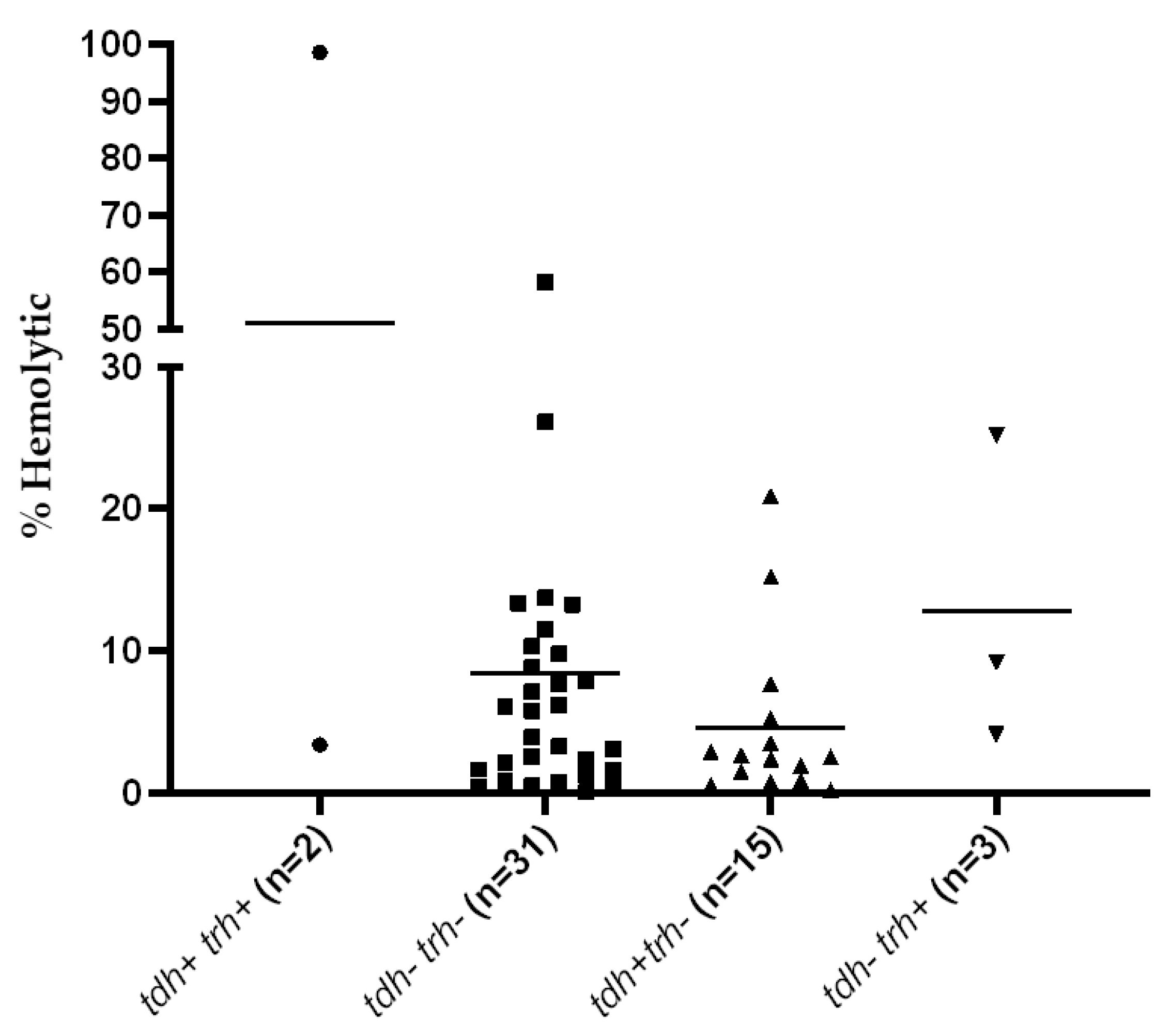
3.1. Detection of Virulence Genes and Hemolytic Activity

In this study, 51 isolates of V. parahaemolyticus isolated from seafood sources were grouped based on the presence of tdh, trh, and the pandemic marker genes detected by GS-PCR (Table 1). The percentages of hemolysis observed among V. parahaemolyticus isolates were varied and the correlation between hemolytic activity and the presence of the tdh gene was not found (r = 0.036, p = 0.802). The correlation between hemolytic activity and the presence of the trh gene was not done due to the small sample size. However, the greatest hemolytic activity (98%) was found in one of the tdh+ trh+ isolates (Figure 1). Furthermore, among 20 tdh+ isolates, 18 isolates exhibited β-hemolytic activity on Wagatsuma blood agar.

3.2. Characterization of Antimicrobial Susceptibility

All V. parahaemolyticus isolates from seafood were susceptible to chloramphenicol, whereas the highest resistance was recorded in response to ampicillin (100%) and followed by erythromycin (43.1%). It is of interest that 74.5% and 31.3% of the isolates demonstrated intermediate resistance to ciprofloxacin and norfloxacin, respectively (Table 2). An MAR index of 0.2 or above was shown by 23 isolates (45%) (Table 1). No significant correlation between the MAR index and the presence of the tdh gene (r = 0.096; p = 0.053), trh gene (r = 0.023; p = 0.871), or hemolytic activity (r = 0.112; p = 0.436) was observed.

3.3. Analysis of CRISPR Repeat Sequences and Patterns

A total of 19/51 (37.3%) of V. parahaemolyticus seafood isolates were positive for CRISPR (Table 1). Overall, the CRISPR element was significantly more prevalent in the tdh+ isolates and occurred less in the tdh− isolates (r = 0.832; p < 0.001). Two or three 28-bp direct repeats (DRs) were found and interspaced by spacers of 31 and/or 32 bp. (Table 3).
Although the CRISPR repeat sequence is highly conserved, the polymorphisms within the terminal repeats were observed in this study (Table 3). The terminal repeats differed in a single nucleotide in the central region and 7 bases in the terminal region of predicted RNA secondary structures. However, these do not affect the based pairing of RNA structure (Figure 2).

3.4. Spacer Sequence Analysis

The spacer sequences analysis revealed 4 different CRISPR spacer types which were designated as Sp1 to Sp4 (Table 4). Sp1, Sp2, and Sp3 shared the best matched with 100, 96.7, and 87.1% homology to fragments of V. alginolyticus plasmid pL300 (Table 4). For Sp4, no homology to foreign genetic elements was detected.
It is of interest that the spacer type was found to be correlated with tdh/trh profile and at least one type of spacer homolog to the V. alginolyticus plasmid (Sp1 to Sp3) was shared by all CRISPR-positive V. parahaemolyticus isolates (Table 3). Further homology analysis of the spacer sequences against publicly available V. parahaemolyticus genomes showed the association of spacer type and tdh/trh profile, although virulence genes of some isolates were unknown (Table 5). These hits are located on a CRISPR array, as they were detected nearby cas gene(s), and some hits have also been addressed as CRISPR region.

3.5. Spacer Origin

To further examine the finding of extrachromosomal origin of CRISPR spacers, the homology found among spacers was compared. This study found that Sp1, Sp2, and Sp3 sequences were matched with 6 mega-plasmid sequences of V. alginolyticus isolates, previously obtained from pipefish caught in the Kiel-Fjord, Germany, which ranged between 280 and 300 kbp in size [28]. Additionally, CRISPR spacers were mapped to represent Kiel alginolyticus megaplasmid, pL300, to locate the position of spacers. This study found that Sp1 to Sp3 spacers were distributed around pL300 (Figure 3). Sp1, Sp2, and Sp3 spacers were matched with the protospacer sequences of genes encoding for the modulator of FtsH protease Hfl, hypothetical protein, and DNA methyltransferase, respectively.
The alignment between pL300 sequences and other plasmids was performed to identify plasmid identity. Apart from V. alginolyticus isolates from Kiel Fjord, this megaplasmid was highly homolog (98.6% identity; E-value of 0.0) to the MYPK1 plasmid of V. alginolyticus strain GS (accession number CP054703.1). Few similarities to other plasmid sequences of Enterobacteria, including Escherichia coli, Klebsiella pneumoniae, Kluyvera ascorbata, and Citrobacter sp. were observed (data not shown). The progressiveMauve alignment results showed the conservation regions among related ancestor plasmids (Figure S1). The conserved CDSs found among these plasmids included the essential genes required for plasmid conjugal transfer and integration (Table S1). A closer look at the protospacer regions among the four plasmids is shown in Figure 4.

3.6. CRISPR-Virulence Typing

In this study, a combination between CRISPR spacer sequence types and tdh/trh virulence genes of V. parahaemolyticus was used to create a dendrogram. CRISPR-virulence typing profiles of V. parahaemolyticus were organized into 4 CRISPR-virulence types (CVTs) (Figure 5). The isolates within the same CVT cluster composed identical virulence profiles and spacer content.
The pandemic isolates with tdh+ trh− genotype was classified in the CVT1 cluster, which contained spacer type 1 and 4, and this was identical to that observed in the reference strain of V. parahaemolyticus RIMD 2210633. The tdhtrh+ and tdhtrh− isolates were classified in the CTV2 and CTV3 clusters, respectively. The differences between CVT1, CVT2, and CVT3 clusters were due to deletion or addition of a single spacer. It is of interest that the tdh+ trh+ isolate in the CVT4 cluster did not have any close association with others. Nevertheless, all 4 clusters shared spacers matched with pL300 of V. alginolyticus, suggesting that these isolates encounter this plasmid before they are diverged.

4. Discussion

In this study, 51 seafood isolates of V. parahaemolyticus previously, obtained from various cities in Southern Thailand between 2000 and 2012, were characterized for their hemolytic characteristics, antibiotic resistance, and CRISPR. All experiments included four hemolytic genotypes, pandemic and non-pandemic, of V. parahaemolyticus isolates. Although environmental and seafood isolates rarely contained tdh/trh, the presence of these isolates has been previously reported [29].
TDH- or TRH-producing V. parahaemolyticus are strongly associated with gastroenteritis. In this study, the result of a quantitative hemolysis assay against human blood demonstrated various levels of hemolytic activity exhibited by seafood isolates, and the highest hemolytic activity was detected in a tdh+ trh+ isolate. A previous study reported that temperature and culture density were the significant factors that affect the hemolysin activation of V. parahaemolyticus [30]. The gastroenteritis also showed a strong correlation with the KP, which is a type of β-hemolysis on the special blood agar and is induced by TDH [31]. A previous study reported that almost all environmental V. parahaemolyticus are tdh-negative and thus negative for KP [32]. In this work, two of the KP-negative V. parahaemolyticus isolates were positive for tdh gene. The previous study suggested that the isolates may possess a single copy of the tdh gene and therefore hemolysin was expressed at a low level [33]. Moreover, the hemolytic activity of V. parahaemolyticus may be phenotypically varied due to the sequence variation of hemolysin genes, which affects their promoter activities [6,34].
The increase in antibiotic-resistant bacteria is a major concern worldwide. Aquatic ecosystems could easily facilitate the exchange of mobile genetic elements among V. parahaemolyticus [35]. All V. parahaemolyticus isolates were resistant to ampicillin which is not recommended empirically to treat V. parahaemolyticus infection [36]. The ampicillin resistance found in this study correlates with other previous reports and this is not a new incident for ampicillin resistance among Vibrio spp. [37]. All V. parahaemolyticus isolates in this study were susceptible to chloramphenicol, which was the first truly broad-spectrum, and a high susceptibility to tetracycline (96.3%), sulfonamide (96.3%), and trimethoprim/sulfamethoxazole (67.2%) was observed. Interestingly, 72.7% and 34.5% of the isolates showed intermediate resistance to ciprofloxacin and norfloxacin, respectively which are the antimicrobial agents recommended to treat human infections [38]. This result was consistent with other studies [39]. The high level of intermediate resistance to both antibiotics may also tend to become resistant. Additionally, four isolates (PSU635, PSU3858, PSU4446) were identified as multidrug resistant (MDR) as they resist at least one agent in three or more antimicrobial classes [40]. This could lead to untreatable illnesses and human health risks. The MAR index value observed in this study indicated the differences in the original sources of the isolates. In this study, nearly half of the V. parahaemolyticus isolates had an MAR index ≥ 0.2, which indicated that these isolates originated from high-risk sources that have been exposed to antibiotics, such as humans and aquaculture, and seafood might be considered as a possible vehicle for pathogen transmission. Previous studies have reported that the V. parahaemolyticus isolates from seafood showed an MAR index > 0.2 [41,42]. Thus, antimicrobial resistance should continue to be monitored to ensure seafood safety.
CRISPR-Cas system is known as a bacterial adaptive immune system against conjugative plasmids, or phages. The virulence and genotypes of V. parahaemolyticus are reported to be associated with CRISPR/Cas evolution [17]. This study found that all pandemic V. parahaemolyticus isolates were identified as CRISPR positive, suggesting the association between CRISPR and the pathogenic evolution of pathogenic V. parahaemolyticus. Previous reports demonstrated high percentages of tdh+ (97.4%) and trh+ (47%) V. parahaemolyticus positive for CRISPR detection, with few reports demonstrating an association with the tdh− or trh− genotypes [15,17]. The result of this study corroborates earlier findings; however, we could detect CRISPR in two of the tdhtrh+ and one of the tdhtrh− isolates. The presence of CRISPR in the tdh− isolates were also reported in a previous study [41]. The CRISPR loci detected in this study is located on the V. parahaemolyticus island-7 (VPAI-7) region of chromosome 2, which encodes the tdh gene and are closely associated with tdh+ V. parahaemolyticus [43,44,45], thus this may be the reason why this CRISPR loci is more prevalent in the tdh+ isolates. Additionally, no significant correlation between the presence of CRISPR and hemolytic activity, or antimicrobial resistance, was observed (p > 0.05).
In this study, the DRs detected in CRISPR loci were 28-bp in length and its sequences have also been reported [16]. These characteristics were identical to those found in the reference strain of V. parahaemolyticus RIMD 2210633. In 2019, Baliga et al. analyzed the DRs of 200 V. parahaemolyticus genomes available in the database and found that 92% possessed a single type of repeat (RU-1) and the minimal set of Cas proteins [16]. The presence of similar DRs was observed in our recent study. However, the CRISPR primers used in this study did not target the Cas genes. Thus, the minimalistic type of CRISPR-Cas system could not be addressed here. Although a previous study reported 3–5 repeat units in the CRISPR loci of V. parahaemolyticus, we demonstrated 2 repeat units in the tdh+ trh+ and the tdhtrh− isolates. The presence of 2 DRs have been reported among tdh+ trh+ isolates from clinical samples [15,17]. Most isolates available in the public databases possess tdh+ trh− genotype, which may acquire more spacers than others. Moreover, further studies are necessary to clarify the types of CRISPR-Cas systems and their activities in the defence system against foreign mobile genetic elements.
This study found that the sequences of CRISPR repeats found among V. parahaemolyticus isolates were not highly identical, especially at the terminal position, however, this does not affect the RNA secondary structures. The polymorphisms in terminal sequences among the isolates in different genotypes were also observed. The variations within the central and terminal repeat sequence were also observed in the CRISPR loci of Streptococcus thermophilus, which suggested that the sequence degeneracy at the 3′-end of the terminal repeat is probably due to the homologous recombination events of DRs [46]. A degenerate repeat is required for the polarized spacer acquisition process [47].
BLAST comparisons of spacers identified matches between spacers and foreign plasmid sequences, which demonstrated the horizontal transfer of V. alginolyticus plasmid to V. parahaemolyticus. Most of the spacer sequences found in this study were similar to those reported in other V. parahaemolyticus isolates from seafood sources [16]. We have previously discussed the similarity of CRISPR spacers found in clinical isolates of V. parahaemolyticus with the V. alginolyticus plasmids [15]. In 2020, all of these plasmids were published as the megaplasmids associated with the Kiel isolates of V. alginolyticus [28]. In continuation of our previous discussion, the spacer sequence and origin were characterized and analyzed in a recent study.
In this study, 3 CRISPR spacers (Sp1 to Sp3) match the coding sequences of V. alginolyticus megaplasmid pL300, and one of these spacers (Sp4) was unique, as it demonstrated no homology to any sequences in the databases. It is often difficult to find sequence homology to spacer sequences because of the limited number of sequence information databases of plasmids and prokaryotic viruses [48]. All V. parahaemolyticus isolates included at least one spacer perfectly matching some region of pL300. One of the homology searches revealed the perfect match between CRISPR spacer and plasmid region, encoded for DNA methyltransferase, an enzyme required to protect DNA from restriction digestion [49]. This gene has been previously reported to be specifically targeted by CRISPR array against the virus to prevent successful host infection and lysis [50]. It is therefore conceivable that V. parahaemolyticus has adapted to its own evolution by avoiding the integration of foreign DNA from V. alginolyticus. Moreover, the results obtained by Progressive Mauve analysis demonstrated that pL300 may originate via the insertion of multiple regions of foreign DNA from bacteria in family Enterobacteriaceae. These conserved regions were found to encode essential genes of conjugative plasmids [49]. In addition, the evolution of conjugative plasmids could be driven through the CRISPR preventive mechanism.
CRISPR spacers can protect against foreign plasmid integration and phage invasions. The influence of these mobile genetic elements on the phenotypes and genotypes of marine Vibrio have been reported. Previous studies demonstrated a strong association of filamentous phages, f237, with recent pandemic strains of V. parahaemolyticus. This phage possesses a unique ORF8 open reading frame that is found only in recent pandemic isolates, and the Zot (Zonula occludens toxin)-like toxin previously described in the V. cholerae is reported to be encoded in the ORF7 [51,52]. A match was found between the CRISPR spacer region of V. anguillarum PF7 with a zot-encoding prophage in V. anguillarum PF4, indicating the contribution of CRISPR to the phage resistance of this bacterium [53]. In this study, no homologous between the CRISPR spacer and any phage regions or endogenous genomic sequences located outside the CRISPR array were found.
The results of this study indicated the association between spacer groups and hemolysin genotypes. Thus, a combination between CRISPR spacer sequences and the presence of hemolysin genes was used to cluster the V. pahaemolyticus isolates. As expected, a close relationship between the tdh+ trh− and the tdhtrh+ isolates were found in this study. The tdhtrh+ isolates were reported to be involved significantly in the pre-pandemic outbreaks, whereas the tdh+ trh− isolates are involved in both pre-pandemic and pandemics [8]. The tdhtrh− isolate was presented as the possible origin of the tdh+ trhV. parahaemolyticus and the tdh+ trh+ isolates were phylogenetically distant to others. Since fewer spacers are less efficient in the protection against foreign genetic elements, further evolutional study is needed to explain this situation. In addition, the position of a spacer in a CRISPR loci indicates the historical timeline of V. parahaemolyticus exposure to foreign genetic elements [54].

5. Conclusions

An analysis of hemolysin genotypes in association with CRISPR loci can help to reveal the genome evolution of V. parahaemolyticus. Our characterization and in-depth analysis of CRISPR among seafood isolates of V. parahaemolyticus showed that these isolates exhibit various virulence phenotypes, and some isolates have become resistant to many antibiotics. More importantly, similar types of CRISPR repeats and spacer sequences were found in isolates harboring the same hemolysin genotypes (tdh and/or trh) and all CRISPR-positive isolates shared spacer(s) homolog to the similar plasmid of V. alginolyticus. In addition, the position of a spacer in a CRISPR loci could indicate an ancestry interaction of V. parahaemolyticus with a foreign genetic element. An in-depth analysis of CRISPR-Cas loci, combined with virulence genes, could provide important data for the evolutionary study of V. parahaemolyticus.

Supplementary Materials

The following are available online at https://0-www-mdpi-com.brum.beds.ac.uk/article/10.3390/microorganisms9061220/s1, Figure S1: The multiple genome alignment by the progressiveMauve between V. alginolyticus pL300 megaplasmid (top) and other plasmids from the blast hit results. Homologous genomes are indicated by the connected-with-lines collinear blocks, Table S1: Details regarding the conserved CDSs residing in the locally collinear blocks (LCBs) obtained from ProgressiveMauve analysis.

Author Contributions

Conceptualization, P.M.-a.; methodology, P.M.-a. and N.J.; software, K.S.; formal analysis, N.J., S.P., K.S. and P.M.-a.; investigation, N.J.; resources, P.M.-a.; data curation, P.M.-a. and S.P.; writing—original draft preparation, N.J. and P.M.-a.; writing—review and editing, P.M.-a.; supervision, P.M.-a.; project administration, P.M.-a.; funding acquisition, P.M.-a. All authors have read and agreed to the published version of the manuscript.

Funding

This research was funded by a Faculty of Science Research Fund, Prince of Songkla University (contract no. 2-2561-02-018).

Institutional Review Board Statement

Not applicable.

Informed Consent Statement

Not applicable.

Data Availability Statement

All relevant data are within the paper and its Supplementary Files.

Acknowledgments

The authors would like to thank Marine Microbiology Research Laboratory, Faculty of Science, Prince of Songkla University for providing V. parahaemolyticus seafood isolates from culture collections.

Conflicts of Interest

The authors declare no conflict of interest. The funders had no role in the design of the study; in the collection, analyses, or interpretation of data; in the writing of the manuscript, or in the decision to publish the results.

References

  1. Nelapati, S.; Nelapati, K.; Chinnam, B.K. Vibrio parahaemolyticus-An emerging foodborne pathogen—A Review. Vet. World 2012, 5, 48–62. [Google Scholar] [CrossRef]
  2. Broberg, C.A.; Calder, T.J.; Orth, K. Vibrio parahaemolyticus cell biology and pathogenicity determinants. Microbes Infect. 2011, 13, 992–1001. [Google Scholar] [CrossRef] [PubMed] [Green Version]
  3. Matsumoto, C.; Okuda, J.; Ishibashi, M.; Iwanaga, M.; Garg, P.; Rammamurthy, T.; Wong, H.C.; Depaola, A.; Kim, Y.B.; Albert, M.J.; et al. Pandemic spread of an O3:K6 clone of Vibrio parahaemolyticus and emergence of related strains evidenced by arbitrarily primed PCR and toxRS sequence analyses. J. Clin. Microbiol. 2000, 38, 578–585. [Google Scholar] [CrossRef] [Green Version]
  4. Meador-Kidd, C.; Parsons, M.; Bopp, C.; Gerner-Smidt, P.; Painter, J.; Vora, G. Virulence gene- and pandemic group-specific marker profiling of clinical Vibrio parahaemolyticus isolates. J. Clin. Microbiol. 2007, 45, 1133–1139. [Google Scholar] [CrossRef] [Green Version]
  5. Nishibuchi, M.; Kaper, J.B. Thermostable direct hemolysin gene of Vibrio parahaemolyticus: A virulence gene acquired by a marine bacterium. Infect. Immun. 1995, 63, 2093–2099. [Google Scholar] [CrossRef] [Green Version]
  6. Kishishita, M.; Matsuoka, N.; Kumagai, K.; Yamasaki, S.; Takeda, Y.; Nishibuchi, M. Sequence variation in the thermostable direct hemolysin-related hemolysin (trh) gene of Vibrio parahaemolyticus. Appl. Environ. Microbiol. 1992, 58, 2449–2457. [Google Scholar] [CrossRef] [PubMed] [Green Version]
  7. Terai, A.; Baba, K.; Shirai, H.; Yoshida, O.; Takeda, Y.; Nishibuchi, M. Evidence for insertion sequence-mediated spread of the thermostable direct hemolysin gene among Vibrio species. J. Bacteriol. 1991, 173, 5036–5046. [Google Scholar] [CrossRef] [Green Version]
  8. Espejo, R.T.; García, K.; Plaza, N. Insight Into the Origin and Evolution of the Vibrio parahaemolyticus Pandemic Strain. Front. Microbiol. 2017, 8, 1397. [Google Scholar] [CrossRef] [PubMed]
  9. Miyamoto, Y.; Kato, T.; Obara, Y.; Akiyama, S.; Takizawa, K.; Yamai, S. In vitro hemolytic characteristic of Vibrio parahaemolyticus: Its close correlation with human pathogenicity. J. Bacteriol. 1969, 100, 1147–1149. [Google Scholar] [CrossRef] [PubMed] [Green Version]
  10. Igbinosa, E.O. Detection and antimicrobial resistance of Vibrio isolates in aquaculture environments: Implications for public health. Microb. Drug Resist. 2016, 22, 238–245. [Google Scholar] [CrossRef] [PubMed]
  11. Jansen, R.; Embden, J.D.; Gaastra, W.; Schouls, L.M. Identification of genes that are associated with DNA repeats in prokaryotes. Mol. Microbiol. 2002, 43, 1565–1575. [Google Scholar] [CrossRef] [PubMed]
  12. Fabre, L.; Zhang, J.; Guigon, G.; Le Hello, S.; Guibert, V.; Accou-Demartin, M.; de Romans, S.; Lim, C.; Roux, C.; Passet, V.; et al. CRISPR typing and subtyping for improved laboratory surveillance of Salmonella infections. PLoS ONE 2012, 7, e36995. [Google Scholar]
  13. Dion, M.B.; Labrie, S.J.; Shah, S.A.; Moineau, S. CRISPRStudio: A User-Friendly software for rapid CRISPR array visualization. Viruses 2018, 10, 602. [Google Scholar] [CrossRef] [Green Version]
  14. Bangpanwimon, K.; Sottisuporn, J.; Mittraparp-Arthorn, P.; Ueaphatthanaphanich, W.; Rattanasupar, A.; Pourcel, C.; Vuddhakul, V. CRISPR-like sequences in Helicobacter pylori and application in genotyping. Gut Pathog. 2017, 9, 65. [Google Scholar] [CrossRef] [Green Version]
  15. Kongrueng, J.; Srinitiwarawong, K.; Nishibuchi, M.; Mittraparp-Arthorn, P.; Vuddhakul, V. Characterization and CRISPR-based genotyping of clinical trh-positive Vibrio parahaemolyticus. Gut Pathog. 2018, 10, 48. [Google Scholar] [CrossRef] [Green Version]
  16. Baliga, P.; Shekar, M.; Venugopal, M. Investigation of direct repeats, spacers and proteins associated with clustered regularly interspaced short palindromic repeat (CRISPR) system of Vibrio parahaemolyticus. Mol. Genet. Genom. 2019, 294, 253–262. [Google Scholar] [CrossRef]
  17. Sun, H.; Li, Y.; Shi, X.; Lin, Y.; Qiu, Y.; Zhang, J.; Liu, Y.; Jiang, M.; Zhang, Z.; Chen, Q.; et al. Association of CRISPR/Cas evolution with Vibrio parahaemolyticus virulence factors and genotypes. Foodborne Pathog. Dis. 2015, 12, 68–73. [Google Scholar] [CrossRef]
  18. Kim, Y.B.; Okuda, J.; Matsumoto, C.; Takahashi, N.; Hashimoto, S.; Nishibuchi, M. Identification of Vibrio parahaemolyticus strains at the species level by PCR targeted to the toxR gene. J. Clin. Microbiol. 1999, 37, 1173–1177. [Google Scholar] [CrossRef] [Green Version]
  19. Wootipoom, N.; Bhoopong, P.; Pomwised, R.; Nishibuchi, M.; Ishibashi, M.; Vuddhakul, V. A decrease in the proportion of infections by pandemic Vibrio parahaemolyticus in Hat Yai Hospital, southern Thailand. J. Med. Microbiol. 2007, 56, 1630–1638. [Google Scholar] [CrossRef]
  20. Tada, J.; Ohashi, T.; Nishimura, N.; Shirasaki, Y.; Ozaki, H.; Fukushima, S.; Takano, J.; Nishibuchi, M.; Takeda, Y. Detection of the thermostable direct hemolysin gene (tdh) and the thermostable direct hemolysin-related hemolysin gene (trh) of Vibrio parahaemolyticus by polymerase chain reaction. Mol. Cell. Probes 1992, 6, 477–487. [Google Scholar] [CrossRef]
  21. Rattanama, P.; Thompson, J.R.; Kongkerd, N.; Srinitiwarawong, K.; Vuddhakul, V.; Mekalanos, J.J. Sigma E regulators control hemolytic activity and virulence in a shrimp pathogenic Vibrio harveyi. PLoS ONE 2012, 7, e32523. [Google Scholar] [CrossRef]
  22. De Souza Santos, M.; Salomon, D.; Li, P.; Krachler, A.-M.; Orth, K. 8—Vibrio parahaemolyticus virulence determinants. In The Comprehensive Sourcebook of Bacterial Protein Toxins, 4th ed.; Joseph, E., Alouf, J., Ladant, D., Popoff, M., Eds.; Elsevier Science: Amsterdam, The Netherlands, 2015; pp. 230–260. [Google Scholar]
  23. The Clinical and Laboratory Standards Institute. Performance Standards for Antimicrobial Susceptibility Testing CLSI Supplement M100S, 26th ed.; The Clinical and Laboratory Standards Institute: Wayne, PA, USA, 2016; pp. 74–75. [Google Scholar]
  24. Tan, C.W.; Rukayadi, Y.; Hasan, H.; Thung, T.Y.; Lee, E.; Rollon, W.D.; Hara, H.; Kayali, A.Y.; Nishibuchi, M.; Radu, S. Prevalence and antibiotic resistance patterns of Vibrio parahaemolyticus isolated from different types of seafood in Selangor, Malaysia. Saudi J. Biol. Sci. 2020, 27, 1602–1608. [Google Scholar] [CrossRef]
  25. Hofacker, I.L.; Fontana, W.; Stadler, P.F.; Bonhoeffer, L.S.; Tacker, M.; Schuster, P. Fast folding and comparison of RNA secondary structures. Mon. Chem. Chem. Mon. 1994, 125, 167–188. [Google Scholar] [CrossRef]
  26. Grant, J.R.; Stothard, P. The CGView Server: A comparative genomics tool for circular genomes. Nucleic Acids Res. 2008, 36, W181–W184. [Google Scholar] [CrossRef]
  27. Darling, A.E.; Mau, B.; Perna, N.T. progressiveMauve: Multiple genome alignment with gene gain, loss and rearrangement. PLoS ONE 2010, 5, e11147. [Google Scholar] [CrossRef] [PubMed] [Green Version]
  28. Chibani, C.M.; Roth, O.; Liesegang, H.; Wendling, C.C. Genomic variation among closely related Vibrio alginolyticus strains is located on mobile genetic elements. BMC Genom. 2020, 21, 354. [Google Scholar] [CrossRef]
  29. Robert-Pillot, A.; Guénolé, A.; Lesne, J.; Delesmont, R.; Fournier, J.M.; Quilici, M.L. Occurrence of the tdh and trh genes in Vibrio parahaemolyticus isolates from waters and raw shellfish collected in two French coastal areas and from seafood imported into France. Int. J. Food Microbiol. 2004, 91, 319–325. [Google Scholar] [CrossRef]
  30. Mahoney, J.C.; Gerding, M.J.; Jones, S.H.; Whistler, C.A. Comparison of the pathogenic potentials of environmental and clinical Vibrio parahaemolyticus strains indicates a role for temperature regulation in virulence. Appl. Environ. Microbiol. 2010, 76, 7459–7465. [Google Scholar] [CrossRef] [PubMed] [Green Version]
  31. Suzuki, N.; Ueda, Y.; Furukawa, T.; Takegaki, Y.; Miyagi, K.; Noda, K.; Hirose, H.; Hashimoto, S.; Yano, S.; Ishibashi, M.; et al. Incidence of Kanagawa phenomenon-positive and -negative Vibrio parahaemolyticus strains isolated from traveller’s diarrhea and their relation to tdh and trh genes. Kansenshogaku Zasshi 1997, 71, 417–420. [Google Scholar] [CrossRef] [Green Version]
  32. Lozano-León, A.; Torres, J.; Osorio, C.R.; Martínez-Urtaza, J. Identification of tdh-positive Vibrio parahaemolyticus from an outbreak associated with raw oyster consumption in Spain. FEMS Microbiol. Lett. 2003, 226, 281–284. [Google Scholar] [CrossRef] [Green Version]
  33. Nishibuchi, M.; Kaper, J.B. Duplication and variation of the thermostable direct haemolysin (tdh) gene in Vibrio parahaemolyticus. Mol. Microbiol. 1990, 4, 87–99. [Google Scholar] [CrossRef]
  34. Okuda, J.; Nishibuchi, M. Manifestation of the Kanagawa phenomenon, the virulence-associated phenotype, of Vibrio parahaemolyticus depends on a particular single base change in the promoter of the thermostable direct haemolysin gene. Mol. Microbiol. 1998, 30, 499–511. [Google Scholar] [CrossRef] [PubMed] [Green Version]
  35. Kümmerer, K. Antibiotics in the aquatic environment—A review—Part I. Chemosphere 2009, 75, 417–434. [Google Scholar] [CrossRef]
  36. Han, F.; Walker, R.D.; Janes, M.E.; Prinyawiwatkul, W.; Ge, B. Antimicrobial Susceptibilities of Vibrio parahaemolyticus and Vibrio vulnificus Isolates from Louisiana Gulf and Retail Raw Oysters. Appl. Environ. Microbiol. 2007, 73, 7096. [Google Scholar] [CrossRef] [PubMed] [Green Version]
  37. Xie, T.; Wu, Q.; Zhang, J.; Xu, X.; Cheng, J. Comparison of Vibrio parahaemolyticus isolates from aquatic products and clinical by antibiotic susceptibility, virulence, and molecular characterisation. Food Control 2017, 71, 315–321. [Google Scholar] [CrossRef]
  38. Okoh, A.I.; Igbinosa, E.O. Antibiotic susceptibility profiles of some Vibrio strains isolated from wastewater final effluents in a rural community of the Eastern Cape Province of South Africa. BMC Microbiol. 2010, 10, 143. [Google Scholar] [CrossRef] [PubMed] [Green Version]
  39. Preeprem, S.; Bhoopong, P.; Srinitiwarawong, K.; Vuddhakul, V.; Mittraparp-arthorn, P. Antibiogram profiles and virulence characteristics of pandemic Vibrio parahaemolyticus isolates from diarrheal patients in Hat Yai hospital, Southern Thailand. Southeast Asian J. Trop. Med. Public Health 2019, 50, 132–145. [Google Scholar]
  40. Basak, S.; Singh, P.; Rajurkar, M. Multidrug Resistant and Extensively Drug Resistant Bacteria: A Study. J. Pathog. 2016, 2016, 4065603. [Google Scholar] [CrossRef] [PubMed] [Green Version]
  41. Baliga, P.; Shekar, M.; Ahamed, S.; Venugopal, M. Antibiotic resistance pattern and its correlation to the presence of tdh gene and CRISPR-Cas system in Vibrio parahaemolyticus strains isolated from seafood. Indian J. Fish. 2019, 66, 100–108. [Google Scholar] [CrossRef] [Green Version]
  42. Elexson, N.; Afsah-Hejri, L.; Rukayadi, Y.; Soopna, P.; Lee, H.Y.; Zainazor, T.T.; Ainy, M.N.; Nakaguchi, Y.; Mitsuaki, N.; Son, R. Effect of detergents as antibacterial agents on biofilm of antibiotics-resistant Vibrio parahaemolyticus isolates. Food Control 2014, 35, 378–385. [Google Scholar] [CrossRef]
  43. Chao, G.; Jiao, X.; Zhou, X.; Wang, F.; Yang, Z.; Huang, J.; Pan, Z.; Zhou, L.; Qian, X. Distribution of genes encoding four pathogenicity islands (VPaIs), T6SS, biofilm, and type I pilus in food and clinical strains of Vibrio parahaemolyticus in China. Foodborne Pathog. Dis. 2010, 7, 649–658. [Google Scholar] [CrossRef]
  44. Hurley, C.C.; Quirke, A.; Reen, F.J.; Boyd, E.F. Four genomic islands that mark post-1995 pandemic Vibrio parahaemolyticus isolates. BMC Genom. 2006, 7, 104. [Google Scholar] [CrossRef] [Green Version]
  45. Makino, K.; Oshima, K.; Kurokawa, K.; Yokoyama, K.; Uda, T.; Tagomori, K.; Iijima, Y.; Najima, M.; Nakano, M.; Yamashita, A.; et al. Genome sequence of Vibrio parahaemolyticus: A pathogenic mechanism distinct from that of V. cholerae. Lancet 2003, 361, 743–749. [Google Scholar] [CrossRef]
  46. Horvath, P.; Romero, D.A.; Coûté-Monvoisin, A.C.; Richards, M.; Deveau, H.; Moineau, S.; Boyaval, P.; Fremaux, C.; Barrangou, R. Diversity, Activity, and Evolution of CRISPR Loci in Streptococcus thermophilus. J. Bacteriol. 2008, 190, 1401. [Google Scholar] [CrossRef] [Green Version]
  47. McGinn, J.; Marraffini, L.A. CRISPR-Cas Systems Optimize Their Immune Response by Specifying the Site of Spacer Integration. Mol. Cell 2016, 64, 616–623. [Google Scholar] [CrossRef]
  48. Li, W.; Bian, X.; Evivie, S.E.; Huo, G.C. Comparative Analysis of Clustered Regularly Interspaced Short Palindromic Repeats (CRISPR) of Streptococcus thermophilus St-I and its Bacteriophage-Insensitive Mutants (BIM) Derivatives. Curr. Microbiol. 2016, 73, 393–400. [Google Scholar] [CrossRef] [PubMed]
  49. Virolle, C.; Goldlust, K.; Djermoun, S.; Bigot, S.; Lesterlin, C. Plasmid transfer by conjugation in gram-negative bacteria: From the cellular to the community level. Genes 2020, 11, 1239. [Google Scholar] [CrossRef] [PubMed]
  50. Nasko, D.J.; Ferrell, B.D.; Moore, R.M.; Bhavsar, J.D.; Polson, S.W.; Wommack, K.E. CRISPR spacers indicate preferential matching of specific virioplankton genes. mBio 2019, 10, e02651-18. [Google Scholar] [CrossRef] [PubMed] [Green Version]
  51. Nasu, H.; Iida, T.; Sugahara, T.; Yamaichi, Y.; Park, K.S.; Yokoyama, K.; Makino, K.; Shinagawa, H.; Honda, T. A filamentous phage associated with recent pandemic Vibrio parahaemolyticus O3:K6 strains. J. Clin. Microbiol. 2000, 38, 2156–2161. [Google Scholar] [CrossRef]
  52. Iida, T.; Hattori, A.; Tagomori, K.; Nasu, H.; Naim, R.; Honda, T. Filamentous phage associated with recent pandemic strains of Vibrio parahaemolyticus. Emerg. Infect. Dis. 2001, 7, 477–478. [Google Scholar] [CrossRef] [PubMed]
  53. Castillo, D.; Kauffman, K.; Hussain, F.; Kalatzis, P.; Rørbo, N.; Polz, M.F.; Middelboe, M. Widespread distribution of prophage-encoded virulence factors in marine Vibrio communities. Nature 2018, 8, 9973. [Google Scholar] [CrossRef] [PubMed]
  54. Sorek, R.; Kunin, V.; Hugenholtz, P. CRISPR-a widespread system that provides acquired resistance against phages in bacteria and archaea. Nature 2008, 6, 181–186. [Google Scholar] [CrossRef] [PubMed]
Figure 1. Quantitative analysis of the hemolytic activity of V. parahaemolyticus seafood isolates. Each point shows the mean value from duplicate experiments. Median values are shown by a horizontal line.
Microorganisms 09 01220 g001
Figure 2. The secondary structures of four CRISPR repeats found in V. parahaemolyticus isolates. Predicted secondary structures were obtained using RNA fold. The dashed boxes indicate the sequence differences between typical repeat (A), and terminal repeat of tdh+ trh− (B), tdh+ trh+ and tdh+ trh− (C), and tdhtrh− (D) V. parahaemolyticus isolates, respectively.
Microorganisms 09 01220 g002
Figure 3. Distribution of protospacers along the representative plasmid pL300 of V. alginolyticus K08M3. The plasmid map is drawn from the GenBank entry CP017915.1 and ORFs are shown as arrows. Red arrows indicate the position of spacer-matching sequences (Sp1 to Sp3) found in the CRISPR region of V. parahaemolyticus analyzed in this study.
Microorganisms 09 01220 g003
Figure 4. A zoomed-in view of the progressiveMauve alignment between V. alginolyticus pL300 megaplasmid (top) and other plasmids from the blast hit results. The protospacer regions are annotated and shown in the red vertical bar. Homologous genomes are indicated by the connected-with-lines collinear blocks.
Microorganisms 09 01220 g004
Figure 5. Dendrogram of the 19 CRISPR-positive V. parahaemolyticus seafood isolates. The maximum likelihood method was used for constructing the phylogenetic tree. The bar represents a 5% dissimilarity between the two sequences. Colors within the squares correspond to the presence of spacer and virulence genes.
Microorganisms 09 01220 g005
Table 1. V. parahaemolyticus isolates used in this study and their characteristics.
Hemolysin
Genotype (n)
IsolatesYear of
Isolation
SourceGS-PCR *Kanagawa
Phenomenon
MAR *
Index
CRISPR-PCR
Seafood isolates
tdh+ trh− (15)PSU1662000Hard clam++0.2+
PSU3582001Mussel
Mussel
Mussel
Mussel
Hard clam
++0.2+
PSU360++0.2+
PSU434++0.2+
PSU474++0.2+
PSU476++0.1+
PSU4792002Hard clam++0.1+
PSU579 Cockle++0.2+
PSU635 Mussel++0.3+
PSU637 Mussel++0.2+
PSU638 Mussel++0.2+
PSU32492006Mussel+0.1+
PSU40672008Cockle++0.1+
PSU48882010Hard clam++0.1+
PSU53822012Shellfish++0.1+
tdhtrh+ (3)PSU3819
PSU3831
2007Crab
Fish
0.2+
0.2+
PSU51242011Shrimp0.1
tdh+ trh+ (2)PSU5822002Cockle+0.2+
PSU44132008Cockle0.1
tdhtrh− (31)PSU513
PSU571
PSU576
PSU578
2002Cockle
Hard clam
Cockle
Cockle
0.1
0.1
0.1
0.1+
PSU8112003Mussel0.1
PSU2463
PSU2467
PSU2471
2005Cockle
Cockle
Cockle
0.2
0.2
0.2
PSU3103
PSU3200
PSU3362
PSU3365
2006Hard clam
Cockle
Mussel
Cockle
0.1
0.2
0.2
0.1
PSU3858
PSU4055
PSU4058
PSU4062
PSU4075
PSU4091
PSU4094
2008Octopus
Mussel
Hard clam
Cockle
Hard clam
Cockle
Cockle
0.4
0.1
0.1
0.2
0.1
0.1
0.1
PSU4415
PSU4418
PSU4425
PSU4446
PSU4459
PSU4460
PSU4575
2009Cockle
Cockle
Cockle
Cockle
Cockle
Cockle
Cockle
0.1
0.1
0.1
0.4
0.1
0.1
0.1
PSU4869
PSU4879
PSU4885
PSU4895
2010Mussel
Mussel
Hard clam
Hard clam
0.2
0.2
0.1
0.2
PSU53792012Shellfish0.1
Clinical isolates
tdh + trh− (3)PSU38722008Clinical++0.3+
PSU39492008Clinical++0.2+
PSU51262011Clinical++0.1+
tdhtrh+ (1)ATCC178021965Clinical0.2
* GS-PCR, Group-Specific PCR; MAR, Multiple Antibiotic Resistance.
Table 2. Antimicrobial susceptibility patterns of V. parahaemolyticus isolates from seafood sources.
Antimicrobial DrugsNo. of Isolates (%)
SusceptibleIntermediateResistant
Ampicillin0051 (100)
Gentamycin12 (23.5)39 (76.5)0
Erythromycin029 (56.9)22 (43.1)
Sulfonamide49 (96)1 (2)1 (2)
Tetracycline49 (96)1 (2)1 (2)
Ciprofloxacin12 (23.5)38 (74.5)1 (2)
Trimethoprim/sulfamethoxazole36 (70.6)13 (25.5)2 (3.9)
Chloramphenicol51 (100)00
Norfloxacin34 (66.7)16 (31.3)1 (2)
Table 3. Characteristics of CRISPR repeat sequences and pattern in CRISPR-positive V. parahaemolyticus isolates.
Hemolysin
Genotype (n)
TypeDirect Repeats (DRs) Sequences *No. of DRsNo. of Spacers
(Sp Type)
CRISPR Locus Pattern (bp) *
Seafood isolates
tdh+ trh− (15)Typical repeat
Terminal repeat
GTGAACTGCCGAATAGGTAGCTGATAAT
GTGAACTGCCGCATAGGTAGAGAGAATC
32 (1, 4)28-32-28-31-28
tdhtrh+ (2)Typical repeat
Terminal repeat
GTGAACTGCCGAATAGGTAGCTGATAAT
GTGAACTGCCGCATAGGTAGAGAGGATC
32 (2, 4)28-31-28-31-28
tdh+ trh+ (1)Typical repeat
Terminal repeat
GTGAACTGCCGAATAGGTAGCTGATAAT
GTGAACTGCCGCATAGGTAGAGAGGATC
21 (3)28-31-28
tdhtrh− (1)Typical repeat
Terminal repeat
GTGAACTGCCGAATAGGTAGCTGATAAT
GTGAACTGCCGAATAGGTAGAGAGGATC
21 (1)28-32-28
Clinical isolates
tdh + trh− (3)Typical repeat
Terminal repeat
GTGAACTGCCGAATAGGTAGCTGATAAT
GTGAACTGCCGCATAGGTAGAGAGAATC
32 (1, 4)28-32-28-31-28
* Underline indicates mutation compared to typical repeat sequence, italic indicates the direct repeats (CDRs) sequence length and bold indicates the spacer length.
Table 4. Homology analysis of V. parahaemolyticus spacers against foreign genetic elements.
Spacer Type (Length)Spacer Sequences (5′ to 3′)Spacer HomologyQuery Cover/
Identities (%)
Accession Number/
Location
Sp1 (32)GAGATACCACAAGCTCAAGCAGATGCTAACAGVibrio alginolyticus strain K08M3 plasmid pL30093/96.7CP017915.1/
184601-184630
Sp2 (31)TCATTCTCACGATCTAATTACAGTTGGTCACVibrio alginolyticus strain K08M3 plasmid pL30093/100CP017915.1/
155899-155927
Sp3 (31)TGCAGACAAACAAAGAGCCATCGACGAGTGCVibrio alginolyticus strain K08M3 plasmid pL300100/87.1CP017915.1/
141985-142015
Sp4 (31)AGTCGGTCAACTGAGAATACGTTGTTGCCAA---
Table 5. Homology analysis of the Sp1 to Sp4 spacers against public genomes of V. parahaemolyticus.
Spacer TypeIsolatesHemolysin
Genotype *
Sources *Identities (%)E-ValueAccession Number
Sp1Vibrio parahaemolyticus strain RIMD 2210633tdh+ trhClinical, Japan1002 × 10−8BA000032.2
Vibrio parahaemolyticus strain VPD14 tdh+ trhShrimp, China1002 × 10−8CP031782.1
Vibrio parahaemolyticus strain FDAARGOS_191 tdh+ trhClinical, India1002 × 10−8CP020428.2
Vibrio parahaemolyticus strain BB220Ptdh+ trhEnvironment, India96.881 × 10−6CP003973.1
Vibrio parahaemolyticus strain FORC_071 tdh+ trhClinical, South Korea1001 × 10−6CP023486.1
Sp2Vibrio parahaemolyticus strain Vp17 tdhunk trhunkClam, India1007 × 10−8MG765521.1
Vibrio parahaemolyticus strain Vp14 tdhunk trhunkOyster, India1007 × 10−8MG765520.1
Vibrio parahaemolyticus strain Vp8 tdhunk trhunkShrimp, India1009 × 10−7MG765517.1
Sp3Vibrio parahaemolyticus strain 10329tdh+ trh+Clinical, USA1007 × 10−8CP045795.1
Vibrio parahaemolyticus strain MAVP-26 tdh+ trh+Clinical, USA1007 × 10−8CP023247.1
Vibrio parahaemolyticus strain ST631 tdh+ trh+Clinical, USA1007 × 10−8CP011885.1
Vibrio parahaemolyticus strain MAVP-QPI tdh+ trh+Clinical, USA1007 × 10−8MF066646.1
Vibrio parahaemolyticus strain MAVP-Q tdh+ trh+Clinical, USA1007 × 10−8CP022472.1
Vibrio parahaemolyticus strain FDAARGOS_662 tdh+ trh+Clinical, USA1007 × 10−8CP044070.1
Vibrio parahaemolyticus strain FDAARGOS_51 tdh+ trh+Clinical, USA1007 × 10−8CP026042.1
Vibrio parahaemolyticus strain 2014V-1125 tdh+ trh+Clinical, USA1007 × 10−8CP046777.1
Vibrio parahaemolyticus strain 2014V-1066 tdh+ trh+Clinical, USA1007 × 10−8CP046780.1
Vibrio parahaemolyticus strain 2015AW-0174 tdh+ trh+Clinical, USA1007 × 10−8CP046753.1
Vibrio parahaemolyticus strain 2010V-1106 tdhunk trh+Clinical, USA1007 × 10−8CP046827.1
Vibrio parahaemolyticus strain 2013V-1146 tdhunk trh+Clinical, USA1007 × 10−8CP046809.1
Vibrio parahaemolyticus strain 2013V-1181 tdhunk trh+Clinical, USA1007 × 10−8CP046784.1
Sp4Vibrio parahaemolyticus strain RIMD 2210633tdh+ trhClinical, Japan1007 × 10−8BA000032.2
Vibrio parahaemolyticus strain VPD14 tdh+ trhShrimp, China1007 × 10−8CP031782.1
Vibrio parahaemolyticus strain FDAARGOS_191 tdh+ trhClinical, India1007 × 10−8CP020428.2
Vibrio parahaemolyticus strain
BB22OP
tdh+ trhEnvironment, Bangladesh1007 × 10−8CP003973.1
Vibrio parahaemolyticus strain FORC_071 tdh+ trhClinical, South Korea1007 × 10−8CP023486.1
Vibrio parahaemolyticus strain Vp14 tdhunk trhunkOyster, India1007 × 10−8MG765520.1
Vibrio parahaemolyticus strain Vp9 tdhunk trhunkOyster, India1007 × 10−8MG765518.1
Vibrio parahaemolyticus strain Vp8tdhunk trhunkShrimp, India1007 × 10−8MG765517.1
* Hemolysin genotypes and sources were indicated based on the results obtained by database search and/or previous publications. Unk—unknown.
Publisher’s Note: MDPI stays neutral with regard to jurisdictional claims in published maps and institutional affiliations.

