Article

Taxonomic Reassessment of the Izumo Lineage of Hynobius utsunomiyaorum: Description of a New Species from Chugoku, Japan

1
Faculty of Science and Technology, Kochi University, 2-5-1 Akebonocho, Kochi City 780-8072, Japan
2
Yasugi City Hall, 878-2, Yasugicho, Yasugi City 692-8686, Japan
3
Kurayoshi City Hall, 722, Aoimachi, Kurayoshi City 682-8611, Japan
4
Faculty of Science and Technology, Oita University, 700 Dannoharu, Oita City 870-1192, Japan
*
Author to whom correspondence should be addressed.
Submission received: 3 June 2021 / Revised: 21 July 2021 / Accepted: 22 July 2021 / Published: 23 July 2021
(This article belongs to the Section Ecology and Conservation)

Abstract

:

Simple Summary

In the study, we investigated the true taxonomic composition of Hynobius utsunomiyaorum from Chugoku District, Japan. Our detailed morphological and molecular comparisons showed that H. utsunomiyaorum is clearly divided into two species: the true H. utsunomiyaorum and the Izumo Lineage of H. utsunomiyaorum, i.e., Hynobius kunibiki sp. nov. This new species does not satisfy the diagnosis of H. utsunomiyaorum, while these two species do not form a monophyletic group based on molecular data. Hynobius setoi is morphologically more similar to the new species than is H. utsunomiyaorum. Previous studies have suggested that H. setoi is distributed across the San’in region, i.e., the northern part of Chugoku District, Japan. However, our research revealed that H. setoi is not distributed in the northern part of Shimane Prefecture, located in the western part of the San’in region in Chugoku District. Rather than H. setoi, we found that the new species was distributed in the northern part of Shimane Prefecture. Additionally, results of phylogenetic analyses using all valid Japanese Hynobius species are provided in our study. Based on these results, we show that Japanese Hynobius species included in the subgenus Hynobius may be divided into four genetic clades. The information in our study will be vital for developing conservation management strategies and policies for these species.

Abstract

Here, we describe a new species of the genus Hynobius from Chugoku, Japan. In populations from central to eastern Shimane Prefecture, the Izumo Lineage of Hynobius utsunomiyaorum was clearly distinguished from the true Hynobius utsunomiyaorum based on morphological and molecular evidence. Thus, we describe the former lineage as a new species, Hynobius kunibiki sp. nov. Morphological comparisons revealed that H. utsunomiyaorum lacks a distinct yellow line on the ventral side of its tail, whereas the new species possesses this yellow line; most H. utsunomiyaorum individuals have distinct white spots on the lateral sides of their body and lack a fifth toe, whereas the new species largely lacks these spots, and all examined individuals had a fifth toe. The two species also differed significantly by several other morphological characteristics. The lentic species Hynobius setoi is morphologically similar to H. kunibiki sp. nov., but they differ significantly by various morphological characteristics. Despite their partial morphological similarity, these two species differed substantially in terms of their genetics. Finally, we show, in a phylogenetic tree including all Japanese Hynobius species, that the subgenus Hynobius can be divided into four genetic clades. Overall, this information will help develop conservation management strategies and policies for these species.

1. Introduction

In the family Hynobiidae (the Asiatic salamanders), the most diverse genus Hynobius includes 55 species, 37 (67.3%) of which are endemic to Japan [1]. The identification of Hynobius species can be challenging without locality and genetic data because they have few distinguishing morphological characteristics [2]. Previous studies have revealed that cryptic species of lotic or lentic Hynobius exist in Japan [3,4]; that are yet to be described [3,5].
The Hiba salamander, Hynobius utsunomiyaorum, was described from Rokunohara, Saijocho Yuki, Shobara City, Hiroshima Prefecture, and has been found in the Chugoku Mountains in eastern Chugoku District, Japan [3]. According to Matsui et al. [3], this species is not distributed in the northern part of Shimane Prefecture; instead, the typical pond breeding species, Hynobius setoi, is found in this area [3]. However, genetic evidence that H. setoi is distributed in northern Shimane Prefecture was not provided by Matsui et al. [3]; such evidence came only from morphological data. On the other hand, Hayashi and Ooi [5] sampled widely from the northern part of Shimane Prefecture and then performed mitochondrial DNA analyses on these samples. Results showed that haplotypes of H. utsunomiyaorum but not H. setoi were found in the study area [5]. However, populations from the northern part of Shimane Prefecture are clearly distinguishable from true H. utsunomiyaorum in terms of their morphology and ecology; thus, Hayashi and Ooi [5] named these populations the Izumo Lineage of H. utsunomiyaorum and suggested that they may be a new species. They concluded that H. setoi is not distributed in the northern part of Shimane Prefecture [5]; however, detailed morphological evidence for these populations is currently lacking.
The Izumo Lineage suggested by Hayashi and Ooi [5] corresponds to the B2b3 clade suggested by Matsui et al. [3]. Based on mitochondrial DNA and allozyme analyses, this lineage paraphyletic with respect to H. utsunomiyaorum [3,6]; therefore, based on the phylogenetic species concept, it should be considered a new species. When a new species is described, essential features must be directly derived from characteristics observed in specimens rather than indirectly inferred [7]; hence, the morphological characteristics that distinguish a species from its close relatives should be described in a “diagnosis.″ If the Izumo Lineage does not satisfy the diagnosis conditions of H. utsunomiyaorum, the two should be considered different species based on the morphological species concept. In herpetology, Frost and Hillis [8] recommend the use of the phylogenetic and evolutionary species concepts. If the Izumo Lineage does not form a monophyly with H. utsunomiyaorum after DNA analysis of samples from the entire distribution range of H. utsunomiyaorum, the two should be considered different species based on the phylogenetic and evolutionary species concepts. Although Matsui et al. [3] concluded that H. setoi is distributed in the northern part of Shimane Prefecture based on its morphology, the phylogenetic analysis of Hayashi and Ooi [5] suggests that H. setoi from the northern part of Shimane Prefecture was most likely mistaken with the Izumo Lineage because the two share a similar morphology. Thus, morphological comparisons between H. setoi and the Izumo Lineage of H. utsunomiyaorum are also required.
In the present study, we used additional DNA sequence data to reconstruct the phylogeny of Hynobius collected from Chugoku. Furthermore, we statistically analyzed morphological characteristics to compare two species and one candidate species, i.e., H. utsunomiyaorum, the Izumo Lineage of H. utsunomiyaorum, and H. setoi. We also evaluated the species validity of the Izumo Lineage of H. utsunomiyaorum using the phylogenetic, evolutionary, and morphological species concepts. We revealed the detailed distribution ranges and borders of H. utsunomiyaorum, the Izumo Lineage of H. utsunomiyaorum, and H. setoi by the sampling from their complete distribution ranges; additionally, we investigated the distribution ranges of Hynobius iwami in the eastern part of Shimane Prefecture, which are found near the distribution ranges of the Izumo Lineage of H. utsunomiyaorum. Finally, we constructed a phylogenetic tree including all Japanese Hynobius species and estimated the phylogenetic relationships in this genus.

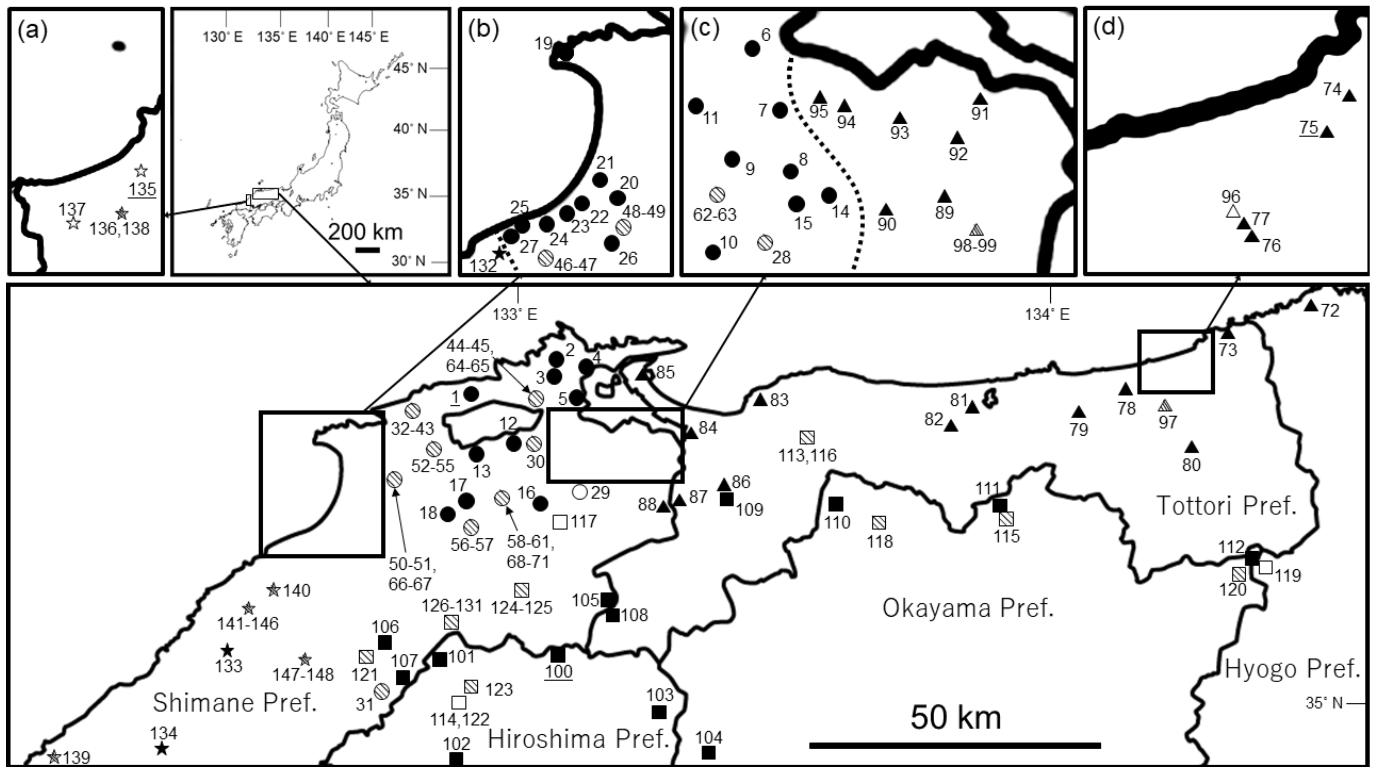
2. Materials and Methods

2.1. Molecular Analysis

For phylogenetic analysis, we sampled 70 individuals (from February 2007 to January 2021) representing the Izumo Lineage of H. utsunomiyaorum (27 individuals), H. setoi (24 individuals), H. utsunomiyaorum (13 individuals), H. iwami (three individuals), Hynobius abuensis (one individual from the type locality), Hynobius tsurugiensis (one individual from the type locality), Hynobius kuishiensis (one individual from the type locality), and Hynobius guttatus (one individual not from the type locality) from 70 localities (Table S1; Figure 1). For samples taken during the breeding season, we removed a single tailbud embryo from each paired egg sac and preserved it in 99.5% ethanol. We also obtained tissue samples by clipping the caudal extremities from larvae preserved in 99.5% ethanol. Compared with other groups of Japanese Hynobius, it is more difficult to collect eggs and larvae from the Hynobius stejnegeri group, including H. tsurugiensis, H. kuishiensis, and H. guttatus; therefore, we obtained tissue samples from the caudal extremities of adults in these three species. We extracted total genomic DNA from the tail tips or single tailbud embryos and preserved it in 99.5% ethanol using a DNeasy Blood and Tissue Kit (Qiagen, Hilden, Germany). For all individual Hynobius, we amplified a 630-bp fragment of the cytochrome b gene using Ex Taq® (TaKaRa, Tokyo, Japan) with primers L14010 (5′-TAHGGWGAHGGATTWGAWGCMACWGC-3′) and H14778 (5′-AARTAYGGGTGRAADGRRAYTTTRTCT-3′) [9]. The reaction mix (with a total volume of 10 μL) contained 1.0 μL of 10× Ex Taq Buffer, 0.8 μL of 25-mM dNTP mix, 0.5 μL of each of the forward and reverse primers (10 pM), 0.05 μL of Taq polymerase, 6.15 μL of distilled deionized water, and 1.0 μL of template DNA. A T100™ thermal cycler (Bio-Rad, Hercules, CA, USA), was utilized with the following conditions: an initial 3-min denaturing step at 94 °C; 40 cycles of 30 s at 94 °C, 45 s at 56 °C, and 90 s at 72 °C; with a final 10-min extension at 72 °C. We purified PCR products with Illustra™ ExoStar™ 1-Step (GE Healthcare, Buckinghamshire, UK) and sequenced them using BigDye® Terminator ver. 3.1 (Applied Biosystems, Foster City, CA, USA) on an ABI 3130xl Genetic Analyzer (Applied Biosystems). We deposited the acquired sequences into the DNA Data Bank of Japan (Table S1). We aligned the DNA sequences using MEGA X [10], and then performed phylogenetic analyses of the aligned sequences from all recognized Japanese Hynobius species, with “Salamandrella keyserlingii″ used as the outgroup (Table S1), using Bayesian inferences (BIs) and maximum likelihood (ML) estimates. We estimated the best-fit nucleotide substitution model using jModelTest 2 [11] based on the corrected Akaike’s information criterion [12] and Bayesian information criterion [13]. We selected the Hasegawa–Kishino–Yano model (gamma distribution with invariant sites) based on both criteria. We constructed Bayesian and ML trees using MrBayes 3.2 [14] and MEGA X [10], respectively. For the Bayesian analysis, we performed two independent MCMC runs for 5,000,000 generations with a sample frequency of 100. We assessed the statistical support for monophyly with posterior probabilities and examined the stationarity of the likelihood scores of sampled trees using Tracer version 1.7 (http://tree.bio.ed.ac.uk/software/tracer/) (accessed on 17 February 2021). The first 25% of generations were discarded as burn-in. We regarded a group as monophyletic when a node had a posterior probability ≥0.95 according to the criterion of Huelsenbeck and Rannala [15]. For ML analysis, we estimated the statistical support for monophyly with 1000 bootstrap replicates; a group was regarded as monophyletic when a node had a bootstrap value ≥80% according to the criterion of Regier et al. [16].

2.2. Morphological Analysis

We sampled 124 individuals across the entire distribution areas of three groups from January 2019 to March 2021, including 31 individuals of the Izumo Lineage of H. utsunomiyaorum (22 males and nine female) from seven populations (Pops. 1–3, 8, 17, 22, and 23), 45 individuals of H. setoi (30 males and 15 females) from six populations (Pops. 75, 76, 81, 82, 84, and 87), and 48 individuals of H. utsunomiyaorum (38 males and 10 females) from four populations (Pops. 100, 102, 111, and 112) (Table S1; Figure 1).
From a conservation perspective, the collected specimens were measured under anesthesia and subsequently returned to their site of capture except for the candidate individuals of type specimens (holotype and two paratypes of both sexes). Before specimens were returned, we captured images of the dorsal, ventral, and lateral sides of all individuals on a black background. We also obtained tissue samples (preserved in 99.9% ethanol) from the tail tips of all individuals as evidence that they had been collected. Salamanders were anesthetized with ethyl 3-aminobenzoate methanesulfonate salt (Sigma-Aldrich, St. Louis, MO, USA) diluted 1000-fold with water [17]. Using a digital caliper, 22 measurements were made to the nearest 0.1 mm on anesthetized specimens, including the following: snout–vent length (SVL), trunk length (TRL), axilla–groin distance (AGD), head length (HL), tail length (TAL), median tail width (MTAW), median tail height (MTAH), vomerine teeth length (VTL), and vomerine teeth width (VTW), head width (HW), forelimb length (FLL), hindlimb length (HLL), second finger length (2FL), third finger length (3FL), third toe length (3TL), fifth toe length (5TL), internarial distance (IND), interorbital distance (IOD), upper eyelid length, snout length (SL), upper eyelid width, and lower jaw length (LJL). For each individual, we also recorded data on the presence and number of distinct white spots on the venter (DWSV) and lateral (DWSL) sides of the body, the presence of a distinct and continuous yellow line on the dorsal side of the tail (DCTYLD), the presence of a distinct yellow line on the ventral side of the tail (DTYLV), and the presence of distinct gular mottling (DGM). The number of costal folds between the adpressed limbs (CFBALN) and the number of costal grooves (CGN) were counted; we used the counting method of Matsui et al. [3] to count CGN.
Prior to performing morphological comparisons among the three groups, we tested for normality using a Shapiro–Wilk test. When data were found to follow a normal distribution, we tested for homoscedasticity using Bartlett’s test. When the variances among the populations were found to be equal, we performed Tukey–Kramer tests; when variances were found not to be equal, we performed Games–Howel tests. When data did not follow a normal distribution and variances among populations were not equal, we performed Steel–Dwass tests. To examine the overall morphological variation among the three groups, we performed canonical discriminant analysis using standardized values for the 22 measurements. We also performed morphological comparisons between males and females for the three groups. Before the analyses, we tested for normality using a Shapiro–Wilk test. When data followed a normal distribution, we tested for homoscedasticity using an F test. When the variances among the groups were equal, we performed Student’s t tests; when variances were not equal, we performed Welch’s t test. When data did not follow a normal distribution and variances among groups were not equal, we performed Brunner–Munzel tests. All statistical analyses were performed in R with α = 0.05 as the significance level [18]. Additionally, we used R to calculate the Euclidean distances among the three groups based on the 22 morphological characters in both sexes.

2.3. Sampling and Measurements of Type Specimens

We collected a holotype and one paratype female from Pop. 1 on 20 December 2019 (Nishinomura, Kamionocho, Matsue City, Shimane Prefecture). We also collected one paratype male from Pop. 17 (Kamocho Chikamatsu, Unnan City, Shimane Prefecture) on 20 January 2021. The collected type specimens were fixed in 10% formalin and then transferred to 70% ethanol. The preserved holotype specimens (TRPM-ARA-0000014) were deposited in Tottori Prefectural Museum (2-124, Higashimachi, Tottori City, Tottori Prefecture, 680-0011, Japan). The two preserved paratype specimens were deposited in Yokosuka City Museum (95, Fukadadai, Yokosuka City, Kanagawa Prefecture, 238-0016, Japan; paratype female: YCM-RA589; paratype male: YCM-RA590). We took 43 measurements from the holotype including SVL, TRL, AGD, HL, TAL, MTAW, MTAH, basal tail width (BTAW), basal tail height (BTAH), VTL, VTW, HW, maximum head width (MXHW), left forelimb length (LFLL), left hindlimb length (LHLL), right forelimb length (RFLL), right hindlimb length (RHLL), left first finger length (L1FL), left second finger length (L2FL), left third finger length (L3FL), left fourth finger length (L4FL), right first finger length (R1FL), right second finger length (R2FL), right third finger length (R3FL), right fourth finger length (R4FL), left first toe length (L1TL), left second toe length (L2TL), left third toe length (L3TL), left fourth toe length (L4TL), left fifth toe length (L5TL), right first toe length (R1TL), right second toe length (R2TL), right third toe length (R3TL), right fourth toe length (R4TL), right fifth toe length (R5TL), IND, IOD, left upper eyelid length (LUEL), right upper eyelid length (RUEL), SL, left upper eyelid width (LUEW), right upper eyelid width (RUEW), and LJL. We also counted the CGN using the method of Matsui et al. [3].

3. Results

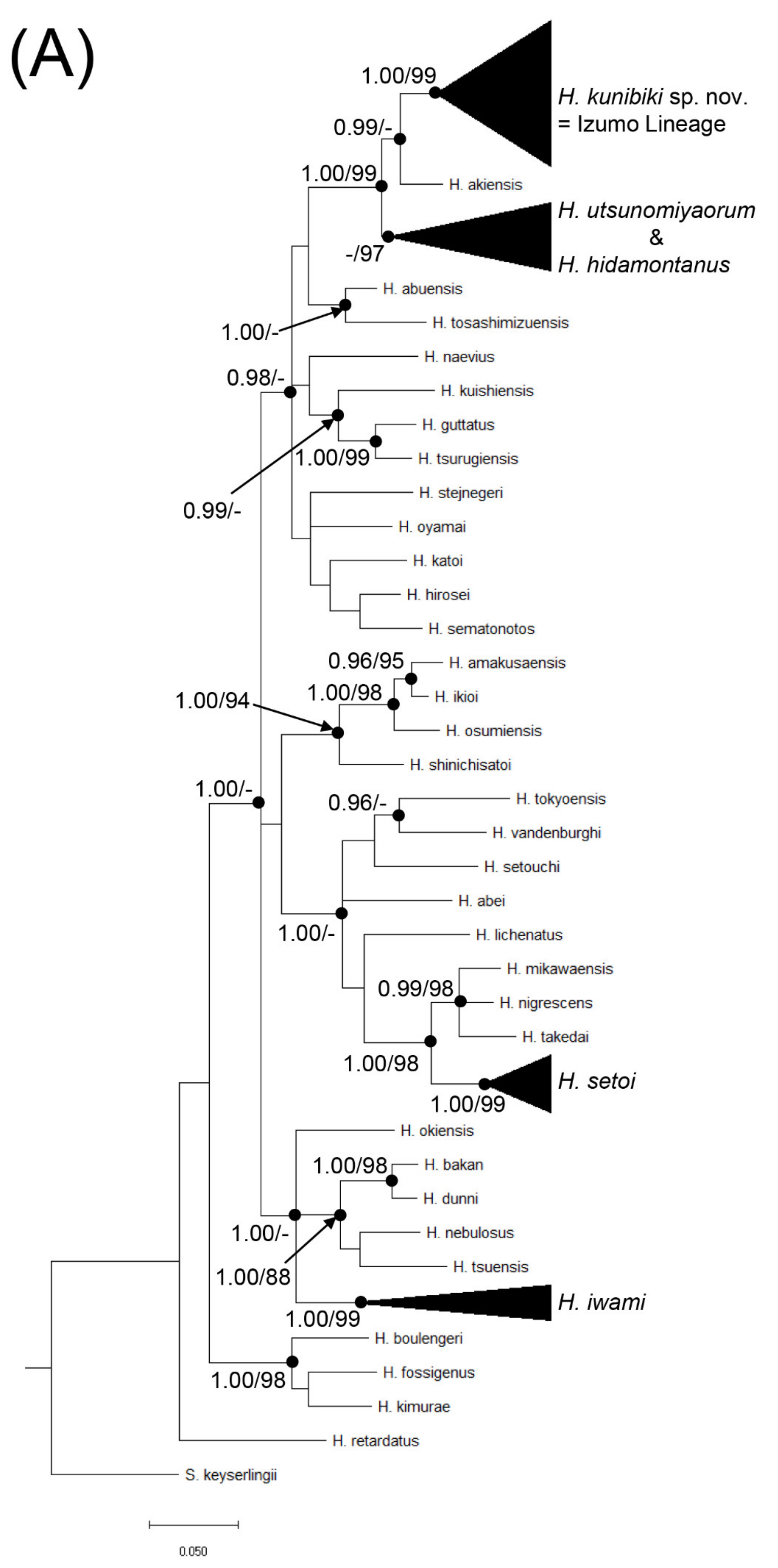
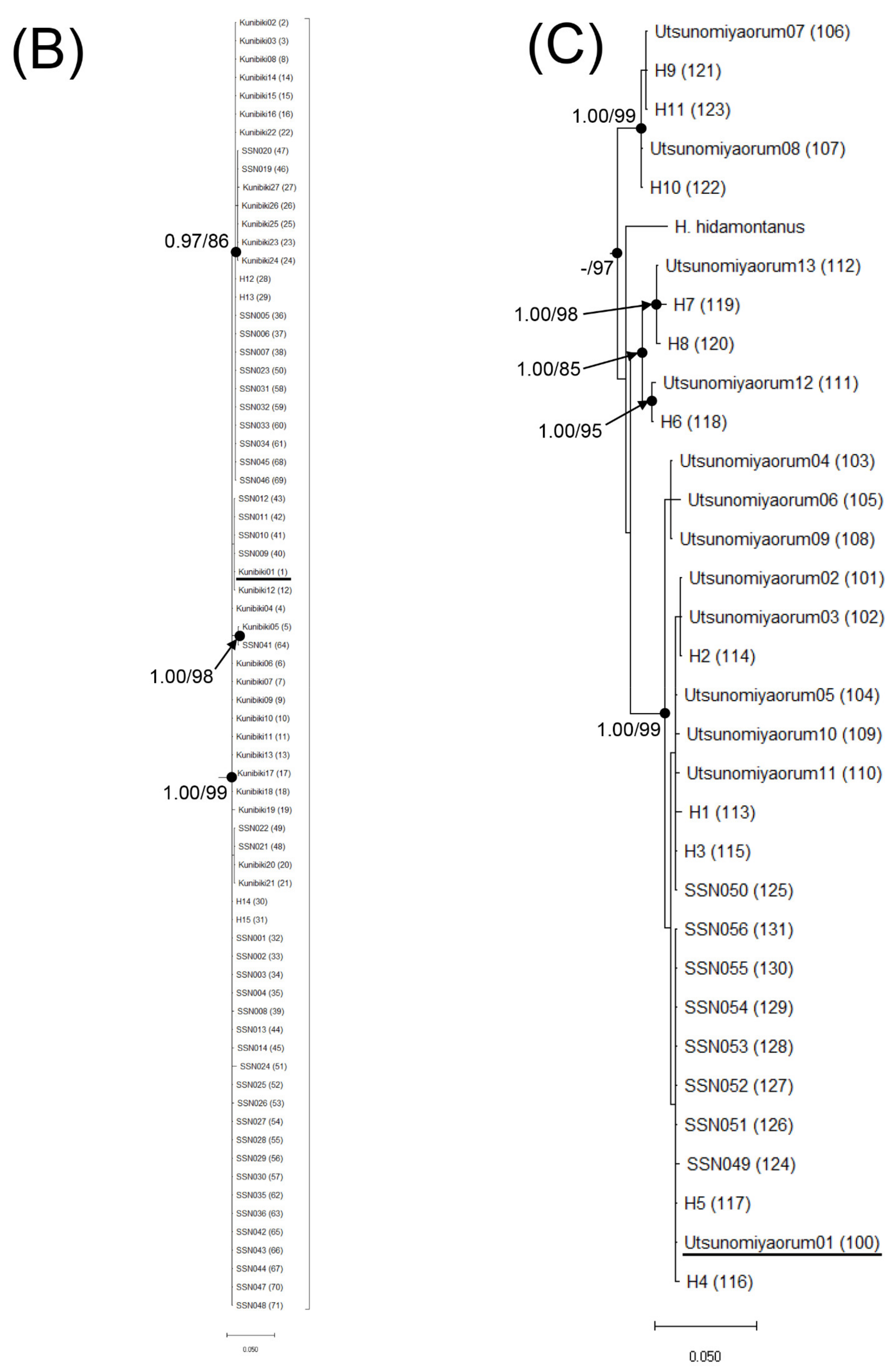
3.1. Molecular Analysis

Phylogenetic reconstructions using BI and ML with the cytochrome b gene recovered nearly identical trees. The Izumo Lineage of H. utsunomiyaorum (Pops. 1–71) and H. utsunomiyaorum (Pops. 100–131) did not form a monophyletic group, whereas the former group and Hynobius akiensis do (Figure 2). Hynobius setoi was largely separated from the two groups (Figure 2); this species formed a monophyletic group which includes Hynobius mikawaensis, Hynobius takedai, and Hynobius nigrescens (Figure 2). Haplotypes of H. setoi were found from Pop. 72 (Tai, Shinonsen Town, Hyogo Prefecture) to Pop. 95 (Higashiizumocho Iya, Matsue City, Shimane Prefecture) (Figure 1 and Figure 2) but were not detected from the northern central part of Shimane Prefecture. In contrast, haplotypes of the Izumo Lineage of H. utsunomiyaorum were found from the central to northern part of Shimane Prefecture (Figure 1 and Figure 2). Indeed, the Izumo Lineage was found from Pop. 14 (Hirosecho Hirose, Yasugi City, Shimane Prefecture) to Pop. 27 (Asayamacho Senyama, Oda City, Shimane Prefecture) (Figure 1 and Figure 2). In addition, haplotypes of H. utsunomiyaorum were detected from Pop. 106 (Nagatani, Iinan Town, Shimane Prefecture) to Pop. 112 (Ogaya, Nishiawakura Village, Okayama Prefecture) (Figure 1 and Figure 2). Finally, haplotypes of H. iwami were newly discovered from Pop. 132 (Asayamacho Asakura, Oda City, Shimane Prefecture) to Pop. 134 (Oda, Sakuraecho, Gotsu City, Shimane Prefecture) (Figure 1 and Figure 2).

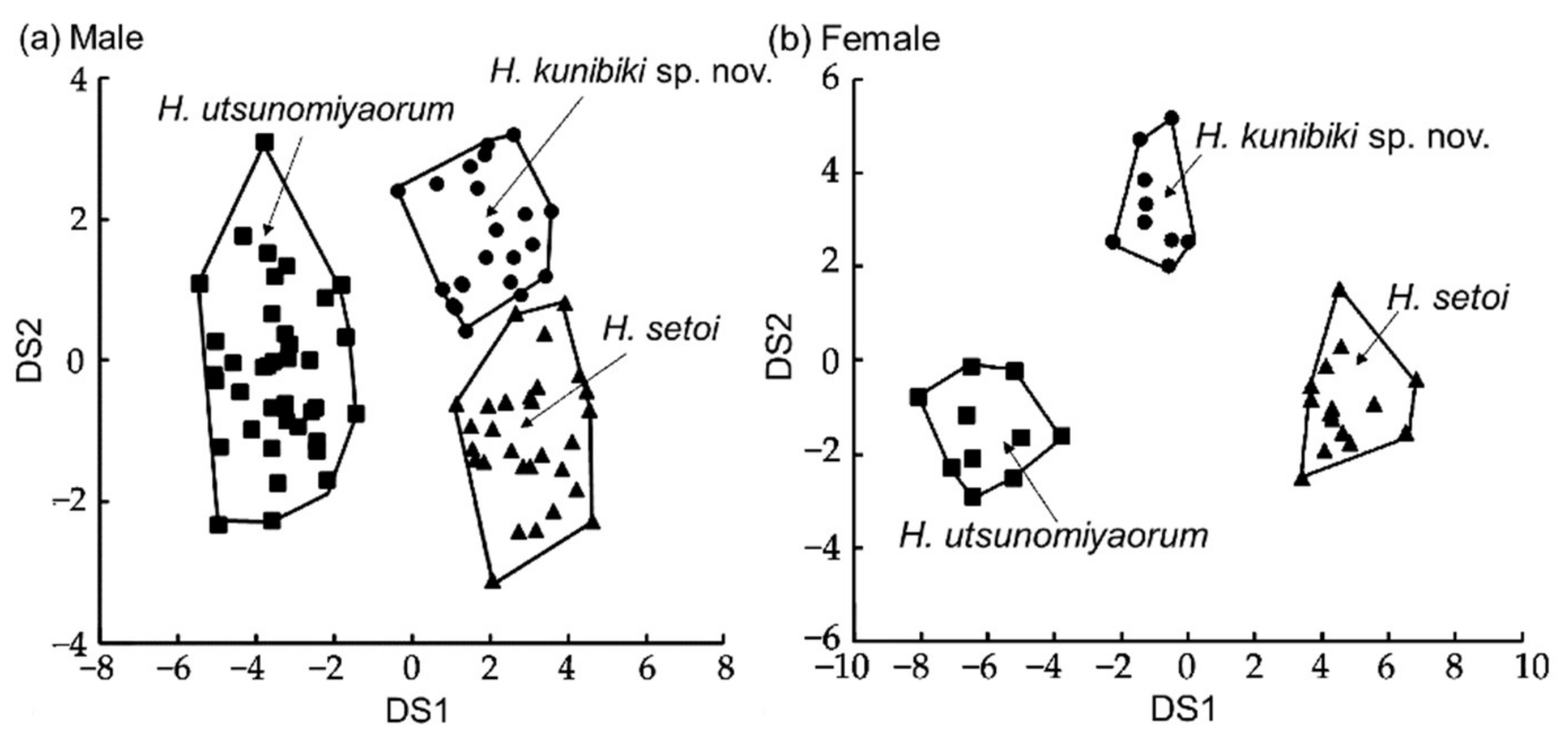
3.2. Morphological Analysis
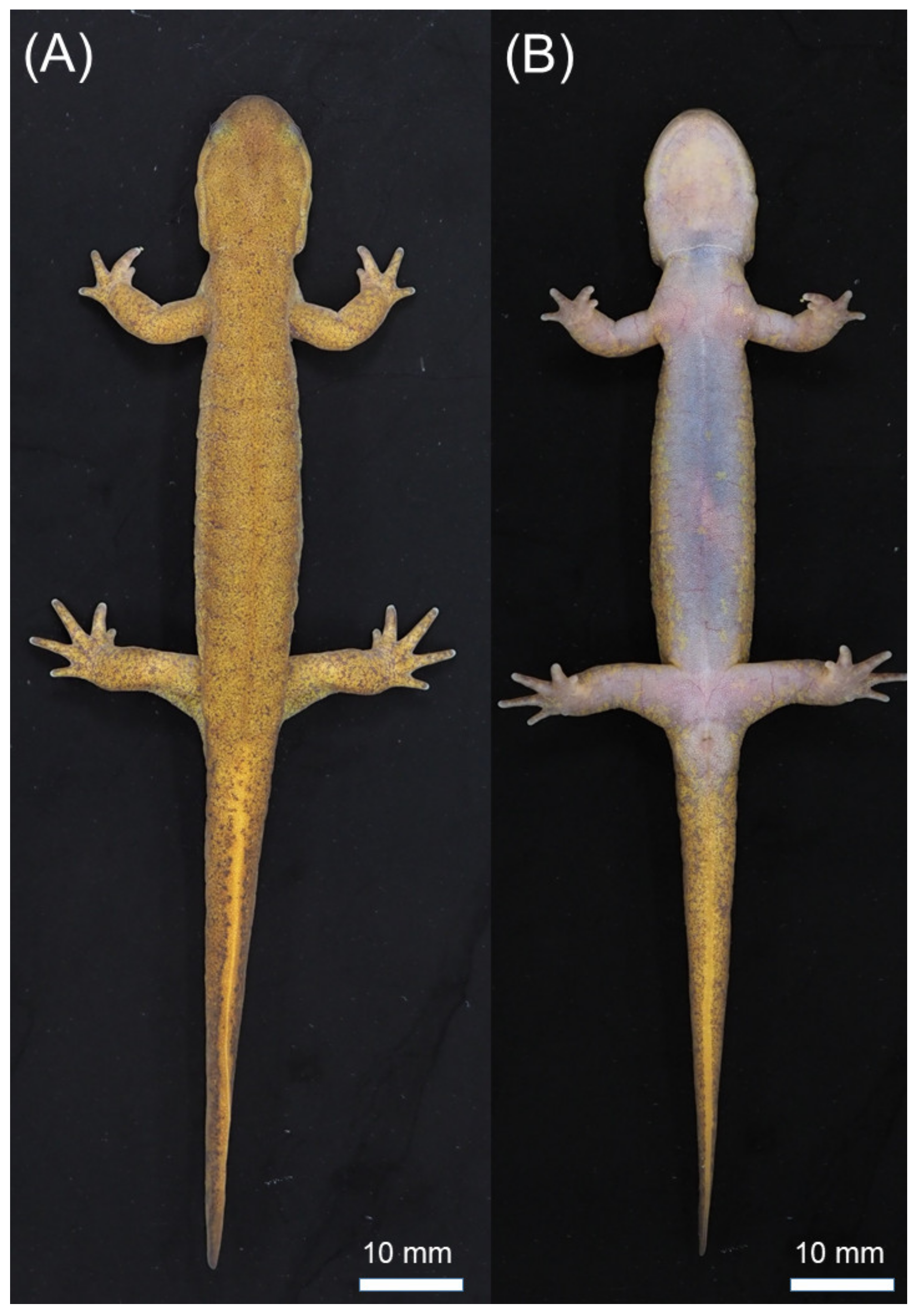
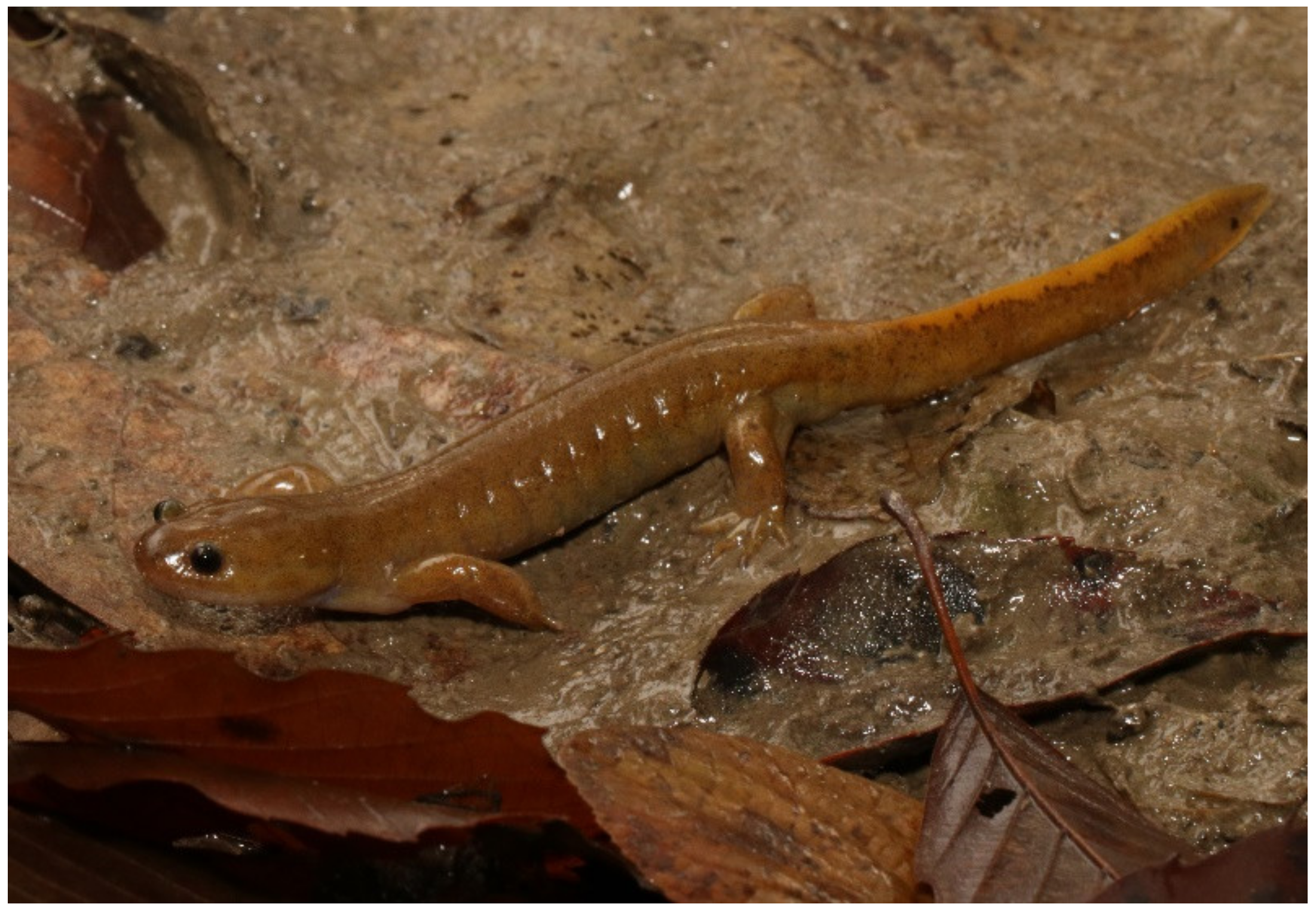
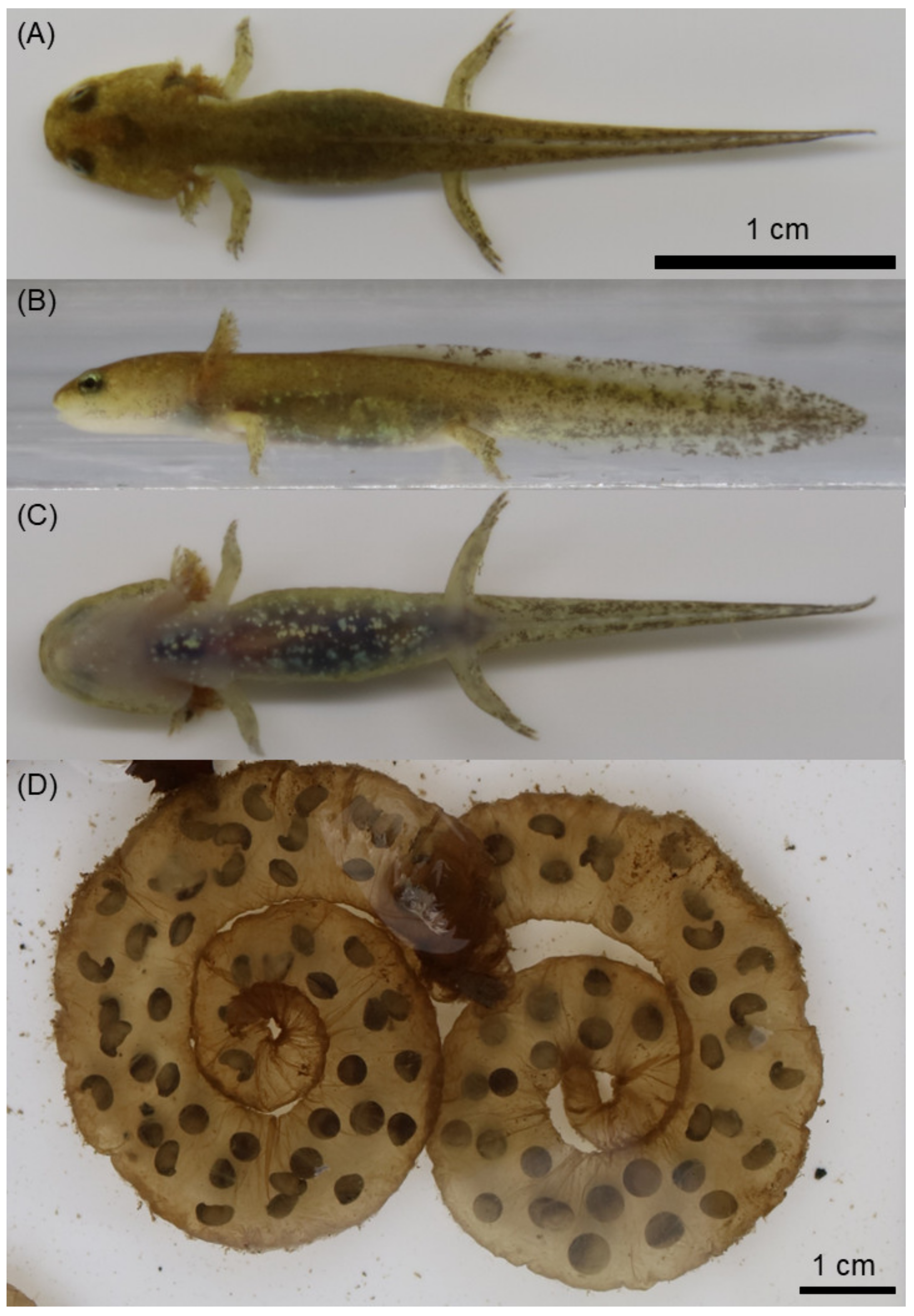
Morphological measurements of the three groups are shown in Table 1. Males of the Izumo Lineage of H. utsunomiyaorum and H. setoi differed significantly in seven morphological characteristics: RTRL (p < 0.05), RHL (p < 0.05), RTAL (p < 0.05), RIND (p < 0.001), RIOD (p < 0.01), RSL (p < 0.0001), and RUEL (p < 0.001). Additionally, females of the Izumo Lineage of H. utsunomiyaorum and H. setoi differed significantly in four morphological characteristics: RVTL (p < 0.01), RFLL (p < 0.05), RIOD (p < 0.05), and RSL (p < 0.01). Furthermore, significant differences were detected between the males of the Izumo Lineage of H. utsunomiyaorum and H. utsunomiyaorum for seven morphological characteristics: SVL (p < 0.0001), RTAL (p < 0.0001), RMTAH (p < 0.0001), RVTL (p < 0.0001), R3FL (p < 0.01), R5TL (p < 0.0001), and RSL (p < 0.01). Significant differences were also detected between the females of the Izumo Lineage of H. utsunomiyaorum and H. utsunomiyaorum for four morphological characteristics: RAGD (p < 0.05), RMTAH (p < 0.05), R3TL (p < 0.05), and R5TL (p < 0.01). Moreover, significant differences existed between the males of H. setoi and H. utsunomiyaorum in 15 morphological characteristics: SVL (p < 0.0001), RTRL (p < 0.01), RHL (p < 0.001), RTAL (p < 0.05), RMTAH (p < 0.0001), RVTL (p < 0.01), RVTW (p < 0.05), R3FL (p < 0.0001), R5TL (p < 0.0001), RIND (p < 0.001), RIOD (p < 0.01), RUEW (p < 0.05), RSL (p < 0.001), RUEL (p < 0.01), and RLJL (p < 0.001). Similarly, significant differences were found between the females of H. setoi and H. utsunomiyaorum in 12 morphological characteristics: SVL (p < 0.01), RAGD (p < 0.05), RHL (p < 0.01), RMTAH (p < 0.001), RVTW (p < 0.05), RFLL (p < 0.05), RHLL (p < 0.01), R2FL (p < 0.05), R3TL (p < 0.01), R5TL (p < 0.0001), RIOD (p < 0.05), and RSL (p < 0.05). Canonical discriminant analyses indicated that the three groups differed according to their males and females (Figure 3). Euclidean distances among the three groups were as follows: Izumo Lineage of H. utsunomiyaorum vs. H. setoi = 4.76 in males and 6.00 in females; Izumo Lineage of H. utsunomiyaorum vs. H. utsunomiyaorum = 11.27 in males and 5.96 in females; and H. setoi vs. H. utsunomiyaorum = 9.84 in males and 10.27 in females.
Results of morphological observations are summarized in Table 2. Males of the Izumo Lineage of H. utsunomiyaorum mostly lacked distinct white spots on the ventral (21/22 = 95.5%) and lateral (21/22 = 95.5%) sides of the body, but they largely possessed a distinct and continuous yellow stripe on the dorsal (22/22 = 100%) side of the tail, a distinct yellow stripe on the ventral (22/22 = 100%) edge of the tail, DGM (18/22 = 81.8%), and 12 costal grooves (18/22 = 81.8%). Females of the Izumo Lineage of H. utsunomiyaorum lacked distinct white spots on the lateral (9/9 = 100%) sides of the body as well as DGM (9/9 = 100%), but they possessed a distinct and continuous yellow stripe on the dorsal (9/9 = 100%) side of the tail and a distinct yellow stripe on the ventral (9/9 = 100%) edge of the tail. Males of H. setoi possessed a distinct and continuous yellow stripe on the dorsal (30/30 = 100%) side of the tail, a distinct yellow stripe on the ventral (30/30 = 100%) edge of the tail, and mostly had 12 costal grooves (24/30 = 80.0%); however, they largely lacked distinct white spots on the ventral (21/30 = 70.0%) and lateral (28/30 = 93.3%) sides of the body. Females of H. setoi possessed a distinct and continuous yellow stripe on the dorsal side of the tail (15/15 = 100%), a distinct yellow stripe on the ventral (15/15 = 100%) edge of the tail, but they lacked DGM (15/15 = 100%), and mostly lacked distinct white spots on the lateral sides of the body (13/15 = 86.7%). Males of H. utsunomiyaorum lacked a distinct yellow stripe on the ventral side of the tail (38/38 = 100%) and mostly lacked a distinct and continuous yellow stripe on the dorsal side of the tail (33/38 = 86.8%) as well as DGM (29/38 = 76.3%); however, they largely possessed distinct white spots on the ventral (30/38 = 78.9%) and lateral (34/38 = 89.5%) sides of the body. Females of H. utsunomiyaorum lacked a distinct yellow stripe on the ventral side of the tail (10/10 = 100%) along with DGM (10/10 = 100%), but they mostly had distinct white spots on the ventral (9/10 = 90.0%) and lateral (9/10 = 90.0%) sides of the body as well as 12 costal grooves (8/10 = 80.0%).
From the perspectives of the three species concepts, we can describe the Izumo Lineage of H. utsunomiyaorum as a new species based on our morphological and molecular analyses.

4. Species Account

Hynobius kunibiki sp. nov.(Figure 4, Figure 5 and Figure 6) Hynobius utsunomiyaorum: Matsui et al. (2019: clade B2b3); Hayashi and Ooi (2020:100). LSID:urn:lsid:zoobank.org:pub:287B9B3A-9E5F-4F6A-BAF6-ADFC3E77B393.
Holotype. An adult male from Nishinomura, Kamionocho, Matsue City, Shimane Prefecture, Chugoku, Japan (35°29′50″ N, 132°55′11″ E; elevation: 100 m above sea level [a.s.l.]; in all cases, datum: WGS84), collected by Takayuki Iwata on 20 December 2019.
Paratype. An adult female from Nishinomura, Kamionocho, Matsue City, Shimane Prefecture, Chugoku, Japan (35° 29′ 50″ N, 132° 55′ 11″ E; elevation: 100 m a.s.l.; in all cases, datum: WGS84), collected by Takayuki Iwata on December 20, 2019. An adult male from Kamocho Chikamatsu, Unnan City, Shimane Prefecture, Chugoku, Japan (35°19′07″ N, 132°55′00″ E; elevation: 90 m a.s.l.; in all cases, datum: WGS84), collected by Takayuki Iwata on January 20, 2021.
Diagnosis. A comparatively large species (with mean SVLs of 58.9 and 58.2 mm in males and females, respectively) within Japanese lentic salamander species complex of Hynobius: distinct yellow stripe on dorsal and ventral edges of tail present; fifth toe of hindlimb present; DWSV absent in adult males (rarely present); distinct white spots on lateral side of body absent in adults (sometimes present); dorsal side yellowish brown to blackish brown; DGM mostly present in males; distinct black spots on dorsum absent in adults (rarely present); V-shaped vomerine teeth series; 12 or 13 (rarely 11) costal grooves; coil-shaped egg sacs.
Comparisons. The new species differs statistically from H. utsunomiyaorum in the following length measurements: SVL, RTAL, RMTAH, RVTL, R3FL, R5TL, and RSL in males; RAGD, RMTAH, R3TL, and R5TL in females; the lengths of these measurements, except for R3TL in females, are significantly longer in H. kunibiki sp. nov. than in H. utsunomiyaorum. The ventral edge of the tail of H. kunibiki sp. nov. has a distinct yellow stripe in both sexes (100%), whereas both sexes of H. utsunomiyaorum do not have this yellow stripe (100%). The dorsal edge of the tail of H. kunibiki sp. nov. has a distinct and continuous yellow stripe in both sexes (100%), whereas H. utsunomiyaorum males mostly do not have this yellow stripe (86.8%). Hynobius kunibiki sp. nov. mostly has no distinct white spots on venter in males (95.5%), but H. utsunomiyaorum males largely have these spots (78.9%). Hynobius kunibiki sp. nov. lacks distinct white spots on the lateral sides of the body in most males (95.5%) and all females (100%), whereas H. utsunomiyaorum males (89.5%) and females (90%) largely have these spots. Hynobius kunibiki sp. nov. mostly has DGM in males (81.8%), but males of H. utsunomiyaorum largely lack this gular mottling (76.3%). Hynobius kunibiki sp. nov. has the fifth toe on the hindlimbs in both sexes (100% in males and females), whereas most individuals of H. utsunomiyaorum do not have this fifth toe (89.5% in males, 70.0% in females). The new species is more morphologically similar to H. setoi than it is to H. utsunomiyaorum (Table 2; Figure 3), but it differs statistically from H. setoi in the following length measurements: RTRL, RHL, RTAL, RIND, RIOD, RSL, and RUEL in males; RVTL, RFLL, RIOD, and RSL in females; the lengths of these measurements, except for RTRL in males, were significantly longer in H. kunibiki sp. nov. than they were in H. setoi. Two lentic Hynobius, H. akiensis and H. iwami, are closely distributed with H. kunibiki sp. nov. The new species differs from H. akiensis by the presence of distinct yellow lines on the dorsal and ventral sides of the tail; it differs from H. iwami by the presence of a fifth toe [3]. Results of molecular analyses (Figure 2) [3] reveal that H. hidamontanus is closely related although its distribution area is largely separated from the habitat of the new species. H. hidamontanus differs from the new species by the absence of the fifth toe [3]. The new species and H. sematonotos are nearly distributed and have relatively closer relationship (Figure 2) in all Japanese Hynobius; however, H. sematonotos differs from the new species by the presence of distinct markings on the dorsum [4].
Description of holotype. A moderately large individual: HL slightly larger than HW; TAL shorter than SVL; body almost cylindrical; rounded snout; gular fold present; tail gradually compressed toward tip; expanded cloaca; webbing between digits absent; four fingers on each forelimb, order of length II = III > IV > I; five toes on each hindlimb, order of length III > IV > II > V > I; V-shaped vomerine teeth; skin smooth and shiny; scattered white spots absent on venter. The holotype had the following measurements (in mm): SVL = 60.0, TRL = 46.7, AGD = 31.2, HL = 13.6, TAL = 49.7, MTAW = 3.5, MTAH = 6.7, BTAW = 6.9, BTAH = 7.1, VTL = 3.2, VTW = 3.4, HW = 10.4, MXHW = 10.7, LFLL = 12.5, RFLL = 11.9, LHLL = 17.3, RHLL = 17.8, L1FL = 0.9, L2FL = 3.1, L3FL = 3.1, L4FL = 1.7, R1FL = 1.2, R2FL = 3.3, R3FL = 3.3, R4FL = 1.8, L1TL = 1.6, L2TL = 3.4, L3TL = 4.9, L4TL = 3.8, L5TL = 1.9, R1TL = 1.4, R2TL = 3.5, R3TL = 4.9, R4TL = 4.2, R5TL = 1.8, IND = 3.4, IOD = 3.9, LUEL = 1.5, RUEL = 1.4, SL = 4.6, LUEW = 2.9, RUEL = 2.7, LJL = 8.2, and CGN = 13.
Variation. Range, mean, and standard deviation of morphometric measurements are presented in Table 1. Morphological variations in skin markings are presented in Table 2. Males had relatively longer RHL (t = 2.69, p < 0.05), RTAL (t = 6.52, p < 0.0001), RMTAW (t = 3.44, p < 0.01), RMTAH (statistic value = −12.93, p < 0.0001), RHW (t = 2.68, p < 0.05), RFLL (t = 2.57, p < 0.05), RHLL (t = 2.12, p < 0.05), RSL (t = 2.58, p < 0.05), and RUEL (t = 3.39, p < 0.01) than females. However, males had relatively shorter RTRL (t = −2.66, p < 0.05) and AGD (t = −5.16, p < 0.0001) than females.
Coloration. Dorsum is uniformly blackish brown or yellowish brown without distinct black spots (rarely present); venter is lighter than dorsum with no distinct white spots in adult males (occasionally present in females); lateral side of head to tail has no distinct white spots (rarely present); iris is dark brown. When preserved, dorsal coloration tended to fade to dark gray.
Etymology. The specific name is derived from “Kunibiki″, with a mythology regarding the formation history of the Shimane Peninsula at which the type locality of the new species is located. The suggested common name in Japanese is Izumo-sanshouo.
Distribution. The new species is endemic to Shimane Prefecture and is known from Oda (including the former Oda City), Yasugi (including the former Hirose Town), Matsue (including the former Matsue City, Higashiizumo, Kashima, Shimane, Shinji, and Tamayu Towns and Yakumo Village), Izumo (including the former Hirata and Izumo Cities and Hikawa, Koryo, Sada, Taki, and Taisha Towns), and Unnan (including the former Daito, Kamo, Kisuki, and Mitoya Towns) Cities in Shimane Prefecture. The dominant vegetation type in the surrounding habitat (Figure 7) is mixed forest of chinquapin (Castanopsis) and live oak (Quercus); the new species breeds in still waters at forest edges.
Larvae and egg sacs. Larvae have distinct black dots on lateral sides of the tail and one pair of balancers during early developmental stages. Claws on the tips of fingers and toes are absent. Egg sacs are coil-shaped. From December to March, adult individuals come to ponds and attach to fallen branches or leaves in still water.
Remarks. The new species is genetically closest to H. akiensis and is not a monophyly with H. utsunomiyaorum based on previous [3] and present results.

5. Discussion

Based on our morphological surveys, H. kunibiki sp. nov. clearly did not satisfy the diagnosis suggested by Matsui et al. [3] in four ways: the fifth toe was always present, clear yellow stripes were always found on the dorsal and ventral sides of the tail, SVL was significantly larger (p < 0.0001), and RTAL was significantly longer (p < 0.0001). In particular, the presence or absence of the distinct yellow line on the ventral side of the tail was absolute and applicable to all individuals of both sexes based on our data (Table 2). Genetically, H. utsunomiyaorum and H. kunibiki sp. nov. are clearly distinguishable and do not form a monophyletic group based both on our data (Figure 2) and that of Matsui et al. [3]. Similarly, the results of phylogenetic analyses using allozyme data based on neighbor-joining and ML methods did not support the monophyly of H. utsunomiyaorum and H. kunibiki sp. nov. [6]. If H. kunibiki sp. nov. is a synonym of H. utsunomiyaorum, polyphyletic species will become valid species. According to Matsui et al. [3], H. kunibiki sp. nov. could not be distinguished from the clade of H. utsunomiyaorum based on small nucleotide polymorphism (SNP) data; hence, Matsui et al. [3] concluded that these two clades were the same species. However, this SNP-based classification is lacking Hynobius hidamontanus despite this species being in the same clade as H. utsunomiyaorum. In addition, this classification was not based on the phylogenetic and morphological species concepts; polyphyletic groups were recognized without detailed morphological comparisons. In contrast, allozyme and mitochondrial data confirm that H. utsunomiyaorum and H. kunibiki sp. nov. are two well-differentiated species [3,6]. Furthermore, H. kunibiki sp. nov. does not satisfy the diagnosis of H. utsunomiyaorum while the two species are clearly distinguishable based on our morphological data (Figure 3; Table 2). Indeed, our data suggest that this new species is more morphologically similar to H. setoi than to H. utsunomiyaorum (especially in males). Thus, H. utsunomiyaorum and H. kunibiki sp. nov. should be distinct species based on the three species concepts.
Individuals from Pop. 31 of Iinan Town (H15) collected by Matsui et al. [3] did not include the haplotype of H. utsunomiyaorum but did contain the haplotype of H. kunibiki sp. nov. (Figure 1 and Figure 2). However, this population is distantly isolated from other populations of H. kunibiki sp. nov. (Figure 1) and our phylogenetic analyses, including samples from Iinan Town (Pops. 106 and 107), did not support the conclusion that the new species was distributed in Iinan Town (Figure 2). Furthermore, detailed information for the voucher specimen from Iinan Town has not been provided here or elsewhere [3]. Thus, the new species we have described may not be distributed in Iinan Town; rather, the distribution range of this species may be limited to the northeastern part of Shimane Prefecture (Figure 1). Hynobius kunibiki sp. nov. is parapatrically distributed with H. setoi and H. iwami (Figure 1), but analyses of nuclear and mitochondrial DNA suggest that H. kunibiki sp. nov. is clearly separated from these two species genetically (Figure 2) [3]. Additionally, H. iwami has no fifth toe; thus, it is clearly separated from H. kunibiki sp. nov. both genetically and morphologically [3].
According to Matsui et al. [3], H. setoi is found at the lowland areas along the Japan Sea from northwestern Hyogo Prefecture to the northeastern part of Shimane Prefecture. Based on our analyses, haplotypes of H. setoi were found from the westernmost part of Hyogo, Tottori, from west to east, and the easternmost part of Shimane Prefectures; however, these haplotypes were not detected from around the northeastern part of Shimane Prefecture (Figure 1 and Figure 2). Similar to our findings, Hayashi and Ooi [5] could not find the haplotypes of H. setoi in the northeastern part of Shimane Prefecture, so individuals of H. setoi from Daitocho Sannoji, Shinobuchi, and Hirosecho Shinoyamasa surveyed by Matsui et al. [3] are unlikely to be H. setoi; thus, the analyses of Matsui et al. [3] may have combined two different species. Consequently, the diagnosis of H. setoi should be carefully applied when attempting to identify this species. Actually, the mean SVL, FLL/SVL, and HLL/SVL suggested by Matsui et al. [3] did not differ significantly between H. setoi and H. kunibiki sp. nov., and both species possessed distinct yellow stripes on the dorsal and ventral sides of their tails (Table 2); therefore, these two species cannot be separated according to the morphology described in the diagnosis of H. setoi by Matsui et al. [3]. In fact, male H. kunibiki sp. nov. were closer to H. setoi than to H. utsunomiyaorum in terms of Euclidean distance, despite H. kunibiki sp. nov. having a closer genetic relationship with H. utsunomiyaorum than it has with H. setoi (Figure 2). Males of H. kunibiki sp. nov. and H. setoi differed significantly in terms of RTRL, RHL, RTAL, RIND, RIOD, RSL, and RUEL; hence, these morphological characteristics will be a useful reference by which to distinguish the two species.
Consistent with the results of Matsui et al. [3], our phylogenetic analyses suggested that H. hidamontanus is a synonym of H. utsunomiyaorum (Figure 2). However, we could not collect samples of H. hidamontanus because it is strictly protected by law in Japan. Therefore, a morphological survey of this species will be required for its synonymization. In the present study, the entire distribution ranges of H. kunibiki sp. nov. and H. setoi were revealed in detail (Table S1; Figure 1), whereas the distribution ranges of H. utsunomiyaorum from the central and eastern part of Hiroshima Prefecture and H. iwami from the western part of Shimane Prefecture were not revealed in detail. Thus, additional research will be required to determine the distribution areas of the latter two species.
We conducted phylogenetic analyses using all Japanese Hynobius species (Figure 2). According to Dubois and Raffaëlli [19], Japanese Hynobius are divided into five supraspecies (i.e., hidamontanus, lichenatus, naevius, nebulosus, and stejnegeri); however, supraspecies hidamontanus and naevius cannot be distinguished genetically (Figure 2). If subgenera of Hynobius were to be divided by supraspecies, four supraspecies would be recognized based on BI: the Hynobius nebulosus supraspecies (including H. bakan, H. dunni, H. iwami, H. nebulosus, H. okiensis, and H. tsuensis), Hynobius lichenatus supraspecies (containing H. abei, H. lichenatus, H. mikawaensis, H. nigrescens, H. setoi, H. setouchi, H. takedai, H. tokyoensis, and H. vandenburghi), Hynobius shinichisatoi supraspecies (including H. amakusaensis, H. ikioi, H. osumiensis, and H. shinichisatoi), and Hynobius naevius supraspecies (comprising H. abuensis, H. akiensis, H. guttatus, H. hidamontanus, H. hirosei, H. katoi, H. kuishiensis, H. kunibiki sp. nov., H. naevius, H. oyamai, H. sematonotos, H. stejnegeri, H. tosashimizuensis, H. tsurugiensis, and H. utsunomiyaorum). Dubois and Raffaëlli [19] used the term supraspecies, but the common morphological characteristics of each supraspecies have not yet been evaluated to the best of our knowledge; hence, this term should not be used without an initial morphological definition. If the supraspecies are valid, further morphological studies among the groups are necessary.
Following this description, the number of Japanese Hynobius spp. is 38, but some taxonomic problems have been left unsolved. Matsui et al. [3] suggested that H. akiensis and H. nebulosus are not monophyletic species, and they may be divided into two species. Further, the study only described male morphology; thus, female morphology of many Hynobius species remains unidentified. The task of clarifying the distribution ranges of lentic Hynobius also needs to be undertaken as detailed distribution ranges of H. setouchi, H. akiensis, and H. vandenburghi are still unknown. Future studies involving taxonomic reexamination and field surveys to clarify the distribution ranges of both sexes of the Japanese Hynobius species are required.
The localities of the four species used in this study have been provided in detail, which will support conservation activities. In particular, H. kunibiki sp. nov. and H. setoi are morphologically similar, so definitive identification of these species will be difficult where DNA data is unavailable. The information in this study is expected to be useful for confirming the distribution ranges of these two species. However, it is possible that overcollection could decimate the Hynobius species following the release of detailed locality information. Indeed, overfishing of H. utsunomiyaorum has already been recognized [20]. Therefore, the conservation status of H. kunibiki sp. nov. must be reassessed, and management plans for its conservation and regulation by national or regional administrations are immediately required to prevent overcollection and possible extinction.

6. Conclusions

The monophyly of the Izumo Lineage of Hynobius utsunomiyaorum and H. utsunomiyaorum was rejected by the molecular evidence. Besides the genetic evidence, the Izumo Lineage of H. utsunomiyaorum did not satisfy the diagnosis from the original description of H. utsunomiyaorum in morphology. Furthermore, significant morphological differences between them were detected in both sexes, so we described the Izumo Lineage of H. utsunomiyaorum as H. kunibiki based on morphological, phylogenetic, and evolutionary species concepts. This new species is limited to the northern part of Shimane Prefecture, and the conservation status should be reassessed for appropriate conservation activities.

Supplementary Materials

The following are available online at https://0-www-mdpi-com.brum.beds.ac.uk/article/10.3390/ani11082187/s1, Table S1. List of specimens using molecular analyses. Population number corresponds to the localities (in Figure 1) where the salamanders were collected. Asterisks after the sampling localities show the type locality of each species.

Author Contributions

Conceptualization, H.S.; Data curation, M.N.; Formal analysis, H.S.; Funding acquisition, H.S., T.I., H.Y. and M.N.; Investigation, T.I. and H.Y.; Methodology, H.S.; Project administration, H.S.; Resources, T.I. and H.Y.; Software, H.S.; Supervision, M.N.; Validation, H.S.; Visualization, H.S.; Writing—original draft, H.S.; Writing—review & editing, H.S. All authors have read and agreed to the published version of the manuscript.

Funding

This research was funded by collaborative research Oita University and TrustBio, grant number 1020CC2017H01003. The APC was funded by the same founder.

Institutional Review Board Statement

This project (also experiments) was performed with the permission from the Oita University, and the code and date of approval given by the university are HKT01 and 28 April 2017, respectively.

Informed Consent Statement

Not applicable.

Data Availability Statement

The data presented in this paper are available on request from the corresponding author.

Acknowledgments

We thank Jun-ichi Naito, Masaru Ishikura, Toshiaki Iwata, and Kazue Iwata for help with field sampling; Nobue Akai, Maki Kirihara, and So Hamada for providing information about the habitat of the species used in this study; and Kei Ichisawa of Tottori Prefectural Museum and Kiyoshi Hagiwara of Yokosuka City Museum, who supported the registration of the type specimens.

Conflicts of Interest

The authors declare no conflict of interest.

References

  1. Frost, D.R. Amphibian Species of the World: An Online Reference; American Museum of Natural History: New York, NY, USA, 2021; Available online: http://research.amnh.org/herpetology/amphibia/index.html (accessed on 15 February 2021).
  2. Matsui, M.; Nishikawa, K.; Misawa, Y.; Kakegawa, M.; Sugahara, T. Taxonomic relationships of an endangered Japanese salamander Hynobius hidamontanus Matsui, 1987 with H. tenuis Nambu, 1991 (Amphibia: Caudata). Curr. Herpetol. 2002, 21, 25–34. [Google Scholar] [CrossRef]
  3. Matsui, M.; Okawa, H.; Nishikawa, K.; Aoki, G.; Eto, K.; Yoshikawa, N.; Tanabe, S.; Misawa, Y.; Tominaga, A. Systematics of the widely distributed Japanese clouded salamander, Hynobius nebulosus (Amphibia: Caudata: Hynobiidae), and Its Closest Relatives. Curr. Herpetol. 2019, 38, 32–90. [Google Scholar] [CrossRef]
  4. Tominaga, A.; Matsui, M.; Nishikawa, K. Two new species of lotic breeding Salamanders (Amphibia, Caudata, Hynobiidae) from Western Japan. Zootaxa 2019, 4550, 525–544. [Google Scholar] [CrossRef] [PubMed]
  5. Hayashi, M.; Ooi, K. Molecular Study on Hynobius in East Area of Shimane Prefecture, Japan: Notes on Haplotype Diversity of mtDNA and Their Distribution. Bull. Hoshizaki Green Found. 2020, 23, 99–104. (In Japanese) [Google Scholar]
  6. Matsui, M.; Nishikawa, K.; Utsunomiya, T.; Tanabe, S. Geographic allozyme variation in the Japanese clouded Salamander, Hynobius nebulosus (Amphibia: Urodela). Biol. J. Linn. Soc. 2006, 89, 311–330. [Google Scholar] [CrossRef] [Green Version]
  7. Dubois, A. The International code of zoological nomenclature must be drastically improved before it is too late. Bionomina 2011, 2, 1–104. [Google Scholar] [CrossRef] [Green Version]
  8. Frost, D.R.; Hillis, D.M. Species in concept and practice: Herpetological applications. Herpetologica 1990, 46, 87–104. [Google Scholar]
  9. Matsui, M.; Tominaga, A.; Hayashi, T.; Misawa, Y.; Tanabe, S. Phylogenetic Relationship and Phylogeography of Hynobius tokyoensis (Amphibia: Caudata) using complete sequences of cytochrome b and control reason genes of mitochondrial DNA. Mol. Phylogenet. Evol. 2007, 44, 204–216. [Google Scholar] [CrossRef] [PubMed]
  10. Kumar, S.; Stecher, G.; Li, M.; Knyaz, C.; Tamura, K. MEGA X: Molecular evolutionary genetics analysis across computing platforms. Mol. Biol. Evol. 2018, 35, 1547–1549. [Google Scholar] [CrossRef] [PubMed]
  11. Darriba, D.; Taboada, G.L.; Doallo, R.; Posada, D. JModelTest 2: More models, new heuristics and parallel computing. Nat. Methods 2012, 9, 772. [Google Scholar] [CrossRef] [PubMed] [Green Version]
  12. Sugiura, N. Further analysts of the data by akaike’s information criterion and the finite corrections. Commun. Stat. Theor. Methods 1978, 7, 13–26. [Google Scholar] [CrossRef]
  13. Schwarz, G. Estimating the dimension of a model. Ann. Statist. 1978, 6, 461–464. [Google Scholar] [CrossRef]
  14. Ronquist, F.; Teslenko, M.; van der Mark, P.; Ayres, D.L.; Darling, A.; Höhna, S.; Larget, B.; Liu, L.; Suchard, M.A.; Huelsenbeck, J.P. MrBayes 3.2: Efficient Bayesian phylogenetic inference and model choice across a large model space. Syst. Biol. 2012, 61, 539–542. [Google Scholar] [CrossRef] [PubMed] [Green Version]
  15. Huelsenbeck, J.P.; Rannala, B. Frequentist properties of Bayesian posterior probabilities of phylogenetic trees under simple and complex substitution models. Syst. Biol. 2004, 53, 904–913. [Google Scholar] [CrossRef] [PubMed] [Green Version]
  16. Regier, J.C.; Mitter, C.; Solis, M.A.; Hayden, J.E.; Landry, B.; Nuss, M.; Simonsen, T.J.; Yen, S.H.; Zwick, A.; Cummings, M.P. A molecular phylogeny for the pyraloid moths (Lepidoptera: Pyraloidea) and its implications for higher-level classification. Syst. Entomol. 2012, 37, 635–656. [Google Scholar] [CrossRef]
  17. Bennett, R.A. A Review of anesthesia and chemical restraint in reptiles. J. Zoo Wildl. Med. 1991, 22, 282–303. [Google Scholar]
  18. Ihaka, R.; Gentleman, R. R: A language for data analysis and graphics. J. Comp. Graph. Stat. 1996, 5, 299–314. [Google Scholar]
  19. Dubois, A.; Raffaëlli, J. A new ergotaxonomy of the order Urodela Duméril, 1805 (Amphibia, Batrachia). Alytes 2012, 28, 77–161. [Google Scholar]
  20. Fujita, H.; Iwata, T.; Teraoka, S. Damage example of eggs of Hynobius nebulosus (high ground type) by overfishing in Eastern Shimane Prefecture. Bull. Hoshizaki Green Found. 2016, 19, 253–255. (In Japanese) [Google Scholar]
Figure 1. Localities for populations sampled in northern Chugoku, Japan. Population numbers match those used for molecular analyses (see Table S1 and Figure 2). Lower mapped area includes the northern part of Chugoku (Tottori, central to eastern part of Shimane, northern part of Okayama, and northeastern part of Hiroshima Prefectures) and northwestern part of Kinki (Hyogo Prefecture). Top left magnified area (a) includes the westernmost part of Shimane Prefecture; (b) includes the north central part of Shimane Prefecture; (c) includes the easternmost part of Shimane Prefecture; (d) includes the eastern part of Tottori Prefecture. The closed symbols correspond to each of four species sampled in this study: H. kunibiki sp. nov. (closed circles), H. setoi (closed triangles), H. utsunomiyaorum (closed squares), and H. iwami (closed stars). The open symbols correspond to each of four species cited from Matsui et al. [3]: H. kunibiki sp. nov. (open circle), H. setoi (open triangle), H. utsunomiyaorum (open squares), and H. iwami (open stars). The open symbols including diagonal lines correspond to each of four species cited from Matsui et al. [3] or Hayashi and Ooi [5] for which the detailed locality is unclear: H. kunibiki sp. nov. (shadow circles), H. setoi (shadow triangles), H. utsunomiyaorum (shadow squares), and H. iwami (shadow stars). For the morphological comparisons, individuals of H. kunibiki sp. nov., H. setoi, and H. utsunomiyaorum were sampled from several localities including all major fragmented distribution areas: H. kunibiki sp. nov. from Pops. 1 (male = 14, female = 6), 2 (male = 2, female = 0), 3 (male = 1, female = 1), 8 (male = 2, female = 0), 17 (male = 2, female = 1), 22 (male = 1, female = 0), and 23 (male = 0, female = 1); H. setoi from Pops. 75 (male = 9, female = 4), 76 (male = 8, female = 2), 81 (male = 5, female = 1), 82 (male = 2, female = 2), 84 (male = 7, female = 5), and 87 (male = 0, female = 1); and H. utsunomiyaorum from Pops. 100 (male = 24, female = 7), 102 (male = 5, female = 1), 111 (male = 4, female = 1), and 112 (male = 5, female = 1). Underlined labels (Pops. 1, 75, 100, and 135) show the type locality of each species.
Animals 11 02187 g001
Figure 2. Bayesian inference phylogenetic tree based on 630-base pair (bp) cytochrome b sequences rooted with Salamandrella keyserlingii. Five phylogenies indicate that phylogenetic relationships of (A) all Japanese Hynobius species, (B) within H. kunibiki sp. nov., (C) within H. utunomiyaorum (also H. hidamontanus), (D) within H. setoi, and (E) within H. iwami, respectively. Scale bars indicate the genetic distance (expected changes per site). Numbers near nodes indicate the supported level based on Bayesian posterior probabilities (BPP) and bootstrap values (BSV), respectively. Underlined labels indicate the type locality of the three species and H. iwami. Numbers appearing in parentheses after the labels correspond to population localities as indicated in Table S1 and Figure 1.
Animals 11 02187 g002aAnimals 11 02187 g002bAnimals 11 02187 g002c
Figure 3. Two-dimensional plots based on canonical discriminant analysis. The x and y axes indicate discriminant score 1 (DS1) and discriminant score 2 (DS2), respectively. The contribution ratios of DS1 and DS2 in males and females were as follows: DS1: 88.90% for males, 83.96% for females; DS2: 11.10% for males, 16.04% for females. Circles, triangles, and squares indicate scores for individuals from the type localities of Hynobius kunibiki sp. nov., H. setoi, and H. utsunomiyaorum, respectively.
Animals 11 02187 g003
Figure 4. Holotype of Hynobius kunibiki sp. nov. (TRPM-ARA-0000014, adult male): (A) dorsal and (B) ventral views.
Animals 11 02187 g004
Figure 5. Live holotype of Hynobius kunibiki sp. nov. (TRPM-ARA-0000014, adult male).
Animals 11 02187 g005
Figure 6. Larva of Hynobius kunibiki sp. nov.: (A) dorsal, (B) lateral, and (C) ventral sides, as well as (D) egg sacs.
Animals 11 02187 g006
Figure 7. Habitat at the type locality of Hynobius kunibiki sp. nov. in Nishinomura, Kamionocho, Matsue City, Shimane Prefecture.
Animals 11 02187 g007
Table 1. Measurements (mm) of snout–vent length (SVL) and character ratios (R = %SVL) of trunk length (TRL) to lower jaw length (LJL) of the three species of Hynobius collected in 2018–2021 (n = number of individuals). Values are reported as means ± standard deviation. Ranges are shown in parentheses. See Materials and Methods section for definitions of morphological traits.
H. kunibiki sp. nov.H. setoiH. utsunomiyaorum
MaleFemaleMaleFemaleMaleFemale
Traitn = 22n = 9n = 30n = 15n = 38n = 10
SVL58.9 ± 3.1558.2 ± 4.2460.7 ± 6.3763.0 ± 6.1653.4 ± 4.1054.6 ± 4.19
(52.7–63.9)(51.0–66.6)(49.0–70.8)(50.5–72.5)(40.0–59.8)(48.0–60.5)
RTRL76.5 ± 1.0077.6 ± 1.2477.4 ± 1.0077.7 ± 2.2076.7 ± 1.6577.0 ± 0.62
(75.7–78.4)(75.6–79.2)(75.7–80.1)(71.0–80.8)(74.3–84.8)(76.2–77.8)
RAGD51.3 ± 2.0655.7 ± 2.3552.3 ± 1.7255.5 ± 2.6352.1 ± 1.6852.8 ± 2.27
(47.7–54.9)(50.8–59.2)(48.1–55.9)(50.7–59.1)(48.9–56.5)(49.0–55.9)
RHL24.2 ± 0.8423.3 ± 0.9123.5 ± 0.9923.0 ± 1.1024.4 ± 1.0324.2 ± 0.73
(22.6–25.7)(21.8–24.6)(22.0–26.1)(21.2–24.8)(22.0–26.5)(22.6–25.0)
RTAL82.0 ± 4.0971.3 ± 4.2878.3 ± 6.1569.5 ± 5.4773.1 ± 7.2870.5 ± 6.51
(74.1–93.1)(62.8–76.4)(67.5–94.7)(59.7–77.0)(52.9–86.0)(58.8–81.4)
RMTAW6.9 ± 0.845.8 ± 0.766.6 ± 1.016.5 ± 0.876.7 ± 0.966.6 ± 0.59
(5.5–8.3)(4.9–7.4)(4.8–8.8)(4.8–7.7)(4.4–8.8)(5.7–7.5)
RMTAH13.0 ± 0.9510.8 ± 1.3813.0 ± 1.3611.3 ± 1.1710.2 ± 1.379.3 ± 0.75
(11.4–14.6)(7.5–12.2)(10.8–15.9)(9.1–13.7)(6.8–12.9)(8.3–10.6)
RVTL5.1 ± 0.465.3 ± 0.504.9 ± 0.484.6 ± 0.634.5 ± 0.504.7 ± 0.38
(4.3–6.1)(4.3–5.9)(3.9–6.1)(3.5–5.6)(3.6–5.9)(4.1–5.4)
RVTW5.4 ± 0.515.4 ± 0.485.4 ± 0.475.1 ± 0.615.7 ± 0.505.6 ± 0.28
(4.4–6.3)(4.7–6.0)(4.4–6.1)(4.3–6.4)(4.7–6.7)(5.0–6.0)
RHW17.7 ± 0.6217.0 ± 0.8717.5 ± 0.8716.6 ± 0.7717.3 ± 0.7316.7 ± 0.71
(16.6–19.2)(15.8–18.2)(16.2–19.0)(15.3–17.6)(15.7–18.8)(15.5–18.2)
RFLL25.5 ± 1.4924.0 ± 1.4524.8 ± 1.7122.3 ± 0.9224.8 ± 1.6925.0 ± 2.50
(23.1–29.5)(21.7–26.1)(22.6–30.6)(20.7–24.2)(21.0–27.9)(21.1–28.8)
RHLL32.4 ± 1.6031.0 ± 1.8732.1 ± 1.3129.4 ± 1.7531.8 ± 1.3031.8 ± 1.50
(29.9–35.2)(27.7–34.8)(29.5–36.3)(25.9–31.9)(29.3–35.6)(29.3–34.2)
R2FL5.2 ± 0.624.7 ± 0.694.9 ± 0.714.3 ± 0.685.1 ± 0.714.9 ± 0.65
(3.9–6.3)(4.1–6.0)(2.9–6.5)(3.1–5.5)(2.9–6.6)(4.0–5.9)
R3FL4.1 ± 0.543.7 ± 0.874.4 ± 0.674.0 ± 0.583.6 ± 0.673.6 ± 0.58
(2.5–4.9)(2.3–5.0)(2.8–5.5)(3.0–5.1)(1.4–4.9)(2.8–4.7)
R3TL7.9 ± 0.647.5 ± 0.618.0 ± 0.567.3 ± 0.848.3 ± 0.828.4 ± 0.59
(7.0–9.1)(6.4–8.3)(7.0–9.1)(5.9–9.1)(5.5–10.4)(7.4–9.4)
R5TL1.9 ± 0.621.8 ± 0.692.2 ± 0.622.3 ± 0.690.2 ± 0.480.3 ± 0.48
(0.3–3.0)(0.5–2.6)(1.1–3.3)(1.1–3.5)(0.0–2.2)(0.0–1.2)
RIND5.3 ± 0.574.9 ± 0.524.7 ± 0.484.6 ± 0.295.2 ± 0.454.9 ± 0.67
(3.9–6.3)(4.2–6.1)(3.7–5.7)(4.1–5.2)(4.4–6.1)(4.3–6.3)
RIOD6.4 ± 0.516.0 ± 0.375.9 ± 0.635.6 ± 0.386.3 ± 0.386.1 ± 0.46
(5.3–7.5)(5.6–6.6)(4.9–7.1)(5.0–6.3)(5.4–6.9)(5.5–7.0)
RUEL3.2 ± 0.253.2 ± 0.193.2 ± 0.303.2 ± 0.283.3 ± 0.283.3 ± 0.27
(2.8–3.8)(2.9–3.4)(2.5–3.9)(2.8–3.8)(2.8–3.9)(2.9–3.8)
RSL6.7 ± 0.326.4 ± 0.356.0 ± 0.345.7 ± 0.296.4 ± 0.386.1 ± 0.53
(6.1–7.3)(5.7–6.8)(5.2–6.7)(5.3–6.3)(5.4–7.4)(5.0–6.6)
RUEW4.6 ± 0.204.3 ± 0.294.4 ± 0.284.2 ± 0.294.6 ± 0.374.4 ± 0.25
(4.2–5.1)(3.9–4.7)(3.8–4.9)(3.7–5.0)(4.1–5.5)(4.1–4.9)
RLJL13.9 ± 0.6913.7 ± 0.6913.4 ± 0.7513.1 ± 0.8514.1 ± 0.7313.4 ± 0.61
(12.9–15.7)(13.1–15.4)(11.5–14.9)(11.5–14.6)(12.7–16.4)(12.2–14.1)
Table 2. Characteristics of skin markings among the three species of Hynobius. The values indicate the number of individuals exhibiting that characteristic with percentages related to each condition in parentheses. See Materials and Methods section for definitions of morphological characteristics.
H. kunibiki sp. nov.H. setoiH. utsunomiyaorum
MaleFemaleMaleFemaleMaleFemale
CharacterConditionn = 22n = 9n = 30n = 15n = 38n = 10
DWSVAbsent21 (95.5%)5 (55.6%)21 (70.0%)7 (46.7%)8 (21.1%)1 (10.0%)
Present1 (4.5%)4 (44.4%)9 (30.0%)8 (53.3%)30 (78.9%)9 (90.0%)
DWSLAbsent21 (95.5%)9 (100%)28 (93.3%)13 (86.7%)4 (10.5%)1 (10.0%)
Present1 (4.5%)0 (0%)2 (6.7%)2 (13.3%)34 (89.5%)9 (90.0%)
DCTYLDAbsent0 (0%)0 (0%)0 (0%)0 (0%)33 (86.8%)6 (60.0%)
Present22 (100%)9 (100%)30 (100%)15 (100%)5 (13.2%)4 (40.0%)
DTYLVAbsent0 (0%)0 (0%)0 (0%)0 (0%)38 (100%)10 (100%)
Present22 (100%)9 (100%)30 (100%)15 (100%)0 (0%)0 (0%)
DGMAbsent4 (18.2%)9 (100%)14 (46.7%)15 (100%)29 (76.3%)10 (100%)
Present18 (81.8%)0 (0%)16 (53.3%)0 (0%)9 (23.7%)0 (0%)
CGN111 (4.5%)1 (11.1%)2 (6.7%)0 (0%)7 (18.4%)1 (10.0%)
1218 (81.8%)4 (44.4%)24 (80.0%)7 (46.7%)23 (60.5%)8 (80.0%)
133 (13.6%)4 (44.4%)4 (13.3%)8 (53.3%)8 (21.1%)1 (10.0%)
CFBALN2.00 (0%)0 (0%)1 (3.3%)0 (0%)0 (0%)0 (0%)
1.51 (4.5%)0 (0%)0 (0%)0 (0%)0 (0%)0 (0%)
1.05 (22.7%)0 (0%)2 (6.7%)0 (0%)3 (7.9%)0 (0%)
0.54 (18.2%)2 (22.2%)2 (6.7%)0 (0%)7 (18.4%)1 (10.0%)
0.06 (27.3%)0 (0%)8 (26.7%)0 (0%)9 (23.7%)3 (30.0%)
−0.54 (18.2%)0 (0%)5 (16.7%)0 (0%)9 (23.7%)1 (10.0%)
−1.02 (9.1%)3 (33.3%)5 (16.7%)0 (0%)8 (21.1%)3 (30.0%)
−1.50 (0%)0 (0%)5 (16.7%)2 (13.3%)1 (2.6%)1 (10.0%)
−2.00 (0%)3 (33.3%)2 (6.7%)8 (53.3%)1 (2.6%)1 (10.0%)
−2.50 (0%)0 (0%)0 (0%)2 (13.3%)0 (0%)0 (0%)
−3.00 (0%)1 (11.1%)0 (0%)3 (20.0%)0 (0%)0 (0%)
Publisher’s Note: MDPI stays neutral with regard to jurisdictional claims in published maps and institutional affiliations.

