Article

Chronic Enteropathy in Dogs—Epidemiologic Aspects and Clinical Characteristics of Dogs Presenting at Two Swedish Animal Hospitals

1
Department of Clinical Sciences, Swedish University of Agricultural Sciences, Almas allé 8, 750 07 Uppsala, Sweden
2
Anicura Albano Animal Hospital, Rinkebyvägen 21B, 182 36 Danderyd, Sweden
3
Departments of Faculty of Veterinary Medicine, Equine and Small Animal Medicine, University of Helsinki, PL 57 Koetilantie 2, 00014 Helsinki, Finland
*
Author to whom correspondence should be addressed.
Submission received: 1 May 2022 / Revised: 31 May 2022 / Accepted: 8 June 2022 / Published: 9 June 2022
(This article belongs to the Special Issue Advances in Small Animal Gastroenterology)

Abstract

:

Simple Summary

Canine chronic enteropathy is characterized by persistent (>3 weeks) or recurring gastrointestinal signs, such as diarrhea, vomiting, loss of appetite, and weight loss. Depending on treatment response, chronic enteropathy is classified as food-responsive-, antibiotic-responsive-, immunosuppressant-responsive-, or non-responsive enteropathy. Information about prevalence and breed disposition of dogs with chronic enteropathy is limited. The aim of this retrospective study was to investigate period prevalence, breed distribution, and characterization of chronic enteropathy in dogs presenting at two Swedish animal hospitals. A total of 814 dogs met inclusion criteria and the period prevalence of chronic enteropathy was 1.1% of the total number of dogs. Breeds with the highest relative risk included Norwegian Lundehund, West Highland White Terrier, and Miniature Poodle. Treatment outcome was classified in 72.9% of dogs, and characterized as immunosuppressant-responsive (55.2%), food-responsive (11.4%), non-responsive (5.2%), and antibiotic-responsive (1.1%). Non-responsive dogs were more likely to present with anemia, hypoproteinemia, hypoalbuminemia, increased C-reactive protein concentrations, and ascites.

Abstract

Information about prevalence and breed predisposition of canine chronic enteropathy (CE) is limited. The aim of this retrospective study was to investigate period prevalence, breed disposition, clinical features, diagnostic results, and treatment response of CE in dogs presenting at two Swedish animal hospitals during 2013–2018. A medical record search was performed to identify CE dogs including those with ≥3 visits because of gastrointestinal disease and/or that had undergone gastroduodenoscopy/colonoscopy during 2013–2018. Dog characteristics, case history, physical examination, laboratory variables, therapeutic protocol, and treatment response were recorded. Inclusion criteria for CE were met by 814 dogs. Period prevalence of CE was 1.1% of total number of dogs. Breeds with the highest relative risk included Norwegian Lundehund, West Highland White Terrier, and Miniature Poodle. Median age at presentation was 3.8 (IQR 1.8–6.8) years. French Bulldogs and Miniature Schnauzers presented at a younger age (<2.5 years) compared to other breeds (p < 0.05). In a subset of dogs, serum hypoalbuminemia (116/662, 17.5%), hypocobalaminemia (98/647, 15.1%), and increased C-reactive protein (CRP) concentrations (145/267, 54.3%) were diagnosed. Treatment outcome was classified in 72.9% of dogs and characterized as immunosuppressant-responsive (55.2%), food-responsive (11.4%), non-responsive (5.2%), and antibiotic-responsive (1.1%). Non-responsive dogs were more likely to present with anemia hypoproteinemia/albuminemia, increased CRP, and ascites (p < 0.05). In conclusion, the prevalence of dogs with CE at Swedish hospitals agreed with earlier reports, but risk breeds differed slightly and, compared to other breeds, a younger age of CE onset was found in two breeds. The largest proportion of dogs was immunosuppressant-responsive and the smallest antibiotic-responsive.

1. Introduction

Canine chronic enteropathy (CE) is characterized by persistent (>3 weeks) or recurring gastrointestinal signs, such as diarrhea, vomiting, loss of appetite, and weight loss [1,2,3]. The pathophysiology of CE is not completely known but is thought to include a complex interplay between host genetics, environmental factors, the intestinal microenvironment, and the immune system [4,5]. Several breeds have been reported to be predisposed to CE, supporting the role of host genetics [6,7,8,9,10,11]. The diagnosis of CE is made after having excluded other medical conditions causing chronic gastrointestinal signs, such as parasitic or infectious disease, endocrine, hepatic, pancreatic, renal disease, or mechanical obstruction [2,12,13]. CE is, depending on treatment response, classified as food-responsive (FRE), antibiotic-responsive (ARE), immunosuppressant-responsive (IRE), or non-responsive (NRE) [1,3,14,15]. If there is a protein loss across the intestinal wall leading to hypoalbuminemia, the condition is termed protein-losing enteropathy (PLE) [2,16,17]. Two different clinical scoring systems, called Canine Inflammatory Bowel Disease Activity Index (CIBDAI) and Canine Chronic Enteropathy Clinical Activity Index (CCECAI), have been developed to evaluate the clinical disease activity in dogs with CE [14,18]. Hypoalbuminemia, hypocobalaminemia, hypovitaminosis D, marked endoscopic lesions in the duodenum, and high CIBDAI are recognized as negative prognostic factors in canine CE [14,18,19,20].
The term inflammatory bowel disease (IBD) is also used in veterinary medicine for dogs where intestinal inflammation has been diagnosed by histological examination [2,21]. Advantages with the use of the term CE instead of IBD is that CE can be used for animals in which intestinal inflammation is suspected, but in which no histological examination have been performed, and the terminology does not infer which treatment that will be needed to control clinical signs [2]. Several treatment interventions have been suggested for dogs with CE, where the use of elimination diets, immunosuppressant treatment with glucocorticoid agents, as well as probiotic treatment, are supported by strong scientific evidence [15,22,23]. Budesonide, which is a nonhalogenated glucocorticoid with less systemic adverse effects, have shown to be as effective as prednisone or induction therapy of IBD in dogs [24].
Information about prevalence of CE in dogs is scarce both at the general practice and referral center level [1], and information about treatment response and breed disposition of dogs with increased risk of CE is limited [6]. Only a few studies have reported treatment outcomes >6 months [3,14,20,22,25,26,27]. The primary aim of this retrospective study was therefore to investigate the period prevalence of canine CE at two Swedish animal hospitals, as well as to investigate dog characteristics, breed disposition, diagnostic results, therapeutic protocol, and treatment response. A secondary aim was to compare laboratory variables between dogs in the different treatment response groups (FRE, IRE, ARE, and NRE).

2. Materials and Methods

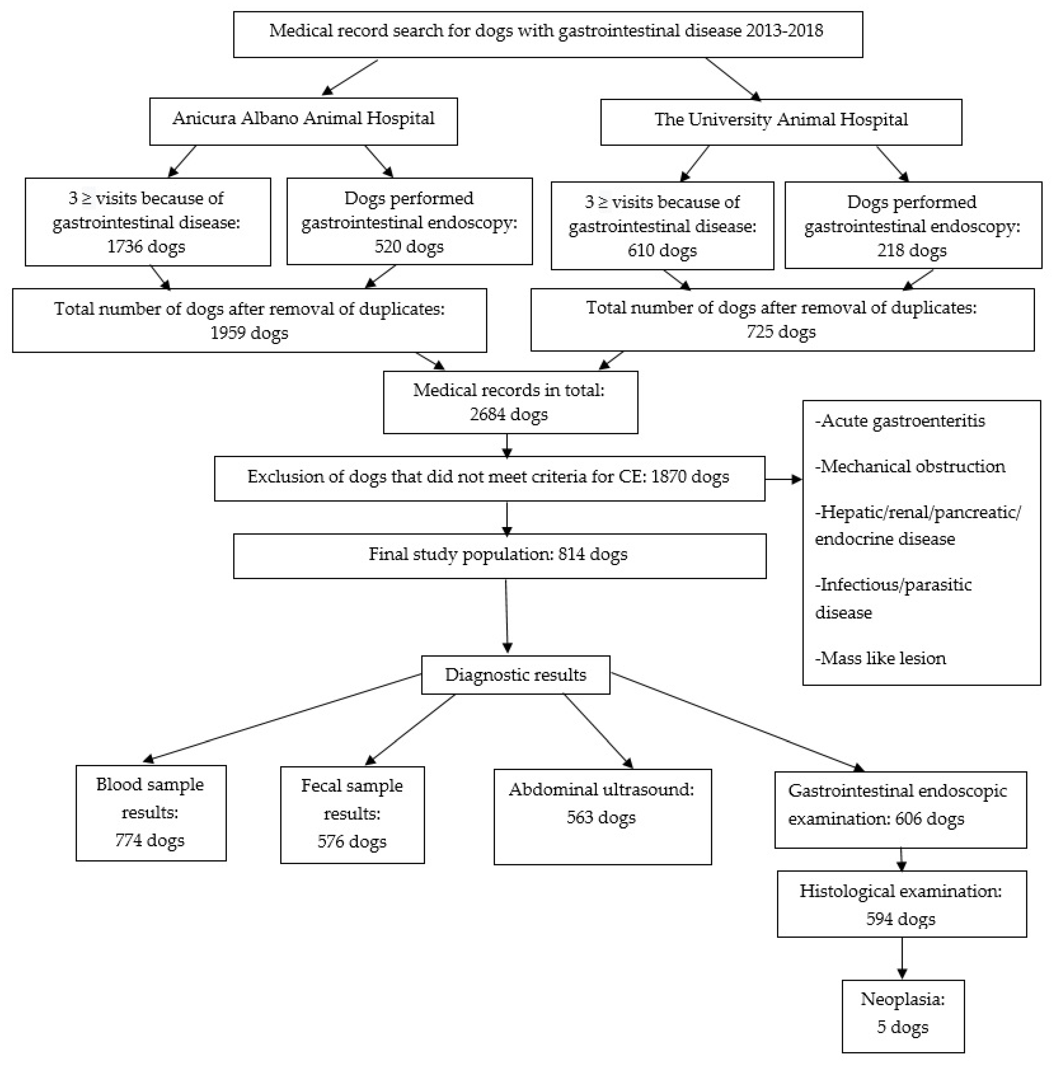
2.1. Study Population

In this retrospective study, a medical record search was performed at two large Swedish animal hospitals: Anicura Albano Animal Hospital (AAAH) in Stockholm and the University Animal Hospital (UAH) at the Swedish University of Agricultural Sciences in Uppsala, to identify dogs with CE. Initially, the diagnostic index of the medical record system Trofast (Version 8.6.0.0, Trofast AB, Västerås, Sweden) was used to search the database for dogs that were presented at either animal hospital because of gastrointestinal signs at ≥3 occasions between January 2013 and December 2018. Dogs were also included if the first visit because of GI signs had taken place before January 2013. A search was performed in the database for diagnostic codes related to vomiting, diarrhea, abdominal pain, weight loss, anorexia, tenesmus, and gastrointestinal inflammation. In addition, a search was performed for dogs that had underwent gastroduodenoscopy/colonoscopy during the same time period. Case duplicates were removed from the resulting data set and the remaining medical records were scrutinized by a veterinarian (JHo). Dogs that had plausible clinical signs of CE, such as persistent (>3 weeks) or recurring diarrhea, vomiting, loss of appetite, or weight loss, were included in the study. Dogs were also included if they still had clinical signs of CE despite a successful treatment of a confirmed intestinal parasitic infection, with negative fecal examination results at the control examination. Gastrointestinal biopsies were not required for the dogs to be included. Dogs were excluded from the study if they had visited the animal hospitals because of acute gastrointestinal disease or if they had been diagnosed with primary endocrine, hepatic, pancreatic or renal disease, infectious or parasitic disease, mechanical obstruction, or a macroscopic neoplastic lesion.

2.2. Database

Dog characteristics (breed, sex, age at first and last visit, body weight (BW) and body condition score (BCS)), case history, physical examination, laboratory variables, and therapeutic protocol and response, were documented in the database. The date of the first visit because of CE was noted, as well as date and reason for the last visit at the animal hospital (including also non-CE related reasons), regardless of being performed during the study period or later. Information about activity level, appetite, stool consistency, and stool frequency were included, as well as information regarding presence of vomiting, abdominal pain, weight loss, or pruritus. Results of laboratory variables including fecal and blood samples, as well as findings from the abdominal ultrasound examination, gastroduodenoscopy/colonoscopy, and histological examination were also recorded. When blood sample results from the initial CE related visit were available in the medical record database, these results were documented together with the reference range used by the laboratory that performed the examinations. Treatment information regarding diet brand, probiotics, type of medication, and concurrent medical conditions was noted from the time of the first CE related visit until the last documented visit in the medical record. In addition, treatment response as well as time and reason for euthanasia/death was documented in the data spread sheet.

2.3. Classification of Dogs

The CE was classified as FRE, IRE, ARE, or NRE depending on treatment response as previously described (1, 2, 14). In short, the dogs were classified with FRE if they had an adequate response to diet change and needed no additional treatment, and classified with IRE if they did not respond solely to diet change but had an adequate response to immunosuppressant treatment. They were classified with ARE if they had an inadequate response to diet change and immunosuppressant treatment, and solely responded to antibiotic treatment. The dogs were classified with NRE if they did not respond to either diet change, immunosuppressant treatment, or antibiotic treatment. For the individual dog, the response was assessed at the discretion of the veterinarian scrutinizing the medical records (JHo), and only classified in the database if the information could clearly be obtained from the medical record (≥2 weeks follow up). If the information regarding treatment response was not available, or if the dog was treated with immunosuppressant and antibiotic treatment concurrently, the dog was categorized as non-classified.

2.4. Statistical Methods

Descriptive statistical calculations were performed in JMP Pro (v16.0, Cary NC). Proportions, medians, and interquartile ranges (IQR) are reported. Relative risk (RR) and 95% confidence intervals for developing CE for each breed was calculated by using the following formula: RR = (number of dogs of a given breed with CE/total number of dogs with CE)/(number of dogs of a given breed/total number of dogs). Differences in age of onset between different breeds and treatment classification groups were tested using Wilcoxon rank-sum test, because age of onset was found not to be normally distributed using the F-test. Numerical laboratory blood analysis data was dichotomized into either above (C-reactive protein [CRP], leukocytes, creatinine, urea, alanine aminotransferase), below (albumin, total protein, leukocytes, hematocrit, calcium, folic acid, cobalamin), or within reference interval. Comparisons between categorical data were performed using either the Chi-2 or the Fischer’s exact two-tailed tests. Subanalyses were performed with pairwise comparisons if the overall p-value was <0.05, and one or both variables included >2 groups using the Fischer’s exact test with Bonferroni correction for multiple comparisons.

3. Results

3.1. Study Population

A flowchart of the different steps in the search process of the medical records at AAAH and UAH is presented in Figure 1. The final study population comprised 814 dogs. In total, 16.2% (132/814) of the dogs were referred to AAAH and UAH from other clinics, and the rest of the dogs presented as primary cases. Thirty of the included dogs had an intestinal parasitic infection at the time of presentation. These dogs had continuous signs of CE despite a successful treatment with negative fecal examination results at the control examination, and they were therefore included in the data set. Five dogs were diagnosed with neoplasia at the histological examination but had no signs of macroscopic neoplastic lesions at the abdominal ultrasound or gastrointestinal endoscopic examination. These five dogs were included in the study population because histological examination was not required for inclusion. The final study population comprised 463 males and 351 females, of which 136 of the male dogs were neutered, and 114 of the female dogs were spayed. In total, 138 different breeds were represented in the data set, and the most common breeds were Mixed Breed (107), Golden Retriever (35), French Bulldog (34), Cavalier King Charles Spaniel (CKCS) (31), Rottweiler (30), Labrador Retriever (27), German Shepherd (25), Miniature Poodle (25), Chihuahua (25), and Miniature Schnauzer (21). Dogs presented at a median age of 3.8 (IQR 1.8–6.7) years, ranging from 0.2–13.8 years. French Bulldogs and Miniature Schnauzers presented at a younger age (below 2.5 years) compared to other breeds (p < 0.05). The median time of follow up from the first visit until the last visit was 2.8 (IQR 0.9–4.6) years. The longest time of follow up was 12.6 years. In total, 30.0% (244/814) of the dogs had been euthanized, and 0.8% (7/814) of the dogs were reported to have died at home. In 47.5% (116/244) of the dogs that were euthanized, CE was the reported reason for euthanasia. For the dogs that were euthanized or were reported to have died at home, the median time of follow up from the first CE related visit until time of death was 2.8 (IQR 0.9–4.6) years. General information about the study population and presenting signs are presented in Table 1.

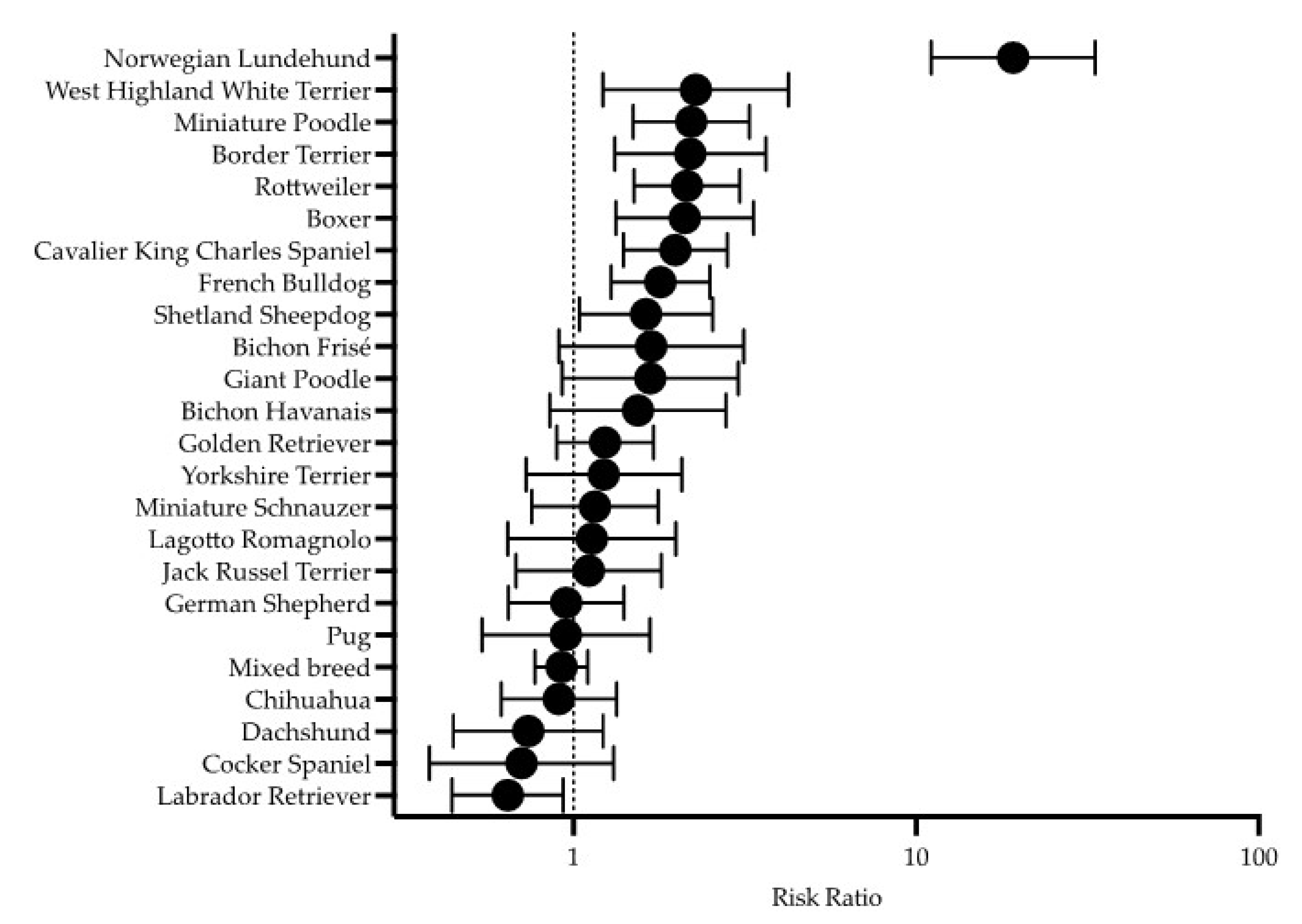
3.2. Period Prevalence and Relative Risk

During the study period, a total of 77,142 dogs visited the animal hospitals: 48,914 dogs at AAAH and 28,228 dogs at UAH. The total number of dogs that presented with gastrointestinal signs during the same period was 18,409 dogs: 12 024 dogs at AAAH and 6,385 dogs at the UAH. The period prevalence of CE was 1.1% (814/77 142) of the total number of dogs that visited the animal hospitals. The CE dogs represented 4.4% (814/18 409) of all dogs that presented at the animal hospitals with gastrointestinal signs during the study period. The breeds with an increased RR of presenting with CE were Norwegian Lundehund, West Highland White Terrier (WHWT), Miniature Poodle, Border Terrier, Rottweiler, Boxer, CKCS, French Bulldog, and Shetland Sheepdog (Figure 2).

3.3. Classification of Dogs

Treatment outcome could be classified in 72.9% (593/814) of the dogs, and their CE was characterized as IRE (449/814, 55.2%), FRE (93/814, 11.4%), NRE (42/814, 5.2%), and ARE (9/814, 1.1%). Twenty-seven percent (221/814) of the dogs were not assigned a classification because of lack of follow up after the treatment start, information about treatment response could not be clearly obtained from the medical record, or due to concurrent initial treatment with immunosuppressant and antibiotic agents. Among the non-responsive dogs, 41/42 (97.5%) of the dogs were euthanized due to CE, and 1/42 (2.4%) of the non-responsive dogs were euthanized due to pneumonia. Non-responsive dogs were more likely to be euthanized due to CE than dogs with FRE, IRE, and dogs that were not assigned a classification (p < 0.008). Norwegian Lundehunds were more likely to be non-responsive than food-responsive (p < 0.008). There was no significant difference in treatment outcome among the other breeds.

3.4. Diagnostic Results
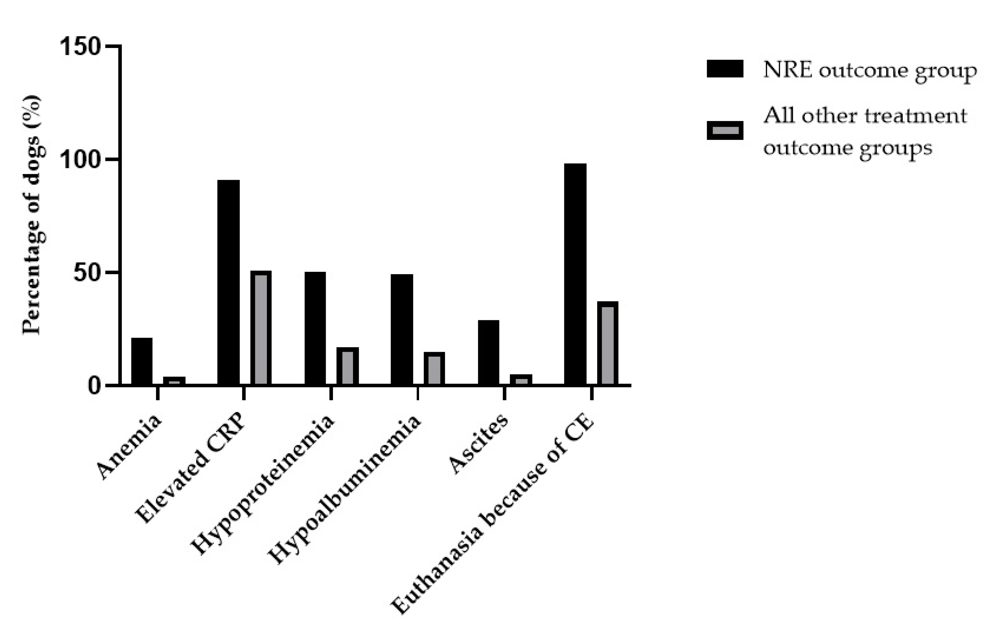
Results of hematology and/or biochemistry were available in 95% (774/814) of the dogs (Table 2). Abnormal results such as anemia (32/632, 5.1%), hypoalbuminemia (116/662, 17.5%), hypoproteinemia (125/655, 19.1%), hypocobalaminemia (98/647, 15.1%), and increased CRP concentrations (145/267, 54.3%) were identified. Dogs with NRE were more likely to present with hypoproteinemia, hypoalbuminemia, and anemia compared to the FRE, IRE, and non-classified outcome groups (p < 0.008). There was no significant difference in total protein, albumin, or hematocrit concentrations among the FRE-, IRE- and non-classified outcome groups. Dogs with NRE were more likely to present with increased CRP concentrations compared to the IRE outcome group (p < 0.008), but no significant difference in CRP concentrations was seen among the NRE, FRE, and non-classified outcome groups. The ARE outcome group was excluded from the multiple comparison test because of an insufficient number of dogs. Results of abdominal ultrasounds were documented in 69.2% (563/814) of the dogs, and in 59.5% (335/563) of these dogs, there was an ultrasonographic abnormality recorded in the gastrointestinal tract. Information regarding presence of ascites was documented in 67.6% (550/814) of the dogs and was identified in 6.7% (37/550) of them (Table 2). Dogs with NRE were more likely to present with ascites compared to the FRE, IRE, and non-classified outcome groups (p < 0.008). A graph that illustrates the characteristics of dogs in the NRE outcome group contrasted to the dogs belonging to the other treatment outcome groups is presented in Figure 3. A gastroscopy and/or colonoscopy was performed in 74.4% (606/814) of the dogs and histological results were available from 98.0% (594/606) of the gastrointestinal endoscopic examinations. Gastrointestinal endoscopy and histological findings are presented in Table 3.

3.5. Treatment

A hydrolyzed diet was initially administered to 69.2% (563/814) of the dogs, whereas 13.0% (106/814) were fed a novel protein diet. The rest of the dogs were given either gastrointestinal diet (111/814, 13.6%), home-prepared food (18/814, 2.2%), or other types of diet that were chosen because of concurrent diseases or due to the dog owner’s preference (14/814, 1.7%). In 82.0% (668/814) of the dogs, immunosuppressive treatment consisted of prednisolone/methylprednisolone, and in 16.2% (132/814), the prednisolone/methylprednisolone was combined with additional immunosuppressive treatment (Table 4). In 63.0% (512/814) of the dogs, gastroprotectant treatment with either proton pump inhibitors, sucralfate, or famotidine were administered (Table 4).
Antibiotic treatment was used in 41.9% (341/814) of the dogs, and the most common antibiotic substances used were metronidazole (325/341, 95.3%) and amoxicillin (40/341, 11.7%). In 10.3% (35/341) of the dogs treated with antibiotics, other antibiotic agents were administered (tylosin, enrofloxacin, amoxicillin plus clavulanic acid, or doxycycline). Among the dogs that received antibiotics, 46.2% (158/341) were not assigned a classification due to concurrent initial treatment with antibiotic and immunosuppressive therapy. In 40.6% (138/341) of the dogs, there was an inadequate response to the antibiotic treatment but an adequate response to immunosuppressant treatment, whereby these dogs were classified in the IRE outcome group. Of the dogs that received antibiotic treatment, 8% (28/341) were classified with NRE, 2.6% (9/341) were classified with ARE, and 2.4% (8/341) were classified in the FRE outcome group. Of the dogs with NRE, 92.9% (39/42) were treated with prednisolone/methylprednisolone, and in 35.7% (15/42) of the dogs, the treatment with prednisolone/methylprednisolone was combined with additional immunosuppressant therapy. In 64.2% (27/42) of the non-responsive dogs, cobalamin supplementation was administered (Table 4). None of the dogs in the NRE outcome group, or any of the other treatment outcome groups, received treatment with albumin or blood products.

4. Discussion

This large retrospective study showed that the period prevalence of CE was approximately 1% of the total number of dogs visiting two Swedish referral animal hospitals. The breeds with the highest RR of developing CE were Norwegian Lundehund, WHWT, and Miniature Poodle. An earlier age of CE onset was seen in French Bulldogs and Miniature Schnauzers, compared to the other breeds. As opposed to previous reports, most of the dogs were classified with IRE and the fewest with ARE. Dogs with NRE were more likely to present with hypoproteinemia, hypoalbuminemia, anemia, and ascites compared to the FRE, IRE, and non-classified outcome group.
The period prevalence of CE was 1.1% in the present study, which corresponds to a previous study of 546 dogs performed at an animal hospital in the UK during a 5.5 year period (2003–2009), where the results indicated a period prevalence of 2% (546/27 463) for CE in dogs [6]. In another retrospective Italian study of 120 dogs, the period prevalence of dogs with signs consistent with CE was 0.9% (120/12 699) during 2013 [28].
The breeds identified with an increased RR of CE in this study were Norwegian Lundehund, WHWT, Miniature Poodle, Border Terrier, Rottweiler, Boxer, CKCS, French Bulldog, and Shetland Sheepdog. Norwegian Lundehunds are known to be predisposed to chronic gastrointestinal disease [13,17,29] and Rottweilers as well as Boxers have been reported to have an increased risk of developing CE in a former study from the UK [6]. Rottweilers have been described to be predisposed to eosinophilic enteritis [30,31], and Boxers and French Bulldogs have been reported to be susceptible to granulomatous colitis, caused by enteroinvasive Escherichia coli [8,32,33]. Boxers and WHWT were mentioned as two of the most commonly represented breeds in a retrospective study of 80 dogs with IBD in Scotland [20], and the results of a previous study from Switzerland indicated an increased proportion of WHWT with IRE and PLE [14]. To the authors’ knowledge, an increased RR of CE in the Miniature Poodle, Border Terrier, CKCS, and Shetland Sheepdog breeds have hitherto not been described. Even though German Shepherd dogs were one of the most represented breeds in the present study population, it was not one of the breeds that was found to have an increased RR of CE as reported in previous studies [6,7,10,11]. A possible reason that could partly explain this finding is that working dogs of the Swedish police force, mainly German Shepherd dogs, are routinely presented at these two animal hospitals for prophylactic interventions. This might lead to a potentially higher proportion of healthy German Shepherd dogs visiting these two animal hospitals than in other animal hospitals or referring clinics in Sweden, leading to a lower proportion of German Shepherd dogs with CE in the present study population.
In the present study, the overall median age of the dogs at the time of the first CE related visit was 3.8 years, which was lower than in other previous studies of 70 dogs from Switzerland and 80 dogs from Scotland: 5.3 years and 4.3 years, respectively [14,20]. There was no significant difference in age at presentation between the different classification groups in the present study. This differs from the results of the study from Switzerland, in which it was reported that dogs with FRE were significantly younger than dogs with IRE [14]. Another study of 203 dogs from the UK demonstrated that dogs in the ARE group were younger than dogs in the FRE and IRE group, respectively [25]. In the present study, the oldest dog in the FRE outcome group was twelve years old at the time of the first visit. This agrees with previous studies that have also reported that some dogs with FRE have presented at an old age: nine years and eleven years, respectively [3,34]. These results support the recommendation that a diet change should be considered regardless of the age of the dog. There was no significant difference in distribution between males and females in the present study population, which agrees with earlier studies (14, 20).
In the present study, the largest proportion of dogs was classified in the IRE outcome group (55.2%), which differs from earlier published studies. In previous studies, the FRE group typically has been the largest group with proportions larger than 50% [14,22,25]. In a retrospective study regarding canine CE in 203 dogs, 64% of the dogs were classified in the FRE group, 16.2% in the ARE group, and 19.2% were considered to have IRE [25]. One reason that could explain why the IRE outcome group is comparably large in the present study might be that both animal hospitals are referral hospitals and 16.2% of the dogs had been referred from general practitioners. It seems likely that many dogs that respond to a dietary intervention are not referred to an animal hospital for further investigation.
The ARE outcome group contained the smallest number of dogs in the present study. In 43% (341/809) of the dogs, antibiotic treatment had been used at some time during their follow up period, but only 1% (8/814) of the dogs responded solely to antibiotic treatment and were therefore classified in the ARE outcome group. Among the dogs that received antibiotics, 46.2% (158/341) of the dogs were not assigned a classification due to difficulties to evaluate treatment response because of concurrent initial treatment with antibiotic and immunosuppressive therapy. The view of clinical use of antibiotic treatment may have shifted over time and become more restricted, which is reflected in changes in local standard operating procedures. The proportion of dogs with ARE in the present study is not in agreement with two earlier studies, where the ARE group constituted of 36.8% and 16.2% of the study populations respectively [3,25]. Swedish authorities also discourage long- term antibiotic treatment for dogs with CE, and immunosuppressive treatment is often started directly after the food trial if the response to diet change is inadequate and other causes of gastrointestinal disease have been excluded [35]. An important exception is the diagnosis of granulomatous colitis where enrofloxacin is recommended [35,36,37]. The use of different antibiotics has been described in dogs with CE, when food trial has failed, or if only a partial response to the diet change is observed [2,15,38,39,40]. The response, however, has shown to be short-lived after the antibiotic treatment has been terminated, with one study reporting relapse of the clinical signs within one month in 86% of the dogs treated with tylosin [41]. Some studies report remission of clinical signs with the use of metronidazole in dogs with CE, but the antibiotic treatment was combined with diet and other drugs which makes it difficult to interpret to what extent the response was due to the antibiotic treatment [15,23,40]. The relapse rate of CE signs after discontinuation of metronidazole has been reported to be 80% with the need for prolonged antibiotic treatment to control CE signs [2,3,25]. In one study, it was demonstrated that prednisone was as effective as a combined treatment with prednisone and metronidazole for induction therapy of canine IBD, putting the usefulness of metronidazole in treating CE in question [40]. The high relapse rate of dogs receiving antibiotics to treat CE can also be caused by an antibiotic-induced dysbiosis, as was reported for tylosin and metronidazole [42,43,44,45].
In the present study, 5.2% (42/814) of the dogs were classified in the NRE outcome group, and of these non-responsive dogs, 97.6% (41/42) were euthanized due to CE. In total, 14.3% (116/814) of dogs in the present study were euthanized due to CE. This result agrees with a retrospective study that reported that 13% (10/80) of the dogs were euthanized due to refractory IBD [20], even though some of the dogs had gone into remission before developing severe relapse and refractoriness to additional treatment. The result of the present study is also comparable to other previous studies where 19% (13/70) and 9% (15/165) of the dogs with CE had an intractable disease [14,26]. The long period of follow up in many dogs of this study enabled accurate assessments of the treatment response. Treatment response has been reported in several earlier studies, but the time of follow up is often less than three months [40,46,47,48,49,50,51,52], even though some studies have a longer follow up than six months [1,3,14,20,22,25,26,27].
In the present study, 17.4% of the dogs had hypoalbuminemia, and dogs with NRE were more likely to present with hypoalbuminemia compared to the FRE, IRE, and non-classified outcome groups (p < 0.008).) This corresponds to a previous study of 70 dogs with CE where hypoalbuminemia was shown to be associated with negative outcomes [14], as well as a previous study of 165 dogs with CE where non-survivors showed significantly lower serum albumin levels compared to survivors [26]. It is also comparable to the results of a previous study of 92 dogs with CE, that demonstrated that the proportion of dogs with normal serum albumin levels was significantly higher in the IRE group than in the NRE group [27]. However, no significant differences in proportion of dogs with hypoalbuminemia were found between the FRE and the IRE outcome group in the present study, which was demonstrated in a previous study of 203 dogs with CE [25]. A possible explanation for this finding is that the CE of the dogs with IRE in the present group was not as severe as the CE of the IRE group in the previous study.
Dogs in the NRE outcome group in the present study were more likely to present with anemia compared to the dogs with FRE, IRE, and the non-classified outcome group, which has also been described in a previous study [53]. Non-responsive dogs also presented with an increased CRP compared to the IRE outcome group, but not compared to the FRE- or non-classified outcome group. Increased CRP has been described as a marker of disease severity in dogs with CE in a previous study [18], but was not significantly associated with negative outcomes in another previous study [14]. Hypocobalaminemia was reported in 15.1% (98/647) of the dogs in the present study. Dogs with NRE were not more likely to present with hypocobalaminemia, as opposed to an earlier study where hypocobalaminemia was associated with negative outcomes [14]. In the present study, a higher proportion of dogs received supplementation with cobalamin compared to the proportion that presented with low cobalamin levels. Substituting dogs with serum cobalamin concentrations close to the lower reference range originates from studies revealing that affected dogs can have subtle cobalamin deficiency as manifested by increased serum methylmalonic acid concentrations despite cobalamin concentrations within the reference range, and that they respond with an increase in serum cobalamin after oral vitamin substitution [54,55].
Because of the retrospective nature of the study, the information in the medical records was sometimes incomplete. Hematology and biochemistry results were not accessible in all dogs, and it was not possible to assess retrospectively the two clinical indexes for disease activity that have been defined previously: CIBDAI and CCECAI [14,18]. Another limitation is that treatment response was subjectively evaluated from the information that was documented in the medical record by the attending clinician.

5. Conclusions

In conclusion, the period prevalence of dogs with CE at two Swedish referral animal hospitals was 1.1%, which is comparable to results of previous studies. Some breeds that were identified with an increased RR of CE in the present study have previously been described as risk breeds, but Miniature Poodle, Border Terrier, CKCS, and Shetland Sheepdog have, to the authors’ knowledge, not previously been described as breeds with an increased risk of CE. Compared to the other breeds, onset of CE was identified at an earlier age in French Bulldogs and Miniature Schnauzers. Hydrolyzed diet and immunosuppressive treatment consisting of prednisolone/methylprednisolone were the most common treatment alternatives. Most dogs with CE were categorized in the IRE outcome group and the smallest proportion of dogs in the ARE outcome group. Even though antibiotic treatment had been used in a comparably large proportion dogs, only 1.1% of the dogs were classified in the ARE outcome group, which differs from previous studies. Dogs with NRE were more likely to present with hypoproteinemia, hypoalbuminemia, anemia, and ascites compared to the dogs with FRE, IRE, and the non-classified outcome group.

Author Contributions

Conceptualization, J.H. (Johanna Holmberg), J.H. (Jens Häggström), L.P., I.L. and T.S.; methodology, J.H. (Johanna Holmberg), J.H. (Jens Häggström), L.P., I.L. and T.S.; formal analysis, J.H. (Jens Häggström) and J.H. (Johanna Holmberg); investigation, J.H. (Johanna Holmberg); resources, C.H.; data curation, J.H. (Johanna Holmberg); writing—original draft preparation, J.H. (Johanna Holmberg); writing—review and editing, J.H. (Johanna Holmberg), J.H. (Jens Häggström), L.P., I.L, T.S. and C.H.; visualization, J.H. (Johanna Holmberg); supervision, J.H. (Jens Häggström), I.L., L.P., C.H. and T.S.; project administration, J.H. (Johanna Holmberg). All authors have read and agreed to the published version of the manuscript.

Funding

This research received no external funding.

Institutional Review Board Statement

Ethical review and approval were waived for this study because this was a retrospective study based on clinical data derived from the authors’ hospitals’ database, and it was aligned with GDPR legislation within the European Union. Therefore, according to Swedish laws, this study did not require authorization.

Informed Consent Statement

Informed consent was obtained from all subjects involved in the study.

Data Availability Statement

The data presented in this study are available on request from the corresponding author.

Conflicts of Interest

The authors declare no conflict of interest.

References

  1. Dandrieux, J.R.S.; Mansfield, C.S. Chronic Enteropathy in Canines: Prevalence, Impact And Management Strategies. Vet. Med. 2019, 10, 203–214. [Google Scholar] [CrossRef] [PubMed] [Green Version]
  2. Dandrieux, J.R.S. Inflammatory bowel disease versus chronic enteropathy in dogs: Are they one and the same? J. Small Anim. Pract. 2016, 57, 589–599. [Google Scholar] [CrossRef] [PubMed]
  3. Dandrieux, J.R.S.; Martinez Lopez, L.M.; Prakash, N.; Mansfield, C.S. Treatment response and long term follow up in nineteen dogs diagnosed with chronic enteropathy in Australia. Aust. Vet. J. 2019, 97, 301–307. [Google Scholar] [CrossRef] [PubMed]
  4. Jergens, A.E.; Simpson, K.W. Inflammatory bowel disease in veterinary medicine. Front. Biosci. 2012, 4, 1404–1419. [Google Scholar] [CrossRef]
  5. Allenspach, K. Clinical Immunology and Immunopathology of the Canine and Feline Intestine. Vet. Clin. N. Am. Small Anim. Pract. 2011, 41, 345–360. [Google Scholar] [CrossRef]
  6. Kathrani, A.; Werling, D.; Allenspach, K. Canine breeds at high risk of developing inflammatory bowel disease in the south-eastern UK. Vet. Rec. 2011, 169, 635. [Google Scholar] [CrossRef] [Green Version]
  7. Allenspach, K.; House, A.; Smith, K.; McNeill, F.M.; Hendricks, A.; Elson-Riggins, J.; Riddle, A.; Steiner, J.M.; Werling, D.; Garden, O.A.; et al. Evaluation of mucosal bacteria and histopathology, clinical disease activity and expression of Toll-like receptors in German shepherd dogs with chronic enteropathies. Vet. Microbiol. 2010, 146, 326–335. [Google Scholar] [CrossRef]
  8. Craven, M.; Mansfield, C.S.; Simpson, K.W. Granulomatous colitis of boxer dogs. Vet. Clin. N. Am. Small Anim. Pract. 2011, 41, 433–445. [Google Scholar] [CrossRef]
  9. Berghoff, N.; Ruaux, C.G.; Steiner, J.M.; Williams, D.A. Gastroenteropathy in Norwegian Lundehund. Compendium 2007, 29, 456. [Google Scholar]
  10. Kathrani, A.; House, A.; Catchpole, B.; Murphy, A.; German, A.; Werling, D.; Allenspach, K. Polymorphisms in the Tlr4 and Tlr5 Gene Are Significantly Associated with Inflammatory Bowel Disease in German Shepherd Dogs. PLoS ONE 2010, 5, e15740. [Google Scholar]
  11. German, A.; Hall, E.; Day, M. Relative deficiency in IgA production by duodenal explants from German shepherd dogs with small intestinal disease. Vet. Immunol. Immunopathol. 2000, 76, 25–43. [Google Scholar] [CrossRef]
  12. Berghoff, N.; Steiner, J.M. Laboratory tests for the diagnosis and management of chronic canine and feline enteropathies. Vet. Clin. N. Am. Small Anim. Pract. 2011, 41, 311–328. [Google Scholar] [CrossRef] [PubMed]
  13. Simpson, K.W.; Jergens, A.E. Pitfalls and Progress in the Diagnosis and Management of Canine Inflammatory Bowel Disease. Vet. Clin. N. Am. Small Anim. Pract. 2011, 41, 381–398. [Google Scholar] [CrossRef]
  14. Allenspach, K.; Wieland, B.; Grone, A.; Gaschen, F. Chronic enteropathies in dogs: Evaluation of risk factors for negative outcome. J. Vet. Intern. Med. 2007, 21, 700–708. [Google Scholar] [CrossRef]
  15. Makielski, K.; Cullen, J.; O’Connor, A.; Jergens, A.E. Narrative review of therapies for chronic enteropathies in dogs and cats. J. Vet. Intern. Med. 2019, 33, 11–22. [Google Scholar] [CrossRef] [PubMed] [Green Version]
  16. Craven, M.D.; Washabau, R.J. Comparative pathophysiology and management of protein-losing enteropathy. J. Vet. Intern. Med. 2019, 33, 383–402. [Google Scholar] [CrossRef]
  17. Dossin, O.; Lavoue, R. Protein-losing enteropathies in dogs. Vet. Clin. N. Am. Small Anim. Pract. 2011, 41, 399–418. [Google Scholar] [CrossRef] [PubMed]
  18. Jergens, A.E.; Schreiner, C.A.; Frank, D.E.; Niyo, Y.; Ahrens, F.E.; Eckersall, P.D.; Benson, T.J.; Evans, R. A scoring index for disease activity in canine inflammatory bowel disease. J. Vet. Intern. Med. 2003, 17, 291–297. [Google Scholar] [CrossRef] [PubMed]
  19. Allenspach, K.; Rizzo, J.; Jergens, A.E.; Chang, Y.M. Hypovitaminosis D is associated with negative outcome in dogs with protein losing enteropathy: A retrospective study of 43 cases. BMC Vet. Res. 2017, 13, 96. [Google Scholar] [CrossRef] [Green Version]
  20. Craven, M.; Simpson, J.W.; Ridyard, A.E.; Chandler, M.L. Canine inflammatory bowel disease: Retrospective analysis of diagnosis and outcome in 80 cases (1995–2002). J. Small Anim. Pract. 2004, 45, 336–342. [Google Scholar] [CrossRef]
  21. Jergens, A.E. Inflammatory bowel disease. Current perspectives. Vet. Clin. North Am. Small Anim. Pract. 1999, 29, 501–521. [Google Scholar] [PubMed]
  22. Mandigers, P.J.J.; Biourge, V.; Van Den Ingh, T.S.G.A.M.; Ankringa, N.; German, A.J.A. A Randomized, Open-Label, Positively-Controlled Field Trial of a Hydrolyzed Protein Diet in Dogs with Chronic Small Bowel Enteropathy. J. Vet. Intern. Med. 2010, 24, 1350–1357. [Google Scholar] [CrossRef] [PubMed]
  23. Rossi, G.; Pengo, G.; Caldin, M.; Palumbo Piccionello, A.; Steiner, J.M.; Cohen, N.D.; Jergens, A.E.; Suchodolski, J.S. Comparison of microbiological, histological, and immunomodulatory parameters in response to treatment with either combination therapy with prednisone and metronidazole or probiotic VSL#3 strains in dogs with idiopathic inflammatory bowel disease. PLoS ONE. 2014, 9, e94699. [Google Scholar]
  24. Dye, T.; Diehl, K.; Wheeler, S.; Westfall, D. Randomized, Controlled Trial of Budesonide and Prednisone for the Treatment of Idiopathic Inflammatory Bowel Disease in Dogs. J. Vet. Intern. Med. 2013, 27, 1385–1391. [Google Scholar] [CrossRef] [Green Version]
  25. Allenspach, K.; Culverwell, C.; Chan, D. Long-term outcome in dogs with chronic enteropathies: 203 cases. Vet. Rec. 2016, 178, 368. [Google Scholar] [CrossRef] [PubMed] [Green Version]
  26. Benvenuti, E.; Pierini, A.; Bottero, E.; Pietra, M.; Gori, E.; Salvadori, S.; Marchetti, V. Immunosuppressant-Responsive Enteropathy and Non-Responsive Enteropathy in Dogs: Prognostic Factors, Short- and Long-Term Follow Up. Animals 2021, 11, 2637. [Google Scholar] [CrossRef]
  27. Pietra, M.; Galiazzo, G.; Bresciani, F.; Morini, M.; Licarini, S.; Turba, M.E.; Amaducci, G.; Bettini, G.; Fracassi, F.; Ostanello, F. Evaluation of Prognostic Factors, including Duodenal P-Glycoprotein Expression, in Canine Chronic Enteropathy. Animals 2021, 11, 2315. [Google Scholar] [CrossRef]
  28. Marchesi, M.C.; Timpano, C.C.; Busechian, S.; Pieramati, C.; Rueca, F. The role of diet in managing inflamatory bowel disease affected dogs: A retrospective cohort study on 76 cases. Vet. Ital. 2017, 53, 297–302. [Google Scholar]
  29. Flesjå, K.; Yri, T. Protein-losing enteropathy in the Lundehund. J. Small Anim. Pract. 1977, 18, 11–23. [Google Scholar] [CrossRef]
  30. Lilliehöök, I.; Gunnarsson, L.; Zakrisson, G.; Tvedten, H. Diseases associated with pronounced eosinophilia: A study of 105 dogs in Sweden. J. Small Anim. Pract. 2000, 41, 248–253. [Google Scholar] [CrossRef]
  31. Sykes, J.E.; Weiss, D.J.; Buoen, L.C.; Blauvelt, M.M.; Hayden, D.W. Idiopathic hypereosinophilic syndrome in 3 Rottweilers. J. Vet. Intern. Med. 2001, 15, 162–166. [Google Scholar] [CrossRef] [PubMed]
  32. Simpson, K.W.; Dogan, B.; Rishniw, M.; Goldstein, R.E.; Klaessig, S.; McDonough, P.L.; German, A.J.; Yates, R.M.; Russell, D.G.; Johnson, S.E.; et al. Adherent and Invasive Escherichia coli Is Associated with Granulomatous Colitis in Boxer Dogs. Infect. Immun. 2006, 74, 4778–4792. [Google Scholar] [CrossRef] [PubMed] [Green Version]
  33. Hall, E.J.; Rutgers, H.C.; Scholes, S.F.E.; Middleton, D.J.; Tennant, B.J.; King, N.M.; Kelly, D.F. Histiocytic ulcerative colitis in boxer dogs in the UK. J. Small Anim. Pract. 1994, 35, 509–515. [Google Scholar] [CrossRef]
  34. Kawano, K.; Shimakura, H.; Nagata, N.; Masashi, Y.; Suto, A.; Suto, Y.; Uto, S.; Ueno, H.; Hasegawa, T.; Ushigusa, T.; et al. Prevalence of food-responsive enteropathy among dogs with chronic enteropathy in Japan. J. Vet. Med. Sci. 2016, 78, 1377–1380. [Google Scholar] [CrossRef] [Green Version]
  35. Läkemedelsverket, Dosering av antibiotika till hund—Ny rekommendation. Inf. Läkemedelsverket 2016, 27, 4–17.
  36. Hostutler, R.A.; Luria, B.J.; Johnson, S.E.; Weisbrode, S.E.; Sherding, R.G.; Jaeger, J.Q.; Guilford, W.G. Antibiotic-responsive histiocytic ulcerative colitis in 9 dogs. J. Vet. Intern. Med. 2004, 18, 499–504. [Google Scholar] [CrossRef]
  37. Mansfield, C.; James, F.; Craven, M.; Davies, D.; O’Hara, A.; Nicholls, P.; Dogan, B.; MacDonough, S.; Simpson, K. Remission of Histiocytic Ulcerative Colitis in Boxer Dogs Correlates with Eradication of Invasive Intramucosal Escherichia coli. J. Vet. Intern. Med. 2009, 23, 964–969. [Google Scholar] [CrossRef] [Green Version]
  38. Westermarck, E.; Skrzypczak, T.; Harmoinen, J.; Steiner, J.M.; Ruaux, C.G.; Williams, D.A.; Eerola, E.; Sundbäck, P.; Rinkinen, M. Tylosin-responsive chronic diarrhea in dogs. J. Vet. Intern. Med. 2005, 19, 177–186. [Google Scholar] [CrossRef]
  39. Kilpinen, S.; Spillmann, T.; Syrjä, P.; Skrzypczak, T.; Louhelainen, M.; Westermarck, E. Effect of tylosin on dogs with suspected tylosin-responsive diarrhea: A placebo-controlled, randomized, double-blinded, prospective clinical trial. Acta Vet. Scand. 2011, 53, 26. [Google Scholar] [CrossRef] [Green Version]
  40. Jergens, A.E.; Crandell, J.; Morrison, J.A.; Deitz, K.; Pressel, M.; Ackermann, M.; Suchodolski, J.; Steiner, J.M.; Evans, R. Comparison of Oral Prednisone and Prednisone Combined with Metronidazole for Induction Therapy of Canine Inflammatory Bowel Disease: A Randomized-Controlled Trial. J. Vet. Intern. Med. 2010, 24, 269–277. [Google Scholar] [CrossRef]
  41. Kilpinen, S.; Spillmann, T.; Westermarck, E. Efficacy of two low-dose oral tylosin regimens in controlling the relapse of diarrhea in dogs with tylosin-responsive diarrhea: A prospective, single-blinded, two-arm parallel, clinical field trial. Acta Vet. Scand. 2014, 56, 43. [Google Scholar] [CrossRef] [PubMed] [Green Version]
  42. Chaitman, J.; Ziese, A.-L.; Pilla, R.; Minamoto, Y.; Blake, A.B.; Guard, B.C.; Isaiah, A.; Lidbury, J.A.; Steiner, J.M.; Unterer, S.; et al. Fecal Microbial and Metabolic Profiles in Dogs with Acute Diarrhea Receiving Either Fecal Microbiota Transplantation or Oral Metronidazole. Front. Vet. Sci. 2020, 7, 192. [Google Scholar] [CrossRef] [PubMed]
  43. Pilla, R.; Gaschen, F.; Barr, J.W.; Olson, E.; Honneffer, J.; Guard, B.C.; Blake, A.B.; Villanueva, D.; Khattab, M.R.; Alshawaqfeh, M.K.; et al. Effects of metronidazole on the fecal microbiome and metabolome in healthy dogs. J. Vet. Intern. Med. 2020, 34, 1853–1866. [Google Scholar] [CrossRef] [PubMed]
  44. Pinna, C.; Vecchiato, C.G.; Grandi, M.; Mammi, L.M.E.; Stefanelli, C.; Biagi, G. In Vitro Evaluation of the Effects of Tylosin on the Composition and Metabolism of Canine Fecal Microbiota. Animals 2020, 10, 98. [Google Scholar] [CrossRef] [PubMed] [Green Version]
  45. Manchester, A.C.; Webb, C.B.; Blake, A.B.; Sarwar, F.; Lidbury, J.A.; Steiner, J.M.; Suchodolski, J.S. Long-term impact of tylosin on fecal microbiota and fecal bile acids of healthy dogs. J. Vet. Intern. Med. 2019, 33, 2605–2617. [Google Scholar] [CrossRef] [PubMed]
  46. Otoni, C.C.; Heilmann, R.M.; García-Sancho, M.; Sainz, A.; Ackermann, M.R.; Suchodolski, J.S.; Steiner, J.M.; Jergens, A.E. Serologic and fecal markers to predict response to induction therapy in dogs with idiopathic inflammatory bowel disease. J. Vet. Intern. Med. 2018, 32, 999–1008. [Google Scholar] [CrossRef]
  47. Garcia-Sancho, M.; Rodriguez-Franco, F.; Sainz, A.; Mancho, C.; Rodríguez, A. Evaluation of clinical, macroscopic, and histopathologic response to treatment in nonhypoproteinemic dogs with lymphocytic-plasmacytic enteritis. J. Vet. Intern. Med. 2007, 21, 11–17. [Google Scholar] [CrossRef]
  48. Heilmann, R.M.; Berghoff, N.; Mansell, J.; Grützner, N.; Parnell, N.K.; Gurtner, C.; Suchodolski, J.S.; Steiner, J.M. Association of fecal calprotectin concentrations with disease severity, response to treatment, and other biomarkers in dogs with chronic inflammatory enteropathies. J. Vet. Intern. Med. 2018, 32, 679–692. [Google Scholar] [CrossRef]
  49. Heilmann, R.M.; Volkmann, M.; Otoni, C.C.; Grützner, N.; Kohn, B.; Jergens, A.E.; Steiner, J.M. Fecal S100A12 concentration predicts a lack of response to treatment in dogs affected with chronic enteropathy. Vet. J. 2016, 215, 96–100. [Google Scholar] [CrossRef]
  50. White, R.; Atherly, T.; Guard, B.; Rossi, G.; Wang, C.; Mosher, C.; Webb, C.; Hill, S.; Ackermann, M.; Sciabarra, P.; et al. Randomized, controlled trial evaluating the effect of multi-strain probiotic on the mucosal microbiota in canine idiopathic inflammatory bowel disease. Gut Microbes 2017, 8, 451–466. [Google Scholar] [CrossRef] [Green Version]
  51. Pietra, M.; Fracassi, F.; Diana, A.; Gazzotti, T.; Bettini, G.; Peli, A.; Morini, M.; Pagliuca, G.; Roncada, P. Plasma concentrations and therapeutic effects of budesonide in dogs with inflammatory bowel disease. Am. J. Vet. Res. 2013, 74, 78–83. [Google Scholar] [CrossRef] [PubMed]
  52. Marchetti, V.; Lubas, G.; Lombardo, A.; Corazza, M.; Guidi, G.; Cardini, G. Evaluation of Erythrocytes, Platelets, and Serum Iron Profile in Dogs with Chronic Enteropathy. Vet. Med. Int. 2010, 2010, 716040. [Google Scholar] [CrossRef] [PubMed] [Green Version]
  53. Volkmann, M.; Steiner, J.M.; Fosgate, G.T.; Zentek, J.; Hartmann, S.; Kohn, B. Chronic Diarrhea in Dogs—Retrospective Study in 136 Cases. J. Vet. Intern. Med. 2017, 31, 1043–1055. [Google Scholar] [CrossRef] [PubMed]
  54. Toresson, L.; Steiner, J.M.; Suchodolski, J.S.; Spillmann, T. Oral Cobalamin Supplementation in Dogs with Chronic Enteropathies and Hypocobalaminemia. J. Vet. Intern. Med. 2016, 30, 101–107. [Google Scholar] [CrossRef]
  55. Berghoff, N.; Suchodolski, J.S.; Steiner, J.M. Association between serum cobalamin and methylmalonic acid concentrations in dogs. Vet. J. 2012, 191, 306–311. [Google Scholar] [CrossRef]
Figure 1. Flowchart describing the establishment of the final study population of 814 dogs and number of individual dogs that had undergone specific tests in this group.
Animals 12 01507 g001
Figure 2. Forest plot showing the relative risk of chronic enteropathy (CE) and 95% confidence intervals breeds represented by ≥10 dogs in the dataset. See statistical methods for details and notice the logarithmically transformed x-axis.
Animals 12 01507 g002
Figure 3. Bar graph that illustrates the characteristics of dogs in the NRE outcome group contrasted to the dogs belonging to the other treatment outcome groups (FRE, IRE, ARE, and non-classified outcome group).
Animals 12 01507 g003
Table 1. Summary of signalment variables, presenting signs, and follow up information by treatment outcome group.
Information Rate
(n = 814)
VariableOverall
(n = 814)
Food Responsive
(n = 93)
Immunosuppressant Responsive
(n = 449)
Antibiotic Responsive
(n = 9)
Non Responsive
(n = 42)
Non Classified
(n = 221)
Study population814/814Sex (male/female)463/35152/41246/2036/323/19136/85
113/814Median BCS (1- 9/9)4 (3–5)4 (4–5)4 (4–5)4(4)3 (3–4)4 (3–4)
702//814Median body weight (kg)11.1 (6.5–25.9)12.3 (6.4–27.6)10.0 (6.3–23.7)9.0 (6.2–14.1)12.2 (7.6–30.2)15.3 (7.5–28.9)
814/814Median age at first visit (years)3.8 (1.8–6.7)3.8 (1.5–6.0)3.7 (1.8–6.6)4.1 (0.8–8.1)5.0 (3.5–9.1)3.9 (1.9–6.8)
814/814Median age at last visit (years)7.6 (4.8–10)7.5 (4.8–9.3)7.9 (5.5–10.6)8.0 (2.5–12.8)6.4 (3.7–9.1)7.0 (4.2–9.5)
251/814Median age at death (years)9.2 (6.5–11.5)8.9 (6.9–10.8)10.3 (7.5–12.4)11.3 (7.9–14.8)6.6 (3.8–9.2)8.8 (6.6–11.2)
814/814Median time of follow up (years)2.8 (0.9–4.6)3.1 (1.1–4.5)3.3 (1.5–5.2)3.3 (1.7–4.3)0.3 (0.1–0.7)2.1 (0.4–3.6)
244/814Euthanasia because of CE (yes/no)116/1283/942/821/141/129/35
Presenting signs759/814Vomiting (yes/no)598/16164/25350/715/429/8150/53
764/814Diarrhea (yes/no)574/19067/25298/1158/135/5166/44
503/814Anorexia (yes/no)282/22133/32157/1082/621/1069/65
269/814Weight loss (yes/no)184/8514/1493/471/122/254/21
469/814Activity (decreased/normal)247/22229/29124/1141/521/1372/61
764/814Hematochezia (yes/no)31/7335/8716/3980/92/388/201
67/814Stool frequency
(increased/normal)
41/268/617/102/03/111/9
250/814Pruritus (yes/no)205/4517/12122/172/06/358/13
Table 2. Summary of laboratory results and ultrasonographic findings in the gastrointestinal (GI) tract by treatment outcome group.
Information Rate
(n = 814)
VariableOverall
(n = 814)
Food Responsive
(n = 93)
Immunosuppressant Responsive
(n = 449)
Antibiotic Responsive
(n = 9)
Non Responsive
(n = 42)
Non Classified
(n = 221)
Blood samples814/814Blood samples taken (yes/no)792/2291/2438/119/042/0212/9
Blood sample results814/814Blood sample results available (yes/no)774/4090/3428/218/142/0206/15
632/814Anemia (yes/no)32/6002/7014/3331/68/317/160
637/814Leukocytosis (yes/no)65/5725/6631/3201/611/2817/152
637/814Leukopenia (yes/no)18/6190/7111/3400/70/397/162
218/814Eosinophilia (yes/no)32/1866/2314/850/32/2010/55
267/814Elevated CRP (yes/no)145/12226/1952/721/020/246/29
608/814Elevated creatinine (yes/no)13/5951/705/3310/73/314/156
486/814Elevated urea (yes/no)28/4583/4415/2760/40/1910/115
621/814Elevated ALT (yes/no)105/51610/6467/2770/76/2922/139
126/814Hypocalcemia (total) (yes/no)15/1111/1910/530/12/32/35
647/814Hypocobalaminemia (yes/no)98/5496/7553/2940/47/2932/147
638/814Low folate (yes/no)194/44418/62122/2210/46/2948/128
655/814Hypoproteinemia (yes/no)125/53013/6661/2922/519/1930/148
662/814Hypoalbuminemia (yes/no)116/5467/7359/2940/720/2130/151
Other clinical signs550/814Ascites (yes/no)37/5131/6720/2790/510/246/138
Diagnostic imaging814/814Abdominal ultrasound (yes/no)563/25169/24305/1445/436/6148/73
563/814Ultrasonographic abnormalities GI tract (yes/no)335/22838/31199/1063/223/1272/77
Table 3. Summary of gastrointestinal (GI) endoscopy findings and histological findings by treatment outcome group.
Information Rate
(n = 814)
VariableOverall
(n = 814)
Food Responsive
(n = 93)
Immunosuppressant Responsive
(n = 449)
Antibiotic Responsive
(n = 9)
Non Responsive
(n = 42)
Non Classified
(n = 221)
GI endoscopy814/814GI endoscopy (yes/no)606/20834/59379/708/124/18161/60
594/814Macroscopic abnormalities (yes/no)593/130/1371/08/024/0160/0
594/814Histological abnormalities (yes/no)576/1828/3362/98/023/1155/5
Histological results stomach566/814Normal stomach (yes/no)82/4848/2236/3180/74/1734/120
566/814Chronic gastritis (yes/no)337/22915/15224/1304/310/1184/70
566/814Lymphoplasmacytic gastritis (yes/no)48/5181/2934/3202/53/188/146
566/814Eosinophilic gastritis
(yes/no)
95/4716/2459/2951/62/1927/127
566/814Neoplasia (yes/no)4/5620/301/3530/72/191/153
Histological results duodenum493/814Normal duodenum (yes/no)106/3876/1763/2534/21/1732/98
493/814Chronic enteritis (yes/no)168/3259/14108/2082/45/1344/86
493/814Lymphoplasmacytic enteritis (yes/no)95/3983/2058/2580/67/1127/103
493/814Eosinophilic enteritis
(yes/no)
123/3705/1886/2300/65/1327/103
493/814Neoplasia (yes/no)1/4920/231/3150/60/180/130
Histological results colon356/814Normal colon (yes/no)32/3244/1218/1970/62/148/95
356/814Chronic colitis (yes/no)204/1528/8130/853/38/855/48
356/814Lymphoplasmacytic colitis (yes/no)53/3032/1427/1880/64/1220/83
356/814Eosinophilic colitis (yes/no)65/2912/1440/1751/52/1420/83
356/814Granulomatous colitis (yes/no)2/3540/160/2152/40/160/103
356/814Neoplasia (yes/no)0/3560/160/2150/60/160/103
Table 4. Summary of treatments used for the dogs in the study population as well as information concerning co-morbidities by treatment outcome group. Other immunosuppressive treatment include cyclosporine, azathioprine, chlorambucil, budesonide, and sulfasalazine/olsalazine. Gastroprotectant treatment included proton pump inhibitors, sucralfate, and/or famotidine. Notice that a dog may have received more than one of the listed medical treatments.
VariableOverall
(n = 814)
Food Responsive
(n = 93)
Immunosuppressant Responsive
(n = 449)
Antibiotic Responsive
(n = 9)
Non Responsive
(n = 42)
Non Classified
(n = 221)
TreatmentHydrolyzed diet 563/81449/93323/4497/926/42158/221
Novel protein diet106/81419/9357/4491/95/4224/221
Gastrointestinal diet111/81423/9350/4491/98/4229/221
Home cooked diet18/8140/9310/4490/91/427/221
Other diet14/8142/939/4490/92/421/221
Prednisolone/methylprednisolone 668/8146/93446/4497/939/42170/221
Budesonide 44/8140/9323/4490/92/4219/221
Cyclosporine 81/8141/9350/4490/98/4222/221
Chlorambucil 11/8140/932/4490/92/427/221
Sulfasalazine/olsalazine20/8140/934/4492/94/4210/221
Prednisolone/methylprednisolone combined with other immunosuppressive treatment132/8141/9372/4492/915/4242/221
Antibiotic treatment 341/8148/93138/4499/928/42158/221
Gastroprotectant treatment512/81448/93299/4495/928/42132/221
Cobalamin supplementation380/81436/93216/4493/927/4298/221
Folic acid supplementation189/81421/93111/4491/912/4244/221
Probiotics 91/8145/9346/4492/97/4231/221
Other diseases/medicationOther diseases 453/81456/93266/4495/915/42111/221
Other medication281/81418/93158/4494/920/4281/221
Publisher’s Note: MDPI stays neutral with regard to jurisdictional claims in published maps and institutional affiliations.

