Article

A Genotype-Phenotype Study of High-Resolution FMR1 Nucleic Acid and Protein Analyses in Fragile X Patients with Neurobehavioral Assessments

by
Dejan B. Budimirovic
1,2,*,
Annette Schlageter
3,
Stela Filipovic-Sadic
3,
Dragana D. Protic
1,4,
Eran Bram
3,
E. Mark Mahone
5,
Kimberly Nicholson
3,
Kristen Culp
3,
Kamyab Javanmardi
3,
Jon Kemppainen
3,
Andrew Hadd
3,
Kevin Sharp
6,
Tatyana Adayev
7,
Giuseppe LaFauci
7,
Carl Dobkin
7,
Lili Zhou
6,
William Ted Brown
7,
Elizabeth Berry-Kravis
6,8,
Walter E. Kaufmann
9,10 and
Gary J. Latham
3,*
1
Departments of Psychiatry and Neurogenetics, Fragile X Clinic, Kennedy Krieger Institute, Baltimore, MD 21205, USA
2
Department of Psychiatry & Behavioral Sciences-Child Psychiatry, Johns Hopkins School of Medicine, Baltimore, MD 21205, USA
3
Asuragen, Inc., Austin, TX 78744, USA
4
School of Medicine, University of Belgrade, 11000 Belgrade, Serbia
5
Department of Neuropsychology, Kennedy Krieger Institute, Baltimore, MD 21205, USA
6
Department of Pediatrics, Rush University Medical Center, Chicago, IL 60612, USA
7
Institute for Basic Research in Developmental Disabilities, Staten Island, NY 10314, USA
8
Departments of Pediatrics, Neurological Sciences and Biochemistry, Rush University Medical Center, Chicago, IL 60612, USA
9
Department of Human Genetics, Emory University School of Medicine, Atlanta, GA 30322, USA
10
Boston Children’s Hospital, Harvard Medical School, Boston, MA 02115, USA
*
Authors to whom correspondence should be addressed.
Submission received: 20 August 2020 / Revised: 25 September 2020 / Accepted: 25 September 2020 / Published: 30 September 2020
(This article belongs to the Special Issue Update on the Treatment of Fragile X Syndrome)

Abstract

:
Fragile X syndrome (FXS) is caused by silencing of the FMR1 gene, which encodes a protein with a critical role in synaptic plasticity. The molecular abnormality underlying FMR1 silencing, CGG repeat expansion, is well characterized; however, delineation of the pathway from DNA to RNA to protein using biosamples from well characterized patients with FXS is limited. Since FXS is a common and prototypical genetic disorder associated with intellectual disability (ID) and autism spectrum disorder (ASD), a comprehensive assessment of the FMR1 DNA-RNA-protein pathway and its correlations with the neurobehavioral phenotype is a priority. We applied nine sensitive and quantitative assays evaluating FMR1 DNA, RNA, and FMRP parameters to a reference set of cell lines representing the range of FMR1 expansions. We then used the most informative of these assays on blood and buccal specimens from cohorts of patients with different FMR1 expansions, with emphasis on those with FXS (N = 42 total, N = 31 with FMRP measurements). The group with FMRP data was also evaluated comprehensively in terms of its neurobehavioral profile, which allowed molecular–neurobehavioral correlations. FMR1 CGG repeat expansions, methylation levels, and FMRP levels, in both cell lines and blood samples, were consistent with findings of previous FMR1 genomic and protein studies. They also demonstrated a high level of agreement between blood and buccal specimens. These assays further corroborated previous reports of the relatively high prevalence of methylation mosaicism (slightly over 50% of the samples). Molecular-neurobehavioral correlations confirmed the inverse relationship between overall severity of the FXS phenotype and decrease in FMRP levels (N = 26 males, mean 4.2 ± 3.3 pg FMRP/ng genomic DNA). Other intriguing findings included a significant relationship between the diagnosis of FXS with ASD and two-fold lower levels of FMRP (mean 2.8 ± 1.3 pg FMRP/ng genomic DNA, p = 0.04), in particular observed in younger age- and IQ-adjusted males (mean age 6.9 ± 0.9 years with mean 3.2 ± 1.2 pg FMRP/ng genomic DNA, 57% with severe ASD), compared to FXS without ASD. Those with severe ID had even lower FMRP levels independent of ASD status in the male-only subset. The results underscore the link between FMR1 expansion, gene methylation, and FMRP deficit. The association between FMRP deficiency and overall severity of the neurobehavioral phenotype invites follow up studies in larger patient cohorts. They would be valuable to confirm and potentially extend our initial findings of the relationship between ASD and other neurobehavioral features and the magnitude of FMRP deficit. Molecular profiling of individuals with FXS may have important implications in research and clinical practice.

1. Introduction

Fragile X syndrome (FXS) is caused by a full mutation (FM, >200 CGGs) expansion in the promoter region of the FMR1 (fragile X mental retardation 1) gene. The expansion of the mutation leads to atypical methylation, transcriptional silencing, and ultimately deficiency of the FMR1-encoded protein (fragile X mental retardation protein, FMRP) [1]. The degree of methylation is influenced by the number of tandem CGG trinucleotide repeats in the 5’ UTR. Normal genotypes (up to 44 CGGs) and the majority of premutation genotypes (PM, 55-200 CGGs) are associated with an unmethylated or partially methylated gene that expresses FMR1 transcripts [2] and its protein (FMRP) in the brain and other tissues [3]. In contrast, in individuals with the FM genotype, FMR1 is typically fully or mostly methylated, while transcript and protein expression are markedly reduced. FMRP interacts with about 4% of total mammalian brain mRNAs [2,4,5] and regulates many proteins involved in synaptic development and function [6]. As an X-linked disorder, females with FXS often have a less severe phenotype than males since FMRP may be expressed from approximately 50% of the cells due to random X-inactivation [7]. Levels of FMRP in the blood of patients with FXS have been positively correlated with cognitive performance, specifically intelligence quotient (IQ) and adaptive behavior [8,9,10].
FMR1 gene expansions are complex, exhibiting size and methylation variability (i.e., mosaicism); skewed X-inactivation may add on another layer of complexity in females. These factors may affect the level of FMRP deficiency and phenotype severity [7,11,12,13]. Both size and methylation mosaicism (MM) can vary in different tissues within an individual [14,15,16], which further complicates the interpretation of phenotypical heterogeneity. For example, previous studies have highlighted differences in the FMR1 genotypes between blood cells (of mesodermal origin) and fibroblast or buccal epithelial cells (of ectodermal origin) from the same individual [14,17]. As FXS mainly affects the brain, these discrepancies are highly relevant: neurons and other cells have an ectodermal origin whereas FMR1 and FMRP assays are generally based on blood samples.
FMR1 genomic profiles are complex and merit characterization in FXS. In particular, FMRP expression in the brain is the ultimate factor determining the severity of the neurobehavioral phenotype [18]. Intriguingly, it is estimated that 10–20% of normal FMRP expression is sufficient for a cognitive performance at the borderline IQ level [19]. Studies applying sensitive methods have revealed that many individuals with FM alleles are size and/or methylation mosaics. These individuals retain the capability of producing at least some FMRP as they are harboring both differentially methylated PM alleles and/or FM alleles [20,21,22].
In addition to genotypical and molecular phenotypical (i.e., FMRP) complexity, FXS is heterogeneous in its physical and neurobehavioral manifestations [23]. This is particularly evident in the range of neurobehavioral abnormalities associated with FM, such as the most prevalent anxiety, along with hyperarousal, impulsivity and other attentional network deficits, aggression, and self-injurious behavior often accompanied by irritability [24]. FXS is also frequently comorbid with autism spectrum disorder (ASD; 50% of male and 20% of female) [25,26,27]. Indeed, evidence supports a partial overlap between the pathogenetic mechanisms that lead to FXS and ASD in the general population [28,29]. Additionally, many proteins that interact with FMRP are also associated with idiopathic ASD [30,31,32]. Lower FMRP levels have also been documented in some research samples of individuals with FXS and ASD (vs. FXS only) and idiopathic ASD [33]; thus, FMRP deficits may impact molecular pathways and synaptic processes [6] underlying some aspects of autistic behavior [29]. Importantly, greater severity and lower functioning is associated with ASD co-morbidity in FXS [26,34]. A higher prevalence of seizures, sleep problems, and co-occurring problem behaviors, especially aggressive/disruptive behavior, is found in the pediatric population with FXS and ASD than in the FXS only population [35]. Nonetheless, it is difficult to determine the selective contribution to ASD as most of these individuals with FXS also show lower cognitive performance [32]. The aforementioned heterogeneity and the difficulties in interpreting blood-based FMR1 and FMRP profiles in FXS is quite compelling, further underscoring the need to improve molecular assays and molecular-phenotypical analyses.
Namely, integration of diagnostic genomic data with complementary FMR1 assays and more accurate FMRP profiles is necessary [36]. The integration of these data can clarify the relationships between genotype and protein expression and the neurobehavioral phenotype [37]. Such links may improve disease prognostics and characterization of response to pharmacological and other therapeutic interventions [11,36], as well as help stratify patients with FXS in clinical trials [38]. Recently, a series of FMR1-based molecular methods quantifying genetic and epigenetic features of the mutation with greater sensitivity, specificity, and breadth have been reported [20,21,39,40,41]. These FMR1 assays can be applied across different specimen types, such as blood and buccal epithelium, and combined with FMRP measurements [42,43]. Specifically, advanced PCR-based methods can reliably amplify small and large FMR1 repeat expansions, detect low-level repeat size mosaicism, determine the number and the sequence context of sequences that interrupt the repeat tract, quantify X-inactivation, and measure skewed FMR1 gene silencing that was masked using less sensitive techniques such as Southern blotting [11,20,21,39,44,45,46]. In addition, more precise FMRP assays allow a better delineation of the range of FMR1 protein levels and, consequently, more accurate FMR1–FMRP correlations [42,43,47]. This integrated approach was recently used to explore repeat instability in a reprogrammed stem cell model of patients with fragile X-associated primary ovarian insufficiency (FXPOI) [42].
In the present investigation, we report results from a comprehensive evaluation of FMR1 and FMRP profiles and their associations with the FXS neurobehavioral phenotype. We compared nine complementary assays for assessing FMR1 DNA, RNA, and protein, using a reference set of well-characterized FMR1 cell lines. We then applied data from the most informative assays to examine present molecular–neurobehavioral relationships in an independent cohort of patients with FXS with and without ASD. In addition to these primary analyses, we also contrasted molecular profiles in matched blood and buccal specimens in order to include their value in informing FXS phenotypical variability.

2. Materials and Methods

2.1. Participants

Two cohorts were included in the molecular and molecular-phenotypical analyses. The first was a Reference Cohort of 11 individuals, with and without FXS, that provided whole blood specimens at Rush University Medical Center (RUMC) under approved Institutional Review Board (IRB) protocols. These five males and six females represented a range of FMR1 genotypes, from normal to FM; their samples were used to generate genomic DNA (gDNA) for different genomic studies (repeat-primed PCR (RP-PCR), methylation PCR (mPCR), AGG interruption analysis, and Southern blot analysis) and to create lymphoblastoid cell lines and other durable control material for evaluating, comparing, and contrasting FMR1 DNA and protein analyses.
The second or Clinical Cohort consisted of 42 patients with a range of FMR1 expansions (Table 1), who were randomly recruited from a pool of patients assessed at the Kennedy Krieger Institute (KKI)’s Fragile X Clinic between 2013–2016. Table 1 presents an overview of the Clinical Cohort, including its demographic features. Detailed genomic profiling was performed for the subset of patients with FM (FM–ALL Subcohort, N = 37; Table 2); most of these participants also had FMRP measurements (FM–FMRP Subcohort, N = 31; Table 1), detailed neurobehavioral assessments, and other genomic assays (Table 2 and Table 3). All protocols were approved by the Johns Hopkins Medicine’s IRB.
Age distribution was comparable among individuals in the Clinical Cohort. The majority of the participants were Caucasian males (% males/% Caucasian): 78/76 for the entire Clinical Cohort, 84/74 for the FM–ALL Subcohort, and 84/77 for the FM–FMRP Subcohort. As shown in Table 1, there was a small subgroup of individuals with PM that consisted of two males and three females.

2.2. Materials

2.2.1. FMR1 Reference Cell Lines

The Reference Cohort consisted of three individuals in the normal FMR1 CGG range, five with PM, and three with FM, each of whom contributed a lymphoblastoid cell line. These lines were established at RUMC using standard techniques (i.e., peripheral blood mononuclear cell Epstein–Barr virus transformation). Transformed cell lines were expanded, cryopreserved, and provided as frozen stocks to the Asuragen team (Austin, TX, USA) for further expansion. Cell lines were grown in suspension at 37 °C in RPMI 1640 (Gibco, Thermo Fisher Scientific, Waltham, MA, USA) supplemented with 15% fetal bovine serum, 2 mM L-glutamine, and penicillin and streptomycin. Viability and total cell counts were determined using a standard trypan blue stain procedure and a phase hemacytometer (VWR, Radnor, PA, USA); bacterial contamination was assessed using a Universal Mycoplasma Detection Kit (ATCC, Manassas, VA, USA). After being grown to confluence (~8 × 10⁵ cells/mL), cells were harvested for DNA, RNA, and total protein isolation.

2.2.2. Whole Blood Collection and Preservation

Whole blood was collected using: (i) EDTA vacutainer tubes, as the whole liquid blood for initial 20 subjects of the Clinical Cohort; (ii) FTA cards, preserved blood saturated spot cards for all (N = 42) subjects (GE Healthcare Life Sciences, Marlborough, MA, USA); and/or (iii) 903 Specimen Collection Paper/Whatman protein saver cards for a total of 36/42 subjects (GE Healthcare Life Sciences, Marlborough, MA, USA). Both the whole liquid blood and FTA cards were analyzed for the first 20/42 subjects. This approach demonstrated that FMR1 CGG repeat quantification and DNA methylation analysis were not affected by the method of the blood collection or storage; thus, the FTA cards were selected as a preferred sample type for the remaining 22/42 subjects in the study. FMRP analysis, however, was not supported by specimens preserved on FTA cards, which prompted the use of the 903 protein saver cards as an additional specimen collection. Cards were spotted with ~125 µL of whole blood (EDTA tubes) and stored at room temperature. Specimens from each subject were shipped to Asuragen for subsequent molecular and protein testing (FMR1 and FMRP analysis).

2.2.3. Matched Blood and Buccal Specimens

Matched whole blood and buccal cell specimens were collected from 42 patients from the Clinical Cohort. Buccal cell samples were collected using ORAcollect·DNA (OCR-100; DNA Genotek, Ottawa, CA) following the manufacturer’s instructions. Blood samples were obtained using standard clinical procedures. All samples were obtained following informed consent and according to protocols approved by the Johns Hopkins Medicine’s IRB.

2.2.4. Genomic DNA Isolation and Primary Characterization

Genomic DNA was isolated from the whole blood specimens (Clinical Cohort, N = 42), FTA cards, buccal swabs, and cell lines described above using the DNeasy Blood and Tissue Kit (Qiagen, Germantown, MD, USA) in accordance with the manufacturer’s instructions. The concentration of gDNA was determined using spectrophotometry (NanoDrop, Thermo Fisher Scientific, Waltham, MA, USA). The level of intact or non-fragmented DNA was assessed by agarose gel electrophoresis (AGE, E-Gels Precast Agarose Gels; Thermo Fisher Scientific, Waltham, MA, USA).
To isolate multiple analytes (DNA, RNA) simultaneously from the cell lines, we used the AllPrep DNA/RNA Mini Kit (Qiagen, Germantown, MD, USA) following the manufacturer’s instructions. Nucleic acid concentration was determined by spectrophotometry (NanoDrop, Thermo Fisher Scientific, Waltham, MA, USA). Additionally, RNA integrity was determined on the 2100 Bioanalyzer (Agilent, Santa Clara, CA, USA) using standard procedures. The minimum RNA Integrity Number (RIN) for all samples was 9.6.

2.3. Molecular Assessments

Table 2 depicts samples and analyses performed with both the Reference Cohort and Clinical Cohort.

2.3.1. CGG Repeat Genotyping and AGG Interruption Analysis

FMR1 genotypes from cell-line and blood sample gDNA were determined using triplet repeat-primed PCR, followed by fragment analysis of amplicons by capillary electrophoresis (CE) on an ABI 3500xl Genetic Analyzer (Thermo Fisher Scientific, Waltham, MA, USA), and Sanger sequencing. PCR-based CGG repeat genotyping was performed with both two-primer and three-primer FMR1 PCR/CE ((FMR1 gene-specific PCR/CE (GS-PCR/CE) and repeat-primed PCR/CE (RP-PCR/CE), AmplideX® PCR/CE FMR1 Kit (Asuragen, Austin, TX, USA)) [20,21]. Cell-line gDNA samples were also analyzed using two-primer FMR1 PCR with products resolved by agarose gel electrophoresis (GS-PCR/AGE) [21]; this permitted sizing of repeat expansions larger than ~200 CGG that cannot be sized within the resolution limits of CE. Samples were annotated as size mosaics if they manifested peaks in two different size categories such as PM and FM with a peak signal intensity greater than 75 relative fluorescence units (RFU) by CE. The number and sequence context of interrupting AGG sequences in the repeat tract was determined using Xpansion Interpreter®, a PCR-based method (Asuragen, Austin, TX, USA) [40,41,45,48].

2.3.2. Methylation PCR Analysis

The AmplideX mPCR FMR1 Kit (Asuragen, Austin, TX, USA) was used to quantify relative allele-specific DNA methylation of the FMR1 gene [39,49] on gDNA from blood and buccal samples or cell lines. Both X-inactivation and methylation of the expanded allele could be assessed using this method, revealing completely or partially methylated states [44]. A sample was defined as a methylation mosaic (MM) if it contained FM or PM fragment peak(s) above 75 RFU by CE that was/were <80% methylated in at least one sample type (buccal swab or whole blood).

2.3.3. Southern Blot Analysis

Southern blot analysis was performed utilizing gDNA isolated either from the whole blood samples or from the cell lines. DNA was digested with EcoRI and NruI and separated on an agarose gel. After the DNA transfer, the membranes were hybridized with the FMR1-specific StB12.3 genomic probe and imaged according to published procedures [50].

2.3.4. FMR1 mRNA Analysis

FMR1 transcript expression was evaluated via quantitative real-time PCR (qPCR) for all cell line samples. A total of 2.5 µg of purified total mRNA was converted to cDNA using a reverse transcription (RT) protocol. The RT reaction was incubated at 42 °C for 45 min followed by 10 min at 93 °C, and then rapidly cooled to 4 °C. FMR1 mRNA specific primers (Integrated DNA Technologies, Coralville, IA, USA) (Forward Primer: 5′-TATGCAGCATGTGATGCAACT-3′, Reverse Primer: 5′-TTGTGGCAGGTTTGTTGGGAT-3′) for use with the KAPA SYBR Fast qPCR kit (KAPA Biosystems, Wilmington, MA, USA) were applied according to the manufacturer’s instructions. Each sample was run at two input concentrations of 31 ng/µL and 6.25 ng/µL, respectively. FMR1 mRNA expression was quantified relative to expression of the control housekeeping gene USP33 [42].

2.3.5. Quantitative FMRP Analysis

FMRP levels were determined using a quantitative FMRP (qFMRP) assay developed by LaFauci et al. [43]. This assay utilizes a recombinant FMRP peptide (GST-SR7) as the standard for quantification of FMRP level. Briefly, for the protein extraction, eight 3 mm punches from blood spotted and dried on Whatman 903 paper were added to 200 µL M-PER solution (Thermo Fisher Scientific, Waltham, MA, USA) supplemented with protease inhibitors (cOmplete, Mini, EDTA-free; Roche Applied Science, Indianapolis, IN, USA). After incubation for 3 h at room temperature, samples were briefly centrifuged at 10,000× g to remove cell debris. Two 50 µL aliquots of the eluate were independently analyzed in the qFMRP assay using liquid bead array (Luminex, Austin, TX, USA) following the method described by Gustin et al. [42]. The FMRP concentration in each sample was determined after comparing the relative fluorescent intensity against an 11-point standard curve constructed using 0.28 to 280 pM of the GST-SR7 peptide, along with a blank. To normalize FMRP concentration across blood specimens, gDNA was used as a proxy for cell count. For each sample, gDNA was quantified using the PicoGreen Quant-iT™ HS kit (Thermo Fisher Scientific, Waltham, MA, USA) and the FMRP level was standardized to the gDNA amount. Where relevant, the total protein concentration of the lysates was determined using a Pierce BCA Protein Assay Kit (Thermo Fisher Scientific, Waltham, MA, USA).

2.4. Clinical Assessments

Table 3 summarizes the neurobehavioral assessments carried out on the FM–FMRP cohort (N = 31). They included (i) presence (diagnosis) of ASD, social anxiety (SA), and/or unspecified anxiety, (ii) level of intellectual functioning/intellectual disability (ID), as determined by a Full Scale Intelligence Quotient (FSIQ), (iii) level of adaptive functioning, (iv) severity of problem behaviors, (v) overall clinical severity, as determined by the Clinical Global Impression-Severity scale (CGI-S), and (vi) use of antipsychotics.
The following assessment tools were employed:
(i)
Diagnostic and Statistical Manual-5th Edition criteria (DSM-5) [51], supplemented by Autism Diagnostic Observation Schedule (ADOS) assessments available in males with FM, were used to diagnose ASD. All males with FM in the study diagnosed with ASD had both DSM-5 and ADOS assessments, respectively. The diagnosis of ASD (and Non-ASD) was made clinically, and confirmed longitudinally, for all subjects in the Clinical Cohort by a clinician (DBB) with expertise in idiopathic ASD, and ASD in FXS [25,26,27,32,34]. Diagnoses of SA and unspecified anxiety were also made using DSM-5 criteria [27,32]. SA include a substantial social inhibition (shyness) accompanied by a broad range of fear of negative evaluation by others, which may be embarrassing, lead to rejection or offend others such as the expression of anger toward others. The “Fragile-X handshake” and various forms of “escape” behaviors in familiar or particularly unfamiliar situations are common as well. (See section (v) for profiling of severity/level of SA diagnosis).
(ii)
A FSIQ was determined by the Stanford–Binet Intelligence Scales-5th Edition (SB-5) for 15/31 individuals, the Wechsler Preschool and Primary Scale of Intelligence (WPPSI) for 1/31, the Wechsler Intelligence Scale for Children (Fourth and Fifth Edition, WISC-IV and WISC-V) for 7/31, The Differential Ability Scales (DAS) for 4/31 and the Mullen Scales of Early Learning for 4/31 [38]. To address the skewed effect of FSIQ-standard or other score testing, raw-score based z-score calculations from the IQ subtests were used by a senior neuropsychologist (EMM) to generate extended FSIQ values [52]. When there were several administrations of a test, the most current one was used for data analysis. Alternatively, if the scores were highly discrepant, estimation was made via interpolation of the two scores. To determine level of ID, the extended FSIQ scores were also used instead of adaptive skill scores because the former, as scaled measures, better reflect the range of cognitive abilities. Subjects were assigned to one of four ID levels: normal range (FSIQ score ≥ 70), mild ID (FSIQ: 55–69), moderate (FSIQ: 35–54), and severe ID (FSIQ < 35).
(iii)
Adaptive functioning was assessed by using adaptive skill scales, which included the Vineland Adaptive Behavior Scales-Second Edition (VABS–II) for most participants, the Adaptive Behavior Assessment System Second and Third Editions (ABAS-2 and -3) for 10 individuals, and Scales of Independent Behavior-Revised (SIB-R) for 2 participants.
(iv)
Problem behaviors were assessed by the Aberrant Behavior Checklist-Community Edition (ABC-C) adapted for FXS (ABC-CFX), which applies a subscale scoring algorithm developed specifically for the disorder and yields six subscales [53]: (i) Irritability, (ii) Lethargy/Social Withdrawal, (iii) Stereotypic Behavior, (iv) Hyperactivity, (v) Inappropriate Speech, and (vi) Social Avoidance [53,54]. The ABC-CFX has been applied as a primary outcome measure in multiple observational and interventional studies in FXS (reviewed in [38]).
(v)
The CGI-S score evaluates the overall impairment of a patient using the clinician’s past experience with patients who have the same diagnosis as a reference. Possible ratings of the CGI-S are as follows: 1—normal, not at all ill, 2—borderline ill, 3—mildly ill, 4—moderately ill, 5—markedly ill, 6—severely ill, and 7—extremely ill. The CGI approach was also applied separately to profile the severity/level of SA (CGI-SANX); based on CGI-SANX scores, two categories were defined: ≥5 (severe) and ≤4 (mild-moderate).
(vi)
A patient’s use of antipsychotics was determined through health records and marked as “there is” or “there is not” (yes/no) use of this class of drugs.

2.5. Statistical Analysis

Statistical analysis was performed using IBM SPSS Statistics version 25 (IBM Corporation, Armonk, NY, USA) or JMP version 14 (SAS Institute, Cary, NC, USA). Descriptive statistics included frequency (percent) of nominal variables and median, mean, standard deviation (SD), and range for continuous variables. Tests of normality and homogeneity of variances were also performed. Depending on data distribution, either parametric or non-parametric tests were applied. The Chi square test was used to test differences between nominal variables (frequencies). Pearson’s correlation coefficient was used as a measurement of the strength of the linear relationship between normally distributed variables. Welch’s t-test for unequal variances and equivalent non-parametric tests (Mann–Whitney) were performed to compare means of two samples. Welch’s t-test maintains type I error rates close to nominal for unequal variances and for unequal sample sizes under normality. Significance was indicated by p ≤ 0.05 and high significance by p ≤ 0.01.

3. Results

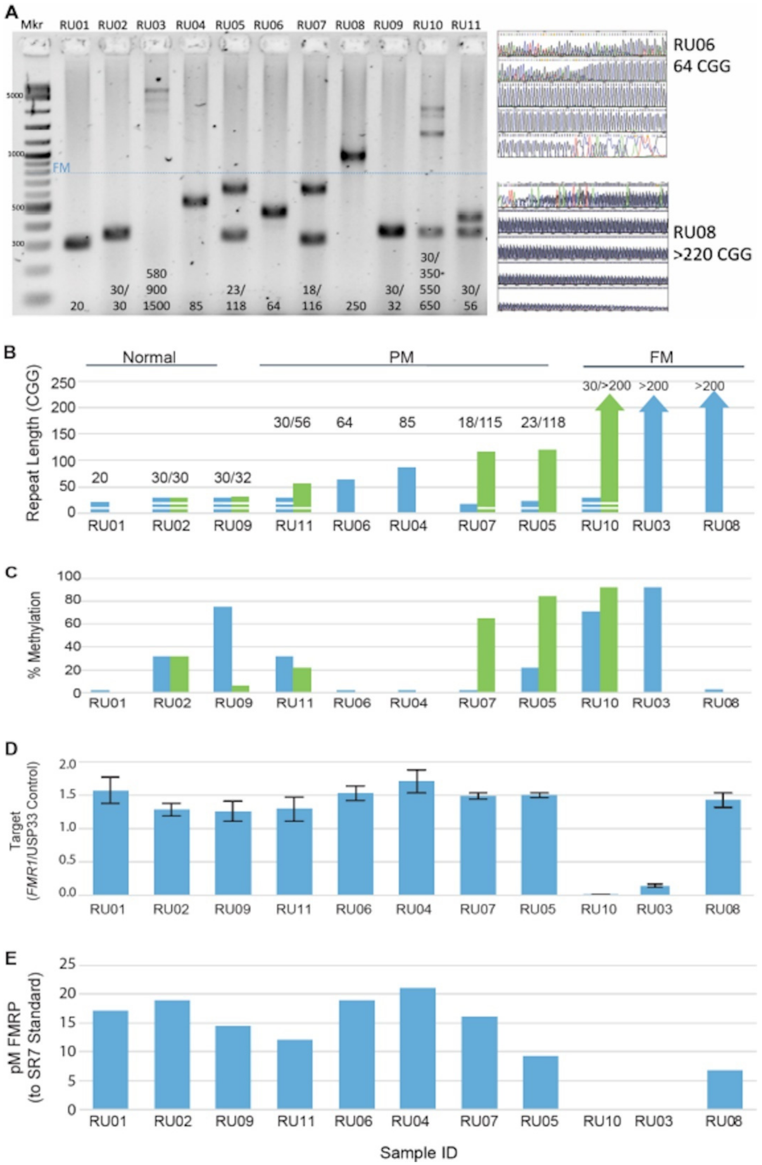
3.1. FMR1 DNA, RNA, and Protein Assays Assessed in a Reference Cell-Line Cohort

High-resolution, sensitive, and specific analyses of the gene and its protein are necessary to understand genotype–phenotype links and their potential clinical impact on FXS. We first compared nine FMR1 DNA, RNA, or protein-based assays (Table 2) using 11 well-characterized lymphoblastoid cell lines (Table 4) from patients with FXS. These nine assays included five FMR1 PCR assays [20,21,39,40,41,42,43,44,45,46] (Figure 1). As shown in Figure 1A,B, the results from both GS-PCR/AGE and RP-PCR/CE were in agreement, both for genotype category (i.e., normal, PM, or FM) and number of CGG repeats (within the known sizing resolution of AGE and CE). Sanger sequencing of selected expanded samples (Figure 1: RU06 and RU08) confirmed those findings. Analysis of interrupting AGG elements were consistent with previous reports demonstrating these interspersions were more highly represented in unexpanded alleles [40,41,55]. We note that the primary CGG sizing and AGG interruption genotypes in the immortalized cell lines were preserved when compared to results using these same assays for the original patient blood samples.
Next, FMR1 mPCR was used to determine the extent of gene methylation and silencing. As expected, the normal and PM cell-lines were unmethylated (males) or partially methylated (females). Two of the three FM cell lines were nearly completely methylated consistent with inactivation of FMR1 in FXS. However, one FM cell line (RU08) was unmethylated (Figure 1C). Southern blot analysis of the original whole blood sample also showed lack of methylation (Table 4). To investigate this finding further, we isolated RNA and quantified the primary FMR1 mRNA isoform on all 11 cell lines. Since a lack of FMR1 methylation would be expected to be associated with transcription of the gene, we anticipated that FMR1 RT-qPCR would demonstrate substantially greater transcript levels for RU08 compared to the other two FM cell-lines with methylated FMR1. Indeed, this was the case (Figure 1D). The level of FMR1 expression was also consistent with methylation status for the other cell-line samples (Figure 1D). We also compared methylation levels of FM cell lines with the corresponding blood samples used for cell immortalization and found nearly identical percent methylation for all three sample pairs.
Finally, we measured FMRP levels for each cell line to assess the impact of repeat expansions and other FMR1 DNA and RNA parameters on translation. Lysates of the 11 cell lines were analyzed by a previously established antibody-based qFMRP liquid bead array assay [42,43]. As shown in Figure 1E, FMRP levels were found to be well above the limit of detection for nine of the 11 cell lines including the unmethylated FM cell line (RU08). The two FM samples (RU03 and RU10) with pronounced gene methylation showed absence of FMRP. Thus, the results indicate that this set of multianalyte assays can quantify FMR1 gene and FMRP parameters across the range of FMR1 categorical genotypes with complementary results. Further, the molecular profiles of each cell line were consistent with the corresponding clinical diagnoses and related presentations for the subjects that provided specimens to generate them (Figure 1 and Table 4).

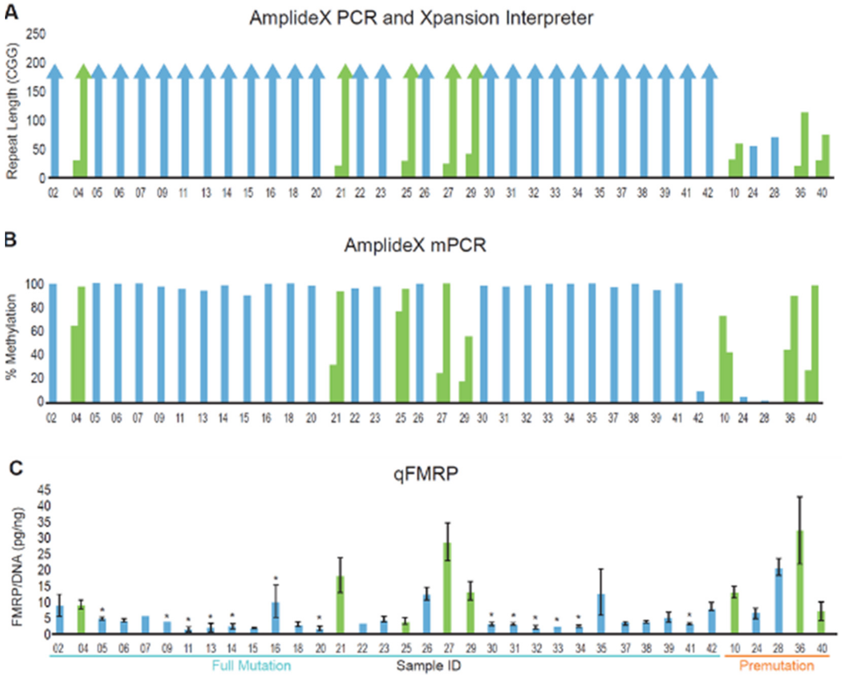
3.2. DNA and Protein Analysis of a Cohort of Patients with FMR1 Triplet Repeat Expansions

Having shown the linkage between FMR1 nucleic acid and protein assays in cell lines immortalized from patients with detailed clinical annotations, we then selected a random group of patients with expanded FMR1 genotypes that had been assessed through the KKI’s Fragile X Clinic (Clinical Cohort, Table 1). The FM–FMRP Subcohort of the Clinical Cohort was further divided into those with FXS-only and those with FXS + ASD. Additional information on the demographics of this cohort are provided in the Methods section and in Table 1. Whole blood (preserved on FTA cards or 903 cards) and buccal samples (preserved in a stabilizing solution), collected from each subject for subsequent molecular and protein testing, allowed comparisons of FMR1 DNA and FMRP profiles on cells from different lineages. Since the full set of FMR1 DNA analyses combined with FMRP quantification were most informative in associating molecular characteristics in FM cases, as shown in our Reference Cohort (Figure 1) and in multiple publications [1,2,4,11,15,19,46], we focused on these measures for subsequent studies.
Using both GS-PCR/CE and RP-PCR/CE, we found that the genotype categories determined for the overall Clinical Cohort were consistent with the original diagnostic test in each case. Specifically, the five PM samples had 56–113 repeats, whereas all 37 FM samples had alleles >200 repeats (Figure 2A,B). Further, the number of CGG repeats was almost always conserved when comparing DNA from whole blood collected in EDTA tubes, whole blood collected on FTA cards, or preserved buccal cells. The differences observed were generally restricted to the appearance of low-level size mosaic peaks (Figure 3 and Figure 4). Overall, slightly more than half of the samples were FM mosaics with evidence of PM alleles. This proportion is roughly in line with previous studies indicating that ~40% of FM samples are allele size mosaics [12,56]. PCR assays that mapped AGG elements in the repeat tract revealed no difference in the interruption pattern when comparing blood and buccal DNA for either unexpanded (most of which had AGG interspersions) or expanded alleles (many of which did not have AGGs).
As expected, the mPCR results indicated partially methylated or unmethylated PM alleles. For example, all three female PM samples were partially methylated and both male PM samples were unmethylated. Further, fully methylated FM alleles were observed in specimens from most subjects with FXS. Indeed, greater than 90% allele-specific methylation was observed for at least one discernible FM peak or near-FM peak in the CE trace for all 37 FM samples. Most samples revealed multiple peaks in the sizeable range of CE; MM was also commonly observed when considering all peaks in a sample, including those with relatively low signal intensity (Figure 3C and Figure 4). Of note, the average methylation of the normal X allele in the six females with FM was 44%, consistent with previous reports [39,47,52] and lyonization, as well as the expectation of a less severe clinical phenotype compared to the 26 FM–FMRP males.
As a next step, we assessed qFMRP levels across all subjects in the Clinical Cohort (Figure 2C). Group analysis of PM compared to FM demonstrated that qFMRP levels were higher in the former; however, three out of five PM were females, who generally have higher qFMRP levels compared to males. In addition, this analysis was limited by having only five individuals with PM. qFMRP levels within the FM–FMRP Subcohort spanned a 30-fold range, when both genders were included (N = 31), and a 12-fold range when only the FM–FMRP male-only subset was analyzed (N = 26). Although our dataset is too limited to definitively untangle the impact of size mosaicism from allele silencing in explaining FMRP expression, we did observe clear trends associating size and MM with FMRP levels. For example, FM alleles only were detected in seven of the ten blood samples with the lowest FMRP levels (i.e., those with less than 2.61 pg FMRP/ng gDNA) in the FM–¬FMRP male Subcohort. Further, these FM alleles were fully methylated in each case. Of the remaining three samples, one had a fully methylated 196 CGG allele and an FM allele, a result consistent with very low FMRP expression (1.2 pg/ng). The other two samples revealed both partially methylated PM and FM alleles; one of them had a 182 CGG PM allele that was only 10% methylated with a fully methylated FM allele. The large size of this PM may have contributed to the low FMRP expression (2.4 pg/ng) in this sample even though the allele was almost completely unmethylated. The remaining sample had a fully methylated FM and 163 CGG allele along with a 32% methylated 122 CGG allele. In this case, the 122 CGG allele may not have been sufficiently abundant in cells and/or efficiently expressed to generate a large amount of FMRP given the low level that was measured (1.48 pg/ng). In contrast to the low-expressing FMRP samples, six of the eight blood samples with FMRP levels higher than the average of 4.17 pg/ng for the Subcohort revealed unmethylated or partially methylated PM alleles.
This analysis shows that epigenetic mosaicism may indicate subpopulations of cells that are competent to express FMRP. Consequently, the FM male-only subset was further analyzed by comparing FMRP levels from subgroups with either appreciable MM (N = 13) or no measurable MM (N = 13) (Supplementary Materials). Of note, the MM subgroup included those with appreciable size mosaicism and MM (as defined in the Methods) but was categorized as MM given the more direct link between methylation status and FMR1 expression. ANOVA demonstrated that FMRP expression was significantly higher in samples with MM compared to those without MM (p = 0.02) (Figure 5).

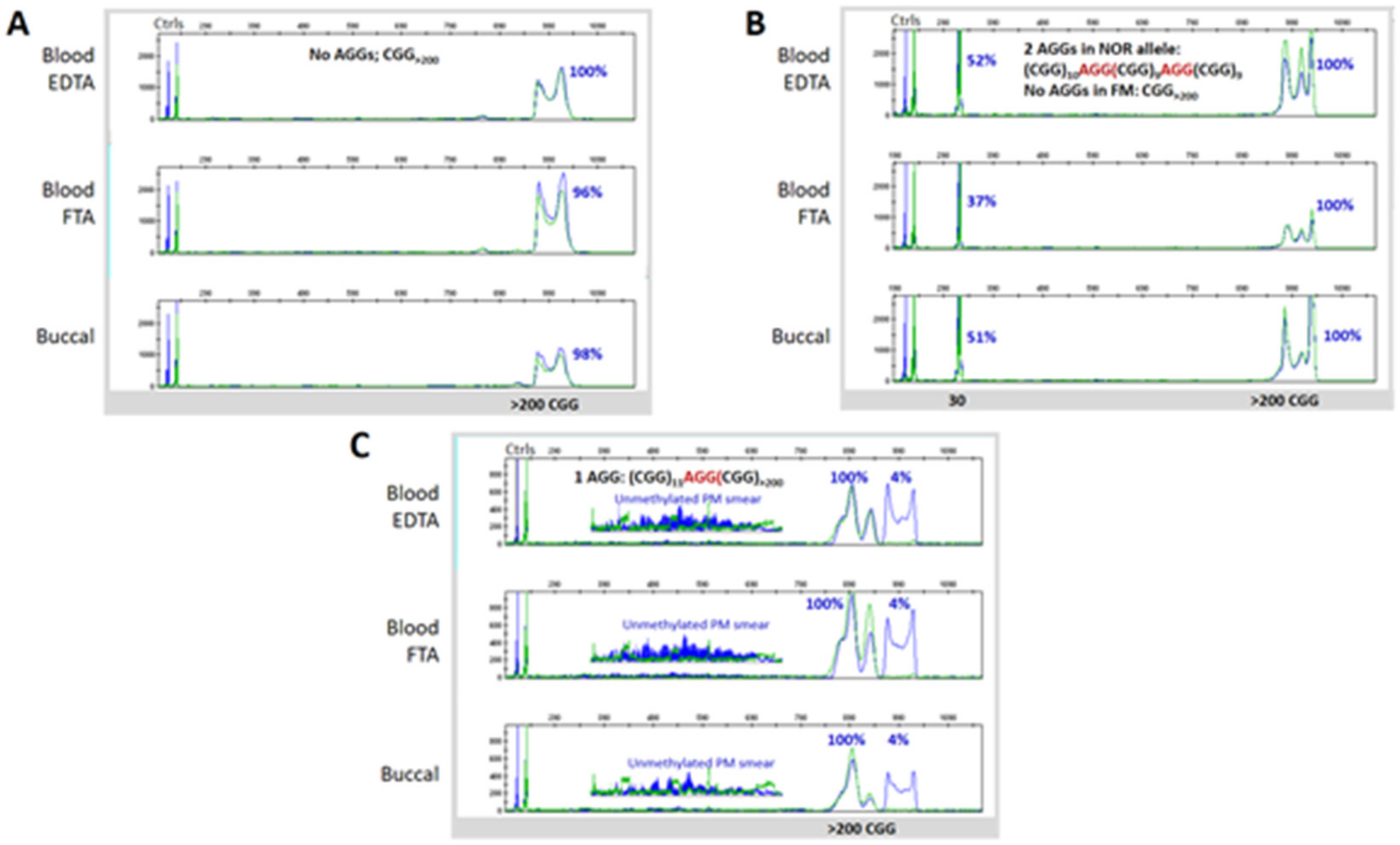
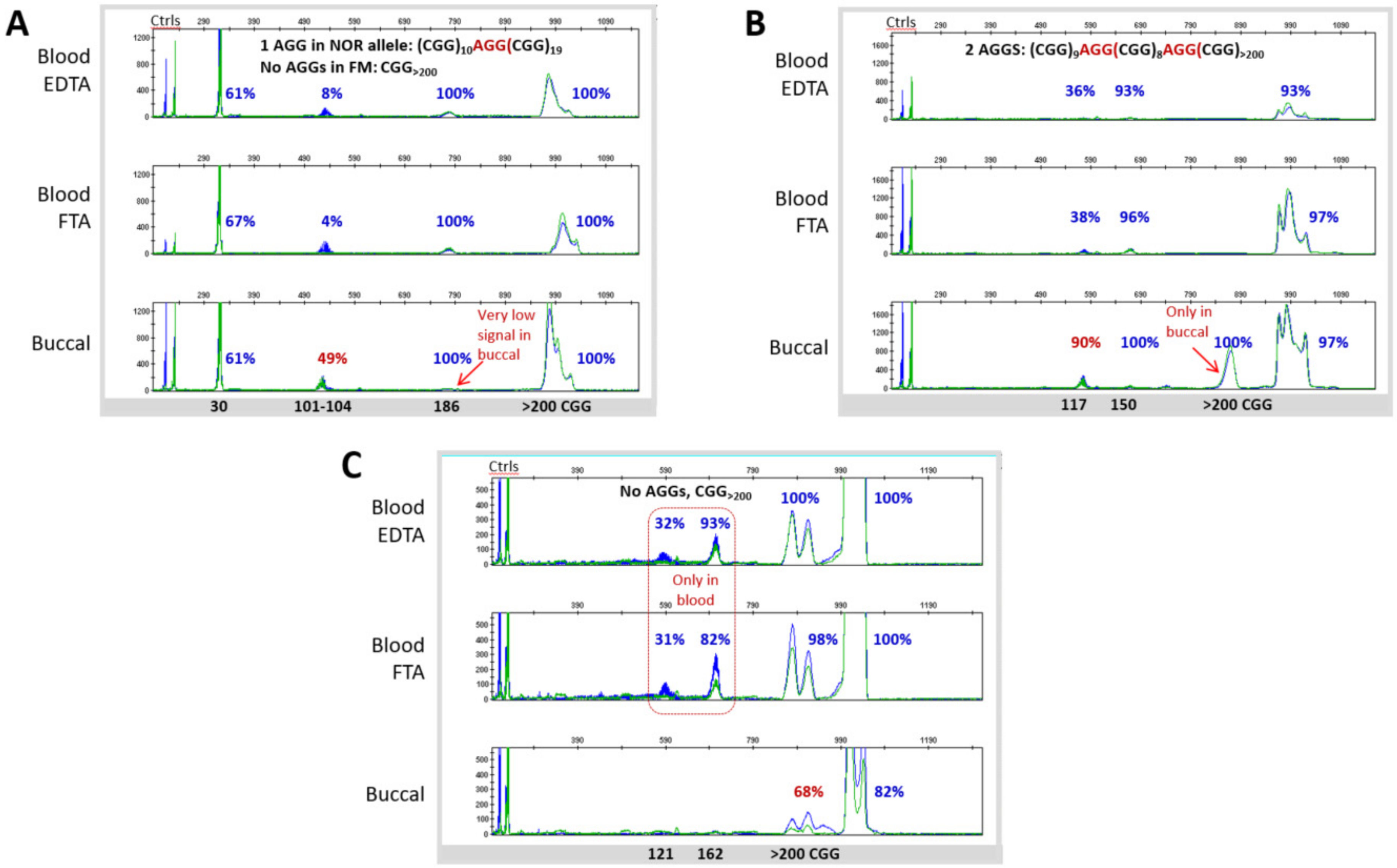
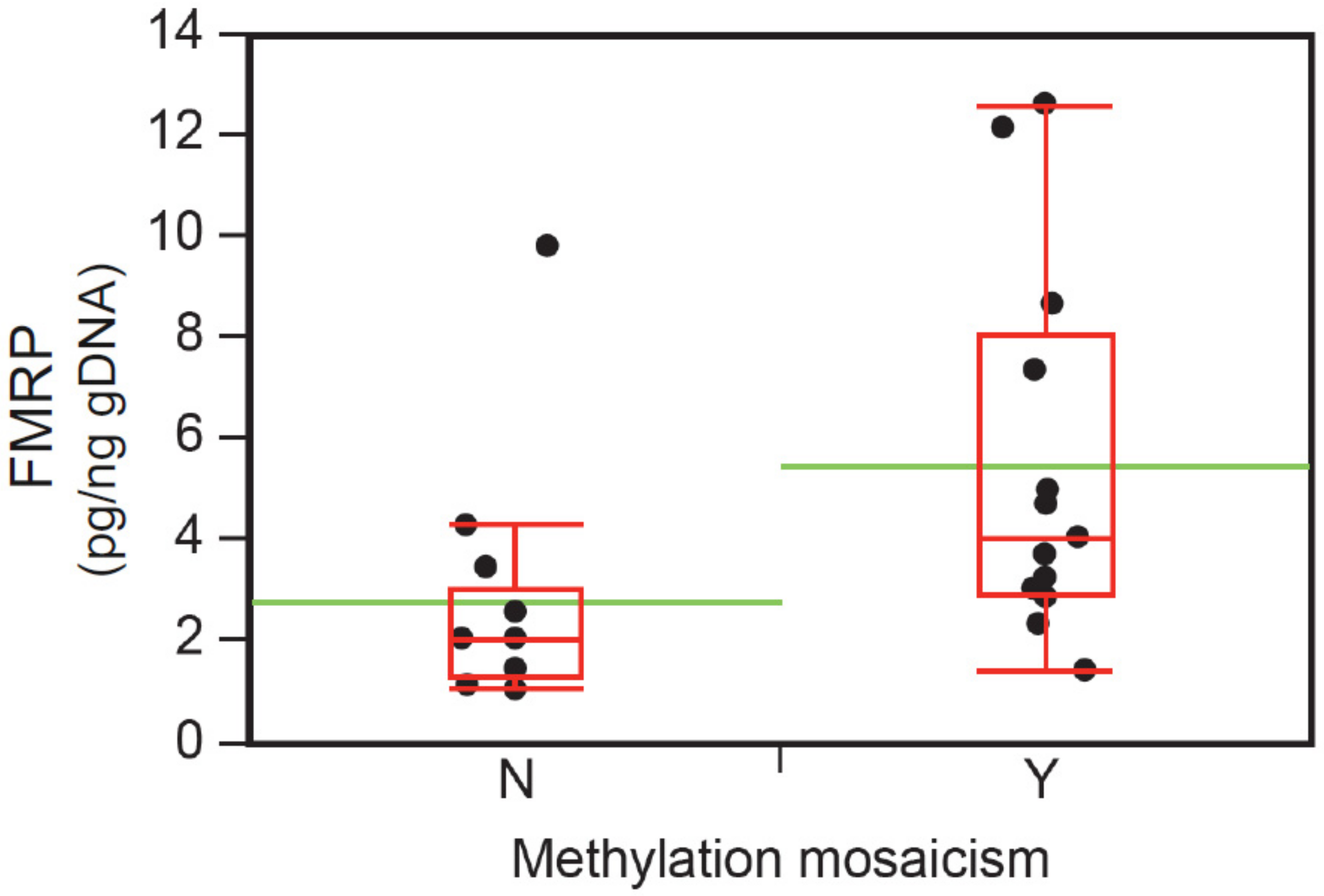
3.3. Blood and Buccal Samples Analyses: Molecular Comparisons within the Overall Clinical Cohort

Heterogeneity in FMR1 repeat size and methylation occurs in many patients with pathogenic repeat expansions. Nonetheless, it can also manifest in different tissues from the same patient, though the potential phenotypic consequences of these differences are not well understood. Consequently, we compared FM DNA profiles across matched blood and buccal specimens from the same individual. Mosaicism, as we defined it, was (i) detected with high analytical sensitivity using assays previously known to report mosaicism down to 1–5% of cell equivalents [21,57,58] and (ii) detected in about 50% of the subjects in our cohort (Supplementary Materials). As a result, this study offered multiple individuals for comparisons of matched blood and buccal specimens. In general, we found that mosaicism was conserved between these specimens from the same patient. Nevertheless, there were a handful of notable exceptions. One example is a ~101–104 CGG size mosaic in sample 04 that was nearly completely unmethylated in the DNA isolated from EDTA tubes or from the corresponding spotted blood cards but 49% methylated in the buccal DNA (Figure 4A). Another example (sample 15) showed mosaic peaks corresponding to 121 and 162 CGG repeats in blood that were absent in the buccal sample (Figure 4C). When differences in mosaicism were observed, however, they were largely constrained to allele-specific methylation in lower-intensity amplicon peaks by CE; repeat length and even peak intensity of size mosaics was conserved in nearly all samples. Thus, a general finding from our study was the primary genotype and epitype was maintained in matched blood and buccal specimens.

3.4. Neurobehavioral Profile of the FM–FMRP Subcohort

Table 5 depicts the neurobehavioral profiles of individuals in the FM–FMRP Subcohort (N = 31). Their age distribution was comparable to the FM–ALL Subcohort (N = 37). Although the number of male and female subjects was not balanced, gender differences in cognitive and adaptive measures, as well as distribution of intellectual functioning, were in line with the literature [35]. The mean FSIQ and adaptive skills composite scores were significantly lower in males than in females (p = 0.016 and p = 0.001, respectively, Mann–Whitney) and all individuals with moderate or severe ID were males. Frequency of SA diagnosis was high for both genders (68%), especially for females (100%). Unspecified anxiety was only found in males (38%), particularly among the lowest functioning (data not shown). Not surprisingly, ASD was present more frequently in males with FXS (46% vs. 20% females), who also had statistically significantly higher mean ABC-CFX total and CGI-S scores than females (p = 0.005 and p = 0.036, respectively, Mann–Whitney). FMRP levels were also significantly lower in males than females (p = 0.005, Mann–Whitney).

3.5. Neurobehavioral Profile of Individuals with ASD in the FM-FMRP Subcohort

In order to determine the influence of ASD status on neurobehavioral profiles in the FM–FMRP Subcohort, we compared males with and without ASD (FXS + ASD and FXS-only). Table 6 depicts these neurobehavioral profiles. Analogous to the differences between males and females, males with ASD had lower level of intellectual functioning, higher proportion of unspecified anxiety diagnosis, and higher mean ABC-CFX total and CGI-S scores than females. FMRP levels were also significantly lower in the FXS + ASD than in the FXS-only group. As expected from the literature, the FXS + ASD group also had higher scores on the ABC-CFX Irritability and Stereotypy subscales and a more prevalent use of antipsychotics. As mentioned above, SA was less prevalent in males FXS + ASD. However, SA severity using a CGI-S adapted measure (CGI-SANX) revealed a closer relationship between SA and ASD. When individuals with mild-moderate CGI-SANX scores (≤4, N = 7) were compared to those with severe CGI-SANX scores (≥5, N = 9), the proportion of males with ASD was higher in the latter (1/7 versus 4/9, respectively). Again, similar to the comparisons between males and females, males with FXS + ASD had lower levels of FMRP than those with FXS-only (Table 6, Figure 6).

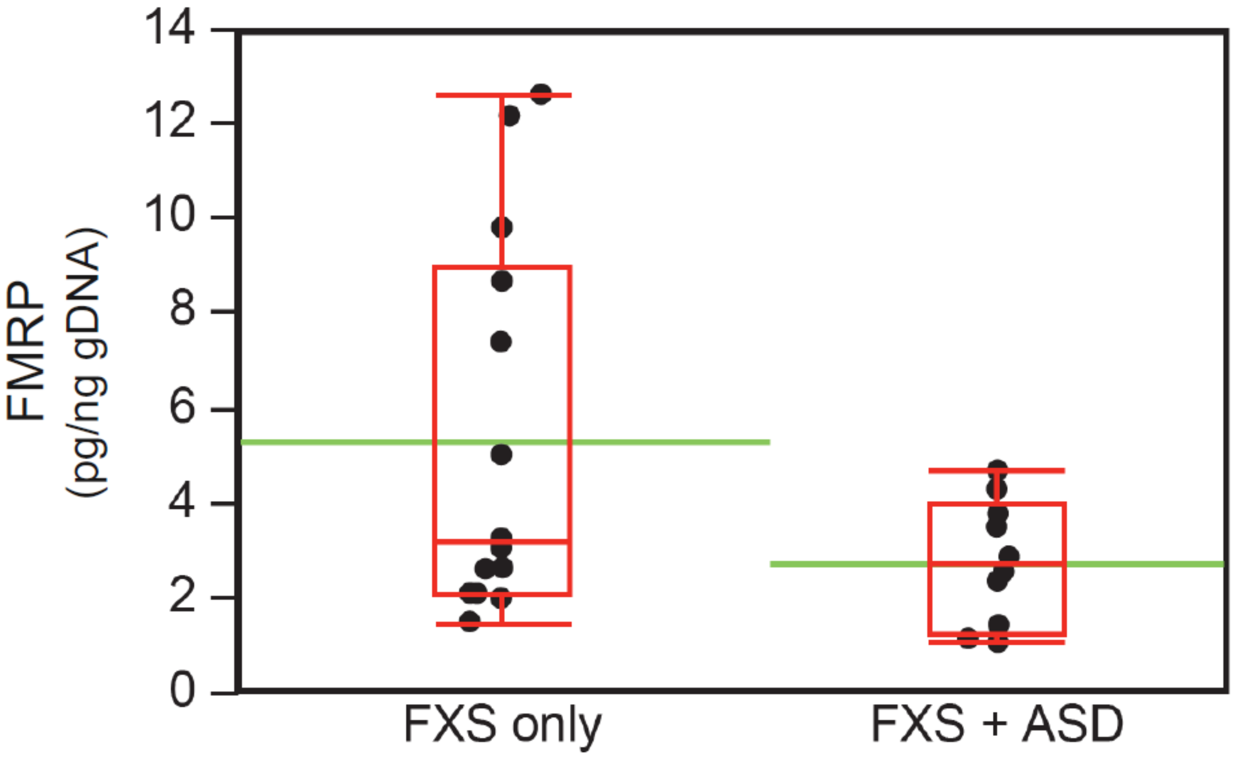
3.6. Intellectual Functioning, ASD Status, and FMRP Levels

Analyses of the FM–FMRP Subcohort presented above have demonstrated groups with lower cognitive functioning, as well as those with ASD, have lower FMRP levels. Thus, we evaluated in greater detail the relationship between intellectual functioning and ASD, and related behaviors, and FMRP in the male subgroups described in Table 6. First, we examined the distribution of FMRP values in those with or without ASD. There were four outliers in the FXS-only; when either two randomly selected or the four were excluded from the analyses, both parametric and nonparametric t-tests showed significant lower levels in the FXS + ASD group (p = 0.05 and p = 0.01, respectively; Welch’s t-test and Mann–Whitney).
Since previous studies have already reported an inverse relationship between intellectual functioning and FMRP levels and a close association between more severe ID and ASD, we also evaluated the role of level of intellectual impairment in the ASD-lower FMRP relationship. An assessment of ID categories and FMRP levels (in pg FMRP/ng gDNA) showed that males with FXS and either mild or moderate ID had significantly higher levels of FMRP than those with severe ID (N = 15, 5.2 ± 3.8 versus N = 10, 2.3 ± 1.1; p = 0.013 and p = 0.019, respectively; Welch’s t-test and Mann–Whitney). These differences were also present when males with FXS + ASD and mild or moderate ID were compared with those with ASD and severe ID (N = 5, 3.3 ± 1.4 versus N = 7, 2.4 ± 1.2) affected by the small subsample (p = 0.4, Mann–Whitney), and when similar groups without ASD were compared (FXS-only and mild + moderate ID versus FXS-only and severe ID: N = 10, 6.1 ± 4.3 versus N = 3, 2.1 ± 0.6, p = 0. 015 and p = 0.07, respectively; Welch’s t-test and Mann–Whitney). There was also a statistical trend towards lower FMRP levels in the FXS + ASD subgroup with mild + moderate ID when compared with FXS-only with mild + moderate ID (N = 5, 3.3 ± 1.4 versus N = 10, 6.1 ± 4.3; p = 0.075, Welch’s t-test), but no differences when groups with severe ID with and without ASD were compared. Another factor that could contribute to the ASD-lower FMRP association is the presence of social behavior impairment that can be interpreted as or associated with ASD. As described above, the group of males with severe SA, by CGI-SANX scores, had a higher proportion of individuals with ASD. This group also had lower FMRP levels than those with mild-moderate SA (3.1 ± 2.2 versus 5.4 ± 3.4; p = 0.05, Mann–Whitney).
Finally, we examined the influence of age, overall clinical severity, as measured by CGI-S, and overall problem behavior severity, as determined by ABC-CFX total score, on FMRP levels within the FXS + ASD group. Younger males (2–11 years, 58%) had two-fold lower FMRP levels (pg FMRP/ng gDNA) (N = 7/15, 3.2 ± 1.2 versus N = 8/15, 6.6 ± 4.4 FXS-only, p = 0.03, Welch’s t-test), adjusted for age- (6.9 ± 0.9 versus 6.9 ± 3.1 FXS-only, p = 0.49, Welch’s t-test) and FSIQ (44.1 ± 17.7 versus 59.6 ± 16.6 FXS-only, p = 0.12, Welch’s t-test); while males with an ABC-CFX total score ≥50 and/or severe CGI-S scores (≥5) also closely linked to FXS + ASD, had significantly lower mean levels of FMRP (p = 0.01 and p = 0.05, respectively; Welch’s t-test).To illustrate the potential clinical impact of the FMR1 and FMRP measurements reported here, one high functioning 7-year old male in the FXS-only group was an outlier with a normal FSIQ of 95 and an adaptive VABS-II Composite score of 79. His FMRP level was 7.4; (mean ± SD) all males (4.2 ± 3.3), FXS-only (5.4 ± 4.0). The subject had unmethylated size and methylation mosaicism on his blood sample and a partially methylated PM smear with an additional PM peak on the buccal sample.

4. Discussion

To our knowledge, this is the first study to integrate high-resolution FMR1 DNA, RNA and protein analyses and to correlate these molecular measures with multiple neurobehavioral parameters, including ASD diagnosis, in order to refine genotype-phenotype correlations in FXS. We first used multiple FMR1 and FMRP assays to deeply profile pathogenic FMR1 CGG expansions, and to determine their impact on transcription and translation in well-characterized cell lines from individuals with a range of FMR1 genotypes. We then applied the most revealing of these assays to an FXS cohort with comprehensive neurobehavioral profiling to investigate molecular profiles and molecular-neurobehavioral correlations.
Using a combination of sensitive and quantitative multi-omic assays, we found that FMR1 CGG repeat length, methylation levels, and FMRP levels were complementary and consistent with our current knowledge of FMR1 translational biology in individuals with normal, PM, and FM alleles [1,2,4,5]. FM expansions were associated with hypermethylation, gene silencing based on FMR1 mRNA levels much lower in individuals with FM than carriers with PM, and FMRP levels lower in males with FM than in females with FM and much lower in subjects with FM overall compared to individuals with PM. Molecular profiles in lymphoblastoid cell lines were corroborated in blood samples with a variety of FMR1 expansions; these expansions included a subject with an FM allele that was unmethylated in both immortalized cell line and original blood specimen and exhibited FMR1 mRNA and FMRP levels similar to those of individuals with PM. Although unmethylated FM alleles are rare, several examples have been described in the primary literature [2,4,15,16,39]. We also observed that blood-based profiles were in general consistent with those in buccal epithelial specimens. These findings are contributory since concordance in patterns of FMR1 mosaicism across tissues continues to be a controversial issue in FXS [46,59,60]. Moreover, very little is known about the correlation between blood FMRP measurements and postmortem brain tissue FMRP levels in the same subject [61,62]. Only a single case study “verified” reduced expression of FMR1 mRNA and FMRP in both peripheral blood and brain leading to the FXS [61]. In our study, a representative sample of males and females with FXS, with their corresponding range of cognitive and behavioral impairments [9,29], served as the basis for examining FMR1 methylation, FMRP levels and their relationship with neurobehavioral features. As expected, samples with complete or near complete FMR1 gene methylation by mPCR exhibited the lowest FMRP values. Females with FXS, not surprisingly, showed lower relative gene methylation and higher FMRP levels. FMRP levels were generally lower for males with FM-only compared to males with PM, females with FM, and males with methylation mosaicism, with the latter groups displaying a wide range of values as described earlier [9,10,19]. A significant finding from this study is that FMRP levels were two-fold higher in males with FXS and appreciable methylation mosaicism compared to males without such mosaicism, expanding our knowledge on the link between methylation and FMR1 silencing. In other published work, Jiraanont and colleagues used FMR1 PCR and mPCR along with Southern blot, RT-qPCR, and FMRP levels to evaluate size- and methylation-mosaicism in 12 males with FXS (7 with FXS + ASD) [11]. They concluded lower FMR1 mRNA and FMRP levels were the main contributors to cognitive impairment and the presence of a normal allele appeared to compensate in some but not all individuals. Pretto et al. [46] studied 18 patients with FXS (9 with FXS + ASD), including 7 with size- and 6 with methylation-mosaicism, demonstrating that FMR1 RNA and FMRP levels correlated in general with FMR1 methylation in peripheral blood cells and fibroblasts although there were differences in CGG expansion and blood samples showed lower methylation. They also reported FSIQ scores were inversely correlated with level of methylation and directly with FMRP levels. However, other aspects of the neurobehavioral phenotype, such as number of seizures and severity of hyperactivity or autistic behavior were not related to methylation or FMRP levels.
Considering the relatively consistent relationship between FMR1 methylation, FMR1 mRNA, and FMRP and the more direct phenotypical implications of protein expression, FMRP levels seem to be the most suitable molecular parameter for molecular-neurobehavioral analyses [1,2]. Our findings confirm previous studies [9,10,29,36] showing FMRP is a biomarker of overall clinical severity in FXS, which may help stratify patients with FXS in clinical trials [38]. We found a strong link between FMRP levels and IQ or level of ID [1,7,9,10,11,19,29,36,46], evident in male–female comparisons and in analysis of males with a range of clinical presentations. Nonetheless, the availability of a sensitive and precise assay such as qFMRP [43], allowed us to detect other phenotypical correlates of FMRP deficiency not reported in the aforementioned studies. For instance, we found two-fold lower levels of FMRP particularly in younger males (age- and IQ-adjusted) with FXS and ASD (57% with severe ASD) than in those with FXS-only, although there was a wide range of protein expression in the latter group. Nonetheless, for total FM–FMRP male-only subset, this finding only held in ones with mild-moderate ID, as those with severe ID had even lower FMRP levels independent of ASD status. Since marked decrease in FMRP is not only the basis for FXS but also a contributor to the ASD phenotype, as the signaling pathways of FMR1 and other genes linked to the behavioral disorder substantially overlap [23,24,28,63], a more definitive answer about the relationship between FMRP levels and ASD status deserves further study. Other relationships between FMRP levels and neurobehavioral parameters included unspecified anxiety only in low functioning males with low FMRP and ASD, and SA in females and higher functioning males without ASD but with higher FMRP levels. Overall, our study replicated an extremely high rate of anxiety in FXS reported in the literature regardless of gender [64]. Anxiety is intertwined in FXS [64,65], and in FXS with ASD [36]. Two potentially clinically meaningful SA subgroups within the male-only group were identified with separately applied CGI-S SA scoring (CGI-SANX)—an approach which has also been used in a second wave of clinical trials in FXS (i.e., the NCT03697161gaboxadol recently completed study). Further study of anxiety in relationship to FMRP and ASD status in FXS may also help stratify patients with FXS in clinical trials [38].
As with ASD in males with severe ID, the relationship between FMRP levels and overall severe problem behavior (ABC-CFX total score ≥50) was also cofounded with greater cognitive impairment. These findings are in line with the recognition, in recent years, of a more severe neurobehavioral phenotype mainly in males with FXS. This is characterized by severe ID, ASD diagnosis, and severe irritability, aggression, agitation, and self-injury types of often anxiety-driven behavior [25,27,66]. Indeed, our sample of males with FXS and ASD [26,34] had significantly elevated ABC-CFX total scores driven by the Irritability subscale [67], and a higher proportion of anxiety diagnosis and CGI-SANX scores, respectively, which together required frequent use of atypical antipsychotic aripiprazole [68,69,70]. A presumed link between anxiety and ABC-CFX Irritability subscale in lower functioning individuals with FXS also led experts in fragile X field to include the latter as an outcome measure in cannabinol clinical trials in FXS (NCT03614663 and NCT03802799). Our findings suggest that the lowest FMRP levels seem to concentrate in this group of male individuals. Availability of sensitive assays like qFMRP used here are key for advancing our understanding of the role of FMRP deficit in FXS, since threshold levels (<70% of FMRP level observed in those with normal CGG repeat numbers) for ID have been recently described [19] and delineating differences among those with the lowest levels will require high power discrimination [38,53,71].

5. Limitations

Although our study, to our knowledge, is the largest molecular–neurobehavioral correlation using multiple and sensitive FMR1 and FMRP assays, the sample size was relatively modest considering the variability and complexity of the molecular and phenotypical measures. Marked neurobehavioral differences between males and females with FXS, coupled with the well-known genetic differences inherent to X-linked disorders (e.g., X-inactivation in females) complicated the molecular and correlational analyses. Given the size of the cohort, the necessity of combining size- and methylation-mosaicism limited the full appreciation of differences between these types of mosaicism. Despite the use of raw score-based z-score calculations for FSIQ, floor effects on cognitive or adaptive skills testing for some subjects were also a limitation for studying range of cognitive impairment. The lack of a formal anxiety measure also limited our ability to subtype anxiety.

6. Conclusions

Our findings underscore the association between FMR1 CGG expansion, gene methylation, FMRP levels, and overall neurobehavioral severity, including an ASD diagnosis. The synergy of FMR1 genomic and protein parameters and the concordance of blood and buccal profiles using a mixture of sensitive and quantitative multi-omic assays support the benefit of characterizing molecular profiles particularly in males with FXS in observational and interventional studies. The benefit of stratifying their genotype–phenotype profiles is further strengthened by two-fold higher FMRP levels and lesser clinical severity in a subset of these males with substantial methylation mosaicism. Thus, the benefit also highlights the potential of FMRP levels, in particular, as a prognostic marker in clinical trials and other relevant studies in males with FXS.

Supplementary Materials

The following are available online at https://0-www-mdpi-com.brum.beds.ac.uk/2076-3425/10/10/694/s1. Title of data: Clinical description and summary of molecular testing results for 31 patients with FM. Description of data: Clinical description (gender, age, level of intellectual disability, estimated extended FISQ, presence of ASD) and summary of molecular testing (FMRP levels, and FMR1 size and methylation data) for 26 male and 5 female patients with FM. * In order to address a skewed effect of the FSIQ-standard score testing, raw-data based subscores were applied instead by a senior neuropsychologist, which allowed an extended FSIQ score to be generated. Briefly, if the SB-5 subtests raw scores were available, they were used for the extended FSIQ scoring. If other than aforementioned IQ standard tests were available, those raw scores were used to estimate the extended FSIQ. When there were several administrations of different tests, the most current administration was used for the consensus. Alternatively, if the scores were highly discrepant, estimation was made somewhere in the middle of the two raw data-based scores. ** Level of methylation mosaicism was evaluated for 26 male patients with FM and scored as: 1-not detected, 2-low level of mosaicism, and 3-high level of mosaicism, based on relative intensities of unmethylated or partially methylated peaks compared to fully methylated peaks using the FMR1 mPCR assay.

Author Contributions

D.B.B. and G.J.L. were responsible for the initial conceptualization of the study. D.B.B. was responsible for patients’ recruitment and retention. He extensively reviewed the data and executed the design of this manuscript, participated in the interpretation of the data, initially drafted the manuscript, developed and modified the tables throughout, critically revised the manuscript, was responsible for its final content, and primarily coordinated all necessary contributions of coauthors of the manuscript throughout the process. A.S. and G.J.L. drafted the manuscript; developed, wrote, and revised the subsections of the manuscript; assisted with the design of the manuscript; generated references in endnote; and applied them in the body text. A.S., S.F.-S., and G.J.L. were responsible for data management, statistical analyses, and preparation of the Figures. E.B., S.F.-S., J.K., K.N., K.C., K.J., A.H., and G.J.L. were all responsible for lab analyses and data collection and interpretation. D.D.P. participated in statistical analyses and interpretation of the data, modified the tables throughout, helped with clinical data management and submission process. E.M.M. generated an extended raw scores analyses of FSIQ scores and critically reviewed the manuscript. T.A., G.L., C.D., and W.T.B. were responsible for lab analyses by providing the IBR protocol, guidance and support in setting up the FMRP assays at Asuragen. K.S., L.Z., and E.B.-K. were responsible for collection of control samples, and E.B.-K. oversaw data collection, participated in interpretation of the data, and critically reviewed and revised the manuscript, and helped to finalize it. W.E.K. critically reviewed and participated in interpretation of the data, statistical analyses, and helped to revise and finalize the manuscript. G.J.L. critically reviewed and revised the manuscript, assisted with the design of the manuscript, helped to finalize the manuscript, participated in interpretation of the data, and coordinated efforts among other coauthors when necessary. All authors have read and agreed to the published version of the manuscript.

Funding

We acknowledge NICHD grant 1R43HD082900-01A1 (Latham PI) subcontract to Kennedy Krieger Institute (PI: Budimirovic).

Acknowledgments

Clinical Trials Unit staff and phlebotomists at the Institute. Anca Toma, and Hilary Rosselot for their assistance with the manuscripts technical support, including their proofreading efforts. Since 2013, these results were presented at the following conferences: 1st through 4th International Conference on FMR1 premutation in 2013, 2015, 2017, and 2019, respectively; ACMG Annual Clinical Genetics Meeting 2014; 15th and 16th International Fragile X Conferences in 2016 and 2018, respectively; Gordon Research Conference “Fragile X and Autism-Related Disorders” in 2016; International Conference on Autism and Fragile-X Associated Disorders in 2019, and American Academy of Child & Adolescent Psychiatry Annual Meeting in 2019.

Conflicts of Interest

D.B.B. has received funding from Seaside, Roche, Neuren, Pfizer, Shire, Lundbeck, Forest, Sunovion, SyneuRX, Alcobra, Akili, Medgenics, Purdue, Supernus as a main sub-investigator, and Ovid and Zynerba Pharmaceuticals as the principal investigator on clinical trials. He also consulted on clinical trial outcome measures focused meetings (Seaside, Ovid). All the above funding has been directed to Kennedy Krieger Institute/the Johns Hopkins Medical Institutions; D.B.B. receives no personal funds, and the Institute has no relevant financial interest in any of the commercial entities listed. A.S., S.F.-S., E.B., K.N., K.C., K.J., J.K., A.H., and G.J.L. are or were employed by Asuragen at the time the work was performed and have or may have stock options in the company. G.J.L., E.B., and A.H. are inventors on one or more patents or patent applications associated with genotyping or epityping FMR1 molecular features. E.B.-K. has received funding from Acadia, Alcobra, AMO, Asuragen, BioMarin, Cydan, Fulcrum, GeneTx, GW, Ionis, Lumos, Marinus, Neuren, Neurotrope, Novartis, Ovid, Roche, Seaside Therapeutics, Ultragenyx, Vtesse/Sucampo/Mallinckrodt, Yamo, and Zynerba Pharmaceuticals to consult on clinical trial design, run clinical trials, or develop testing standards or biomarkers, all of which is directed to RUMC in support of rare disease programs; E.B.-K. receives no personal funds, and RUMC has no relevant financial interest in any of the commercial entities listed. G.L. and W.T.B. declare no conflicts of interest for the patent “System and Method for Quantifying Fragile X Mental 1Protein in tissue and blood samples” (United States Patent # 8628934). W.E.K. receives no personal funds and has no relevant financial interest in any of the commercial entities listed. He is Chief Medical Officer of Anavex Life Sciences Corp. and has been consultant to AveXis, EryDel, GW, Marinus, Neuren, Newron, Ovid, Stalicla, and Zynerba. The other co-authors declare that they have no competing interests.

Abbreviations

ABAS-II and ABAS-III—Adaptive Behavior Assessment System Second and Third Editions; ABC-CFX—Aberrant Behavior Checklist-Community adapted for Fragile X; ADHD—Attention deficit hyperactivity disorder; AGE—Agarose gel electrophoresis; AGG—adenine-guanine-guanine; ASD—Autism Spectrum Disorders; ADOS—Autism Diagnostic Observation Schedule; CE—Capillary electrophoresis; CGI-S—Clinical Global Impression-Severity; CGI-SANX—CGI applied to profile the severity/level of social anxiety; FM—full mutation; DAS—Differential Ability Scales; df—Degrees of Freedom; FMRP—Fragile X mental retardation protein; FSIQ—full scale intelligence quotient; FX—fragile X; FXS—fragile X syndrome; gDNA—genomic DNA; GS-PCR—gene-specific PCR; GS-PCR/AGE—gene-specific PCR with products resolved by agarose gel electrophoresis; ID—intellectual disability; IQ—intelligence quotient; IRB—Institutional Review Board; KKI—the Kennedy Krieger Institute; MM-methylation mosaicism; mPCR—methylation PCR; PBS—phosphate-buffered saline; PM—premutation; RIN—RNA Integrity Number; RP-PCR—Repeat-primed PCR; RP-PCR/CE—Repeat-primed PCR with products resolved by capillary electrophoresis; RT-qPCR—Reverse transcription quantitative PCR (or quantitative reverse transcription PCR); RFU—relative fluorescence units; RUMC—Rush University Medical Center; SA-social anxiety; SB-5—Stanford–Binet Intelligence Scales-5th Edition; SD—Standard Deviation; SIB-R—Scales of Independent Behavior-Revised; VABS-II—Vineland Adaptive Behavior Scales-Second Edition; WB—whole blood; WISC-IV and WISC-V—Wechsler Intelligence Scale for Children, Fourth and Fifth Edition; WPPSI—Wechsler Preschool and Primary Scale of Intelligence.

References

  1. Kaufmann, W.E.; Reiss, A.L. Molecular and cellular genetics of fragile X syndrome. Am. J. Med. Genet. 1999, 88, 11–24. [Google Scholar] [CrossRef]
  2. Bagni, C.; Tassone, F.; Neri, G.; Hagerman, R. Fragile X syndrome: Causes, diagnosis, mechanisms, and therapeutics. J. Clin. Investig. 2012, 122, 4314–4322. [Google Scholar] [CrossRef] [PubMed] [Green Version]
  3. Gholizadeh, S.; Halder, S.K.; Hampson, D.R. Expression of fragile X mental retardation protein in neurons and glia of the developing and adult mouse brain. Brain Res. 2015, 1596, 22–30. [Google Scholar] [CrossRef] [PubMed]
  4. Banerjee, A.; Ifrim, M.F.; Valdez, A.N.; Raj, N.; Bassell, G.J. Aberrant RNA translation in fragile X syndrome: From FMRP mechanisms to emerging therapeutic strategies. Brain Res. 2018, 1693 Pt A, 24–36. [Google Scholar] [CrossRef]
  5. Darnell, J.C.; Klann, E. The translation of translational control by FMRP: Therapeutic targets for FXS. Nat. Neurosci. 2013, 16, 1530–1536. [Google Scholar] [CrossRef] [Green Version]
  6. Sidorov, M.S.; Auerbach, B.D.; Bear, M.F. Fragile X mental retardation protein and synaptic plasticity. Mol. Brain 2013, 6, 15. [Google Scholar] [CrossRef] [Green Version]
  7. Baker, E.K.; Arpone, M.; Vera, S.A.; Bretherton, L.; Ure, A.; Kraan, C.M.; Bui, M.; Ling, L.; Francis, D.; Hunter, M.F.; et al. Intellectual Functioning and Behavioural Features Associated With Mosaicism in Fragile X Syndrome. J. Neurodev. Disord. 2019, 11, 41. [Google Scholar] [CrossRef]
  8. Lessard, M.; Chouiali, A.; Drouin, R.; Sebire, G.; Corbin, F. Quantitative measurement of FMRP in blood platelets as a new screening test for fragile X syndrome. Clin. Genet. 2012, 82, 472–477. [Google Scholar] [CrossRef]
  9. Loesch, D.Z.; Huggins, R.M.; Hagerman, R.J. Phenotypic variation and FMRP levels in fragile X. Ment. Retard. Dev. Disabil. Res. Rev. 2004, 10, 31–41. [Google Scholar] [CrossRef]
  10. Kaufmann, W.E.; Abrams, M.T.; Chen, W.; Reiss, A.L. Genotype, Molecular Phenotype, and Cognitive Phenotype: Correlations in Fragile X Syndrome. Am. J. Med. Genet. 1999, 83, 286–295. [Google Scholar] [CrossRef]
  11. Jiraanont, P.; Kumar, M.; Tang, H.T.; Espinal, G.; Hagerman, P.J.; Hagerman, R.J.; Chutabhakdikul, N.; Tassone, F. Size and methylation mosaicism in males with Fragile X syndrome. Expert Rev. Mol. Diagn. 2017, 17, 1023–1032. [Google Scholar] [CrossRef] [Green Version]
  12. Nolin, S.L.; Glicksman, A.; Houck, G.E., Jr.; Brown, W.T.; Dobkin, C.S. Mosaicism in fragile X affected males. Am. J. Med. Genet. 1994, 51, 509–512. [Google Scholar] [CrossRef] [PubMed]
  13. Tassone, F.; Hagerman, R.J.; Gane, L.W.; Taylor, A.K. Strong similarities of the FMR1 mutation in multiple tissues: Postmortem studies of a male with a full mutation and a male carrier of a premutation. Am. J. Med. Genet. 1999, 84, 240–244. [Google Scholar] [CrossRef]
  14. Dobkin, C.S.; Nolin, S.L.; Cohen, I.; Sudhalter, V.; Bialer, M.G.; Ding, X.H.; Jenkins, E.C.; Zhong, N.; Brown, W.T. Tissue differences in fragile X mosaics: Mosaicism in blood cells may differ greatly from skin. Am. J. Med. Genet. 1996, 64, 296–301. [Google Scholar] [CrossRef]
  15. Hagerman, R.J.; Hull, C.E.; Safanda, J.F.; Carpenter, I.; Staley, L.W.; O’Connor, R.A.; Seydel, C.; Mazzocco, M.M.; Snow, K.; Thibodeau, S.N.; et al. High functioning fragile X males: Demonstration of an unmethylated fully expanded FMR-1 mutation associated with protein expression. Am. J. Med. Genet. 1994, 51, 298–308. [Google Scholar] [CrossRef]
  16. Taylor, A.K.; Tassone, F.; Dyer, P.N.; Hersch, S.M.; Harris, J.B.; Greenough, W.T.; Hagerman, R.J. Tissue heterogeneity of the FMR1 mutation in a high-functioning male with fragile X syndrome. Am. J. Med. Genet. 1999, 84, 233–239. [Google Scholar] [CrossRef]
  17. Maddalena, A.; Yadvish, K.N.; Spence, W.C.; Howard-Peebles, P.N. A fragile X mosaic male with a cryptic full mutation detected in epithelium but not in blood. Am. J. Med. Genet. 1996, 64, 309–312. [Google Scholar] [CrossRef]
  18. Ciaccio, C.; Fontana, L.; Milani, D.; Tabano, S.; Miozzo, M.; Esposito, S. Fragile X syndrome: A review of clinical and molecular diagnoses. Ital. J. Pediatr. 2017, 43, 39. [Google Scholar] [CrossRef] [Green Version]
  19. Kim, K.; Hessl, D.; Randol, J.L.; Espinal, G.M.; Schneider, A.; Protic, D.; Aydin, E.Y.; Hagerman, R.J.; Hagerman, P.J. Association between IQ and FMR1 protein (FMRP) across the spectrum of CGG repeat expansions. PLoS ONE 2019, 14, e0226811. [Google Scholar] [CrossRef] [Green Version]
  20. Chen, L.; Hadd, A.; Sah, S.; Filipovic-Sadic, S.; Krosting, J.; Sekinger, E.; Pan, R.; Hagerman, P.J.; Stenzel, T.T.; Tassone, F.; et al. An information-rich CGG repeat primed PCR that detects the full range of fragile X expanded alleles and minimizes the need for southern blot analysis. J. Mol. Diagn. 2010, 12, 589–600. [Google Scholar] [CrossRef]
  21. Filipovic-Sadic, S.; Sah, S.; Chen, L.; Krosting, J.; Sekinger, E.; Zhang, W.; Hagerman, P.J.; Stenzel, T.T.; Hadd, A.G.; Latham, G.J.; et al. A novel FMR1 PCR method for the routine detection of low abundance expanded alleles and full mutations in fragile X syndrome. Clin. Chem. 2010, 56, 399–408. [Google Scholar] [CrossRef] [PubMed] [Green Version]
  22. Yrigollen, C.M.; Martorell, L.; Durbin-Johnson, B.; Naudo, M.; Genoves, J.; Murgia, A.; Polli, R.; Zhou, L.; Barbouth, D.; Rupchock, A.; et al. AGG interruptions and maternal age affect FMR1 CGG repeat allele stability during transmission. J. Neurodev. Disord. 2014, 6, 24. [Google Scholar] [CrossRef] [PubMed] [Green Version]
  23. Kover, S.T.; Pierpont, E.I.; Kim, J.S.; Brown, W.T.; Abbeduto, L. A neurodevelopmental perspective on the acquisition of nonverbal cognitive skills in adolescents with fragile X syndrome. Dev. Neuropsychol. 2013, 38, 445–460. [Google Scholar] [CrossRef] [PubMed]
  24. Hagerman, R.J.; Polussa, J. Treatment of the psychiatric problems associated with fragile X syndrome. Curr. Opin. Psychiatry 2015, 28, 107–112. [Google Scholar] [CrossRef]
  25. Kaufmann, W.E.; Kidd, S.A.; Andrews, H.F.; Budimirovic, D.B.; Esler, A.; Haas-Givler, B.; Stackhouse, T.; Riley, C.; Peacock, G.; Sherman, S.L.; et al. Autism Spectrum Disorder in Fragile X Syndrome: Cooccurring Conditions and Current Treatment. Pediatrics 2017, 139 (Suppl. 3), S194–S206. [Google Scholar] [CrossRef] [Green Version]
  26. Budimirovic, D.; Haas-Givler, B.; Blitz, R.; Esler, A.; Kaufmann, W.; Sudhalter, V.; Stackhouse, T.; Scharfenaker, S.; Berry-Kravis, E. Consensus of the Fragile X Clinical and Research Consortium on Clinical Practices: Autism Spectrum Disorder. In Fragile X Syndrome; National Fragile X Foundation: McLean, VA, USA, 2014; pp. 1–15. Available online: https://fragilex.org/wp-content/uploads/2012/08/Autism-Spectrum-Disorder-in-Fragile-X-Syndrome-2014-Nov.pdf (accessed on 20 August 2020).
  27. Budimirovic, D.B.; Kaufmann, W.E. What can we learn about autism from studying fragile X syndrome? Dev. Neurosci. 2011, 33, 379–394. [Google Scholar] [CrossRef] [Green Version]
  28. Bagni, C.; Zukin, R.S. A Synaptic Perspective of Fragile X Syndrome and Autism Spectrum Disorders. Neuron 2019, 101, 1070–1088. [Google Scholar] [CrossRef] [Green Version]
  29. Loesch, D.Z.; Bui, Q.M.; Dissanayake, C.; Clifford, S.; Gould, E.; Bulhak-Paterson, D.; Tassone, F.; Taylor, A.K.; Hessl, D.; Hagerman, R.; et al. Molecular and Cognitive Predictors of the Continuum of Autistic Behaviours in Fragile X. Neurosci. Biobehav. Rev. 2007, 31, 315–326. [Google Scholar] [CrossRef] [Green Version]
  30. Ascano, M.; Mukherjee, N.; Bandaru, P.; Miller, J.B.; Nusbaum, J.D.; Corcoran, D.L.; Langlois, C.; Munschauer, M.; Dewell, S.; Hafner, M.; et al. FMRP targets distinct mRNA sequence elements to regulate protein expression. Nature 2012, 492, 382–386. [Google Scholar] [CrossRef]
  31. Iossifov, I.; Ronemus, M.; Levy, D.; Wang, Z.; Hakker, I.; Rosenbaum, J.; Yamrom, B.; Lee, Y.H.; Narzisi, G.; Leotta, A.; et al. De novo gene disruptions in children on the autistic spectrum. Neuron 2012, 74, 285–299. [Google Scholar] [CrossRef] [Green Version]
  32. Kaufmann, W.E.; Capone, G.T.; Clarke, M.; Budimirovic, D.B. Autism in Genetic Intellectual Disability: Insights into Idiopathic Autism; The Humana Press Inc.: Totowa, NJ, USA, 2008. [Google Scholar]
  33. Fatemi, S.H.; Folsom, T.D. The role of fragile X mental retardation protein in major mental disorders. Neuropharmacology 2011, 60, 1221–1226. [Google Scholar] [CrossRef] [PubMed] [Green Version]
  34. Budimirovic, D.B.; Subramanian, M. Neurobiology of Autism and Intellectual Disability: Fragile X Syndrome. In Neurobiology of Disease, 2nd ed.; Oxford University Press: New York, NY, USA, 2016; pp. 375–384. [Google Scholar]
  35. Hagerman, R.J.; Berry-Kravis, E.; Hazlett, H.C.; Bailey, D.B.; Moine, H.; Kooy, R.F.; Tassone, F.; Gantois, I.; Sonenberg, N.; Mandel, J.L.; et al. Fragile X syndrome. Nat. Rev. Dis. Primers 2017, 3, 17065. [Google Scholar] [CrossRef] [PubMed]
  36. Niu, M.; Han, Y.; Dy, A.B.C.; Du, J.; Jin, H.; Qin, J.; Zhang, J.; Li, Q.; Hagerman, R.J. Autism Symptoms in Fragile X Syndrome. J. Child Neurol. 2017, 32, 903–909. [Google Scholar] [CrossRef]
  37. Zafarullah, M.; Tassone, F. Molecular Biomarkers in Fragile X Syndrome. Brain Sci. 2019, 9, 96. [Google Scholar] [CrossRef] [PubMed] [Green Version]
  38. Budimirovic, D.B.; Berry-Kravis, E.; Erickson, C.A.; Hall, S.S.; Hessl, D.; Reiss, A.L.; King, M.K.; Abbeduto, L.; Kaufmann, W.E. Updated report on tools to measure outcomes of clinical trials in fragile X syndrome. J. Neurodev. Disord. 2017, 9, 14. [Google Scholar] [CrossRef] [PubMed]
  39. Chen, L.; Hadd, A.G.; Sah, S.; Houghton, J.F.; Filipovic-Sadic, S.; Zhang, W.; Hagerman, P.J.; Tassone, F.; Latham, G.J. High-resolution methylation polymerase chain reaction for fragile X analysis: Evidence for novel FMR1 methylation patterns undetected in Southern blot analyses. Genet. Med. 2011, 13, 528–538. [Google Scholar] [CrossRef] [Green Version]
  40. Nolin, S.L.; Glicksman, A.; Ding, X.; Ersalesi, N.; Brown, W.T.; Sherman, S.L.; Dobkin, C. Fragile X analysis of 1112 prenatal samples from 1991 to 2010. Prenat. Diagn. 2011, 31, 925–931. [Google Scholar] [CrossRef]
  41. Nolin, S.L.; Sah, S.; Glicksman, A.; Sherman, S.L.; Allen, E.; Berry-Kravis, E.; Tassone, F.; Yrigollen, C.; Cronister, A.; Jodah, M.; et al. Fragile X AGG analysis provides new risk predictions for 45-69 repeat alleles. Am. J. Med. Genet. A 2013, 161A, 771–778. [Google Scholar] [CrossRef] [PubMed] [Green Version]
  42. Gustin, S.L.F.; Wang, G.; Baker, V.M.; Latham, G.; Sebastiano, V. Use of human-derived stem cells to create a novel, in vitro model designed to explore FMR1 CGG repeat instability amongst female premutation carriers. J. Assist. Reprod. Genet. 2018, 35, 1443–1455. [Google Scholar] [CrossRef]
  43. LaFauci, G.; Adayev, T.; Kascsak, R.; Kascsak, R.; Nolin, S.; Mehta, P.; Brown, W.T.; Dobkin, C. Fragile X screening by quantification of FMRP in dried blood spots by a Luminex immunoassay. J. Mol. Diagn. 2013, 15, 508–517. [Google Scholar] [CrossRef] [Green Version]
  44. Hadd, A.G.; Filipovic-Sadic, S.; Zhou, L.; Williams, A.; Latham, G.J.; Berry-Kravis, E.; Hall, D.A. A methylation PCR method determines FMR1 activation ratios and differentiates premutation allele mosaicism in carrier siblings. Clin. Epigenetics 2016, 8, 130. [Google Scholar] [CrossRef] [PubMed] [Green Version]
  45. Nolin, S.L.; Glicksman, A.; Ersalesi, N.; Dobkin, C.; Brown, W.T.; Cao, R.; Blatt, E.; Sah, S.; Latham, G.J.; Hadd, A.G. Fragile X full mutation expansions are inhibited by one or more AGG interruptions in premutation carriers. Genet. Med. 2015, 17, 358–364. [Google Scholar] [CrossRef] [PubMed] [Green Version]
  46. Pretto, D.; Yrigollen, C.M.; Tang, H.T.; Williamson, J.; Espinal, G.; Iwahashi, C.K.; Durbin-Johnson, B.; Hagerman, R.J.; Hagerman, P.J.; Tassone, F. Clinical and molecular implications of mosaicism in FMR1 full mutations. Front. Genet. 2014, 5, 318. [Google Scholar] [CrossRef] [PubMed] [Green Version]
  47. Adayev, T.; LaFauci, G.; Dobkin, C.; Caggana, M.; Wiley, V.; Field, M.; Wotton, T.; Kascsak, R.; Nolin, S.L.; Glicksman, A.; et al. Fragile X protein in newborn dried blood spots. BMC Med. Genet. 2014, 15, 119. [Google Scholar] [CrossRef] [PubMed] [Green Version]
  48. Latham, G.J.; Coppinger, J.; Hadd, A.G.; Nolin, S.L. The role of AGG interruptions in fragile X repeat expansions: A twenty-year perspective. Front. Genet. 2014, 5, 244. [Google Scholar] [CrossRef] [Green Version]
  49. Grasso, M.; Boon, E.M.; Filipovic-Sadic, S.; van Bunderen, P.A.; Gennaro, E.; Cao, R.; Latham, G.J.; Hadd, A.G.; Coviello, D.A. A novel methylation PCR that offers standardized determination of FMR1 methylation and CGG repeat length without southern blot analysis. J. Mol. Diagn. 2014, 16, 23–31. [Google Scholar] [CrossRef] [PubMed] [Green Version]
  50. Tassone, F.; Pan, R.; Amiri, K.; Taylor, A.K.; Hagerman, P.J. A rapid polymerase chain reaction-based screening method for identification of all expanded alleles of the fragile X (FMR1) gene in newborn and high-risk populations. J. Mol. Diagn. 2008, 10, 43–49. [Google Scholar] [CrossRef] [Green Version]
  51. Association, A.P. Diagnostic and Statistical Manual of Mental Disorders, 5th ed.; APA Publishing: Arlington, VA, USA, 2013. [Google Scholar]
  52. Sansone, S.M.; Schneider, A.; Bickel, E.; Berry-Kravis, E.; Prescott, C.; Hessl, D. Improving IQ measurement in intellectual disabilities using true deviation from population norms. J. Neurodev. Disord. 2014, 6, 16. [Google Scholar] [CrossRef] [Green Version]
  53. Sansone, S.M.; Widaman, K.F.; Hall, S.S.; Reiss, A.L.; Lightbody, A.; Kaufmann, W.E.; Berry-Kravis, E.; Lachiewicz, A.; Brown, E.C.; Hessl, D. Psychometric study of the Aberrant Behavior Checklist in Fragile X Syndrome and implications for targeted treatment. J. Autism Dev. Disord. 2012, 42, 1377–1392. [Google Scholar] [CrossRef] [Green Version]
  54. Budimirovic, D.B.; Bukelis, I.; Cox, C.; Gray, R.M.; Tierney, E.; Kaufmann, W.E. Autism spectrum disorder in Fragile X syndrome: Differential contribution of adaptive socialization and social withdrawal. Am. J. Med. Genet. A 2006, 140A, 1814–1826. [Google Scholar] [CrossRef]
  55. Eichler, E.E.; Holden, J.J.; Popovich, B.W.; Reiss, A.L.; Snow, K.; Thibodeau, S.N.; Richards, C.S.; Ward, P.A.; Nelson, D.L. Length of uninterrupted CGG repeats determines instability in the FMR1 gene. Nat. Genet. 1994, 8, 88–94. [Google Scholar] [CrossRef] [PubMed]
  56. Cohen, I.L.; Nolin, S.L.; Sudhalter, V.; Ding, X.H.; Dobkin, C.S.; Brown, W.T. Mosaicism for the FMR1 gene influences adaptive skills development in fragile X-affected males. Am. J. Med. Genet. 1996, 64, 365–369. [Google Scholar] [CrossRef]
  57. Juusola, J.S.; Anderson, P.; Sabato, F.; Wilkinson, D.S.; Pandya, A.; Ferreira-Gonzalez, A. Performance evaluation of two methods using commercially available reagents for PCR-based detection of FMR1 mutation. J. Mol. Diagn. 2012, 14, 476–486. [Google Scholar] [CrossRef] [PubMed]
  58. Seneca, S.; Lissens, W.; Endels, K.; Caljon, B.; Bonduelle, M.; Keymolen, K.; De Rademaeker, M.; Ullmann, U.; Haentjens, P.; Van Berkel, K.; et al. Reliable and sensitive detection of fragile X (expanded) alleles in clinical prenatal DNA samples with a fast turnaround time. J. Mol. Diagn. 2012, 14, 560–568. [Google Scholar] [CrossRef]
  59. Genç, B.; Müller-Hartmann, H.; Zeschnigk, M.; Deissler, H.; Schmitz, B.; Majewski, F.; von Gontard, A.; Doerfler, W. Methylation Mosaicism of 5′-(CGG)(n)-3′ Repeats in Fragile X, Premutation and Normal Individuals. Nucleic Acids Res. 2000, 28, 2141–2152. [Google Scholar] [CrossRef]
  60. Bonarrigo, F.A.; Russo, S.; Vizziello, P.; Menni, F.; Cogliati, F.; Giorgini, V.; Monti, F.; Milani, D. Think About It: FMR1 Gene Mosaicism. J. Child Neurol. 2014, 29, NP74–NP77. [Google Scholar] [CrossRef]
  61. Pretto, D.I.; Hunsaker, M.R.; Cunningham, C.L.; Greco, C.M.; Hagerman, R.J.; Noctor, S.C.; Hall, D.A.; Hagerman, P.J.; Tassone, F. Intranuclear inclusions in a fragile X mosaic male. Transl. Neurodegener. 2013, 2, 10. [Google Scholar] [CrossRef] [Green Version]
  62. LaFauci, G.; Adayev, T.; Kascsak, R.; Brown, W.T. Detection and Quantification of the Fragile X Mental Retardation Protein 1 (FMRP). Genes 2016, 7, 121. [Google Scholar] [CrossRef] [PubMed]
  63. Steinberg, J.; Webber, C. The roles of FMRP-regulated genes in autism spectrum disorder: Single- and multiple-hit genetic etiologies. Am. J. Hum. Genet. 2013, 93, 825–839. [Google Scholar] [CrossRef] [PubMed] [Green Version]
  64. Cordeiro, L.; Ballinger, E.; Hagerman, R.; Hessl, D. Clinical assessment of DSM-IV anxiety disorders in fragile X syndrome: Prevalence and characterization. J. Neurodev. Disord. 2011, 3, 57–67. [Google Scholar] [CrossRef] [PubMed] [Green Version]
  65. Talisa, V.B.; Boyle, L.; Crafa, D.; Kaufmann, W.E. Autism and anxiety in males with fragile X syndrome: An exploratory analysis of neurobehavioral profiles from a parent survey. Am. J. Med. Genet. A 2014, 164A, 1198–1203. [Google Scholar] [CrossRef] [PubMed]
  66. Eckert, E.M.; Dominick, K.C.; Pedapati, E.V.; Wink, L.K.; Shaffer, R.C.; Andrews, H.; Choo, T.H.; Chen, C.; Kaufmann, W.E.; Tartaglia, N.; et al. Pharmacologic Interventions for Irritability, Aggression, Agitation and Self-Injurious Behavior in Fragile X Syndrome: An Initial Cross-Sectional Analysis. J. Autism Dev. Disord. 2019, 49, 4595–4602. [Google Scholar] [CrossRef] [PubMed]
  67. Berry-Kravis, E.; Hagerman, R.; Visootsak, J.; Budimirovic, D.; Kaufmann, W.E.; Cherubini, M.; Zarevics, P.; Walton-Bowen, K.; Wang, P.; Bear, M.F.; et al. Arbaclofen in fragile X syndrome: Results of phase 3 trials. J. Neurodev. Disord. 2017, 9, 3. [Google Scholar] [CrossRef] [PubMed] [Green Version]
  68. Kucka, M.; Tomic, M.; Bjelobaba, I.; Stojilkovic, S.S.; Budimirovic, D.B. Paliperidone and aripiprazole differentially affect the strength of calcium-secretion coupling in female pituitary lactotrophs. Sci. Rep. 2015, 5, 8902. [Google Scholar] [CrossRef] [Green Version]
  69. Berry-Kravis, E.; Sumis, A.; Hervey, C.; Mathur, S. Clinic-based retrospective analysis of psychopharmacology for behavior in fragile X syndrome. Int. J. Pediatr. 2012, 2012, 843016. [Google Scholar] [CrossRef] [Green Version]
  70. Erickson, C.A.; Stigler, K.A.; Wink, L.K.; Mullett, J.E.; Kohn, A.; Posey, D.J.; McDougle, C.J. A Prospective Open-Label Study of Aripiprazole in Fragile X Syndrome. Psychopharmacology 2011, 216, 85–90. [Google Scholar] [CrossRef] [PubMed]
  71. Erickson, C.A.; Kaufmann, W.E.; Budimirovic, D.B.; Lachiewicz, A.; Haas-Givler, B.; Miller, R.M.; Weber, J.D.; Abbeduto, L.; Hessl, D.; Hagerman, R.J.; et al. Best Practices in Fragile X Syndrome Treatment Development. Brain Sci. 2018, 8, 224. [Google Scholar] [CrossRef] [Green Version]
Figure 1. (A) GS-PCR/AGE and Sanger sequencing for repeat sizing. (B) RP-PCR/CE repeat sizing and AGG interruptions (represented as horizontal column breaks, determined using Xpansion Interpreter PCR). (C) Methylation percentage assessed with mPCR. (D) FMR1 mRNA expression levels for matched cell lines (RT-qPCR), and (E) FMRP levels normalized to the recombinant FMRP peptide GST-SR7. In (B,C), the second (usually longer) CGG allele for female cell lines is shown in green; all others are given in blue.
Brainsci 10 00694 g001
Figure 2. (A) FMR1 genotype (top panel) and (B) methylation status (middle panel) correlated with (C) FMRP levels (lower panel). Samples were annotated for FMR1 CGG repeat length and degree of methylation using AmplideX PCR/CE FMR1 and mPCR technologies. Male and female samples are colored in blue and green, respectively. Bottom panel: Error bars represent standard deviations. FM samples without methylation mosaicism are indicated with an asterisk above the bar.
Brainsci 10 00694 g002
Figure 3. Blood samples collected in EDTA tubes or spotted on FTA paper used only for DNA analyses were compared with cheek swab specimens. DNA analyses from three representative FM subjects are shown. (A) Male with FXS (sample 09) with a fully methylated expansion and no detectable size or methylation mosaicism. (B) Female with FXS (sample 19) with random X-inactivation of the normal allele and a fully methylated FM allele. (C) Male with FXS (sample 02) with FM, PM size and methylation mosaicism in both blood and buccal gDNA. Capillary electrophoresis traces show CGG repeat length (blue, undigested) and methylation status (green, digested).
Brainsci 10 00694 g003
Figure 4. Level of concordance in primary allele CGG sizing and methylation status in blood and buccal specimen from subjects with FXS. (A) Female with FXS (sample 04) with PM size mosaicism and differential relative methylation in blood and buccal gDNA. (B) Male with FXS (sample 06) with an additional FM peak in buccal cells compared to blood, along with methylation mosaicism. (C) Male with FXS (sample 15) with a blood-only PM size mosaicism and reduced relative methylation of the FM in the buccal sample. Capillary electrophoresis traces show CGG repeat length (blue, undigested) and methylation status (green, digested).
Brainsci 10 00694 g004
Figure 5. FMRP levels and methylation mosaicism (MM) status from whole blood specimens. In the FM–FMRP Subcohort males (N = 26), those with MM (Y) had significantly higher FMRP levels as measured by the qFMRP assay. One-way ANOVA (p = 0.02), where the green line shows the mean for each group, and box plots represent the median and quantiles. Y (yes), N (no) methylation mosaicism.
Brainsci 10 00694 g005
Figure 6. FMRP levels in whole blood specimens from males with FXS with and without ASD. qFMRP measurements showed significantly higher levels in the FXS-only group than in those with FXS + ASD (p = 0.04, Welch’s t-test). The box plots outline median and percentiles 25 and 75 for each group, while the green line represents the mean for each group.
Brainsci 10 00694 g006
Table 1. Characteristics of the clinical cohort.
Clinical CohortFM–ALL SubcohortFM–FMRP Subcohort *
N = 42N = 37N = 31
GenderMaleFemaleMaleFemaleMaleFemale
N339316265
Age years
Mean (SD)14.4 (11.9)14.7 (10.9)14.7 (12.6)11.8(7.3)13.8 (11.9)13.2 (7.2)
Median101010109.510
Range2.6–47.15–392.6-47.15–262.6-47.19–26
Race, N
Caucasian258235204
African American313121
Asian4/4/3/
Hispanic1/1/1/
FMR1 expansions, N
FM316316265
PM23////
Abbreviations: FM—full mutation, FMRP—fragile X mental retardation protein, N—number of participants, PM—premutation, SD—standard deviation. * Detailed clinical neurobehavioral assessments of the FM–FMRP Subcohort are presented in Table 3.
Table 2. Summary of samples and molecular analyses.
MaterialsDNA Genotype
Repeat Size Analysis
DNA Epitype Methylation AnalysisRNA
Expression Analysis
Protein Quantification
Reference Cohort
Cell lines (11/11)
GS-PCR/CE
GS-PCR/AGE
RP-PCR/CE
AGG interruption Sanger Sequencing Southern Blot
mPCRRT-qPCRqFMRP
Clinical Cohort
WB (20/42)
FTA cards (42/42)
903 protein saver cards (36/42)
Buccal (42/42)
GS-PCR/CE
RP-PCR/CE
AGG interruption
mPCR qFMRP (36/42)
The number of subjects or cell lines tested is given in parentheses. Abbreviations: WB—whole liquid blood, FTA—FTA blood spot cards, GS-PCR/CE—Gene-Specific PCR/CE, GS-PCR/AGE—Gene-Specific PCR/Agarose Gel Electrophoresis, RP-PCR/CE—Repeat-Primed PCR/CE, AGG—adenine-guanine-guanine, mPCR—methylation PCR, RT-qPCR—Reverse transcription-quantitative PCR, qFMRP—quantitative FMRP.
Table 3. Neurobehavioral assessments of the FM–FMRP subcohort.
DomainMeasureCategories
DiagnosisDSM-5 criteriaASD *
Anxiety **
Intellectual FunctioningFSIQNormal range
Mild ID
Moderate ID
Severe ID
Problem Behaviors (Parent report)ABC-CFX(i) Irritability
(ii) Lethargy/Social Withdrawal
(iii) Stereotypic Behavior
(iv) Hyperactivity
(v) Inappropriate Speech
(vi) Social Avoidance
Overall Clinical SeverityCGI-SSeverity Score Range
Not at all ill (1)
To
Extremely ill (7)
Antipsychotic UseHealth RecordsYes
No
Abbreviations: DSM-5—Diagnostic and Statistical Manual, 5th edition; ASD—autism spectrum disorder; FSIQ—full scale intellectual quotient; ABC-CFX—Aberrant Behavior Checklist-Community (fragile X version); CGI-S—clinical global impression-severity. * Subcohort also received Autism Diagnostic Observation Schedule (ADOS) assessments. ** Includes social and unspecified anxiety diagnoses. A separate CGI-S scoring approach was also applied to profile for severity of social anxiety diagnosis (CGI-SANX).
Table 4. Genetic and clinical characteristics of 11 subjects who provided blood cells for immortalized lymphoblastoid cell lines used in this study.
SubjectGenderCGG RepeatClinical AnnotationCGG Repeat
Genotype *Genotype **
Cell Line
Whole Blood
RU01M20Clinically normal20
RU02F30/30Clinically normal30/30
RU03M>200 (~900)FXS and moderate intellectual impairment,
fully methylated FM
>200
RU04M78Clinically unaffected,
Grandchild with FXS,
unmethylated PM allele
85
RU05F23/118,
124–169,
minor >200
Chronic fatigue syndrome,
Daughter with FXS,
with low-level mosaicism for an FM allele of >200 repeats. Both methylated and unmethylated expanded alleles apparent by Southern blot analysis.
23/113,
118,
FM not present
RU06M62Clinically unaffected
Grandson with FXS
unmethylated PM
64
41, 51
RU07F18/116 (>200)Mild tremor,
Son with FXS
PM allele with repeat length of 115 with low-level mosaicism and an FM allele of >200 repeats.
Both methylated and unmethylated expanded alleles apparent by Southern blot analysis.
18/115
RU08M~180 mosaicism and >200Male with normal development but mild symptoms of FXTAS and grandchildren with FXS, donor subject has alleles with unmethylated CGG repeat lengths of about 180 and >200 by Southern blot analysis.>200, 86
RU09F30/32Clinically normal30/32
RU10F30/>200Female with FXS and mild ID, a fully methylated FM allele with CGG repeat length of ~60030/>200
RU11F30/56Clinically unaffected relatives with FXS30/56
* Genotype determined using FMR1-specific repeat-primed PCR (RP-PCR) or a combination of RP-PCR and Southern blot analysis. Values are given as the number of CGG repeats and include minor alleles; ** Genotype determined from cell lines with RP-PCR assay FXTAS—fragile X-associated tremor ataxia syndrome.
Table 5. Neurobehavioral profiles by gender in the FM–FMRP Subcohort.
VariableNFM–FMRP Subcohort§ F (df)/p
N = 31 Chi-Square (df)
MalesFemales
N = 26N = 5
Age years, mean (SD)3114.0 (11.9)13.2 (7.2)0.01 (1) §0.58
FSIQ, mean (SD)3143.6 (18.3)74.8 (19.1)1.36 (1) §0.016 *
Adaptive Skills Composite, mean (SD)3160.8 (12.0)79.6 (13.2)11.47 (1) §0.001 *
Intellectual functioning ¥, N31
Normal range IQ, N (% of N)31 (4)2 (40)35.69 (1) <0.0001 *
Mild ID, N (% of N)74 (16)3 (60)39.24 (1) <0.0001 *
Moderate ID, N (% of N)1111 (42)0 (0)50.66 (1) <0.0001 *
Severe ID (% of N)1010 (38)0 (0)44.48 (1) <0.0001 *
CGI-S (overall), mean (SD)315.0 (0.9)4.0 (0.7)5.7 (1) §0.036*
Anxiety, total N31
Social Anxiety, N (% of N)2116 (62)5 (100)0.24 (1) 0.63
Unspecified anxiety, N (% of N)1010 (38)0 (0)0.24 (1) 0.63
ASD, N (%)3112 (46)1 (20)1.18 (1) 0.28
ABC-CFX, mean (SD)3059 (19.6)28.7 (12.6)14.0 (1) §0.005 *
Antipsychotics, N (% of N)3110 (38)0 (0)0.8 (1) 0.09
FMRP, mean pg/ng (SD)314.2 (3.3)14.3 (9.5)19.7 (1) §0.005 *
Abbreviations: FM—full mutation, ASD—autism spectrum disorder, FSIQ—full scale intelligence quotient, raw-score based z-score calculations from the IQ subtests as the basis for an extended FSIQ values, IQ—intelligence quotient, ID—intellectual disability, CGI-S—Clinical Global Impression-Severity, ABC-CFX—Aberrant Behavior Checklist-Community (fragile X version), FMRP—fragile X mental retardation protein, N—number of participants, SD—standard deviation, § F—statistic ratio for one-way ANOVA, df—degrees of freedom. Chi-Square test was used, * statistically significant continuous data, Mann–Whitney, * Chi-Square statistically significant ¥ Chi-Square Intellectual functioning: normal range IQ (FSIQ score > 70); mild ID (FSIQ: 55–69), and moderate (FSIQ: 35–54) and severe ID (FSIQ < 35). FMRP levels are provided in pg FMRP per ng of gDNA to normalize for cell count.
Table 6. Neurobehavioral profiles by ASD status in males in the FM–FMRP Subcohort.
NFM–FMRP Males§ F (df)/p
N = 26 Chi-Square (df)
FXS-OnlyFXS + ASD
N = 14N = 12
Age in years, mean (SD)2617.1 (15.1)9.9 (4.9)2.5 (1) §0.13
FSIQ, mean (SD)2649.8 (17.5)36.4 (17.0)3.6 (1) §0.03 *
Adaptive Skills Composite, mean (SD)2662.3 (13.9)58.2 (10.5)1.8 (1) §0.17
Intellectual functioning ¥, N26
Normal range IQ, N (% of N)11 (7)0 (0)5.3 (1) 0.02 *
Mild ID, N (% of N)43 (21)1 (8)5.8 (1) 0.01 *
Moderate ID, N (% of N)117 (50)4 (33)5.3 (1) 0.02 *
Severe ID, N (% of N)103 (21)7 (58)27.1 (1) <0.0001 *
CGI-S (overall), mean (SD)264.6 (0.9)5.9 (0.7)6.6 (1) §0.03 *
Anxiety, N26
Social Anxiety, N (%)1611 (79)5 (42)7.9 (1) 0.05 *
Unspecified Anxiety, N (%)103 (21)7 (58)7.9 (1) 0.05 *
ABC-CFX, mean (SD)2651.7 (17.0)67.4 (19.7)4.8 (1) §0.04 *
ABC Irritability2617.2 (9.2)26 (11.7)4.6 (1) §0.04 *
ABC Unresponsive Lethargy266.8 (4.6)8.7 (3.7)1.4 (1) §0.2
ABC Stereotypy266.6 (3.8)10.2 (4.7)4.6 (1) §0.04 *
ABC Hyperactivity2615.4 (8.6)18.8 (8.1)1.1 (1) §0.3
ABC Inappropriate Speech264.8 (3.3)5.8 (3.7)0.6 (1) §0.4
ABC Social Avoid263.9 (3.0)4.0 (2.6)0.02 (1) §0.9
Antipsychotics, N (%)263 (21)7 (58)3.7 (1) 0.05 *
FMRP [pg/ng], mean (SD)265.4 (4.0)2.8 (1.3)4.6 (1) §0.04 *
Abbreviations: FM—full mutation, FXS—fragile X syndrome, ASD—autism spectrum disorder, FSIQ—full scale intelligence quotient, Extended raw-score based scoring of the FSIQ, ID—intellectual disability, CGI-S—Clinical Global Impression-Severity, ABC-CFX—Aberrant Behavior Checklist-Community (fragile X version), FMRP—fragile X mental retardation protein, N—number of participants, SD—standard deviation, § F—statistic ratio for one-way ANOVA, df—degrees of freedom. Chi-Square test, * statistically significant continuous data with randomly selected FXS-only outliers included, Welch’s t-test, * Chi-Square statistically significant; ¥ Intellectual functioning: normal (FSIQ score > 70); mild ID (FSIQ: 55–69), moderate (FSIQ: 35–54) and severe ID (FSIQ < 35). FMRP levels are provided in pg FMRP per ng of gDNA to normalize for cell count.

