
Molecular Mechanisms and Clinical Challenges of Glioma Invasion


Department of Neurosurgery, Hamamatsu University School of Medicine, Hamamatsu 431-3192, Japan
*
Author to whom correspondence should be addressed.
Brain Sci. 2022, 12(2), 291; https://0-doi-org.brum.beds.ac.uk/10.3390/brainsci12020291
Submission received: 27 January 2022
/
Revised: 16 February 2022
/
Accepted: 17 February 2022
/
Published: 20 February 2022
(This article belongs to the Special Issue Advance in Glioma Invasion)
Abstract
:Glioma is the most common primary brain tumor, and its prognosis is poor. Glioma cells are highly invasive to the brain parenchyma. It is difficult to achieve complete resection due to the nature of the brain tissue, and tumors that invade the parenchyma often recur. The invasiveness of tumor cells has been studied from various aspects, and the related molecular mechanisms are gradually becoming clear. Cell adhesion factors and extracellular matrix factors have a strong influence on glioma invasion. The molecular mechanisms that enhance the invasiveness of glioma stem cells, which have been investigated in recent years, have also been clarified. In addition, it has been discussed from both basic and clinical perspectives that current therapies can alter the invasiveness of tumors, and there is a need to develop therapeutic approaches to glioma invasion in the future. In this review, we will summarize the factors that influence the invasiveness of glioma based on the environment of tumor cells and tissues, and describe the impact of the treatment of glioma on invasion in terms of molecular biology, and the novel therapies for invasion that are currently being developed.
1. Introduction
Gliomas are primary brain tumors that arise in the brain parenchyma and have histologically similar features to normal glial cells. Of these, glioblastoma is the most common tumor in adults and is a biologically aggressive tumor characterized by high cell density, pleomorphic tumors with mitosis, and either microvascular proliferation or necrosis [1]. The extent of resection is the most important independent predictor of overall survival (OS) and progression-free survival (PFS) [2], and an extent of resection of 78% or higher is required to improve prognosis [3]. Although the development of assistive technologies, such as awake surgery, intraoperative navigation, intraoperative magnetic resonance imaging, and 5-amino levulinic acid (5-ALA), has improved the removal rate [4,5], the prognosis for standard treatment remains unsatisfactory at approximately 15 months [2]. Tumor cells are highly infiltrative and often invade important brain regions, making it very difficult to obtain a negative tumor margin. Even if a total resection is achieved, patients often suffer from recurrence around the extraction cavity. Therefore, the control of invasive lesions is one of the issues to be solved in the treatment of glioma. This article will review the molecular mechanisms of glioma invasion, the impact of glioma treatment on invasion, and the developing treatments for glioma invasion.
2. The Characteristics of Glioma Invasion
2.1. Cell Dynamics
Unlike metastatic brain tumors, glial-derived tumors are prone to invade the normal brain. “Cell migration” is defined as the movement of cells from their original location to another, whereas “cell invasion” is defined as the ability of cells to navigate through the extracellular matrix within a tissue or to infiltrate neighboring tissues [6]. Tumor cell invasion involves four steps: (1) detachment from the primary tumor mass, (2) adhesion to the extracellular matrix (ECM), (3) degradation of the ECM, and (4) movement and stretching of the invading cells [7,8]. The invasion of glioma follows the nature of neural progenitor cells [9], while glioma cells take the form of individual cells and cell clusters [7,8,10] and infiltrate along blood vessels and nerve fibers by saltatory migration [11,12,13]. The migration speed has been measured to be 51 to 100 μm/h [13,14,15]. Cell division is often observed at the bifurcation of blood vessels [14].
2.2. Invasion to the Corpus Callosum and Subventricular Zone
The infiltration of glioma often presents with a butterfly appearance. Invasion through the corpus callosum is seen in 14% of cases, and gliomas presenting with invasion of the corpus callosum are more aggressive [16,17]. In highly invasive gliomas, invasion of the subventricular zone is also often seen. In particular, glioma stem cells (GSCs) are prone to invade the subventricular zone [18]. Invasion of the subventricular zone has a poor prognosis and high recurrence rate [19,20,21]. Involvement of the subventricular zone is associated with a high expression of pleiotrophin (PTN), also known as a heparin-binding growth-associated molecule, which is exerted by neural progenitor cells. PTN binds to secreted protein acidic and rich in cysteine (SPARC)/SPARC-like protein 1 (SPARCL1) and heat shock protein 90B (HSP90B) as the partners, activates Rho/Rho-associated protein kinase (ROCK) signaling, and promotes cell migration [22]. In addition, there are two types of PTNs: immobilized pleiotrophin (PTN18), which promotes migration via the cell surface receptor, the protein tyrosine phosphatase receptor zeta (PTPRZ1), and soluble pleiotrophin (PTN15), which is mainly involved in promoting glioblastoma proliferation [23]. PTN is a strong binder of glycosaminoglycans (GAGs) and has been shown to interact with a variety of receptors, including proteoglycans PTPRZ and syndecans and GAG non-containing integrin and nucleolin [24]. These interactions depend on the sulfation density of GAGs and activate many intracellular kinases, which are involved in cell activation and transformation [24,25].
Glioma infiltration of the subventricular zone is also related to C-X-C motif chemokine receptor type 4 (CXCR4)/C-X-C motif chemokine ligand 12 (CXCL12 or stromal derived factor-1, SDF-1) [26]. The CXCR4/CXCL12 axis upregulates the downstream phosphoinositide-3 kinase/serine-threonine protein kinase B/nuclear factor-kappa B (PI3K/Akt/NF-κB) pathway and is involved in cell survival, migration, and stemness [27]. Compared to non-invasive tumor cells, gliomas have higher expression of CXCR4 [28]. CXCL12 induces the invadopodia formation and the expression of membrane type-2 matrix metalloproteinase (MT2-MMP), which degrades the surrounding ECM and is involved in glioma invasion [29,30]. Invadopodia consist of actin-rich protrusions that facilitate the invasion of tumor cells from the tumor cell mass to the surrounding healthy parenchyma [31]. MMPs are enriched and secreted at the tips of invadopodia, mediating the degradation of the ECM [32].
3. Hypoxia
One of the mechanisms of cancer cell invasion is hypoxia-driven motility, which is enhanced in hypoxic conditions [33]. Although oxygen is essential for the maintenance of cell life, cancer cells proliferate so rapidly that insufficient angiogenesis results in the formation of hypoxic areas. Hypoxia-inducible factor (HIF)-1α is an important factor in hypoxic conditions. Semenza et al. found that HIF-1α is upregulated in cancer cells under hypoxic conditions [34]. HIF-1α is normally subjected to prolyl hydroxylation under normal oxygen conditions, and is further degraded by proteosomes after ubiquitination by von Hippel–Lindau. In contrast, in hypoxic conditions, HIF-1α is not hydroxylated and is transferred to the nucleus. As a result, it binds to HIF-1β and causes various gene expressions related to angiogenesis, migration, cell survival, and glucose metabolism [35].
Activation of the HIF-1 pathway is a common feature in glioma, and HIF-1 regulates target genes in activators of angiogenesis and invasion in glioma [36]. In hypoxic areas, HIF accumulates and enhances glioblastoma invasiveness through increased delta like non-canonical Notch ligand 1 (DLK1) expression [37]. HIF-1α stabilizes and upregulates the Notch intracellular domain (NICD) to activate the Notch pathway, which is involved in maintaining GSCs [38] and enhancing cell invasion [39]. Activation of the PI3K/Akt/mTOR pathway by HIF-1α has also been reported to cause enhanced invasiveness [40]. Thus, the HIF-1 pathway contributes significantly to the invasiveness of glioma.
4. Factors Associated with ECM
Tumor cell adhesion and degradation to the ECM, which is important for tumor cell invasion, involves various factors at the cell surface and in the intercellular space. The tumor microenvironment provides gliomas with invasion, proliferation, and resistance to treatment. The extracellular matrix plays roles in scaffolding and the maintenance of tissue homeostasis, and its degradation and changes have a major impact on cancer invasion. Factors involved in ECM adhesion include integrin, brain-specific angiogenesis inhibitor (BAI1), cysteine-rich 61/connective tissue growth factor/nephroblastoma overexpressed (CCN1), proteoglycans, fibronectin, laminin, cadherin, collagen, CD44, and factors involved in degradation include protease, such as matrix metalloproteinases (MMPs), a tissue inhibitor of MMP (TIMP) [8]. In this section, we describe MMPs, integrin, CCN1, and proteoglycans. The ECM and cell surface factors involved in glioma invasion are summarized in Figure 1.
4.1. Matrix Metalloproteinases (MMPs)
MMPs are members of the zinc-dependent endoproteases family that play a role in ECM remodeling by degrading the proteins responsible for various ECM structures. In malignant tumors, MMPs promote invasion and metastasis behavior in the epithelial-mesenchymal transition (EMT) [41]. MMP-2 and MMP-9 are the most well-studied and major promoters of tumor cell invasion. The expression of MMP-2 in glioma has been reported to be an important key molecule involved in malignancy and invasion [42,43]. MMPs are promoted by transforming growth factor (TGF)-β, a key molecule in the EMT, while TIMPs are suppressed by TGF-β [41,44,45]. In addition, tumor cells use aerobic glycolysis in energy metabolism despite adequate oxygenation; excess extracellular lactate enhances the expression of MMP-2 and integrin αvβ3 via high expression of TGF-β2, which enhances glioma cell migration [46,47]. Heparanase (HPSE) degrades heparan sulfate and shortens the heparan sulfate chains on Syndecan-1, which makes the core protein susceptible to degradation by protease. Furthermore, HPSE is involved in tumor metastasis and angiogenesis by regulating the expression of downstream effector genes such as HGF, MMP-9, and VEGF [48].
4.2. Integrin
Integrins are cell surface proteins that are key molecules involved in cell-extracellular matrix adhesion and cell-cell adhesion, and play a role in initiating various signaling cascades through the binding of α and β subunits. Twenty-four heterodimers are formed from 18 α-subunits and 8 β-subunits, and the ligand preference is determined by collagen-, laminin-, RGD motif-binding-, and leucocyte-specific receptors. Binding of integrins to the ECM promotes proliferation, invasion, and metastasis. Integrins enhance pathways such as the PI3K/AKT pathway, RAS, or small GTPases and mitogen-activated protein kinase (MAPK). In glioblastomas, αvβ3 and αvβ5 are upregulated, and αvβ3 co-localizes with MMP-2 in tumor cells [49]. TGF-β promotes glioma cell migration via αvβ3 integrin expression [50]. Collagen accumulation and crosslinking increase ECM stiffness and integrin clustering promotes focal adhesions and drives tumor invasion [51,52].
4.3. Cysteine-Rich 61/Connective Tissue Growth Factor/Nephroblastoma Overexpressed (CCN1)
The Cysteine-rich 61/connective tissue growth factor/nephroblastoma overexpressed (CCN) protein family is found on the ECM and cell surface and is involved in cell-matrix interactions, such as cell proliferation, attachment, migration, differentiation, wound healing, and angiogenesis. CCN1 interacts with α6β1, αvβ3, αvβ5, and αIIβ3 integrins to trigger downstream signals such as PI3K/Akt, TGF-β, and vascular endothelial growth factor (VEGF) signaling [53,54,55]. CCN1 has been reported to be overexpressed in 48 to 69% of primary gliomas and is associated with PFS and OS [55,56]. CCN1 is secreted by differentiated glioblastoma cells rather than glioma stem cells, which promotes the migration of macrophages into the tumor and contributes to GSC-dependent tumor progression [57].
4.4. Proteoglycans
The major extracellular matrix components of the adult brain are glycosaminoglycan hyaluronic acid, proteoglycans of the lectican family, and link proteins [44]. Proteoglycans (PGs) are molecules consisting of a core protein and GAG side chains such as chondroitin sulfate (CS) and heparan sulfate (HS). Chondroitin sulfate proteoglycans (CSPGs) are critical regulators of brain tumor histopathology, and the low content of CSPGs is related to the active invasion of glioma cells [58]. Extracellular proteoglycans can bind to matrix proteins, trapping ligands such as growth factors [59].
The lectical subfamily of CSPG includes aggrecan, syndecan, neurocan, versican, and brevican. Syndecan-1, a transmembrane HS proteoglycan, is particularly upregulated in glioblastoma and is activated in an NFκB-dependent manner. Syndecan-1 interacts with HPSE and enhances growth factor signaling to promote the growth of glioma cells [60,61,62]. Moreover, brevican, a member of the lectican family of CSPG is upregulated in glio mas and its expression induces glioma invasion, which is especially enriched in the glioma stem cell (GSC) niche [63]. The HPSE is an endo-β-D-glucuronidase that degrades the heparan sulfate side chain of HSPG. It is an important regulator of ECM remodeling and is involved in the growth and invasion of glioma [64]. HPSE upregulates extracellular signal-regulated kinase (ERK) and AKT pathways to increase glioma cell proliferation and worsen prognosis [64,65].
5. Glioma Stem Cells (GCS)
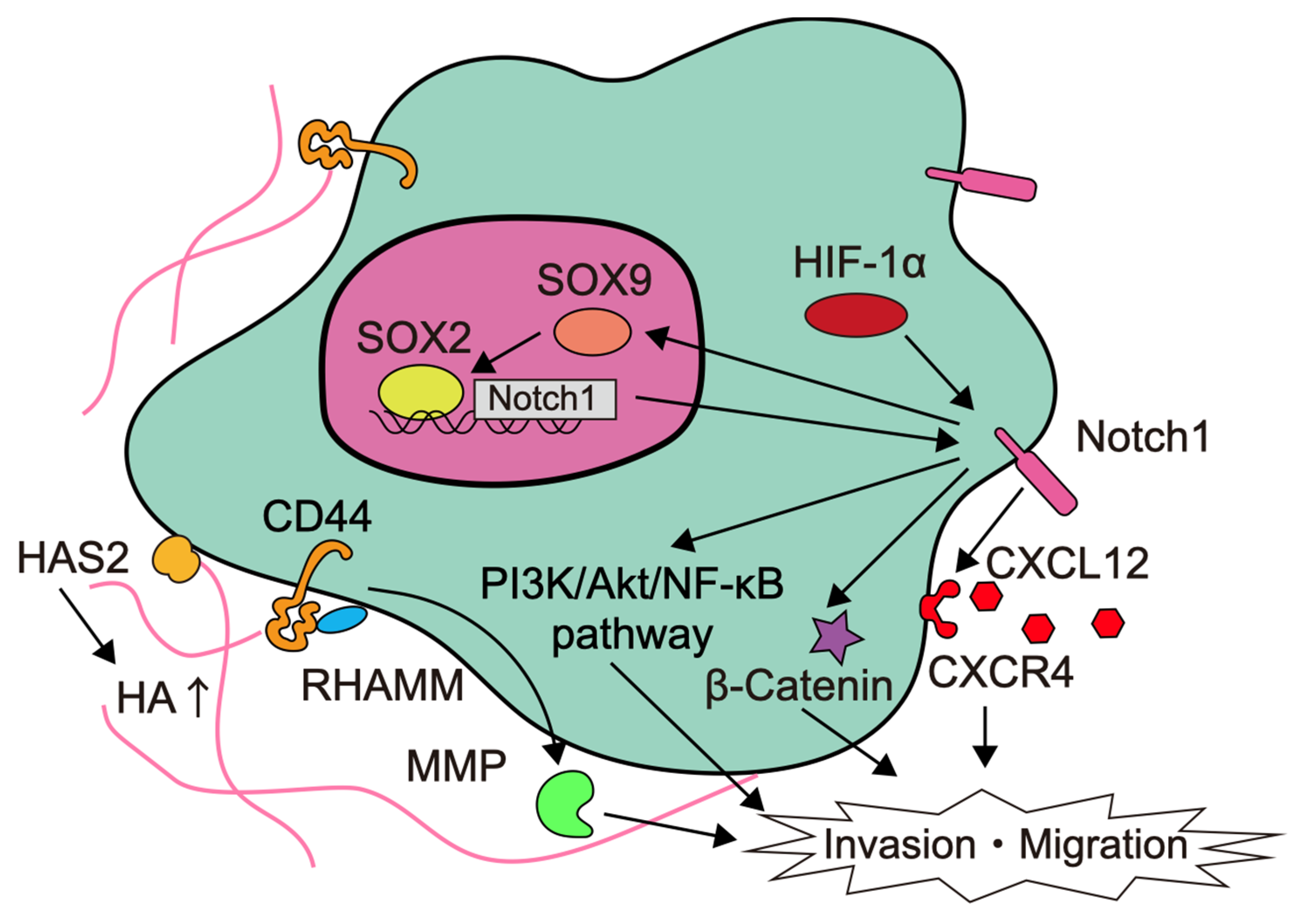
Glioma stem cells (GSCs), also called glioma-initiating cells, are cellular subpopulations that, like normal stem cells, are capable of self-renewal and differentiation to produce secondary tumors. A heterogeneity is created by cells that have a tendency to differentiate from the GSC at the top of the hierarchy [66]. Among them, GSCs are responsible for the distinctive feature of glioma invasion [67]. GSCs are characterized by the presence of CD133, CD44, leucine-rich repeat-containing G protein-coupled receptor 5 (LGR5) as surface markers [68]. The factors involved in the invasion of glioma stem cells are summarized in Figure 2.
The expression of delta-like canonical Notch ligand (DLL)-1, Notch1, nestin, and Sox2 is upregulated in GSCs compared to conventional cell lines [69]. In particular, Notch is a signal that functions in cell fate determination during tissue construction, and is involved in the maintenance and regulation of neural stem cells and progenitor cells differentiation during central nervous system development [70]. In white-matter GSCs, Notch-induced transcription factor Sox9 upregulates Sox2 and attenuates Notch1 promoter methylation to enhance Notch1 expression. This positive feedback loop increases invasiveness and worsens the prognosis [71]. Notch1 also activates the PI3K/Akt pathway and stimulates β-catenin and NF-κB signaling to promote the migratory and invasive properties of glioma [69,72,73]. In addition, Notch1 signaling upregulates CXCR4 expression and activates the CXCL12/CXCR4 autocrine/paracrine loop to enhance GSC survival and invasiveness [69,74]. Thus, signaling through increased Notch expression in glioblastoma, especially in GSCs, is thought to be highly relevant to tumor invasion, and Notch inhibitors are being developed [70,75].
CD44 is a transmembrane glycoprotein that mediates cell-cell or cell-matrix interactions with hyaluronic acid (HA) as the main ligand, and is strongly involved in tumor progression, apoptosis evasion, multidrug resistance, and cell invasion. CD44-mediated signaling has been implicated in MMP-mediated matrix degradation, tumor growth, and tumor invasion [76]. HA is a major component of white matter, a frequent route of glioma invasion, and increases the invasiveness of glioblastoma in a dose-dependent manner [77,78]. CD44 binding with HA stimulates a number of signaling pathways, such as PI3K/Akt/mTOR, Ras, focal adhesion kinase (FAK), and ERK signaling, and induces MMP-9 expression [79,80]. Receptors for HA-mediated cell motility (RHAMM), CD44, and osteopontin, which is a ligand for CD44, are involved in HA-mediated migration, invasion, proliferation, radiation therapy, and chemotherapy resistance [80,81]. Tumor-associated mesenchymal stem cells interact with glioblastoma and increase invasiveness by remodeling the ECM. Tumor-associated mesenchymal stem cells increase HA levels in the ECM by upregulating HA synthase-2 (HAS2) expression [82].
6. Epithelial-Mesenchymal Transition (EMT)
Cell invasion requires a reduction in the adhesive connections that maintain cell-to-cell adhesion. The EMT is a reversible change found in cells of epithelial origin, and mesenchymal phenotype changes are associated with increased cell motility and resistance to apoptosis [83]. In general, the EMT produces cell detachment from the basement membrane and the formation of a mass of mesenchymal cells at sites away from the origin. EMTs are classified into three subtypes according to the biological setting. Type 1 EMT is associated with embryogenesis and gives rise to the mesoderm and endoderm and to mobile neural crest cells. Type 2 EMT is a program that begins as part of a tissue repairment that normally generates fibroblasts and other related cells in order to reconstruct tissues following trauma and inflammatory injury. Unlike these subtypes, type 3 EMT occurs in neoplastic cells. Carcinoma cells undergoing a type 3 EMT may invade and metastasize [83]. The term proneural–mesenchymal transition is also used for glioma, as well as EMTs for other aggressive cancers [84]. The EMT is an important driver of invasiveness and recurrence of glioblastoma, with cellular reprogramming causing cytoskeletal remodeling and loss of adhesion molecules [85]. The main executors of the EMT are EMT-activated transcription factors (EMT-TF), such as SNAIL, TWIST, and ZEB family [86]. SNAIL induces MMP-9 expression triggered by TGF-β [87]. Notch signaling is required for the conversion of hypoxic stimuli into the EMT [39].
TGF-β is a major key molecule that induces the EMT via various transcription factors [88]. TGF-β is an important cytokine that maintains homeostasis, and the TGF-β pathway acts as an oncogenic factor to induce angiogenesis, immunosuppression, cell invasion, and proliferation in tumor progression, including glioblastoma [89,90]. TGF-β1 activates a variety of downstream signaling pathways, including PI3K, Smads, and MAPK, which are key players of the TGF-β-induced EMT [83,86]. Proteolytic degradation by MMPs plays a central role in the EMT process, and the EMT-related pathway is one of the regulatory mechanisms for MMP expression. In oral squamous cell carcinoma, TGF-β1 facilitates MT1-MMP-mediated MMP-9 activation and stimulates invasion of the tumor in collaboration with MT1-MMP and MMP-2 [91]. Elevated TGF-β activity is associated with poor prognosis in glioma patients [89,92]. In addition, TGF-β is also involved in tumor initiation and recurrence via CD44 and inhibitors of DNA-binding protein (Id)-1 [93].
7. Effect of Glioma Therapy on Tumor Invasion
As mentioned above, various molecular signaling pathways are intricately involved in the invasion of glioma cells. In the current treatment of glioma, the preclinical impact on glioma invasion is being studied.
7.1. Radiation Therapy
The standard of care for glioma is postoperative chemotherapy and radiation therapy. Radiation therapy is the main treatment modality for glioma lesions that cannot be safely resected. Whether or not the post-radiation microenvironment enhances invasiveness is inconclusive. It has been reported that radiotherapy coupled with temozolomide (TMZ) treatment has an additional effect of inhibiting the proliferation and migration of glioma spheroids [94]. It is thought that the radiation-induced tumor bed effect reduces blood flow, pH, and hypoxia, making the environment unsuitable for tumor cell survival [95,96]. However, it has been pointed out that radiation increases the invasiveness and motility of glioma [97,98]. Tsuji et al. reported that the secretion of CXCL12, VEGF-A, TGF-β1, and TNFα is enhanced in the brain after irradiation, and that the microenvironment in the brain in the chronic phase after irradiation is suitable for tumor cell growth and invasion [99]. In experiments using cell lines, various changes have been observed in invasiveness caused by radiation [100,101]. Radiation-induced damage of the tumor microenvironment may create a tumor-susceptive niche that promotes the proliferation and invasion of the residual glioma cells [102]. In a model experiment, multiple radiations altered glycosylated components (PG and GAG) in normal brain tissue, reduced CSPG expression and CS in normal brain tissue, and promoted residual glioma cell adhesion and proliferation [103]. In molecular biology, radiation has been reported to activate MMP-2 and MMP-9 through p53, resulting in increased invasiveness [104,105]. It was also suggested that radiation increases the activity of MMP-2 and MMP-9 through the expression of integrin αvβ3 [106]. PI3K-mediated activation of the Rho signaling pathway is associated with radiation-induced invasion [98]. HIF-1α is also an important molecule that contributes to radiation-induced enhancement of invasiveness. It has been reported that irradiation stabilizes HIF-1α by destabilizing prolyl hydroxylases (PHD)-2 and protein von Hippel-Lindau (pVHL) [107]. Ionizing radiation enhances the invasive capacity of GSCs through stabilization of HIF-1α and activation of junction-mediated protein [108].
However, although radiation therapy may change the tumor microenvironment to increase the invasiveness of tumors, radiation is still an important modality for the treatment of tumors. It has been reported that high linear energy transfer (LET) irradiation, such as alpha and carbon radiation, suppresses migration, and future development of radiotherapy is expected [109,110].
7.2. Temozolomide (TMZ)
TMZ is an alkylating oral anticancer drug that has been used in conjunction with postoperative radiation therapy as the standard treatment for glioma [2]. It is one of few anticancer drugs that can pass through the blood-brain barrier (BBB) and is the mainstay of postoperative chemoradiotherapy in the current treatment of glioma. It is unclear whether TMZ enhances the invasion of glioma. MMP-2 secretion and invadopodia formation is enhanced by radiation and TMZ therapy [111,112]. There are reports that the expression of CXCR4 and VEGF and the activity of MMP-2 and MMP-9 were reduced by TMZ [113,114].
7.3. Anti-VEGF Therapy
VEGF is a stimulator of angiogenesis that is frequently expressed in glioblastoma; it is commonly attributed to the autocrine and paracrine production of VEGF-A. Inhibiting VEGF signaling suppresses the tumor growth of glioma xenografts in model mice [115,116]. Anti-VEGF antibody is a monoclonal antibody to VEGF and has a certain effect on the tumor control of primary or relapsed glioblastoma [117,118,119]. Although it improved PFS in primary and recurrent glioblastoma, it was not effective in improving OS.
There is some preclinical evidence that antiangiogenic therapies promote glioma cell invasiveness. Anti-VEGF therapy induces a vascular gradient, which, in turn, induces tumor hypoxia, macrophage infiltration, mesenchymal transition, stem cell marker expression, and increased invasiveness [120]. It has been reported that hypoxia induced by anti-VEGF enhances angiogenesis, tumor survival, invasion, and resistance to therapy. Keunen et al. reported that, in a rat-patient-derived xenograft model, bevacizumab treatment resulted in a decrease in tumor volume and tumor blood flow, but a 68% increase in infiltrating cells, which was associated with the enhanced expression of HIF-1 and activation of the PI3K pathway and Wnt-signaling pathway [121]. Shimizu et al. also reported that bevacizumab upregulates δ-catenin in glioma cells and increases invasiveness [116]. It has been reported that suppression of VEGF increased CD44 expression and that GSCs became invasive [122]. Administration of bevacizumab causes the dose-dependent accumulation of collagen, MMP-2, and MMP-9, which play important roles in the adhesion process of tumor cell invasion and degradation of the cellular matrix [123]. Although invasiveness is perhaps enhanced by anti-VEGF therapy, the prognosis is not necessarily poor [118,119,124]. Combination with drugs that suppress invasion, such as γ-secretase inhibitor, has been used in an attempt to mitigate the bevacizumab-induced invasive effect [125].
7.4. Glucocorticoid
Glioma is often associated with prominent cerebral edema, which can cause mass effect and elevated intracranial pressure, affecting the prognosis [126]. Glioblastoma-induced cerebral edema has been conventionally treated with dexamethasone (DEX). Glioblastoma-induced brain edema is associated with vasogenic edema due to extravasation by disruption of the BBB, and DEX improves edema by increasing the expression of tight junction genes that regulate the endothelial permeability of the BBB [127]. The effect of DEX on the invasive properties of glioma is not well understood. Luedi et al. reported that DEX increases invasion, proliferation, and angiogenesis in GSCs, and that patients with a high DEX-regulated gene signature derived from DEX-treated GSCs showed worse prognosis [128]. It has also been reported that glucocorticoid receptor-β interacts with β-catenin and is involved in proliferation and migration [129]. DEX also affects the tumor microenvironment. The combination of TMZ and DEX affects proteoglycan structure and composition in normal brain tissue, resulting in worsened brain ECM, which is favorable for the progression of residual glioma cells; a high DEX dose results in downregulation of the transcription of PG-coding genes, whereas a high DEX dose and TMZ predominantly affects the polysaccharide GAG chains of the molecules [130]. In contrast, DEX inhibits migration via the suppression of glucocorticoid receptor-dependent ERK1/2 MAPK pathway and MMP-2 secretion [131,132]. Guan et al. reported that DEX inhibits cell proliferation and promotes migration and invasion by upregulating aquaporin-1 (AQP1) in C6 cells [133].
8. Treatment of Invasive Glioma and Its Future Development
The suppression of glioma invasion is one of the therapeutic approaches in gliomas that spread invasively to the brain and offer limited resection. However, treatments that inhibit invasion are still being studied at present. In this section, we will discuss treatments for invasion that are undergoing clinical trials and therapeutic development.
8.1. Tumor-Treating Fields (TTF)
Tumor-treating fields (TTF) is a non-invasive treatment that uses electrode pads on the scalp to deliver a weak, sustained 100 to 300 kHz mid-frequency current to brain tumors. TTF works by selectively inhibiting the mitosis of brain tumor cells and can prolong survival by 4.9 months [134]. In addition to inhibiting cell proliferation, TTF has also been reported to inhibit EMT, endothelial cell angiogenesis, and migration by downregulating PI3K/Akt/NF-κB pathways [135].
8.2. Molecular Target Drugs
As our knowledge of the invasion of various cancers increases, therapies that target the molecular mechanisms that lead to cancer invasion are being developed. The major therapeutic agents that are currently in development are summarized in Table 1.
MMP inhibitor: Degradation of the ECM has been well observed in tumor tissues, and MMPs have been considered a good target for tumor invasion. However, the selectivity, low bioavailability, and low metabolic profile of broad-spectrum MMP inhibitors limited the efficacy of MMP inhibitors and did not justify continuation of the clinical trial [136]. MMP inhibitors were expected to have an effect on the invasiveness of tumor cells, but clinical trials for gliomas to date have not shown positive results. In a mouse model of colorectal carcinoma, AB0041 and AB0046, which are monoclonal anti-MMP-9 antibodies, were found to inhibit tumor growth and metastasis [137]. Since bevacizumab increases the expression of MMP-9 [138], anti-MMP-9 therapy may be effective against bevacizumab-induced resistance and invasiveness. Clinical trials of GS5745, a monoclonal antibody of MMP-9, are in progress.
Integrin inhibitor: Cilengitide was one of the earliest integrin antagonists to enter clinical trials [139]. In a rat model, bevacizumab treatment caused tumor cells to invade the brain parenchyma along with blood vessels, but cilengitide administration suppressed the invasion of tumor borders, suggesting the involvement of integrin in bevacizumab-induced glioma invasion [140]. The phase 2 study of cilengitide showed prolongation of median OS by adding cilengitide to standard treatment, and development was expected [141]; however, phase 3 studies such as CENTRIC failed to show OS prolongation [142] and development was discontinued. The addition of cilengitide to oncolytic virus therapy has been shown to enhance antitumor effects, and cilengitide may undergo re-evaluation as virus therapy develops [143].
Notch inhibitor: The expression of Notch is increased in GSCs, and Notch inhibition by γ-secretase inhibitor has been attempted. γ-secretase cleaves Notch to form the NICD, which translocates to the nucleus. γ-secretase inhibitors can inhibit Notch signaling. Jin et al. showed that MRK-003, an inhibitor of Notch and Akt phosphorylation, suppresses invasion but not mitosis when combined with MK-2206, an Akt phosphorylation inhibitor [144]. The Notch pathway blockade by γ-secretase inhibitor inhibited tumor growth and neurosphere formation in culture, and also prolonged survival in xenograft mice [145]. However, in a phase 2 study, RO4929097, a γ-secretase inhibitor, was evaluated for 6 month PFS and neurosphere formation in patients with recurrent glioblastoma, but failed to demonstrate efficacy [146]. Although no effective treatment for GSCs has been established yet, their treatment may remain an attractive strategy.
TGF-β inhibitor: TGF-β inhibitor has been reported to have inhibitory effects on the metastasis of breast, colon, pancreatic, and gastric cancers [147,148,149,150]. LY2109761, a TGF-β receptor inhibitor, not only enhanced the effects of radiotherapy but also inhibited cell migration via the SMAD4 signaling pathway [150]. In glioblastoma treatment, Zhang et al. reported that LY2109761 suppressed migration in vitro, and LY2109761 added to RT + TMZ significantly inhibited tumor growth in model mice [151]. Since it is not an invasive model, however, the inhibition of invasion in vivo was inconclusive. A phase 2b study of AP-12009 (trabedersen), a phosphorothioate antisense oligodeoxynucleotide, a specific target for the mRNA of TGFβ2, showed safety against chemotherapy in recurrent high-grade glioma and significant improvement in the 14-month tumor control rate in anaplastic astrocytoma. However, the phase 3 study, SAPPHIRE, was discontinued due to a lack of patients [152]. Compared with chemoradiotherapy, galunisertib, a TGF-β receptor inhibitor, had a higher disease control rate but shorter PFS and no difference in efficacy [153].
PI3K inhibitor: Tumor metastasis and invasion are enhanced by activating the PI3K/Akt pathway through regulating the expression of MMP [154]. In glioblastoma, the PI3K pathway is activated by changes in epidermal growth factor receptor (EGFR) amplification and phosphatase and tensin homolog (PTEN) mutation; and PTEN changes are a poor prognostic factor in glioblastoma [155]. It has been reported that a number of inhibitory agents of PI3K were preclinically effective in inhibiting cell proliferation and invasion [156,157,158,159]. The PI3K inhibitor PX-866 was well tolerated in a phase 1 trial, but failed to meet its predefined efficacy endpoint in a phase 2 trial in patients with recurrent glioblastoma [160]. A phase 1 study of voxtalisib, the PI3K/mTOR inhibitor, plus TMZ with or without radiotherapy, in patients with high-grade gliomas demonstrated favorable safety; however, no conclusion could be drawn regarding the efficacy because of the small number of patients and short follow-up [161]. Buparlisib, on oral pan-PI3K inhibitor, has high penetration across the BBB. A phase 2 study of buparlisib in recurrent glioblastoma patients showed minimal efficacy. The lack of tumor response was explained by incomplete PI3K inhibition in the tumor tissue [162].

Table 1.
Molecular-targeted drugs and clinical trials related to glioma invasion.
| Inhibitor | Target Molecule | Clinical Trial Phase | Reference | |
|---|---|---|---|---|
| MMP inhibitor | AG3340 (prinomastat) | MMP | Phase 2 | NCT00004200 |
| GS5745 (andecaliximab) | MMP-9 | Phase 1, ongoing | NCT03631836 | |
| Integrin inhibitor | cilengitide | integrin | Phase 3 | Stupp et al. (2014) [142] |
| Notch inhibitor | RO4929097 | γ-secretase inhibitor | Phase 2 | Peereboom et al. (2021) [146] |
| TGF-β inhibitor | AP-12009 (trabedersen) | TGF-β2 | Phase 3 | Bogdahn et al. (2011) [152] |
| LY2157299 (galunisertib) | TGF-β receptor I | Phase 1b/2a | Wick et al. (2020) [153] | |
| PI3K inhibitor | PX-866 | PI3K | Phase 2 | Pitz et al. (2015) [160] |
| XL-765, SAR245409 (voxtalisib) | PI3K/mTOR | Phase 1 | Wen et al. (2015) [161] | |
| NVP-BKM120 (buparlisib) | PI3K | Phase 2 | Wen et al. (2019) [162] |
8.3. Stem Cell Therapy
In recent years, research has been conducted on therapies using stem cells for drug delivery. Neural stem cells, mesenchymal stem cells, and induced pluripotent stem cells all accumulate in tumors [163,164]. Radioisotope transporters, tumor lytic viruses, suicide genes, immunomodulatory agents, anti-angiogenic factors apoptosis-inducing agents, etc., are placed on the stem cells, injected locally or intravenously, and accumulate in the tumor to produce a therapeutic effect [165,166,167,168].
In suicide gene therapy, tumor cells expressing genes such as herpes simplex virus thymidine kinase (HSV-TK) and cytosine deaminase (CD) can metabolize the prodrugs, resulting in apoptosis [169]. In phase 3 clinical trial using fibroblasts with suicide genes incorporated, the significant improvement in PFS, median survival and survival rate was not observed, suggesting that the low diffusibility of suicide genes is a problem [170]. Therefore, the use of stem cells, which have a high ability to accumulate in tumors, as a vehicle for the delivery of suicide genes to glioma cells infiltrating the brain parenchyma, is being attempted for therapeutic development [165,171].
8.4. Viral Therapy
Antitumor therapy using oncolytic viruses (viral therapy) is a field derived from gene therapy. Proliferative viruses selectively proliferate in the tumor cells and exhibit antitumor effects through tumor lysis and inducing tumor immunity. It has been reported that the oncolytic virus has antitumor effects in mouse models of invasive tumors using GSCs due to its extensive distribution and infectivity [67]. Microenvironmental changes such as increased tumor vascular permeability and elevated expression of inflammatory cytokine genes induced by viral administration induce the resistance to viral therapy [172]. Therefore, there has been an attempt to modify the microenvironment and enhance the antitumor effects of viral therapy [143,173,174]. It is also known that tumor lytic virus activates Notch signaling in non-infected cells, and it has been reported that the combination of oncolytic virus and Notch inhibitor suppressed the cell proliferation of non-infected cells and enhanced the effect of tumor lytic virus [175]. In the future, viral therapy will continue to evolve with various studies and improvements in the approach to invasive lesions.
9. Conclusions
The high invasiveness of gliomas is due to a complex combination of factors, including hypoxia, ECM, cancer stem cells, EMT, etc. The impact of glioma treatment on invasiveness is not yet fully understood, and further research is needed in this area. Establishing a treatment for glioma invasion will be one of the main topics of therapeutic development in the future.
Author Contributions
Conceptualization, T.O. and K.K.; writing—original draft preparation, T.O.; writing—review and editing, S.K. and K.K.; supervision, S.K. and K.K. All authors have read and agreed to the published version of the manuscript.
Funding
Please add: This research received no external funding.
Institutional Review Board Statement
Not applicable.
Informed Consent Statement
Not applicable.
Data Availability Statement
Not applicable.
Acknowledgments
We thank Chris Rowthorn of Eibunkousei Company (https://www.eibunkousei.net/ (accessed on 16 February 2022)) for editing a draft of this manuscript.
Conflicts of Interest
The authors declare no conflict of interest.
References
- Louis, D.N.; Perry, A.; Reifenberger, G.; von Deimling, A.; Figarella-Branger, D.; Cavenee, W.K.; Ohgaki, H.; Wiestler, O.D.; Kleihues, P.; Ellison, D.W. The 2016 World Health Organization Classification of Tumors of the Central Nervous System: A summary. Acta Neuropathol. 2016, 131, 803–820. [Google Scholar] [CrossRef] [Green Version]
- Stupp, R.; Mason, W.P.; van den Bent, M.J.; Weller, M.; Fisher, B.; Taphoorn, M.J.B.; Belanger, K.; Brandes, A.A.; Marosi, C.; Bogdahn, U.; et al. Radiotherapy plus Concomitant and Adjuvant Temozolomide for Glioblastoma. N. Engl. J. Med. 2005, 352, 987–996. [Google Scholar] [CrossRef]
- Sanai, N.; Polley, M.Y.; McDermott, M.W.; Parsa, A.T.; Berger, M.S. An extent of resection threshold for newly diagnosed glioblastomas. J. Neurosurg. 2011, 115, 3–8. [Google Scholar] [CrossRef] [Green Version]
- Stummer, W.; Pichlmeier, U.; Meinel, T.; Wiestler, O.D.; Zanella, F.; Reulen, H.-J.; ALA-Glioma Study Group. Fluorescence-guided surgery with 5-aminolevulinic acid for resection of malignant glioma: A randomised controlled multicentre phase III trial. Lancet Oncol. 2006, 7, 392–401. [Google Scholar] [CrossRef]
- Kuhnt, D.; Ganslandt, O.; Schlaffer, S.-M.; Buchfelder, M.; Nimsky, C. Quantification of Glioma Removal by Intraoperative High-Field Magnetic Resonance Imaging: An Update. Neurosurgery 2011, 69, 852–863. [Google Scholar] [CrossRef] [PubMed] [Green Version]
- Hua, X.; Huang, M.; Deng, X.; Xu, J.; Luo, Y.; Xie, Q.; Xu, J.; Tian, Z.; Li, J.; Zhu, J.; et al. The inhibitory effect of compound ChlA-F on human bladder cancer cell invasion can be attributed to its blockage of SOX2 protein. Cell Death Differ. 2020, 27, 632–645. [Google Scholar] [CrossRef] [PubMed]
- Onishi, M.; Ichikawa, T.; Kurozumi, K.; Date, I. Angiogenesis and invasion in glioma. Brain Tumor Pathol. 2011, 28, 13–24. [Google Scholar] [CrossRef]
- Shimizu, T.; Kurozumi, K.; Ishida, J.; Ichikawa, T.; Date, I. Adhesion molecules and the extracellular matrix as drug targets for glioma. Brain Tumor Pathol. 2016, 33, 97–106. [Google Scholar] [CrossRef] [PubMed]
- Fayzullin, A.; Sandberg, C.J.; Spreadbury, M.; Saberniak, B.M.; Grieg, Z.; Skaga, E.; Langmoen, I.A.; Vik-Mo, E.O. Phenotypic and Expressional Heterogeneity in the Invasive Glioma Cells. Transl. Oncol. 2019, 12, 122–133. [Google Scholar] [CrossRef] [PubMed]
- Lagerweij, T.; Dusoswa, S.; Negrean, A.; Hendrikx, E.M.L.; De Vries, H.E.; Kole, J.; Garcia-Vallejo, J.J.; Mansvelder, H.; Vandertop, W.P.; Noske, D.P.; et al. Optical clearing and fluorescence deep-tissue imaging for 3D quantitative analysis of the brain tumor microenvironment. Angiogenesis 2017, 20, 533–546. [Google Scholar] [CrossRef] [PubMed]
- Jabouille, A.; Delugin, M.; Pineau, R.; Dubrac, A.; Soulet, F.; Lhomond, S.; Pallares-Lupon, N.; Prats, H.; Bikfalvi, A.; Chevet, E.; et al. Glioblastoma invasion and cooption depend on IRE1α endoribonuclease activity. Oncotarget 2015, 6, 24922–24934. [Google Scholar] [CrossRef] [PubMed] [Green Version]
- Monzo, P.; Chong, Y.K.; Guetta-Terrier, C.; Krishnasamy, A.; Sathe, S.R.; Yim, E.; Ng, W.H.; Ang, B.T.; Tang, C.; Ladoux, B.; et al. Mechanical confinement triggers glioma linear migration dependent on formin FHOD3. Mol. Biol. Cell 2016, 27, 1246–1261. [Google Scholar] [CrossRef]
- Tamura, R.; Miyoshi, H.; Sampetrean, O.; Shinozaki, M.; Morimoto, Y.; Iwasawa, C.; Fukaya, R.; Mine, Y.; Masuda, H.; Maruyama, T.; et al. Visualization of spatiotemporal dynamics of human glioma stem cell invasion. Mol. Brain 2019, 12, 45. [Google Scholar] [CrossRef] [PubMed]
- Farin, A.; Suzuki, S.O.; Weiker, M.; Goldman, J.E.; Bruce, J.N.; Canoll, P. Transplanted glioma cells migrate and proliferate on host brain vasculature: A dynamic analysis. Glia 2006, 53, 799–808. [Google Scholar] [CrossRef]
- Prahl, L.; Stanslaski, M.R.; Vargas, P.; Piel, M.; Odde, D.J. Predicting Confined 1D Cell Migration from Parameters Calibrated to a 2D Motor-Clutch Model. Biophys. J. 2020, 118, 1709–1720. [Google Scholar] [CrossRef]
- Chaichana, K.L.; Jusue-Torres, I.; Lemos, A.M.; Gokaslan, A.; Cabrera-Aldana, E.E.; Ashary, A.; Olivi, A.; Quinones-Hinojosa, A. The butterfly effect on glioblastoma: Is volumetric extent of resection more effective than biopsy for these tumors? J. Neuro-Oncol. 2014, 120, 625–634. [Google Scholar] [CrossRef] [Green Version]
- Shen, S.; Feng, S.; Liu, H.; Jiang, J.; Yu, X. Associations of histological and molecular alterations with invasion of the corpus callosum in gliomas. Acta Neurochir. 2020, 162, 1691–1699. [Google Scholar] [CrossRef]
- Sadahiro, H.; Yoshikawa, K.; Ideguchi, M.; Kajiwara, K.; Ishii, A.; Ikeda, E.; Owada, Y.; Yasumoto, Y.; Suzuki, M. Pathological features of highly invasive glioma stem cells in a mouse xenograft model. Brain Tumor Pathol. 2014, 31, 77–84. [Google Scholar] [CrossRef] [PubMed]
- Jafri, N.F.; Clarke, J.L.; Weinberg, V.; Barani, I.J.; Cha, S. Relationship of glioblastoma multiforme to the subventricular zone is associated with survival. Neuro-Oncology 2013, 15, 91–96. [Google Scholar] [CrossRef] [PubMed] [Green Version]
- Mistry, A.M.; Dewan, M.C.; White-Dzuro, G.A.; Brinson, P.R.; Weaver, K.D.; Thompson, R.C.; Ihrie, R.A.; Chambless, L.B. Decreased survival in glioblastomas is specific to contact with the ventricular-subventricular zone, not subgranular zone or corpus callosum. J. Neuro-Oncol. 2017, 132, 341–349. [Google Scholar] [CrossRef] [Green Version]
- Yamaki, T.; Shibahra, I.; Matsuda, K.-I.; Kanemura, Y.; Konta, T.; Kanamori, M.; Yamakawa, M.; Tominaga, T.; Sonoda, Y. Relationships between recurrence patterns and subventricular zone involvement or CD133 expression in glioblastoma. J. Neuro-Oncol. 2020, 146, 489–499. [Google Scholar] [CrossRef] [PubMed]
- Qin, E.Y.; Cooper, D.D.; Abbott, K.; Lennon, J.; Nagaraja, S.; Mackay, A.; Jones, C.; Vogel, H.; Jackson, P.K.; Monje, M. Neural Precursor-Derived Pleiotrophin Mediates Subventricular Zone Invasion by Glioma. Cell 2017, 170, 845–859.e819. [Google Scholar] [CrossRef]
- Lu, K.V.; Jong, K.A.; Kim, G.Y.; Singh, J.; Dia, E.Q.; Yoshimoto, K.; Wang, M.Y.; Cloughesy, T.F.; Nelson, S.F.; Mischel, P.S. Differential Induction of Glioblastoma Migration and Growth by Two Forms of Pleiotrophin. J. Biol. Chem. 2005, 280, 26953–26964. [Google Scholar] [CrossRef] [Green Version]
- Wang, X. Pleiotrophin: Activity and mechanism. Adv. Clin. Chem. 2020, 98, 51–89. [Google Scholar] [CrossRef] [PubMed]
- Ryan, E.; Shen, D.; Wang, X. Pleiotrophin interacts with glycosaminoglycans in a highly flexible and adaptable manner. FEBS Lett. 2021, 595, 925–941. [Google Scholar] [CrossRef]
- Goffart, N.; Kroonen, J.; Di Valentin, E.; Dedobbeleer, M.; Denne, A.; Martinive, P.; Rogister, B. Adult mouse subventricular zones stimulate glioblastoma stem cells specific invasion through CXCL12/CXCR4 signaling. Neuro-Oncology 2015, 17, 81–94. [Google Scholar] [CrossRef] [Green Version]
- Shi, Y.; Riese, D.J.I.; Shen, J. The Role of the CXCL12/CXCR4/CXCR7 Chemokine Axis in Cancer. Front. Pharmacol. 2020, 11, 574667. [Google Scholar] [CrossRef] [PubMed]
- Ehtesham, M.; A Winston, J.; Kabos, P.; Thompson, R.C. CXCR4 expression mediates glioma cell invasiveness. Oncogene 2006, 25, 2801–2806. [Google Scholar] [CrossRef] [PubMed] [Green Version]
- Zhang, J.; Sarkar, S.; Yong, V. The chemokine stromal cell derived factor-1 (CXCL12) promotes glioma invasiveness through MT2-matrix metalloproteinase. Carcinogenesis 2005, 26, 2069–2077. [Google Scholar] [CrossRef] [Green Version]
- Chen, L.; Zhu, M.; Yu, S.; Hai, L.; Zhang, L.; Zhang, C.; Zhao, P.; Zhou, H.; Wang, S.; Yang, X. Arg kinase mediates CXCL12/CXCR4-induced invadopodia formation and invasion of glioma cells. Exp. Cell Res. 2020, 389, 111893. [Google Scholar] [CrossRef]
- Paz, H.; Pathak, N.; Yang, J. Invading one step at a time: The role of invadopodia in tumor metastasis. Oncogene 2014, 33, 4193–4202. [Google Scholar] [CrossRef] [Green Version]
- Artym, V.V.; Zhang, Y.; Seillier-Moiseiwitsch, F.; Yamada, K.; Mueller, S.C. Dynamic Interactions of Cortactin and Membrane Type 1 Matrix Metalloproteinase at Invadopodia: Defining the Stages of Invadopodia Formation and Function. Cancer Res. 2006, 66, 3034–3043. [Google Scholar] [CrossRef] [PubMed] [Green Version]
- Plasswilm, L.; Tannapfel, A.; Cordes, N.; Demir, R.; Höper, K.; Bauer, J.; Höper, J. Hypoxia-Induced Tumour Cell Migration in an in vivo Chicken Model. Pathobiology 2000, 68, 99–105. [Google Scholar] [CrossRef] [PubMed]
- Semenza, G.L.; Wang, G.L. A nuclear factor induced by hypoxia via de novo protein synthesis binds to the human erythropoietin gene enhancer at a site required for transcriptional activation. Mol. Cell. Biol. 1992, 12, 5447–5454. [Google Scholar] [CrossRef]
- Semenza, G.L. Targeting HIF-1 for cancer therapy. Nat. Rev. Cancer 2003, 3, 721–732. [Google Scholar] [CrossRef]
- Kaur, B.; Khwaja, F.W.; Severson, E.A.; Matheny, S.L.; Brat, D.J.; Van Meir, E.G. Hypoxia and the hypoxia-inducible-factor pathway in glioma growth and angiogenesis. Neuro-Oncology 2005, 7, 134–153. [Google Scholar] [CrossRef] [PubMed] [Green Version]
- Grassi, E.S.; Pantazopoulou, V.; Pietras, A. Hypoxia-induced release, nuclear translocation, and signaling activity of a DLK1 intracellular fragment in glioma. Oncogene 2020, 39, 4028–4044. [Google Scholar] [CrossRef] [PubMed] [Green Version]
- Qiang, L.; Wu, T.; Zhang, H.-W.; Lu, N.; Hu, R.; Wang, Y.-J.; Zhao, L.; Chen, F.-H.; Wang, X.-T.; You, Q.-D.; et al. HIF-1α is critical for hypoxia-mediated maintenance of glioblastoma stem cells by activating Notch signaling pathway. Cell Death Differ. 2012, 19, 284–294. [Google Scholar] [CrossRef] [Green Version]
- Sahlgren, C.; Gustafsson, M.V.; Jin, S.; Poellinger, L.; Lendahl, U. Notch signaling mediates hypoxia-induced tumor cell migration and invasion. Proc. Natl. Acad. Sci. USA 2008, 105, 6392–6397. [Google Scholar] [CrossRef] [Green Version]
- Huang, W.; Ding, X.; Ye, H.; Wang, J.; Shao, J.; Huang, T. Hypoxia enhances the migration and invasion of human glioblastoma U87 cells through PI3K/Akt/mTOR/HIF-1α pathway. NeuroReport 2018, 29, 1578–1585. [Google Scholar] [CrossRef]
- Smith, B.N.; Bhowmick, N.A. Role of EMT in Metastasis and Therapy Resistance. J. Clin. Med. 2016, 5, 17. [Google Scholar] [CrossRef] [PubMed] [Green Version]
- Wang, M.; Wang, T.; Liu, S.; Yoshida, D.; Teramoto, A. The expression of matrix metalloproteinase-2 and-9 in human gliomas of different pathological grades. Brain Tumor Pathol. 2003, 20, 65–72. [Google Scholar] [CrossRef]
- Sincevičiūtė, R.; Vaitkienė, P.; Urbanavičiūtė, R.; Steponaitis, G.; Tamašauskas, A.; Skiriutė, D. MMP2 is associated with glioma malignancy and patient outcome. Int. J. Clin. Exp. Pathol. 2018, 11, 3010–3018. [Google Scholar] [PubMed]
- Ferrer, V.P.; Moura-Neto, V.; Mentlein, R. Glioma infiltration and extracellular matrix: Key players and modulators. Glia 2018, 66, 1542–1565. [Google Scholar] [CrossRef]
- Santibanez, J.F.; Obradović, H.; Kukolj, T.; Krstić, J. Transforming growth factor-β, matrix metalloproteinases, and urokinase-type plasminogen activator interaction in the cancer epithelial to mesenchymal transition. Dev. Dyn. 2018, 247, 382–395. [Google Scholar] [CrossRef] [Green Version]
- Baumann, F.; Leukel, P.; Doerfelt, A.; Beier, C.P.; Dettmer, K.; Oefner, P.J.; Kastenberger, M.; Kreutz, M.; Nickl-Jockschat, T.; Bogdahn, U.; et al. Lactate promotes glioma migration by TGF-β2–dependent regulation of matrix metalloproteinase-2. Neuro-Oncology 2009, 11, 368–380. [Google Scholar] [CrossRef] [PubMed] [Green Version]
- Seliger, C.; Leukel, P.; Moeckel, S.; Jachnik, B.; Lottaz, C.; Kreutz, M.; Brawanski, A.; Proescholdt, M.; Bogdahn, U.; Bosserhoff, A.; et al. Lactate-Modulated Induction of THBS-1 Activates Transforming Growth Factor (TGF)-beta2 and Migration of Glioma Cells In Vitro. PLoS ONE 2013, 8, e78935. [Google Scholar] [CrossRef] [Green Version]
- Sanderson, R.D.; Elkin, M.; Rapraeger, A.C.; Ilan, N.; Vlodavsky, I. Heparanase regulation of cancer, autophagy and inflammation: New mechanisms and targets for therapy. FEBS J. 2017, 284, 42–55. [Google Scholar] [CrossRef]
- Bello, L.; Francolini, M.; Marthyn, P.; Zhang, J.; Carroll, R.S.; Nikas, D.C.; Strasser, J.F.; Villani, R.; Cheresh, D.A.; Black, P.M. αvβ3 and αvβ5 Integrin Expression in Glioma Periphery. Neurosurgery 2001, 49, 380–389. [Google Scholar] [CrossRef]
- Platten, M.; Wick, W.; Bode, C.W.; Aulwurm, S.; Dichgans, J.; Weller, M. Transforming Growth Factors β1 (TGF-β1) and TGF-β2 Promote Glioma Cell Migration via Up-Regulation of αVβ3 Integrin Expression. Biochem. Biophys. Res. Commun. 2000, 268, 607–611. [Google Scholar] [CrossRef]
- Levental, K.; Yu, H.; Kass, L.; Lakins, J.N.; Egeblad, M.; Erler, J.; Fong, S.F.; Csiszar, K.; Giaccia, A.; Weninger, W.; et al. Matrix Crosslinking Forces Tumor Progression by Enhancing Integrin Signaling. Cell 2009, 139, 891–906. [Google Scholar] [CrossRef] [Green Version]
- Ulrich, T.A.; de Juan Pardo, E.M.; Kumar, S. The Mechanical Rigidity of the Extracellular Matrix Regulates the Structure, Motility, and Proliferation of Glioma Cells. Cancer Res. 2009, 69, 4167–4174. [Google Scholar] [CrossRef] [Green Version]
- Grzeszkiewicz, T.M.; Kirschling, D.J.; Chen, N.; Lau, L.F. CYR61 Stimulates Human Skin Fibroblast Migration through Integrin αvβ5 and Enhances Mitogenesis through Integrin αvβ3, Independent of Its Carboxyl-terminal Domain. J. Biol. Chem. 2001, 276, 21943–21950. [Google Scholar] [CrossRef] [Green Version]
- Walsh, C.T.; Radeff-Huang, J.; Matteo, R.; Hsiao, A.; Subramaniam, S.; Stupack, D.; Brown, J.H. Thrombin receptor and RhoA mediate cell proliferation through integrins and cysteine-rich protein 61. FASEB J. 2008, 22, 4011–4021. [Google Scholar] [CrossRef] [PubMed] [Green Version]
- Ishida, J.; Kurozumi, K.; Ichikawa, T.; Otani, Y.; Onishi, M.; Fujii, K.; Shimazu, Y.; Oka, T.; Shimizu, T.; Date, I. Evaluation of extracellular matrix protein CCN1 as a prognostic factor for glioblastoma. Brain Tumor Pathol. 2015, 32, 245–252. [Google Scholar] [CrossRef] [PubMed]
- Xie, D.; Yin, N.; Wang, H.-J.; Liu, G.-T.; Elashoff, R.; Black, K.; Koeffler, H.P. Levels of expression of CYR61 and CTGF are prognostic for tumor progression and survival of individuals with gliomas. Clin. Cancer Res. 2004, 10, 2072–2081. [Google Scholar] [CrossRef] [Green Version]
- Uneda, A.; Kurozumi, K.; Fujimura, A.; Fujii, K.; Ishida, J.; Shimazu, Y.; Otani, Y.; Tomita, Y.; Hattori, Y.; Matsumoto, Y.; et al. Differentiated glioblastoma cells accelerate tumor progression by shaping the tumor microenvironment via CCN1-mediated macrophage infiltration. Acta Neuropathol. Commun. 2021, 9, 29. [Google Scholar] [CrossRef]
- Silver, D.J.; Siebzehnrubl, F.; Schildts, M.J.; Yachnis, A.T.; Smith, G.M.; Smith, A.A.; Scheffler, B.; Reynolds, B.A.; Silver, J.; Steindler, D.A. Chondroitin Sulfate Proteoglycans Potently Inhibit Invasion and Serve as a Central Organizer of the Brain Tumor Microenvironment. J. Neurosci. 2013, 33, 15603–15617. [Google Scholar] [CrossRef] [Green Version]
- Wade, A.; Robinson, A.E.; Engler, J.R.; Petritsch, C.; James, C.D.; Phillips, J.J. Proteoglycans and their roles in brain cancer. FEBS J. 2013, 280, 2399–2417. [Google Scholar] [CrossRef] [PubMed] [Green Version]
- Naganuma, H.; Satoh, E.; Asahara, T.; Amagasaki, K.; Watanabe, A.; Satoh, H.; Kuroda, K.; Zhang, L.; Nukui, H. Quantification of thrombospondin-1 secretion and expression of ?v?3 and ?3?1 integrins and syndecan-1 as cell-surface receptors for thrombospondin-1 in malignant glioma cells. J. Neuro-Oncol. 2004, 70, 309–317. [Google Scholar] [CrossRef]
- Ramani, V.C.; Purushothaman, A.; Stewart, M.D.; Thompson, C.A.; Vlodavsky, I.; Au, J.L.-S.; Sanderson, R.D. The heparanase/syndecan-1 axis in cancer: Mechanisms and therapies. FEBS J. 2013, 280, 2294–2306. [Google Scholar] [CrossRef] [PubMed] [Green Version]
- Shi, S.; Zhong, D.; Xiao, Y.; Wang, B.; Wang, W.; Zhang, F.; Huang, H. Syndecan-1 knockdown inhibits glioma cell proliferation and invasion by deregulating a c-src/FAK-associated signaling pathway. Oncotarget 2017, 8, 40922–40934. [Google Scholar] [CrossRef] [PubMed]
- Dwyer, C.A.; Bi, W.L.; Viapiano, M.S.; Matthews, R.T. Brevican knockdown reduces late-stage glioma tumor aggressiveness. J. Neuro-Oncol. 2014, 120, 63–72. [Google Scholar] [CrossRef] [Green Version]
- Hong, X.; Jiang, F.; Kalkanis, S.N.; Zhang, Z.G.; Zhang, X.; Zheng, X.; Jiang, H.; Mikkelsen, T.; Chopp, M. Increased chemotactic migration and growth in heparanase-overexpressing human U251n glioma cells. J. Exp. Clin. Cancer Res. 2008, 27, 23–28. [Google Scholar] [CrossRef] [PubMed] [Green Version]
- Kundu, S.; Xiong, A.; Spyrou, A.; Wicher, G.; Marinescu, V.D.; Edqvist, P.-H.D.; Zhang, L.; Essand, M.; Dimberg, A.; Smits, A.; et al. Heparanase Promotes Glioma Progression and Is Inversely Correlated with Patient Survival. Mol. Cancer Res. 2016, 14, 1243–1253. [Google Scholar] [CrossRef] [Green Version]
- Gimple, R.C.; Bhargava, S.; Dixit, D.; Rich, J.N. Glioblastoma stem cells: Lessons from the tumor hierarchy in a lethal cancer. Genes Dev. 2019, 33, 591–609. [Google Scholar] [CrossRef]
- Wakimoto, H.; Kesari, S.; Farrell, C.J.; Curry, W.T., Jr.; Zaupa, C.; Aghi, M.; Kuroda, T.; Stemmer-Rachamimov, A.; Shah, K.; Liu, T.-C.; et al. Human Glioblastoma–Derived Cancer Stem Cells: Establishment of Invasive Glioma Models and Treatment with Oncolytic Herpes Simplex Virus Vectors. Cancer Res. 2009, 69, 3472–3481. [Google Scholar] [CrossRef] [Green Version]
- Zhang, J.; Cai, H.; Sun, L.; Zhan, P.; Chen, M.; Zhang, F.; Ran, Y.; Wan, J. LGR5, a novel functional glioma stem cell marker, promotes EMT by activating the Wnt/β-catenin pathway and predicts poor survival of glioma patients. J. Exp. Clin. Cancer Res. 2018, 37, 225. [Google Scholar] [CrossRef]
- Yi, L.; Zhou, X.; Li, T.; Liu, P.; Hai, L.; Tong, L.; Ma, H.; Tao, Z.; Xie, Y.; Zhang, C.; et al. Notch1 signaling pathway promotes invasion, self-renewal and growth of glioma initiating cells via modulating chemokine system CXCL12/CXCR4. J. Exp. Clin. Cancer Res. 2019, 38, 339. [Google Scholar] [CrossRef] [Green Version]
- Teodorczyk, M.; Schmidt, M.H.H. Notching on Cancer’s Door: Notch Signaling in Brain Tumors. Front. Oncol. 2014, 4, 341. [Google Scholar] [CrossRef] [Green Version]
- Wang, J.; Xu, S.-L.; Duan, J.-J.; Yi, L.; Guo, Y.-F.; Shi, Y.; Li, L.; Yang, Z.-Y.; Liao, X.-M.; Cai, J.; et al. Invasion of white matter tracts by glioma stem cells is regulated by a NOTCH1–SOX2 positive-feedback loop. Nat. Neurosci. 2019, 22, 91–105. [Google Scholar] [CrossRef]
- Zhang, X.; Chen, T.; Zhang, J.; Mao, Q.; Li, S.; Xiong, W.; Qiu, Y.; Xie, Q.; Ge, J. Notch1 promotes glioma cell migration and invasion by stimulating β-catenin and NF-κB signaling via AKT activation. Cancer Sci. 2011, 103, 181–190. [Google Scholar] [CrossRef]
- Li, L.; Tang, P.; Li, S.; Qin, X.; Yang, H.; Wu, C.; Liu, Y. Notch signaling pathway networks in cancer metastasis: A new target for cancer therapy. Med. Oncol. 2017, 34, 180. [Google Scholar] [CrossRef] [PubMed]
- Calinescu, A.-A.; Yadav, V.N.; Carballo, E.; Kadiyala, P.; Tran, D.; Zamler, D.; Doherty, R.; Srikanth, M.; Lowenstein, P.R.; Castro, M.G. Survival and Proliferation of Neural Progenitor–Derived Glioblastomas Under Hypoxic Stress is Controlled by a CXCL12/CXCR4 Autocrine-Positive Feedback Mechanism. Clin. Cancer Res. 2017, 23, 1250–1262. [Google Scholar] [CrossRef] [Green Version]
- Xu, R.; Shimizu, F.; Hovinga, K.; Beal, K.; Karimi, S.; Droms, L.; Peck, K.K.; Gutin, P.; Iorgulescu, J.B.; Kaley, T.; et al. Molecular and Clinical Effects of Notch Inhibition in Glioma Patients: A Phase 0/I Trial. Clin. Cancer Res. 2016, 22, 4786–4796. [Google Scholar] [CrossRef] [PubMed] [Green Version]
- Bourguignon, L.Y.; Earle, C.; Shiina, M. Hyaluronan-CD44 interaction promotes HPV 16 E6 oncogene-mediated oropharyngeal cell carcinoma survival and chemoresistance. Matrix Biol. 2019, 78–79, 180–200. [Google Scholar] [CrossRef]
- Radotra, B.; McCormick, D. Glioma invasion in vitro is mediated by CD44-hyaluronan interactions. J. Pathol. 1997, 181, 434–438. [Google Scholar] [CrossRef]
- Sherpa, A.D.; Guilfoyle, D.N.; Naik, A.A.; Isakovic, J.; Irie, F.; Yamaguchi, Y.; Hrabe, J.; Aoki, C.; Hrabetova, S. Integrity of White Matter is Compromised in Mice with Hyaluronan Deficiency. Neurochem. Res. 2020, 45, 53–67. [Google Scholar] [CrossRef]
- Park, M.J.; Kim, M.S.; Park, I.C.; Kang, H.S.; Yoo, H.; Park, S.H.; Rhee, C.H.; Hong, S.I.; Lee, S.H. PTEN sup-presses hyaluronic acid-induced matrix metalloproteinase-9 expression in U87MG glioblastoma cells through focal adhesion kinase dephosphorylation. Cancer Res. 2002, 62, 6318–6322. [Google Scholar] [PubMed]
- Kim, M.-S.; Park, M.-J.; Moon, E.-J.; Kim, S.-J.; Lee, C.-H.; Yoo, H.; Shin, S.-H.; Song, E.-S.; Lee, S.-H. Hyaluronic acid induces osteopontin via the phosphatidylinositol 3-kinase/Akt pathway to enhance the motility of human glioma cells. Cancer Res. 2005, 65, 686–691. [Google Scholar]
- Pibuel, M.A.; Poodts, D.; Díaz, M.; Hajos, S.E.; Lompardía, S.L. The scrambled story between hyaluronan and glioblastoma. J. Biol. Chem. 2021, 296, 100549. [Google Scholar] [CrossRef]
- Lim, E.-J.; Suh, Y.; Yoo, K.-C.; Lee, J.-H.; Kim, I.-G.; Kim, M.-J.; Chang, J.H.; Kang, S.-G.; Lee, S.-J. Tumor-associated mesenchymal stem-like cells provide extracellular signaling cue for invasiveness of glioblastoma cells. Oncotarget 2017, 8, 1438–1448. [Google Scholar] [CrossRef] [PubMed] [Green Version]
- Kalluri, R.; Weinberg, R.A. The basics of epithelial-mesenchymal transition. J. Clin. Investig. 2009, 119, 1420–1428. [Google Scholar] [CrossRef] [PubMed] [Green Version]
- Fedele, M.; Cerchia, L.; Pegoraro, S.; Sgarra, R.; Manfioletti, G. Proneural-Mesenchymal Transition: Phenotypic Plasticity to Acquire Multitherapy Resistance in Glioblastoma. Int. J. Mol. Sci. 2019, 20, 2746. [Google Scholar] [CrossRef] [PubMed] [Green Version]
- Iwadate, Y. Epithelial-mesenchymal transition in glioblastoma progression. Oncol. Lett. 2016, 11, 1615–1620. [Google Scholar] [CrossRef] [Green Version]
- Lu, Y.-B.; Sun, T.-J.; Chen, Y.-T.; Cai, Z.-Y.; Zhao, J.-Y.; Miao, F.; Yang, Y.-N.; Wang, S.-X. Targeting the Epithelial-to-Mesenchymal Transition in Cancer Stem Cells for a Better Clinical Outcome of Glioma. Technol. Cancer Res. Treat. 2020, 19, 1533033820948053. [Google Scholar] [CrossRef]
- Jordà, M.; Olmeda, D.; Vinyals, A.; Valero, E.; Cubillo, E.; Llorens, A.; Cano, A.; Fabra, A. Upregulation of MMP-9 in MDCK epithelial cell line in response to expression of the Snail transcription factor. J. Cell Sci. 2005, 118, 3371–3385. [Google Scholar] [CrossRef] [Green Version]
- Moustakas, A.; Heldin, C.-H. Mechanisms of TGFβ-Induced Epithelial–Mesenchymal Transition. J. Clin. Med. 2016, 5, 63. [Google Scholar] [CrossRef]
- Bruna, A.; Darken, R.S.; Rojo, F.; Ocana, A.; Peñuelas, S.; Arias, A.; Paris, R.; Tortosa, A.; Mora, J.; Baselga, J.; et al. High TGFβ-Smad Activity Confers Poor Prognosis in Glioma Patients and Promotes Cell Proliferation Depending on the Methylation of the PDGF-B Gene. Cancer Cell 2007, 11, 147–160. [Google Scholar] [CrossRef] [Green Version]
- Massague, J. TGFbeta in Cancer. Cell 2008, 134, 215–230. [Google Scholar] [CrossRef] [Green Version]
- Yamahana, H.; Terashima, M.; Takatsuka, R.; Asada, C.; Suzuki, T.; Uto, Y.; Takino, T. TGF-β1 facilitates MT1-MMP-mediated proMMP-9 activation and invasion in oral squamous cell carcinoma cells. Biochem. Biophys. Rep. 2021, 27, 101072. [Google Scholar] [CrossRef]
- Iwadate, Y.; Matsutani, T.; Hirono, S.; Shinozaki, N.; Saeki, N. Transforming growth factor-β and stem cell markers are highly expressed around necrotic areas in glioblastoma. J. Neuro-Oncol. 2016, 129, 101–107. [Google Scholar] [CrossRef]
- Anido, J.; Sáez-Borderías, A.; Gonzàlez-Juncà, A.; Rodón, L.; Folch, G.; Carmona, M.A.; Prieto-Sánchez, R.M.; Barba, I.; Martinez-Saez, E.; Prudkin, L.; et al. TGF-β Receptor Inhibitors Target the CD44high/Id1high Glioma-Initiating Cell Population in Human Glioblastoma. Cancer Cell 2010, 18, 655–668. [Google Scholar] [CrossRef] [PubMed] [Green Version]
- Fehlauer, F.; Muench, M.; Richter, E.; Rades, D. The inhibition of proliferation and migration of glioma sphe-roids exposed to temozolomide is less than additive if combined with irradiation. Oncol. Rep. 2007, 17, 941–945. [Google Scholar]
- Kim, I.H.; Lemmon, M.J.; Brown, J.M. The influence of irradiation of the tumor bed on tumor hypoxia: Meas-urements by radiation response, oxygen electrodes, and nitroimidazole binding. Radiat. Res. 1993, 135, 411–417. [Google Scholar] [CrossRef] [PubMed]
- Baumann, M.; Würschmidt, F.; Twardy, A.; Beck-Bornholdt, H.-P. Impact of Tumor Stroma on Expression of the Tumor Bed Effect in R1H Rat Rhabdomyosarcoma. Radiat. Res. 1994, 140, 432–436. [Google Scholar] [CrossRef]
- Hegedus, B.; Zach, J.; Czirók, A.; Lovey, J.; Vicsek, T. Irradiation and Taxol Treatment Result in Non-Monotonous, Dose-Dependent Changes in the Motility of Glioblastoma Cells. J. Neuro-Oncol. 2004, 67, 147–157. [Google Scholar] [CrossRef]
- Zhai, G.G.; Malhotra, R.; Delaney, M.; Latham, D.; Nestler, U.; Zhang, M.; Mukherjee, N.; Song, Q.; Robe, P.; Chakravarti, A. Radiation Enhances the Invasive Potential of Primary Glioblastoma Cells via Activation of the Rho Signaling Pathway. J. Neuro-Oncol. 2006, 76, 227–237. [Google Scholar] [CrossRef] [PubMed]
- Tsuji, Y.; Nonoguchi, N.; Okuzaki, D.; Wada, Y.; Motooka, D.; Hirota, Y.; Toho, T.; Yoshikawa, N.; Furuse, M.; Kawabata, S.; et al. Chronic pathophysiological changes in the normal brain parenchyma caused by radiotherapy accelerate glioma progression. Sci. Rep. 2021, 11, 22110. [Google Scholar] [CrossRef]
- Cordes, N.; Hansmeier, B.; Beinke, C.; Meineke, V.; Van Beuningen, D. Irradiation differentially affects substratum-dependent survival, adhesion, and invasion of glioblastoma cell lines. Br. J. Cancer 2003, 89, 2122–2132. [Google Scholar] [CrossRef] [Green Version]
- Nakamura, J.L.; Haas-Kogan, D.A.; Pieper, R.O. Glioma Invasiveness Responds Variably to Irradiation in a Co-Culture Model. Int. J. Radiat. Oncol. Biol. Phys. 2007, 69, 880–886. [Google Scholar] [CrossRef]
- Grigorieva, E.V. Radiation Effects on Brain Extracellular Matrix. Front. Oncol. 2020, 10, 576701. [Google Scholar] [CrossRef] [PubMed]
- Politko, M.O.; Tsidulko, A.Y.; Pashkovskaya, O.A.; Kuper, K.E.; Suhovskih, A.V.; Kazanskaya, G.M.; Klyushova, L.S.; Sokolov, D.K.; Volkov, A.M.; Kliver, E.E.; et al. Multiple Irradiation Affects Cellular and Extracellular Components of the Mouse Brain Tissue and Adhesion and Proliferation of Glioblastoma Cells in Experimental System In Vivo. Int. J. Mol. Sci. 2021, 22, 13350. [Google Scholar] [CrossRef]
- Trog, D.; Yeghiazaryan, K.; Fountoulakis, M.; Friedlein, A.; Moenkemann, H.; Haertel, N.; Schueller, H.; Breipohl, W.; Schild, H.; Leppert, D.; et al. Pro-invasive gene regulating effect of irradiation and combined temozolomide-radiation treatment on surviving human malignant glioma cells. Eur. J. Pharmacol. 2006, 542, 8–15. [Google Scholar] [CrossRef]
- Pei, J.; Park, I.-H.; Ryu, H.-H.; Li, S.-Y.; Li, C.-H.; Lim, S.-H.; Wen, M.; Jang, W.-Y.; Jung, S. Sublethal dose of irradiation enhances invasion of malignant glioma cells through p53-MMP 2 pathway in U87MG mouse brain tumor model. Radiat. Oncol. 2015, 10, 164. [Google Scholar] [CrossRef] [Green Version]
- Wild-Bode, C.; Weller, M.; Rimner, A.; Dichgans, J.; Wick, W. Sublethal irradiation promotes migration and invasiveness of glioma cells: Implications for radiotherapy of human glioblastoma. Cancer Res. 2001, 612, 744–2750. [Google Scholar]
- Kim, Y.-H.; Yoo, K.-C.; Cui, Y.-H.; Uddin, N.; Lim, E.-J.; Kim, M.-J.; Nam, S.-Y.; Kim, I.-G.; Suh, Y.; Lee, S.-J. Radiation promotes malignant progression of glioma cells through HIF-1alpha stabilization. Cancer Lett. 2014, 354, 132–141. [Google Scholar] [CrossRef]
- Gauthier, L.R.; Saati, M.; Bensalah-Pigeon, H.; Ben M’Barek, K.; Gitton-Quent, O.; Bertrand, R.; Busso, D.; Mouthon, M.-A.; Collura, A.; Junier, M.-P.; et al. The HIF1α/JMY pathway promotes glioblastoma stem-like cell invasiveness after irradiation. Sci. Rep. 2020, 10, 18742. [Google Scholar] [CrossRef] [PubMed]
- Goetze, K.; Scholz, M.; Taucher-Scholz, G.; Mueller-Klieser, W. The impact of conventional and heavy ion irradiation on tumor cell migration in vitro. Int. J. Radiat. Biol. 2007, 83, 889–896. [Google Scholar] [CrossRef]
- Wank, M.; Schilling, D.; Reindl, J.; Meyer, B.; Gempt, J.; Motov, S.; Alexander, F.; Wilkens, J.J.; Schlegel, J.; Schmid, T.E.; et al. Evaluation of radiation-related invasion in primary patient-derived glioma cells and validation with established cell lines: Impact of different radiation qualities with differing LET. J. Neuro-Oncol. 2018, 139, 583–590. [Google Scholar] [CrossRef] [PubMed]
- Mao, L.; Whitehead, C.; Paradiso, L.; Kaye, A.H.; Morokoff, A.; Luwor, R.; Stylli, S.S. Enhancement of invadopodia activity in glioma cells by sublethal doses of irradiation and temozolomide. J. Neurosurg. 2018, 129, 598–610. [Google Scholar] [CrossRef]
- Dinevska, M.; Gazibegovic, N.; Morokoff, A.P.; Kaye, A.H.; Drummond, K.J.; Mantamadiotis, T.; Stylli, S.S. Inhibition of Radiation and Temozolomide-Induced Glioblastoma Invadopodia Activity Using Ion Channel Drugs. Cancers 2020, 12, 2888. [Google Scholar] [CrossRef] [PubMed]
- Khazaei, M.; Pazhouhi, M.; Sariri, R.; Khazaei, M.R.; Moradi, M.T. Synergistic effect of temozolomide and thymoquinone on human glioblastoma multiforme cell line (U87MG). J. Cancer Res. Ther. 2018, 14, 1023–1028. [Google Scholar] [CrossRef] [PubMed]
- Mirabdaly, S.; Komi, D.E.A.; Shakiba, Y.; Moini, A.; Kiani, A. Effects of temozolomide on U87MG glioblastoma cell expression of CXCR4, MMP2, MMP9, VEGF, anti-proliferatory cytotoxic and apoptotic properties. Mol. Biol. Rep. 2020, 47, 1187–1197. [Google Scholar] [CrossRef] [PubMed]
- Stefanik, D.F.; Fellows, W.K.; Rizkalla, L.R.; Rizkalla, W.M.; Stefanik, P.P.; DeLeo, A.B.; Welch, W.C. Monoclonal antibodies to vascular endothelial growth factor (VEGF) and the VEGF receptor, FLT-1, inhibit the growth of C6 glioma in a mouse xenograft. J. Neuro-Oncol. 2001, 55, 91–100. [Google Scholar] [CrossRef]
- Shimizu, T.; Ishida, J.; Kurozumi, K.; Ichikawa, T.; Otani, Y.; Oka, T.; Tomita, Y.; Hattori, Y.; Uneda, A.; Matsumoto, Y.; et al. δ-Catenin Promotes Bevacizumab-Induced Glioma Invasion. Mol. Cancer Ther. 2019, 18, 812–822. [Google Scholar] [CrossRef] [Green Version]
- Friedman, H.S.; Prados, M.D.; Wen, P.Y.; Mikkelsen, T.; Schiff, D.; Abrey, L.E.; Yung, W.A.; Paleologos, N.; Nicholas, M.K.; Jensen, R.; et al. Bevacizumab Alone and in Combination With Irinotecan in Recurrent Glioblastoma. J. Clin. Oncol. 2009, 27, 4733–4740. [Google Scholar] [CrossRef] [Green Version]
- Chinot, O.L.; Wick, W.; Mason, W.; Henriksson, R.; Saran, F.; Nishikawa, R.; Carpentier, A.F.; Hoang-Xuan, K.; Kavan, P.; Cernea, D.; et al. Bevacizumab plus Radiotherapy–Temozolomide for Newly Diagnosed Glioblastoma. N. Engl. J. Med. 2014, 370, 709–722. [Google Scholar] [CrossRef] [Green Version]
- Gilbert, M.R.; Dignam, J.J.; Armstrong, T.S.; Wefel, J.S.; Blumenthal, D.T.; Vogelbaum, M.A.; Colman, H.; Chakravarti, A.; Pugh, S.; Won, M.; et al. A Randomized Trial of Bevacizumab for Newly Diagnosed Glioblastoma. N. Engl. J. Med. 2014, 370, 699–708. [Google Scholar] [CrossRef] [Green Version]
- Piao, Y.; Liang, J.; Holmes, L.; Zurita, A.J.; Henry, V.; Heymach, J.V.; De Groot, J.F. Glioblastoma resistance to anti-VEGF therapy is associated with myeloid cell infiltration, stem cell accumulation, and a mesenchymal phenotype. Neuro Oncol. 2012, 14, 1379–1392. [Google Scholar] [CrossRef] [Green Version]
- Keunen, O.; Johansson, M.; Oudin, A.; Sanzey, M.; Rahim, S.A.A.; Fack, F.; Thorsen, F.; Taxt, T.; Bartoš, M.; Jirik, R.; et al. Anti-VEGF treatment reduces blood supply and increases tumor cell invasion in glioblastoma. Proc. Natl. Acad. Sci. USA 2011, 108, 3749–3754. [Google Scholar] [CrossRef] [PubMed] [Green Version]
- Nishikawa, M.; Inoue, A.; Ohnishi, T.; Yano, H.; Kanemura, Y.; Kohno, S.; Ohue, S.; Ozaki, S.; Matsumoto, S.; Suehiro, S.; et al. CD44 expression in the tumor periphery predicts the responsiveness to bevacizumab in the treatment of recurrent glioblastoma. Cancer Med. 2021, 10, 2013–2025. [Google Scholar] [CrossRef]
- Zhang, W.; Fulci, G.; Buhrman, J.S.; Stemmer-Rachamimov, A.O.; Chen, J.W.; Wojtkiewicz, G.; Weissleder, R.; Rabkin, S.; Martuza, R.L. Bevacizumab With Angiostatin-armed oHSV Increases Antiangiogenesis and Decreases Bevacizumab-induced Invasion in U87 Glioma. Mol. Ther. 2012, 20, 37–45. [Google Scholar] [CrossRef] [Green Version]
- Nowosielski, M.; Wiestler, B.; Goebel, G.; Hutterer, M.; Schlemmer, H.P.; Stockhammer, G.; Wick, W.; Bendszus, M.; Radbruch, A. Progression types after antiangiogenic therapy are related to outcome in recurrent glioblastoma. Neurology 2014, 82, 1684–1692. [Google Scholar] [CrossRef] [PubMed]
- Pan, E.; Supko, J.G.; Kaley, T.J.; Butowski, N.A.; Cloughesy, T.; Jung, J.; Desideri, S.; Grossman, S.; Ye, X.; Park, D.M. Phase I study of RO4929097 with bevacizumab in patients with recurrent malignant glioma. J. Neuro-Oncol. 2016, 130, 571–579. [Google Scholar] [CrossRef] [PubMed] [Green Version]
- Stummer, W. Mechanisms of tumor-related brain edema. Neurosurg. Focus 2007, 22, E8. [Google Scholar] [CrossRef] [PubMed] [Green Version]
- Hue, C.D.; Cho, F.S.; Cao, S.; Dale Bass, C.R.; Meaney, D.F.; Morrison, I.B., 3rd. Dexamethasone Potentiates in Vitro Blood-Brain Barrier Recovery after Primary Blast Injury by Glucocorticoid Receptor-Mediated Upregulation of ZO-1 Tight Junction Protein. J. Cereb. Blood Flow Metab. 2015, 35, 1191–1198. [Google Scholar] [CrossRef] [Green Version]
- Luedi, M.M.; Singh, S.K.; Mosley, J.C.; Hassan, I.S.A.; Hatami, M.; Gumin, J.; Andereggen, L.; Sulman, E.P.; Lang, F.F.; Stueber, F.; et al. Dexamethasone-mediated oncogenicity in vitro and in an animal model of glioblastoma. J. Neurosurg. 2018, 129, 1446–1455. [Google Scholar] [CrossRef] [Green Version]
- Yin, Y.; Zhang, X.; Li, Z.; Deng, L.; Jiao, G.; Zhang, B.; Xie, P.; Mu, H.; Qiao, W.; Zou, J. Glucocorticoid receptor β regulates injury-mediated astrocyte activation and contributes to glioma pathogenesis via modulation of β-catenin/TCF transcriptional activity. Neurobiol. Dis. 2013, 59, 165–176. [Google Scholar] [CrossRef]
- Tsidulko, A.; Bezier, C.; De La Bourdonnaye, G.; Suhovskih, A.V.; Pankova, T.M.; Kazanskaya, G.M.; Aidagulova, S.; Grigorieva, E. Conventional Anti-glioblastoma Chemotherapy Affects Proteoglycan Composition of Brain Extracellular Matrix in Rat Experimental Model in vivo. Front. Pharmacol. 2018, 9, 1104. [Google Scholar] [CrossRef]
- Lin, Y.-M.; Jan, H.-J.; Lee, C.-C.; Tao, H.-Y.; Shih, Y.-L.; Wei, H.-W.; Lee, H.-M. Dexamethasone reduced invasiveness of human malignant glioblastoma cells through a MAPK phosphatase-1 (MKP-1) dependent mechanism. Eur. J. Pharmacol. 2008, 593, 1–9. [Google Scholar] [CrossRef]
- Piette, C.; Deprez, M.; Roger, T.; Noel, A.; Foidart, J.-M.; Munaut, C. The Dexamethasone-induced Inhibition of Proliferation, Migration, and Invasion in Glioma Cell Lines Is Antagonized by Macrophage Migration Inhibitory Factor (MIF) and Can Be Enhanced by Specific MIF Inhibitors. J. Biol. Chem. 2009, 284, 32483–32492. [Google Scholar] [CrossRef] [Green Version]
- Guan, Y.; Chen, J.; Zhan, Y.; Lu, H. Effects of dexamethasone on C6 cell proliferation, migration and invasion through the upregulation of AQP1. Oncol. Lett. 2018, 15, 7595–7602. [Google Scholar] [CrossRef] [PubMed]
- Stupp, R.; Taillibert, S.; Kanner, A.A.; Read, W.; Steinberg, D.M.; Lhermitte, B.; Toms, S.; Idbaih, A.; Ahluwalia, M.S.; Fink, K.; et al. Effect of Tumor-Treating Fields Plus Maintenance Temozolomide vs. Maintenance Temozolomide Alone on Survival in Patients With Glioblastoma: A randomized clinical Trial. JAMA 2017, 318, 2306–2316. [Google Scholar] [CrossRef] [PubMed] [Green Version]
- Kim, E.H.; Song, H.S.; Yoo, S.H.; Yoon, M. Tumor treating fields inhibit glioblastoma cell migration, invasion and angiogenesis. Oncotarget 2016, 7, 65125–65136. [Google Scholar] [CrossRef] [PubMed] [Green Version]
- Vandenbroucke, R.; Libert, C. Is there new hope for therapeutic matrix metalloproteinase inhibition? Nat. Rev. Drug Discov. 2014, 13, 904–927. [Google Scholar] [CrossRef]
- Marshall, D.C.; Lyman, S.K.; McCauley, S.; Kovalenko, M.; Spangler, R.; Liu, C.; Lee, M.; O’Sullivan, C.; Barry-Hamilton, V.; Ghermazien, H.; et al. Selective Allosteric Inhibition of MMP9 Is Efficacious in Preclinical Models of Ulcerative Colitis and Colorectal Cancer. PLoS ONE 2015, 10, e0127063. [Google Scholar] [CrossRef] [Green Version]
- Lucio-Eterovic, A.K.; Piao, Y.; De Groot, J.F. Mediators of Glioblastoma Resistance and Invasion during Antivascular Endothelial Growth Factor Therapy. Clin. Cancer Res. 2009, 15, 4589–4599. [Google Scholar] [CrossRef] [Green Version]
- Kurozumi, K.; Ichikawa, T.; Onishi, M.; Fujii, K.; Date, I. Cilengitide Treatment for Malignant Glioma: Current Status and Future Direction. Neurol. medico-chirurgica 2012, 52, 539–547. [Google Scholar] [CrossRef] [Green Version]
- Ishida, J.; Onishi, M.; Kurozumi, K.; Ichikawa, T.; Fujii, K.; Shimazu, Y.; Oka, T.; Date, I. Integrin Inhibitor Suppresses Bevacizumab-Induced Glioma Invasion. Transl. Oncol. 2014, 7, 292–302.e291. [Google Scholar] [CrossRef] [Green Version]
- Nabors, L.B.; Fink, K.L.; Mikkelsen, T.; Grujicic, D.; Tarnawski, R.; Nam, D.H.; Mazurkiewicz, M.; Salacz, M.; Ashby, L.; Zagonel, V.; et al. Two cilengitide regimens in combination with standard treatment for patients with newly diagnosed glioblastoma and unmethylated MGMT gene promoter: Results of the open-label, controlled, randomized phase II CORE study. Neuro-Oncology 2015, 17, 708–717. [Google Scholar] [CrossRef] [PubMed]
- Stupp, R.; Hegi, M.; Gorlia, T.; Erridge, S.C.; Perry, J.; Hong, Y.-K.; Aldape, K.D.; Lhermitte, B.; Pietsch, T.; Grujicic, D.; et al. Cilengitide combined with standard treatment for patients with newly diagnosed glioblastoma with methylated MGMT promoter (CENTRIC EORTC 26071-22072 study): A multicentre, randomised, open-label, phase 3 trial. Lancet Oncol. 2014, 15, 1100–1108. [Google Scholar] [CrossRef] [Green Version]
- Fujii, K.; Kurozumi, K.; Ichikawa, T.; Onishi, M.; Shimazu, Y.; Ishida, J.; Chiocca, E.A.; Kaur, B.; Date, I. The integrin inhibitor cilengitide enhances the anti-glioma efficacy of vasculostatin-expressing oncolytic virus. Cancer Gene Ther. 2013, 20, 437–444. [Google Scholar] [CrossRef] [PubMed] [Green Version]
- Jin, R.; Nakada, M.; Teng, L.; Furuta, T.; Sabit, H.; Hayashi, Y.; Demuth, T.; Hirao, A.; Sato, H.; Zhao, G.; et al. Combination therapy using Notch and Akt inhibitors is effective for suppressing invasion but not proliferation in glioma cells. Neurosci. Lett. 2013, 534, 316–321. [Google Scholar] [CrossRef] [PubMed]
- Fan, X.; Khaki, L.; Zhu, T.S.; Soules, M.E.; Talsma, C.E.; Gul, N.; Koh, C.; Zhang, J.; Li, Y.-M.; Maciaczyk, J.; et al. NOTCH Pathway Blockade Depletes CD133-Positive Glioblastoma Cells and Inhibits Growth of Tumor Neurospheres and Xenografts. Stem Cells 2010, 28, 5–16. [Google Scholar] [CrossRef] [Green Version]
- Peereboom, D.M.; Ye, X.; Mikkelsen, T.; Lesser, G.J.; Lieberman, F.S.; Robins, H.I.; Ahluwalia, M.S.; E Sloan, A.; A Grossman, S. A Phase II and Pharmacodynamic Trial of RO4929097 for Patients With Recurrent/Progressive Glioblastoma. Neurosurgery 2021, 88, 246–251. [Google Scholar] [CrossRef] [PubMed]
- Ge, R.; Rajeev, V.; Ray, P.; Lattime, E.; Rittling, S.; Medicherla, S.; Protter, A.; Murphy, A.; Chakravarty, J.; Dugar, S.; et al. Inhibition of Growth and Metastasis of Mouse Mammary Carcinoma by Selective Inhibitor of Transforming Growth Factor-β Type I Receptor KinaseIn vivo. Clin. Cancer Res. 2006, 12, 4315–4330. [Google Scholar] [CrossRef] [Green Version]
- Melisi, D.; Ishiyama, S.; Sclabas, G.M.; Fleming, J.B.; Xia, Q.; Tortora, G.; Abbruzzese, J.L.; Chiao, P.J. LY2109761, a novel transforming growth factor β receptor type I and type II dual inhibitor, as a therapeutic approach to suppressing pancreatic cancer metastasis. Mol. Cancer Ther. 2008, 7, 829–840. [Google Scholar] [CrossRef] [Green Version]
- Zhang, B.; Halder, S.K.; Zhang, S.; Datta, P.K. Targeting transforming growth factor-β signaling in liver metastasis of colon cancer. Cancer Lett. 2009, 277, 114–120. [Google Scholar] [CrossRef] [Green Version]
- Yang, T.; Huang, T.; Zhang, D.; Wang, M.; Wu, B.; Shang, Y.; Sattar, S.; Ding, L.; Liu, Y.; Jiang, H.; et al. TGF-β receptor inhibitor LY2109761 enhances the radiosensitivity of gastric cancer by inactivating the TGF-β/SMAD4 signaling pathway. Aging 2019, 11, 8892–8910. [Google Scholar] [CrossRef]
- Zhang, M.; Herion, T.W.; Timke, C.; Han, N.; Hauser, K.; Weber, K.J.; Peschke, P.; Wirkner, U.; Lahn, M.; Huber, P.E. Trimodal Glioblastoma Treatment Consisting of Concurrent Radiotherapy, Temozolomide, and the Novel TGF-β Receptor I Kinase Inhibitor LY2109761. Neoplasia 2011, 13, 537-IN14. [Google Scholar] [CrossRef] [PubMed] [Green Version]
- Bogdahn, U.; Hau, P.; Stockhammer, G.; Venkataramana, N.K.; Mahapatra, A.K.; Suri, A.; Balasubramaniam, A.; Nair, S.; Oliushine, V.; Parfenov, V.; et al. Targeted therapy for high-grade glioma with the TGF- 2 inhibitor trabedersen: Results of a randomized and controlled phase IIb study. Neuro-Oncology 2011, 13, 132–142. [Google Scholar] [CrossRef] [Green Version]
- Wick, A.; Desjardins, A.; Suarez, C.; Forsyth, P.; Gueorguieva, I.; Burkholder, T.; Cleverly, A.L.; Estrem, S.T.; Wang, S.; Lahn, M.M.; et al. Phase 1b/2a study of galunisertib, a small molecule inhibitor of transforming growth factor-beta receptor I, in combination with standard temozolomide-based radiochemotherapy in patients with newly diagnosed malignant glioma. Investig. New Drugs 2020, 38, 1570–1579. [Google Scholar] [CrossRef] [PubMed] [Green Version]
- Dilly, A.-K.; Ekambaram, P.; Guo, Y.; Cai, Y.; Tucker, S.C.; Fridman, R.; Kandouz, M.; Honn, K.V. Platelet-type 12-lipoxygenase induces MMP9 expression and cellular invasionviaactivation of PI3K/Akt/NF-κB. Int. J. Cancer 2013, 133, 1784–1791. [Google Scholar] [CrossRef] [PubMed] [Green Version]
- Smith, J.S.; Tachibana, I.; Passe, S.M.; Huntley, B.K.; Borell, T.J.; Iturria, N.; O’Fallon, J.R.; Schaefer, P.L.; Scheithauer, B.W.; James, C.D.; et al. PTEN Mutation, EGFR Amplification, and Outcome in Patients With Anaplastic Astrocytoma and Glioblastoma Multiforme. JNCI J. Natl. Cancer Inst. 2001, 93, 1246–1256. [Google Scholar] [CrossRef] [Green Version]
- Koul, D.; Shen, R.; Kim, Y.-W.; Kondo, Y.; Lu, Y.; Bankson, J.; Ronen, S.M.; Kirkpatrick, D.L.; Powis, G.; Yung, W.K.A. Cellular and in vivo activity of a novel PI3K inhibitor, PX-866, against human glioblastoma. Neuro-Oncology 2010, 12, 559–569. [Google Scholar] [CrossRef]
- Nan, Y.; Guo, L.; Song, Y.; Wang, L.; Yu, K.; Huang, Q.; Zhong, Y. Combinatorial therapy with adenoviral-mediated PTEN and a PI3K inhibitor suppresses malignant glioma cell growth in vitro and in vivo by regulating the PI3K/AKT signaling pathway. J. Cancer Res. Clin. Oncol. 2017, 143, 1477–1487. [Google Scholar] [CrossRef]
- Zhong, S.; Xue, J.; Cao, J.-J.; Sun, B.; Sun, Q.-F.; Bian, L.-G.; Hu, L.-Y.; Pan, S.-J. The therapeutic value of XL388 in human glioma cells. Aging 2020, 12, 22550–22563. [Google Scholar] [CrossRef]
- Omeljaniuk, W.J.; Krętowski, R.; Ratajczak-Wrona, W.; Jabłońska, E.; Cechowska-Pasko, M. Novel Dual PI3K/mTOR Inhibitor, Apitolisib (GDC-0980), Inhibits Growth and Induces Apoptosis in Human Glioblastoma Cells. Int. J. Mol. Sci. 2021, 22, 11511. [Google Scholar] [CrossRef]
- Pitz, M.W.; Eisenhauer, E.A.; MacNeil, M.V.; Thiessen, B.; Easaw, J.C.; Macdonald, D.R.; Eisenstat, D.D.; Kakumanu, A.S.; Salim, M.; Chalchal, H.; et al. Phase II study of PX-866 in recurrent glioblastoma. Neuro-Oncology 2015, 17, 1270–1274. [Google Scholar] [CrossRef] [Green Version]
- Wen, P.Y.; Omuro, A.; Ahluwalia, M.S.; Fathallah-Shaykh, H.M.; Mohile, N.; Lager, J.; Laird, A.D.; Tang, J.; Jiang, J.; Egile, C.; et al. Phase I dose-escalation study of the PI3K/mTOR inhibitor voxtalisib (SAR245409, XL765) plus temozolomide with or without radiotherapy in patients with high-grade glioma. Neuro-Oncology 2015, 17, 1275–1283. [Google Scholar] [CrossRef] [Green Version]
- Wen, P.Y.; Touat, M.; Alexander, B.M.; Mellinghoff, I.K.; Ramkissoon, S.; McCluskey, C.S.; Pelton, K.; Haidar, S.; Basu, S.S.; Gaffey, S.C.; et al. Buparlisib in Patients With Recurrent Glioblastoma Harboring Phosphatidylinositol 3-Kinase Pathway Activation: An Open-Label, Multicenter, Multi-Arm, Phase II Trial. J. Clin. Oncol. 2019, 37, 741–750. [Google Scholar] [CrossRef] [PubMed]
- Koizumi, S.; Gu, C.; Amano, S.; Yamamoto, S.; Ihara, H.; Tokuyama, T.; Namba, H. Migration of mouse-induced pluripotent stem cells to glioma-conditioned medium is mediated by tumor-associated specific growth factors. Oncol. Lett. 2011, 2, 283–288. [Google Scholar] [CrossRef] [PubMed] [Green Version]
- Yamazoe, T.; Koizumi, S.; Yamasaki, T.; Amano, S.; Tokuyama, T.; Namba, H. Potent tumor tropism of induced pluripotent stem cells and induced pluripotent stem cell-derived neural stem cells in the mouse intracerebral glioma model. Int. J. Oncol. 2015, 46, 147–152. [Google Scholar] [CrossRef] [Green Version]
- Yamasaki, T.; Wakao, S.; Kawaji, H.; Koizumi, S.; Sameshima, T.; Dezawa, M.; Namba, H. Genetically Engineered Multilineage-Differentiating Stress-Enduring Cells as Cellular Vehicles against Malignant Gliomas. Mol. Ther. Oncolytics 2017, 6, 45–56. [Google Scholar] [CrossRef] [Green Version]
- Krueger, T.E.G.; Thorek, D.L.J.; Denmeade, S.R.; Isaacs, J.T.; Brennen, W.N. Concise Review: Mesenchymal Stem Cell-Based Drug Delivery: The Good, the Bad, the Ugly, and the Promise. STEM CELLS Transl. Med. 2018, 7, 651–663. [Google Scholar] [CrossRef] [Green Version]
- Chastkofsky, M.I.; Pituch, K.C.; Katagi, H.; Zannikou, M.; Ilut, L.; Xiao, T.; Han, Y.; Sonabend, A.M.; Curiel, D.T.; Bonner, E.R.; et al. Mesenchymal Stem Cells Successfully Deliver Oncolytic Virotherapy to Diffuse Intrinsic Pontine Glioma. Clin. Cancer Res. 2021, 27, 1766–1777. [Google Scholar] [CrossRef] [PubMed]
- Tutter, M.; Schug, C.; Schmohl, K.A.; Urnauer, S.; Kitzberger, C.; Schwenk, N.; Petrini, M.; Zach, C.; Ziegler, S.; Bartenstein, P.; et al. Regional Hyperthermia Enhances Mesenchymal Stem Cell Recruitment to Tumor Stroma: Implications for Mesenchymal Stem Cell-Based Tumor Therapy. Mol. Ther. 2021, 29, 788–803. [Google Scholar] [CrossRef]
- Kurozumi, K.; Tamiya, T.; Ono, Y.; Otsuka, S.; Kambara, H.; Adachi, Y.; Ichikawa, T.; Hamada, H.; Ohmoto, T. Apoptosis induction with 5-fluorocytosine/cytosine deaminase gene therapy for human malignant glioma cells mediated by adenovirus. J. Neuro-Oncol. 2004, 66, 117–127. [Google Scholar] [CrossRef] [Green Version]
- Rainov, N.G. A Phase III Clinical Evaluation of Herpes Simplex Virus Type 1 Thymidine Kinase and Ganciclovir Gene Therapy as an Adjuvant to Surgical Resection and Radiation in Adults with Previously Untreated Glioblastoma Multiforme. Hum. Gene Ther. 2000, 11, 2389–2401. [Google Scholar] [CrossRef]
- Kenmochi, H.; Yamasaki, T.; Koizumi, S.; Sameshima, T.; Namba, H. Nicotine does not affect stem cell properties requisite for suicide gene therapy against glioma. Neurol. Res. 2020, 42, 818–827. [Google Scholar] [CrossRef] [PubMed]
- Kurozumi, K.; Hardcastle, J.; Thakur, R.; Yang, M.; Christoforidis, G.; Fulci, G.; Hochberg, F.H.; Weissleder, R.; Carson, W.; Chiocca, E.A.; et al. Effect of Tumor Microenvironment Modulation on the Efficacy of Oncolytic Virus Therapy. JNCI J. Natl. Cancer Inst. 2007, 99, 1768–1781. [Google Scholar] [CrossRef] [PubMed] [Green Version]
- Hardcastle, J.; Kurozumi, K.; Dmitrieva, N.; Sayers, M.P.; Ahmad, S.; Waterman, P.; Weissleder, R.; Chiocca, E.A.; Kaur, B. Enhanced Antitumor Efficacy of Vasculostatin (Vstat120) Expressing Oncolytic HSV-1. Mol. Ther. 2010, 18, 285–294. [Google Scholar] [CrossRef]
- Tomita, Y.; Kurozumi, K.; Yoo, J.Y.; Fujii, K.; Ichikawa, T.; Matsumoto, Y.; Uneda, A.; Hattori, Y.; Shimizu, T.; Otani, Y.; et al. Oncolytic Herpes Virus Armed with Vasculostatin in Combination with Bevacizumab Abrogates Glioma Invasion via the CCN1 and AKT Signaling Pathways. Mol. Cancer Ther. 2019, 18, 1418–1429. [Google Scholar] [CrossRef] [PubMed] [Green Version]
- Otani, Y.; Yoo, J.Y.; Chao, S.; Liu, J.; Jaime-Ramirez, A.C.; Lee, T.J.; Hurwitz, B.; Yan, Y.; Dai, H.; Glorioso, J.C.; et al. Oncolytic HSV–Infected Glioma Cells Activate NOTCH in Adjacent Tumor Cells Sensitizing Tumors to Gamma Secretase Inhibition. Clin. Cancer Res. 2020, 26, 2381–2392. [Google Scholar] [CrossRef] [Green Version]
Figure 1.
Summary of the ECM and cell surface factors involved in glioma invasion.

Figure 2.
Summary of the factors involved in glioma stem cell invasion.

Publisher’s Note: MDPI stays neutral with regard to jurisdictional claims in published maps and institutional affiliations. |
© 2022 by the authors. Licensee MDPI, Basel, Switzerland. This article is an open access article distributed under the terms and conditions of the Creative Commons Attribution (CC BY) license (https://creativecommons.org/licenses/by/4.0/).
Share and Cite
MDPI and ACS Style
Oishi, T.; Koizumi, S.; Kurozumi, K. Molecular Mechanisms and Clinical Challenges of Glioma Invasion. Brain Sci. 2022, 12, 291. https://0-doi-org.brum.beds.ac.uk/10.3390/brainsci12020291
AMA Style
Oishi T, Koizumi S, Kurozumi K. Molecular Mechanisms and Clinical Challenges of Glioma Invasion. Brain Sciences. 2022; 12(2):291. https://0-doi-org.brum.beds.ac.uk/10.3390/brainsci12020291
Chicago/Turabian StyleOishi, Tomoya, Shinichiro Koizumi, and Kazuhiko Kurozumi. 2022. "Molecular Mechanisms and Clinical Challenges of Glioma Invasion" Brain Sciences 12, no. 2: 291. https://0-doi-org.brum.beds.ac.uk/10.3390/brainsci12020291
Note that from the first issue of 2016, this journal uses article numbers instead of page numbers. See further details here.