Neuromodulation Strategies in Post-Traumatic Stress Disorder: From Preclinical Models to Clinical Applications

 , ,
, , 
Abstract
:1. Introduction
2. Preclinical Models
3. Neurocircuitry
4. Stimulation in Preclinical Models
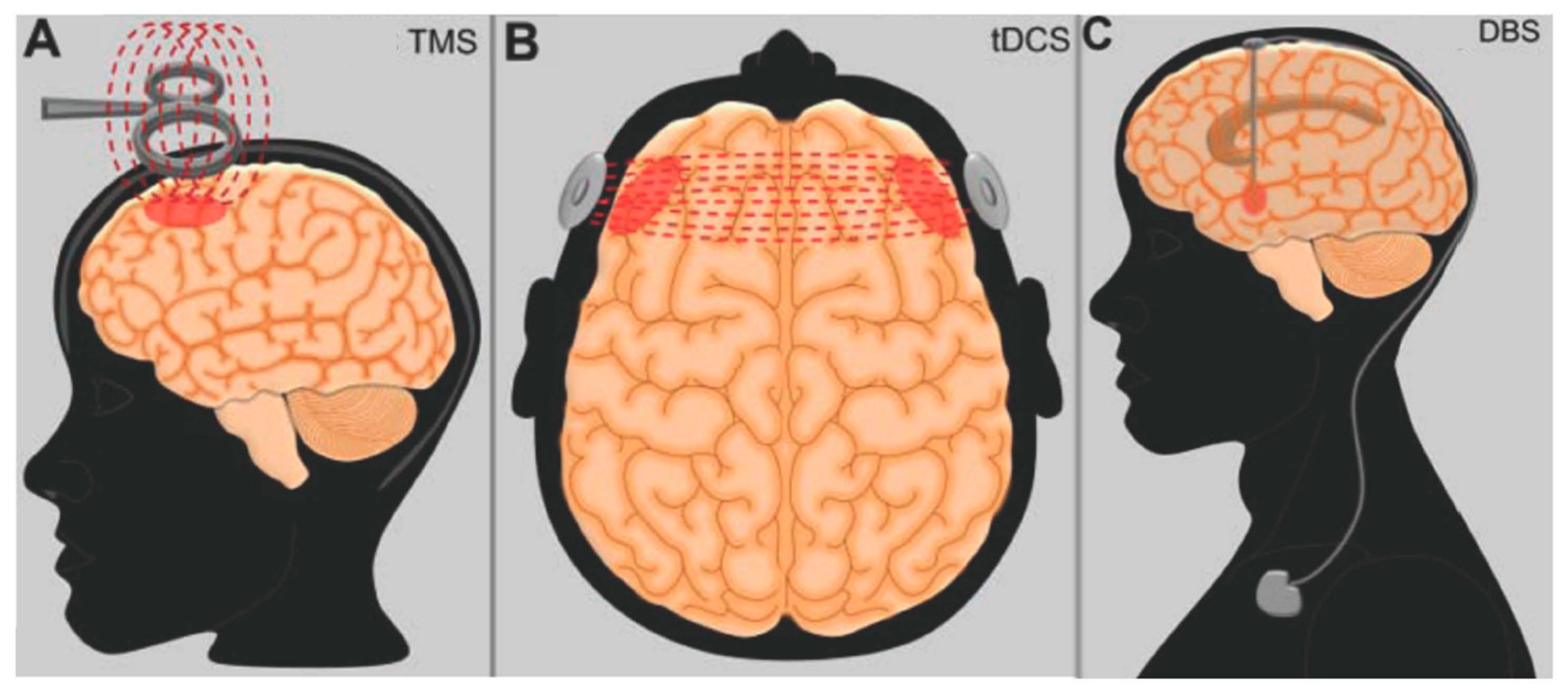
5. Neuromodulation Studies in Clinical Practice
6. Conclusions
Author Contributions
Funding
Conflicts of Interest
References
- Breslau, N.; Davis, G.C.; Andreski, P.; Peterson, E. Traumatic events and posttraumatic stress disorder in an urban population of young adults. Arch. Gen. Psychiatry 1991, 48, 216–222. [Google Scholar] [CrossRef] [PubMed]
- Breslau, N.; Kessler, R.C.; Chilcoat, H.D.; Schultz, L.R.; Davis, G.C.; Andreski, P. Trauma and posttraumatic stress disorder in the community: The 1996 Detroit Area Survey of Trauma. Arch. Gen. Psychiatry 1998, 55, 626–632. [Google Scholar] [CrossRef]
- Fairbank, J.A.; Ebert, L.; Costello, E.J. Epidemiology of traumatic events and post-traumatic stress disorder. In Post Traumatic Stress Disorder: Diagnosis, Management and Treatment; Nutt, D., Davidson, J.R.T., Zohar, J., Eds.; Martin Dunitz Ltd.: London, UK, 2000. [Google Scholar]
- Kessler, R.C.; Sonnega, A.; Bromet, E.; Hughes, M.; Nelson, C.B. Posttraumatic stress disorder in the National Comorbidity Survey. Arch. Gen. Psychiatry 1995, 52, 1048–1060. [Google Scholar] [CrossRef]
- Trivedi, R.B.; Post, E.P.; Sun, H.; Pomerantz, A.; Saxon, A.J.; Piette, J.D.; Maynard, C.; Arnow, B.; Curtis, I.; Fihn, S.D.; et al. Prevalence, comorbidity, and prognosis of mental health among US veterans. Am. J. Public Health 2015, 105, 2564–2569. [Google Scholar] [CrossRef]
- Fulton, J.J.; Calhoun, P.S.; Wagner, H.R.; Schry, A.R.; Hair, L.P.; Feeling, N.; Elbogen, E.; Beckham, J.C. The prevalence of posttraumatic stress disorder in Operation Enduring Freedom/Operation Iraqi Freedom (OEF/OIF) Veterans: A meta-analysis. J. Anxiety Disord 2015, 31, 98–107. [Google Scholar] [CrossRef] [PubMed]
- Stewart, C.L.; Wrobel, T.A. Evaluation of the efficacy of pharmacotherapy and psychotherapy in treatment of combat-related post-traumatic stress disorder: A meta-analytic review of outcome studies. Mil. Med. 2009, 174, 460–469. [Google Scholar] [CrossRef] [PubMed]
- Powers, M.B.; Halpern, J.M.; Ferenschak, M.P.; Gillihan, S.J.; Foa, E.B. A meta-analytic review of prolonged exposure for posttraumatic stress disorder. Clin. Psychol Rev. 2010, 30, 635–641. [Google Scholar] [CrossRef]
- Bradley, R.; Greene, J.; Russ, E.; Dutra, L.; Westen, D. A multidimensional meta-analysis of psychotherapy for PTSD. Am. J. Psychiatry 2005, 162, 214–227. [Google Scholar] [CrossRef]
- Bisson, J.I.; Ehlers, A.; Matthews, R.; Pilling, S.; Richards, D.; Turner, S. Psychological treatments for chronic post-traumatic stress disorder. Systematic review and meta-analysis. Br. J. Psychiatry 2007, 190, 97–104. [Google Scholar] [CrossRef]
- Katzman, M.A.; Bleau, P.; Blier, P.; Chokka, P.; Kjernisted, K.; Van Ameringen, M.; Canadian Anxiety Guidelines Initiative Group on behalf of the Anxiety Disorders Association of Canada/Association Canadienne des troubles anxieux and McGill University; Antony, M.M.; Bouchard, S.; Brunet, A.; et al. Canadian clinical practice guidelines for the management of anxiety, posttraumatic stress and obsessive-compulsive disorders. BMC Psychiatry 2014, 14 (Suppl. 1), S1. [Google Scholar] [CrossRef]
- Davidson, J.R.; Connor, K.M.; Hertzberg, M.A.; Weisler, R.H.; Wilson, W.H.; Payne, V.M. Maintenance therapy with fluoxetine in posttraumatic stress disorder: A placebo-controlled discontinuation study. J. Clin. Psychopharmacol. 2005, 25, 166–169. [Google Scholar] [CrossRef] [PubMed]
- Murphy, D.; Smith, K.V. Treatment efficacy for veterans with posttraumatic stress disorder: Latent class trajectories of treatment response and their predictors. J. Trauma Stress 2018, 31, 753–763. [Google Scholar] [CrossRef] [PubMed]
- VanElzakker, M.B.; Dahlgren, M.K.; Davis, F.C.; Dubois, S.; Shin, L.M. From Pavlov to PTSD: The extinction of conditioned fear in rodents, humans, and anxiety disorders. Neurobiol. Learn. Mem. 2014, 113, 3–18. [Google Scholar] [CrossRef] [PubMed] [Green Version]
- Bonanno, G.A. Loss, trauma, and human resilience: Have we underestimated the human capacity to thrive after extremely aversive events? Am. Psychol. 2004, 59, 20–28. [Google Scholar] [CrossRef] [PubMed]
- Van Minnen, A.; Wessel, I.; Dijkstra, T.; Roelofs, K. Changes in PTSD patients’ narratives during prolonged exposure therapy: A replication and extension. J. Trauma Stress 2002, 15, 255–258. [Google Scholar] [CrossRef]
- Foa, E.B. Psychosocial treatment of posttraumatic stress disorder. J. Clin. Psychiatry 2000, 61, 43–48; discussion 49–51. [Google Scholar] [PubMed]
- Eysenck, H.J. The conditioning model of neurosis. Behav. Brain Sci. 1979, 2, 155–199. [Google Scholar] [CrossRef]
- Blechert, J.; Michael, T.; Vriends, N.; Margraf, J.; Wilhelm, F.H. Fear conditioning in posttraumatic stress disorder: Evidence for delayed extinction of autonomic, experiential, and behavioural responses. Behav. Res. Ther. 2007, 45, 2019–2033. [Google Scholar] [CrossRef]
- Orr, S.P.; Metzger, L.J.; Lasko, N.B.; Macklin, M.L.; Peri, T.; Pitman, R.K. De novo conditioning in trauma-exposed individuals with and without posttraumatic stress disorder. J. Abnorm. Psychol. 2000, 109, 290–298. [Google Scholar] [CrossRef]
- Pitman, R.K.; Orr, S.P. Test of the conditioning model of neurosis: Differential aversive conditioning of angry and neutral facial expressions in anxiety disorder patients. J. Abnorm. Psychol. 1986, 95, 208–213. [Google Scholar] [CrossRef]
- Reznikov, R.; Binko, M.; Nobrega, J.N.; Hamani, C. Deep brain stimulation in animal models of fear, anxiety, and posttraumatic stress disorder. Neuropsychopharmacology 2016, 41, 2810–2817. [Google Scholar] [CrossRef] [PubMed]
- Reznikov, R.; Diwan, M.; Nobrega, J.N.; Hamani, C. Towards a better preclinical model of PTSD: Characterizing animals with weak extinction, maladaptive stress responses and low plasma corticosterone. J. Psychiatr. Res. 2015, 61, 158–165. [Google Scholar] [CrossRef] [PubMed]
- Blanchard, R.J.; Blanchard, D.C. Passive and active reactions to fear-eliciting stimuli. J. Comp. Physiol. Psychol. 1969, 68, 129–135. [Google Scholar] [CrossRef] [PubMed]
- Lissek, S.; Powers, A.S.; McClure, E.B.; McClure, E.B.; Phelps, E.A.; Woldehawariat, G.; Grillon, C.; Pine, D.S. Classical fear conditioning in the anxiety disorders: A meta-analysis. Behav. Res. Ther. 2005, 43, 1391–1424. [Google Scholar] [CrossRef] [PubMed]
- Myers, K.M.; Davis, M. Mechanisms of fear extinction. Mol. Psychiatry 2007, 12, 120–150. [Google Scholar] [CrossRef] [PubMed]
- Rodgers, R.J.; Dalvi, A. Anxiety, defence and the elevated plus-maze. Neurosci. Biobehav. Rev. 1997, 21, 801–810. [Google Scholar] [CrossRef]
- Besnard, A.; Sahay, A. Adult hippocampal neurogenesis, fear generalization, and stress. Neuropsychopharmacology 2016, 41, 24–44. [Google Scholar] [CrossRef]
- Ahmadizadeh, M.J.; Rezaei, M. Unilateral right and bilateral dorsolateral prefrontal cortex transcranial magnetic stimulation in treatment post-traumatic stress disorder: A randomized controlled study. Brain Res. Bull. 2018, 140, 334–340. [Google Scholar] [CrossRef]
- LeDoux, J. The amygdala. Curr. Biol. 2007, 17, 868–874. [Google Scholar] [CrossRef]
- Garrido, M.I.; Barnes, G.R.; Sahani, M.; Dolan, R.J. Functional evidence for a dual route to amygdala. Curr. Biol. 2012, 22, 129–134. [Google Scholar] [CrossRef]
- Romanski, L.M.; Clugnet, M.C.; Bordi, F.; LeDoux, J.E. Somatosensory and auditory convergence in the lateral nucleus of the amygdala. Behav. Neurosci. 1993, 107, 444–450. [Google Scholar] [CrossRef] [PubMed]
- Johnson, L.R.; McGuire, J.; Lazarus, R.; Palmer, A.A. Pavlovian fear memory circuits and phenotype models of PTSD. Neuropharmacology 2012, 62, 638–646. [Google Scholar] [CrossRef] [PubMed]
- LeDoux, J.E.; Iwata, J.; Cicchetti, P.; Reis, D.J. Different projections of the central amygdaloid nucleus mediate autonomic and behavioral correlates of conditioned fear. J. Neurosci. 1988, 8, 2517–2529. [Google Scholar] [CrossRef] [PubMed] [Green Version]
- Assareh, N.; Sarrami, M.; Carrive, P.; McNally, G.P. The organization of defensive behavior elicited by optogenetic excitation of rat lateral or ventrolateral periaqueductal gray. Behav. Neurosci. 2016, 130, 406–414. [Google Scholar] [CrossRef] [PubMed]
- Hsu, D.T.; Chen, F.L.; Takahashi, L.K.; Kalin, N.H. Rapid stress-induced elevations in corticotropin-releasing hormone mRNA in rat central amygdala nucleus and hypothalamic paraventricular nucleus: An in situ hybridization analysis. Brain Res. 1998, 788, 305–310. [Google Scholar] [CrossRef]
- Dekeyzer, S.; De Kock, I.; Nikoubashman, O.; Vanden Bossche, S.; Van Eetvelde, R.; De Groote, J.; Acou, M.; Wiesmann, M.; Deblaere, K.; Achten, E. “Unforgettable”—a pictorial essay on anatomy and pathology of the hippocampus. Insights Imaging 2017, 8, 199–212. [Google Scholar] [CrossRef] [PubMed]
- Giap, B.T.; Jong, C.N.; Ricker, J.H.; Cullen, N.K.; Zafonte, R.D. The hippocampus: Anatomy, pathophysiology, and regenerative capacity. J. Head Trauma Rehabil. 2000, 15, 875–894. [Google Scholar] [CrossRef]
- Fanselow, M.S.; Dong, H.W. Are the dorsal and ventral hippocampus functionally distinct structures? Neuron 2010, 65, 7–19. [Google Scholar] [CrossRef]
- Maren, S.; Aharonov, G.; Fanselow, M.S. Neurotoxic lesions of the dorsal hippocampus and Pavlovian fear conditioning in rats. Behav. Brain Res. 1997, 88, 261–274. [Google Scholar] [CrossRef]
- Wood, J.N.; Grafman, J. Human prefrontal cortex: Processing and representational perspectives. Nat. Rev. Neurosci. 2003, 4, 139–147. [Google Scholar] [CrossRef]
- Diekhof, E.K.; Geier, K.; Falkai, P.; Gruber, O. Fear is only as deep as the mind allows: A coordinate-based meta-analysis of neuroimaging studies on the regulation of negative affect. Neuroimage 2011, 58, 275–285. [Google Scholar] [CrossRef] [PubMed]
- Britton, J.C.; Phan, K.L.; Taylor, S.F.; Fig, L.M.; Liberzon, I. Corticolimbic blood flow in posttraumatic stress disorder during script-driven imagery. Biol. Psychiatry 2005, 57, 832–840. [Google Scholar] [CrossRef] [PubMed]
- Bremner, J.D.; Narayan, M.; Staib, L.H.; Southwick, S.M.; McGlashan, T.; Charney, D.S. Neural correlates of memories of childhood sexual abuse in women with and without posttraumatic stress disorder. Am. J. Psychiatry 1999, 156, 1787–1795. [Google Scholar] [PubMed]
- Shin, L.M.; Wright, C.I.; Cannistraro, P.A.; Wedig, M.M.; McMullin, K.; Martis, B.; Macklin, M.L.; Lasko, N.B.; Cavanagh, S.R.; Krangel, T.S.; et al. A functional magnetic resonance imaging study of amygdala and medial prefrontal cortex responses to overtly presented fearful faces in posttraumatic stress disorder. Arch. Gen. Psychiatry 2005, 62, 273–281. [Google Scholar] [CrossRef] [PubMed]
- Shin, L.M.; Orr, S.P.; Carson, M.A.; Rauch, S.L.; Macklin, M.L.; Lasko, N.B.; Peters, P.M.; Metzger, L.J.; Dougherty, D.D.; Cannistraro, P.A.; et al. Regional cerebral blood flow in the amygdala and medial prefrontal cortex during traumatic imagery in male and female Vietnam veterans with PTSD. Arch. Gen. Psychiatry 2004, 61, 168–176. [Google Scholar] [CrossRef] [PubMed]
- Sripada, R.K.; King, A.P.; Garfinkel, S.N.; Wang, X.; Sripada, C.S.; Welsh, R.C.; Liberzon, I. Altered resting-state amygdala functional connectivity in men with posttraumatic stress disorder. J. Psychiatry Neurosci. 2012, 37, 241–249. [Google Scholar] [CrossRef] [PubMed] [Green Version]
- Carrion, V.G.; Haas, B.W.; Garrett, A.; Song, S.; Reiss, A.L. Reduced hippocampal activity in youth with posttraumatic stress symptoms: An FMRI study. J. Pediatr. Psychol. 2010, 35, 559–569. [Google Scholar] [CrossRef]
- Thomaes, K.; Dorrepaal, E.; Draijer, N.P.; de Ruiter, M.B.; Elzinga, B.M.; van Balkom, A.J.; Smoor, P.L.; Smit, J.; Veltman, D.J. Increased activation of the left hippocampus region in Complex PTSD during encoding and recognition of emotional words: A pilot study. Psychiatry Res. 2009, 171, 44–53. [Google Scholar] [CrossRef]
- Hamani, C.; Nobrega, J.N. Preclinical studies modeling deep brain stimulation for depression. Biol. Psychiatry 2012, 72, 916–923. [Google Scholar] [CrossRef]
- Uylings, H.B.; Groenewegen, H.J.; Kolb, B. Do rats have a prefrontal cortex? Behav. Brain Res. 2003, 146, 3–17. [Google Scholar] [CrossRef]
- Milad, M.R.; Quirk, G.J. Neurons in medial prefrontal cortex signal memory for fear extinction. Nature 2002, 420, 70–74. [Google Scholar] [CrossRef] [PubMed] [Green Version]
- Corcoran, K.A.; Quirk, G.J. Recalling safety: Cooperative functions of the ventromedial prefrontal cortex and the hippocampus in extinction. CNS Spectr. 2007, 12, 200–206. [Google Scholar] [CrossRef] [PubMed]
- Amano, T.; Unal, C.T.; Pare, D. Synaptic correlates of fear extinction in the amygdala. Nat. Neurosci. 2010, 13, 489–494. [Google Scholar] [CrossRef] [PubMed] [Green Version]
- Royer, S.; Martina, M.; Pare, D. An inhibitory interface gates impulse traffic between the input and output stations of the amygdala. J. Neurosci. 1999, 19, 10575–10583. [Google Scholar] [CrossRef] [PubMed]
- Likhtik, E.; Popa, D.; Apergis-Schoute, J.; Fidacaro, G.A.; Pare, D. Amygdala intercalated neurons are required for expression of fear extinction. Nature 2008, 454, 642–645. [Google Scholar] [CrossRef] [Green Version]
- Ranck, J.B., Jr. Which elements are excited in electrical stimulation of mammalian central nervous system: A review. Brain Res. 1975, 98, 417–440. [Google Scholar] [CrossRef]
- Lozano, A.M.; Dostrovsky, J.; Chen, R.; Ashby, P. Deep brain stimulation for Parkinson’s disease: Disrupting the disruption. Lancet Neurol. 2002, 1, 225–231. [Google Scholar] [CrossRef]
- Hamani, C.; Temel, Y. Deep brain stimulation for psychiatric disease: Contributions and validity of animal models. Sci. Transl. Med. 2012, 4, 142–148. [Google Scholar] [CrossRef]
- Cleren, C.; Tallarida, I.; Guiniec, E.L.; Janin, F.; Nachon, O.; Canini, F.; Spennato, G.; Moreau, J.L.; Garcia, R. Low-frequency stimulation of the ventral hippocampus facilitates extinction of contextual fear. Neurobiol. Learn. Mem. 2013, 101, 39–45. [Google Scholar] [CrossRef]
- Deschaux, O.; Thevenet, A.; Spennato, G.; Arnaud, C.; Moreau, J.L.; Garcia, R. Low-frequency stimulation of the hippocampus following fear extinction impairs both restoration of rapid eye movement sleep and retrieval of extinction memory. Neuroscience 2010, 170, 92–98. [Google Scholar] [CrossRef]
- Garcia, R.; Spennato, G.; Nilsson-Todd, L.; Moreau, J.L.; Deschaux, O. Hippocampal low-frequency stimulation and chronic mild stress similarly disrupt fear extinction memory in rats. Neurobiol. Learn. Mem. 2008, 89, 560–566. [Google Scholar] [CrossRef] [PubMed]
- Farinelli, M.; Deschaux, O.; Hugues, S.; Thevenet, A.; Garcia, R. Hippocampal train stimulation modulates recall of fear extinction independently of prefrontal cortex synaptic plasticity and lesions. Learn. Mem. 2006, 13, 329–334. [Google Scholar] [CrossRef] [PubMed]
- Rodriguez-Romaguera, J.; Do Monte, F.H.; Quirk, G.J. Deep brain stimulation of the ventral striatum enhances extinction of conditioned fear. Proc. Natl. Acad. Sci. USA 2012, 109, 8764–8769. [Google Scholar] [CrossRef] [PubMed] [Green Version]
- Sui, L.; Huang, S.; Peng, B.; Ren, J.; Tian, F.; Wang, Y. Deep brain stimulation of the amygdala alleviates fear conditioning-induced alterations in synaptic plasticity in the cortical-amygdala pathway and fear memory. J. Neural. Transm. 2014, 121, 773–782. [Google Scholar] [CrossRef] [PubMed]
- Saldivar-Gonzalez, J.A.; Posadas-Andrews, A.; Rodriguez, R.; Gómez, C.; Hernández-Manjarrez, M.E.; Ortiz-León, S.; Martínez-Pineda, A.; Gómez-Laguna, D.; Salgado, V.; Manjarrez, J.; et al. Effect of electrical stimulation of the baso-lateral amygdala nucleus on defensive burying shock probe test and elevated plus maze in rats. Life Sci. 2003, 72, 819–829. [Google Scholar] [CrossRef]
- Langevin, J.P.; De Salles, A.A.; Kosoyan, H.P.; Krahl, S.E. Deep brain stimulation of the amygdala alleviates post-traumatic stress disorder symptoms in a rat model. J. Psychiatr. Res. 2010, 44, 1241–1245. [Google Scholar] [CrossRef] [PubMed]
- Vidal-Gonzalez, I.; Vidal-Gonzalez, B.; Rauch, S.L.; Quirk, G.J. Microstimulation reveals opposing influences of prelimbic and infralimbic cortex on the expression of conditioned fear. Learn. Mem. 2006, 13, 728–733. [Google Scholar] [CrossRef] [PubMed] [Green Version]
- Maroun, M.; Kavushansky, A.; Holmes, A.; Wellman, C.; Motanis, H. Enhanced extinction of aversive memories by high-frequency stimulation of the rat infralimbic cortex. PLoS ONE 2012, 7, e35853. [Google Scholar] [CrossRef]
- Zheng, X.; Deschaux, O.; Lavigne, J.; Le Guisquet, A.M.; Belzung, C.; El-Hage, W. Prefrontal high-frequency stimulation prevents sub-conditioning procedure-provoked, but not acute stress-provoked, reemergence of extinguished fear. Neurobiol. Learn. Mem. 2013, 101, 33–38. [Google Scholar] [CrossRef]
- Deschaux, O.; Motanis, H.; Spennato, G.; Moreau, J.L.; Garcia, R. Re-emergence of extinguished auditory-cued conditioned fear following a sub-conditioning procedure: Effects of hippocampal and prefrontal tetanic stimulations. Neurobiol. Learn. Mem. 2011, 95, 510–518. [Google Scholar] [CrossRef]
- Reznikov, R.; Bambico, F.R.; Diwan, M.; Nachon, O.; Cleren, C.; Moreau, J.L.; Garcia, R. prefrontal cortex deep brain stimulation improves fear and anxiety-like behavior and reduces basolateral amygdala activity in a preclinical model of posttraumatic stress disorder. Neuropsychopharmacology 2018, 43, 1099–1106. [Google Scholar] [CrossRef] [PubMed]
- Van’t Wout, M.; Longo, S.M.; Reddy, M.K.; Philip, N.S.; Bowker, M.T.; Greenberg, B.D. Transcranial direct current stimulation may modulate extinction memory in posttraumatic stress disorder. Brain Behav. 2017, 7, e00681. [Google Scholar] [CrossRef] [PubMed] [Green Version]
- Zwanzger, P.; Steinberg, C.; Rehbein, M.A.; Bröckelmann, A.K.; Dobel, C.; Zavorotnyy, M.; Domschke, K.; Junghöfer, M. Inhibitory repetitive transcranial magnetic stimulation (rTMS) of the dorsolateral prefrontal cortex modulates early affective processing. Neuroimage 2014, 101, 193–203. [Google Scholar] [CrossRef] [PubMed]
- Baek, K.; Chae, J.H.; Jeong, J. The effect of repetitive transcranial magnetic stimulation on fear extinction in rats. Neuroscience 2012, 200, 159–165. [Google Scholar] [CrossRef] [PubMed]
- Wang, H.N.; Bai, Y.H.; Chen, Y.C.; Zhang, R.G.; Wang, H.H.; Zhang, Y.H.; Gan, J.L.; Peng, Z.W.; Tan, Q.R. Repetitive transcranial magnetic stimulation ameliorates anxiety-like behavior and impaired sensorimotor gating in a rat model of post-traumatic stress disorder. PLoS ONE 2015, 10, e0117189. [Google Scholar] [CrossRef] [PubMed]
- Legrand, M.; Troubat, R.; Brizard, B.; Le Guisquet, A.M.; Belzung, C.; El-Hage, W. Prefrontal cortex rTMS reverses behavioral impairments and differentially activates c-Fos in a mouse model of post-traumatic stress disorder. Brain Stimul. 2019, 12, 87–95. [Google Scholar] [CrossRef] [PubMed]
- Liu, G.; Feng, D.; Wang, J.; Zhang, H.; Peng, Z.; Cai, M.; Yang, J.; Zhang, R.; Wang, H.; Wu, S.; et al. rTMS ameliorates PTSD symptoms in rats by enhancing glutamate transmission and synaptic plasticity in the ACC via the PTEN/Akt signalling pathway. Mol. Neurobiol. 2018, 55, 3946–3958. [Google Scholar] [CrossRef]
- Sparta, D.R.; Jennings, J.H.; Ung, R.L.; Stuber, G.D. Optogenetic strategies to investigate neural circuitry engaged by stress. Behav. Brain Res. 2013, 255, 19–25. [Google Scholar] [CrossRef] [Green Version]
- Sparta, D.R.; Smithuis, J.; Stamatakis, A.M.; Jennings, J.H.; Kantak, P.A.; Ung, R.L. Stuber GDInhibition of projections from the basolateral amygdala to the entorhinal cortex disrupts the acquisition of contextual fear. Front. Behav. Neurosci. 2014, 8, 129. [Google Scholar] [CrossRef] [PubMed]
- Tovote, P.; Fadok, J.P.; Luthi, A. Neuronal circuits for fear and anxiety. Nat. Rev. Neurosci. 2015, 16, 317–331. [Google Scholar] [CrossRef]
- Johansen, J.P.; Wolff, S.B.; Luthi, A.; LeDoux, J.E. Controlling the elements: An optogenetic approach to understanding the neural circuits of fear. Biol. Psychiatry 2012, 71, 1053–1060. [Google Scholar] [CrossRef] [PubMed]
- Gafford, G.M.; Ressler, K.J. Mouse models of fear-related disorders: Cell-type-specific manipulations in amygdala. Neuroscience 2016, 321, 108–120. [Google Scholar] [CrossRef] [PubMed] [Green Version]
- Allsop, S.A.; Vander Weele, C.M.; Wichmann, R.; Tye, K.M. Optogenetic insights on the relationship between anxiety-related behaviors and social deficits. Front. Behav. Neurosci. 2014, 8, 241. [Google Scholar] [CrossRef] [PubMed]
- Kozel, F.A. Clinical repetitive transcranial magnetic stimulation for posttraumatic stress disorder, generalized anxiety disorder, and bipolar disorder. Psychiatr. Clin. North. Am. 2018, 41, 433–446. [Google Scholar] [CrossRef] [PubMed]
- Fryml, L.D.; Pelic, C.G.; Acierno, R.; Tuerk, P.; Yoder, M.; Borckardt, J.J.; Juneja, N.; Schmidt, M.; Beaver, K.L.; George, M.S. Exposure therapy and simultaneous repetitive transcranial magnetic stimulation: A controlled pilot trial for the treatment of posttraumatic stress disorder. J. ECT 2018. [Google Scholar] [CrossRef] [PubMed]
- Kozel, F.A.; Motes, M.A.; Didehbani, N.; DeLaRosa, B.; Bass, C.; Schraufnagel, C.D.; Jones, P.; Morgan, C.R.; Spence, J.S.; Kraut, M.A.; et al. Repetitive TMS to augment cognitive processing therapy in combat veterans of recent conflicts with PTSD: A randomized clinical trial. J. Affect. Disord. 2018, 229, 506–514. [Google Scholar] [CrossRef] [PubMed]
- Boggio, P.S.; Rocha, M.; Oliveira, M.O.; Fecteau, S.; Cohen, R.B.; Campanhã, C.; Ferreira-Santos, E.; Meleiro, A.; Corchs, F.; Zaghi, S.; et al. Noninvasive brain stimulation with high-frequency and low-intensity repetitive transcranial magnetic stimulation treatment for posttraumatic stress disorder. J. Clin. Psychiatry 2010, 71, 992–999. [Google Scholar] [CrossRef]
- Carpenter, L.L.; Conelea, C.; Tyrka, A.R.; Welch, E.S.; Greenberg, B.D.; Price, L.H.; Niedzwiecki, M.; Yip, A.G.; Barnes, J.; Philip, N.S. 5Hz Repetitive transcranial magnetic stimulation for posttraumatic stress disorder comorbid with major depressive disorder. J. Affect. Disord. 2018, 235, 414–420. [Google Scholar] [CrossRef]
- Fryml, L.D.; Sahlem, G.; Fox, J.; Short, E.B. The role of rTMS for patients with severe PTSD and depression. Evid Based Ment. Health 2018, 21, 39–40. [Google Scholar] [CrossRef]
- Nam, D.H.; Pae, C.U.; Chae, J.H. Low-frequency, repetitive transcranial magnetic stimulation for the treatment of patients with posttraumatic stress disorder: A double-blind, sham-controlled study. Clin. Psychopharmacol Neurosci. 2013, 11, 96–102. [Google Scholar] [CrossRef]
- Oznur, T.; Akarsu, S.; Celik, C.; Bolu, A.; Ozdemir, B.; Akcay, B.D.; Ozselek, S.; Bozkurt, A.; Ozmenler, K.N. Is transcranial magnetic stimulation effective in treatment-resistant combat related posttraumatic stress disorder? Neurosciences 2014, 19, 29–32. [Google Scholar] [PubMed]
- Philip, N.S.; Barredo, J.; van’t Wout-Frank, M.; Tyrka, A.R.; Price, L.H.; Carpenter, L.L. Network mechanisms of clinical response to transcranial magnetic stimulation in posttraumatic stress disorder and major depressive disorder. Biol. Psychiatry 2018, 83, 263–272. [Google Scholar] [CrossRef] [PubMed]
- Philip, N.S.; Ridout, S.J.; Albright, S.E.; Sanchez, G.; Carpenter, L.L. 5-Hz Transcranial Magnetic Stimulation for Comorbid Posttraumatic Stress Disorder and Major Depression. J. Trauma Stress 2016, 29, 93–96. [Google Scholar] [CrossRef] [PubMed] [Green Version]
- Yesavage, J.A.; Fairchild, J.K.; Mi, Z.; Biswas, K.; Davis-Karim, A.; Phibbs, C.S.; Forman, S.D.; Thase, M.; Williams, L.M.; Etkin, A.; et al. Effect of Repetitive Transcranial Magnetic Stimulation on Treatment-Resistant Major Depression in US Veterans: A Randomized Clinical Trial. JAMA Psychiatry 2018, 75, 884–893. [Google Scholar] [CrossRef] [PubMed]
- Saunders, N.; Downham, R.; Turman, B.; Kropotov, J.; Clark, R.; Yumash, R.; Szatmary, A. Working memory training with tDCS improves behavioral and neurophysiological symptoms in pilot group with post-traumatic stress disorder (PTSD) and with poor working memory. Neurocase 2015, 21, 271–278. [Google Scholar] [CrossRef] [PubMed]
- van’t Wout-Frank, M.; Shea, M.T.; Larson, V.C.; Greenberg, B.D.; Philip, N.S. Combined transcranial direct current stimulation with virtual reality exposure for posttraumatic stress disorder: Feasibility and pilot results. Brain Stimul. 2018. [Google Scholar] [CrossRef]
- Hamani, C.; Lozano, A.M. Hardware-related complications of deep brain stimulation: A review of the published literature. Stereotact. Funct. Neurosurg. 2006, 84, 248–251. [Google Scholar] [CrossRef]
- Awan, N.R.; Lozano, A.; Hamani, C. Deep brain stimulation: Current and future perspectives. Neurosurg. Focus 2009, 27, E2. [Google Scholar] [CrossRef]
- Langevin, J.P.; Koek, R.J.; Schwartz, H.N.; Chen, J.W.Y.; Sultzer, D.L.; Mandelkern, M.A.; Kulick, A.D.; Krahl, S.E. Deep brain stimulation of the basolateral amygdala for treatment-refractory posttraumatic stress disorder. Biol. Psychiatry 2016, 79, e82–e84. [Google Scholar] [CrossRef]


© 2019 by the authors. Licensee MDPI, Basel, Switzerland. This article is an open access article distributed under the terms and conditions of the Creative Commons Attribution (CC BY) license (http://creativecommons.org/licenses/by/4.0/).
Share and Cite
Gouveia, F.V.; Gidyk, D.C.; Giacobbe, P.; Ng, E.; Meng, Y.; Davidson, B.; Abrahao, A.; Lipsman, N.; Hamani, C. Neuromodulation Strategies in Post-Traumatic Stress Disorder: From Preclinical Models to Clinical Applications. Brain Sci. 2019, 9, 45. https://0-doi-org.brum.beds.ac.uk/10.3390/brainsci9020045
Gouveia FV, Gidyk DC, Giacobbe P, Ng E, Meng Y, Davidson B, Abrahao A, Lipsman N, Hamani C. Neuromodulation Strategies in Post-Traumatic Stress Disorder: From Preclinical Models to Clinical Applications. Brain Sciences. 2019; 9(2):45. https://0-doi-org.brum.beds.ac.uk/10.3390/brainsci9020045
Chicago/Turabian StyleGouveia, Flavia V., Darryl C. Gidyk, Peter Giacobbe, Enoch Ng, Ying Meng, Benjamin Davidson, Agessandro Abrahao, Nir Lipsman, and Clement Hamani. 2019. "Neuromodulation Strategies in Post-Traumatic Stress Disorder: From Preclinical Models to Clinical Applications" Brain Sciences 9, no. 2: 45. https://0-doi-org.brum.beds.ac.uk/10.3390/brainsci9020045