
Methods to Determine Chain-Breaking Antioxidant Activity of Nanomaterials beyond DPPH•. A Review


1
Istituto per la Sintesi Organica e la Fotoreattività, Consiglio Nazionale delle Ricerche (ISOF-CNR), Via P. Gobetti 101, 40129 Bologna, Italy
2
Department of Chemistry “G. Ciamician”, University of Bologna, Via S. Giacomo 11, 40126 Bologna, Italy
*
Author to whom correspondence should be addressed.
Antioxidants 2021, 10(10), 1551; https://0-doi-org.brum.beds.ac.uk/10.3390/antiox10101551
Submission received: 13 September 2021
/
Revised: 27 September 2021
/
Accepted: 28 September 2021
/
Published: 29 September 2021
(This article belongs to the Special Issue Nanoantioxidants)
Abstract
:This review highlights the progress made in recent years in understanding the mechanism of action of nanomaterials with antioxidant activity and in the chemical methods used to evaluate their activity. Nanomaterials represent one of the most recent frontiers in the research for improved antioxidants, but further development is hampered by a poor characterization of the ‘‘antioxidant activity’’ property and by using oversimplified chemical methods. Inhibited autoxidation experiments provide valuable information about the interaction with the most important radicals involved in the lipid oxidation, namely alkylperoxyl and hydroperoxyl radicals, and demonstrate unambiguously the ability to stop the oxidation of organic materials. It is proposed that autoxidation methods should always complement (and possibly replace) the use of assays based on the quenching of stable radicals (such as DPPH• and ABTS•+). The mechanisms leading to the inhibition of the autoxidation (sacrificial and catalytic radical trapping antioxidant activity) are described in the context of nanoantioxidants. Guidelines for the selection of the appropriate testing conditions and of meaningful kinetic analysis are also given.
Keywords:
nanomaterial; antioxidant; radicals; autoxidation; nanoantioxidants; oxygen; assays; reactive oxygen species; ROS; catalysis1. Introduction
Autoxidation, or peroxidation, is the spontaneous reaction of organic molecules with O2 that leads to the formation of peroxides, epoxides, aldehydes and ketones as well as to simpler breakdown fragments or to insoluble polymers. These products have usually unwanted characteristics, such as bad smell (i.e., the rancid odour of oxidized lipids), chemico-physical characteristics that are very different from those of the non-oxidized substrate and, most importantly, toxic effects due to their strong electrophilicity. Although autoxidation occurs in disparate organic substrates including plastic, lubricants, fuels, edible oils, meat, cosmetics and cellular membranes, in all cases the reaction occurs via a radical chain mechanism involving carbon and oxygen centered radicals [1]. The importance of stopping or delaying the autoxidation is of paramount importance for many industrial applications and is still attracting the interest of many researchers. Antioxidants can be defined as molecules able to slow down the oxidative radical chain, therefore preventing the damage that can be caused to oxidizable substrates by the effects of oxygen. Antioxidants comprise an incredibly vast and heterogeneous family of small organic and inorganic molecules, as well as macromolecules and enzymes, all capable of interfering at different stages of the autoxidation reaction.
Antioxidants can be divided into two groups, depending on their mechanism of action: (i) preventive antioxidants, that interfere with the initiation process by retarding or stopping the initial formation of radical species and (ii) chain-breaking antioxidants, that slow down the autoxidation by competing with the propagation reactions; that is, they react with radicals faster than the oxidizable substrate. In addition to these direct antioxidants, compounds that do not themselves possess antioxidant activity, but can stimulate and increase the efficacy of the endogenous antioxidant defences in biological systems, are usually classified as indirect antioxidants [2].
Although antioxidants have been known from a long time, the research of improved natural or synthetic antioxidants is still a hot topic because of their important practical implications. Antioxidants may suffer from low stability under O2 and in biological systems they can be degraded before reaching the target sites, or they can have adverse health effects that limit their use [3,4]. Sometimes, it would be desirable removing antioxidants from the homogeneous system to which they were added, after their effect [5].
In this context, nanotechnology has opened up new possibilities to exploit the nearly infinite and innovative properties of nanomaterials, possibly in combination with some conventional natural or synthetic compounds, with the aim of obtaining pioneering “nanoantioxidants” with enhanced properties [6,7,8,9]. More interestingly, many categories of nanoantioxidants have potent radical scavenging and quenching capacities in combination with interesting mechanical, optical, magnetic, catalytic, and optical properties increasing their potential practical use [10,11].
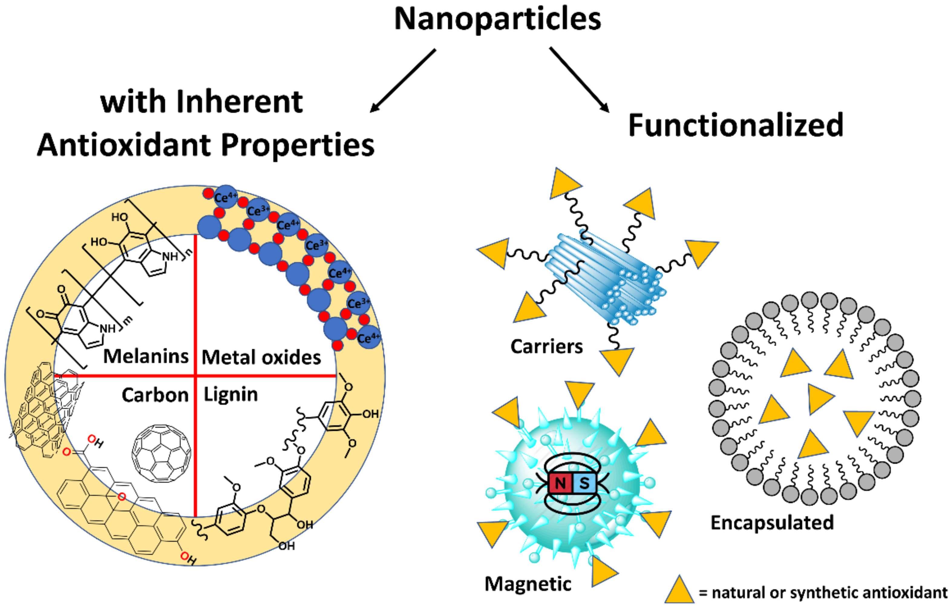
Antioxidant nanoparticle can be classified into two broad categories: (i) nanoparticles with inherent antioxidant properties; (ii) inert nanoparticle functionalized with antioxidants. The latter can be of very different types, such as a core with antioxidants covalently bound on the surface (e.g., in the case of magnetic nanoantioxidants), or passive carriers able to deliver and release small-molecular antioxidants (e.g., nano-encapsulated, nanotubes or mesoporous materials) [12,13,14,15,16] (Figure 1).
We believe that this classification is still useful to inspire the research in this field, as it is demonstrated by the selected recent examples reported in the next section. However, while there is an enormous effort in the synthesis and physical characterization of these new materials, much less is known regarding the mechanism of radical trapping and the ability to stop the autoxidation of easily oxidizable substrates. In the Section 5 we will show how these two fundamental properties can be easily measured by studying the inhibition of the autoxidation of standard substrates.
2. Recent Examples of Nanomaterials Used for Their Antioxidant Activity
2.1. Inherent Antioxidant Nanoparticles
The most common nanoparticles (NPs) with intrinsic antioxidant properties are based on metal oxides. CeO2 nanoparticles (CeNPs) have been used as efficient antioxidants thanks to the ability of the metal to cycle between Ce3+ and Ce4+ ionic states. The lack of oxygen atoms on the surface of the nanoparticle leads to the presence of electrons on the 4f orbitals of some cerium atoms. Not all cerium (IV) atoms are converted to cerium (III), thus, Ce3+ and Ce4+ exist simultaneously on the surface to form a redox pair [17].
Cerium oxide nanoparticles have been widely used for solid oxide fuel cells, electrochromic thin films, sensors, catalysts, and biomedical materials [18,19]. In 2019, Shi and co-workers proposed to use CeNPs for the treatment of ocular inflammation [20] while in 2020, Hyeon and co-workers have synthesized heterostructured CeO2/Mn3O4 nanocrystals for the protection of hematopoietic intestinal stem cells from irradiation-induced ROS damage [21].
Despite the very promising properties of metal nanoparticles as antioxidants [22], the environmental impact of their long-term use have been questioned, [23]. This problem can be attenuated using biodegradable nanoparticles. Lignin, as a polymeric polyphenol, is considered a promising natural antioxidant agent and several studies have been developed to explore this property [24,25]. However, it has a limited solubility in organic solvents, so various approaches are used to decrease its molecular weight and increase its content of –OH groups. Studies to obtain and characterize lignin nanoparticles are quite recent with only a few reports in the literature so far [26,27].
In 2018, Puglia et al. prepared spherical lignin nanoparticles by dissolving pristine alkali lignin into ethylene glycol, followed by addition of different acids. Higher radical trapping by lignin nanoparticles aqueous solution with respect of pristine lignin was revealed [28]. In 2019, Cheng et al. synthesized lignin nanoparticles by adding an anti-solvent to a colloidal dispersion of lignin in DMSO. They observed how the size of the nanoparticles and consequently, both the content and accessibility of phenolic hydroxyl group, affect the radical trapping activity of the latter [29]. Lastly, in 2020, Rezende and co-workers isolated pure lignin from a non-food biomass resource. These lignin nanoparticles also showed a radical trapping activity about 1.5 times higher than lignin in solution, and 3–4 times higher than butylated hydroxytoluene (BHT) and butylated hydroxyanisole (BHA), respectively, indicating their potential applicability as active antioxidant materials in dermocosmetics products [30].
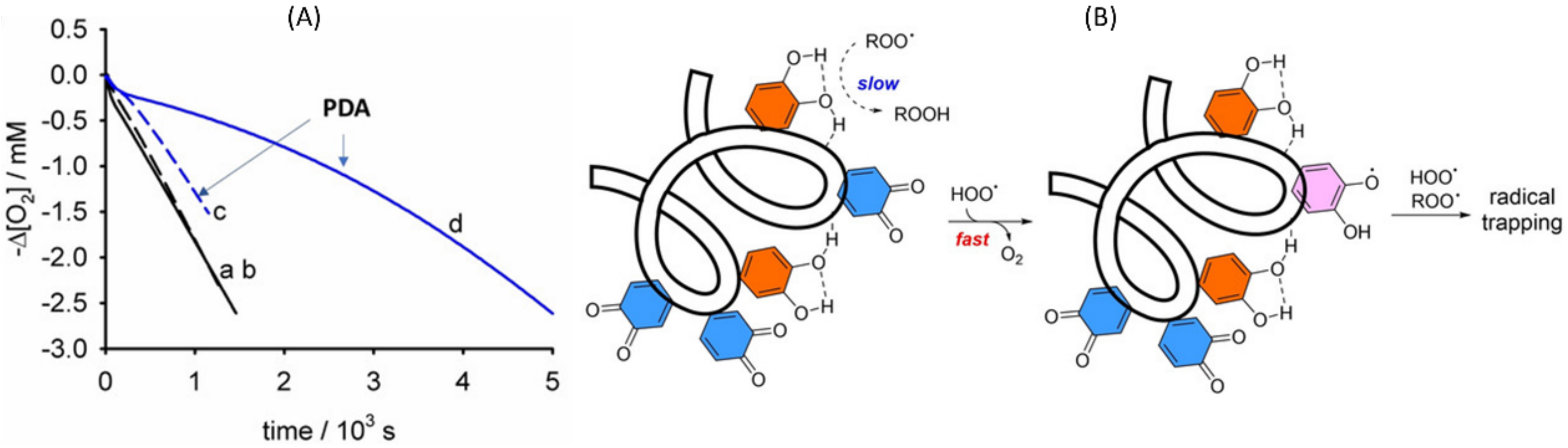
Advancement in nanotechnology has revealed several nanoparticles with biological origins, such as melanin nanoparticles as potent antioxidants by themselves [31]. Melanins have been proposed to possess antioxidant activity [32,33], that has been related, for instance, to their anti-inflammatory [34], wound regeneration [35] and anti-ischemic activity [31]. We have recently explained the antioxidant properties of polydopamine (PDA) in the synergic trapping of alkylperoxyl and hydroperoxyl radicals. The key reaction explaining this peculiar antioxidant activity is the reduction of the ortho-quinone moieties present in PDA by the reaction with HOO• [36].
Due to their native biocompatibility and their biodegradability, melanins are gaining increasing attention ranging from nanomedicine to nanocosmetics [37]. Inspired by PDA nanoparticles, nanoparticles based on the autoxidation of serotonin (5-hydroxytryptamine) and of 1,8-dihydroxynaphthalene (DHN) have been designed and developed, to obtain, respectively, polyserotonin and DHN allomelanin, a type of nitrogen-free melanin. Polyserotonin nanoparticles, obtained from oxidative polymerization of the well-known neurotransmitter serotonin, was used as promising nanomaterials for cancer therapeutics by testing its photothermal properties, drug loading and release, and biocompatibility [38]. Artificial allomelanin nanoparticles instead, showed radiation protection in human skin cells via radical scavenging [39].
Finally, another example of intrinsically active nanoantioxidants concerns carbon nanomaterials, whose structures are based primarily on sp2-hybridized carbon bonding, and include fullerenes [40], graphenes [41], carbon nanotubes [42] and their derivatives [43]. It was demonstrated that they exhibit strong ROS-scavenging properties [44]. Carbon nanomaterials possess such properties due to their conjugated π-system, which permits the scavenging of free radicals by addition to double bonds [45].
2.2. Functionalized Nanoparticles
The antioxidant properties of functionalized nanoparticles strictly depend on the compounds used to decorate the surface. The nanoparticle structure, in addition to acting as a support, may provide specific features to the final materials, that would be impossible to obtain with small-molecule antioxidants, such as magnetic properties. Surface functionalization has been typically performed by exploiting natural or synthetic antioxidants including glutathione [46], carotenoids [47], gallic acid [48], curcumin [49], rosmarinic acid [50], caffeic acid [51], α-tocopherol analogues [52], and BHT [53] but also biomacromolecules such as protamine sulfate polyelectrolyte (PSP) and SOD enzyme [54]. Recently, nanoantioxidants functionalized with surface-bound hindered nitroxides have also been proposed [55,56]. Nitroxides derived from TEMPO (2,2,6,6-tetramethylpiperidine 1-oxyl) are a class of persistent radicals characterized by high stability in water under air [57] that exhibit antioxidant activity and no in vitro toxicity and can be conveniently used to obtain different kinds of nanoantioxidants.
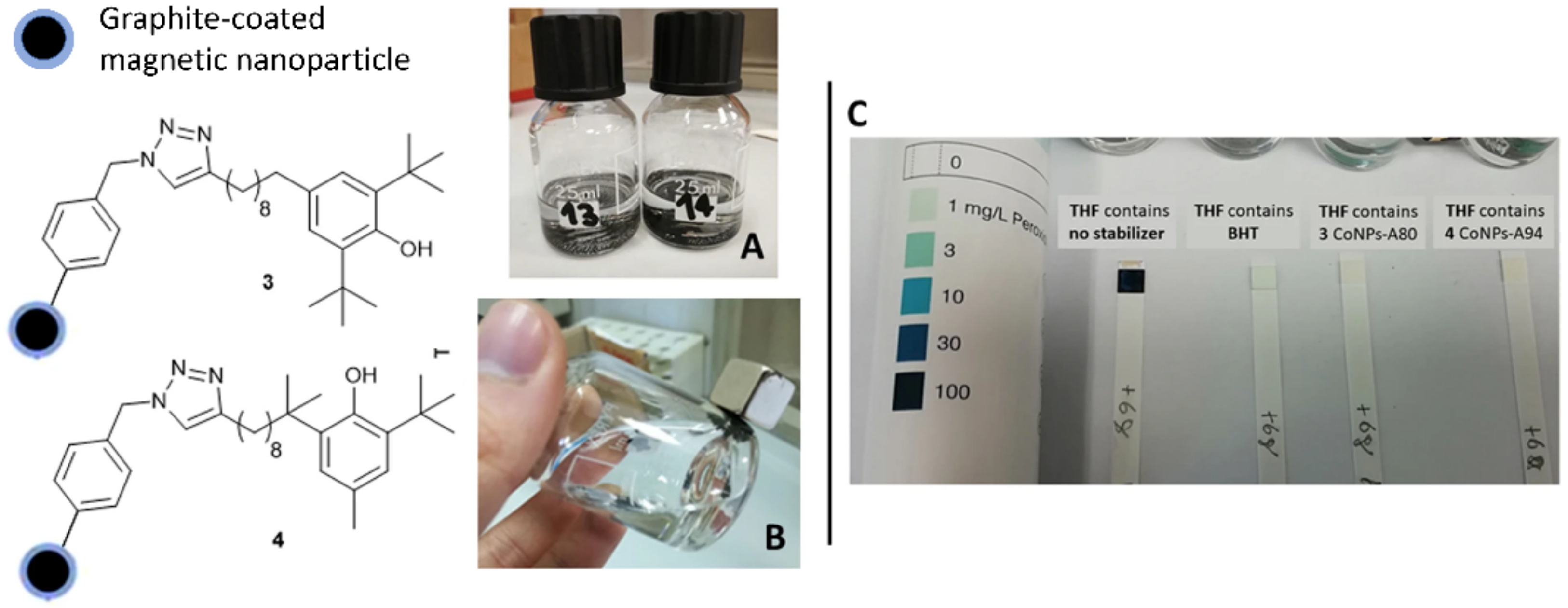
The surface functionalization of Fe3O4 nanoparticles with gallic acid allowed to obtain magnetically separable, efficient and low cost nanoantioxidant with potential applications in polymer, cosmetics, biomedical and food industry [58]. In 2019, magnetite-quercetin complex with free radical scavenging capacity and super-paramagnetic behaviour were obtained and showed potential antifungal and antibacterial effects [59]. Carbon-coated cobalt nanomagnets decorated by α-tocopherol units or BHT (see the example reported in Figure 2) are able to effectively counteract the autoxidation of organic substrates, and can be efficiently controlled by an external magnet [5,60].
On the other hand, nano-encapsulated materials allow for better stability and serve as carriers for the controlled release of the compounds they contain. This is the case of ascorbic acid in liposomes [61] and in porous silica nanoparticles [62]. Halloysite is a natural clay with an intrinsic nanotubular structure that has been used to load a diarylamine antioxidant for rubber stabilization [63], and various natural phenolic antioxidants such as curcumin [49], silibinin [64], resveratrol [65], and quercetin [52] to improve their stability and to obtain controlled antioxidant delivery systems.
The examples reported above demonstrate how research on nanoantioxidants is a trending topic that span very different fields. In most cases, however, the methods used to detect their antioxidant properties are not up to par with the other characterizations carried out on those types of materials, for example regarding the size of the nanoparticles, the percentage of loading or the release capacity. These latter data are obtained using appropriate and well-established techniques that rarely make inherent mistakes. Instead, regarding antioxidant properties, simplified methods such as the DPPH• and ABTS•+ assays are almost exclusively used, whereas the ability of nanomaterials to block the autoxidation of organic substrates is only seldom investigated. In the next sections, we explain how to improve the understanding of our chemical knowledge on nanoantioxidants by using inhibited autoxidation experiments.
3. Why You Should Not Use Simplified Methods?
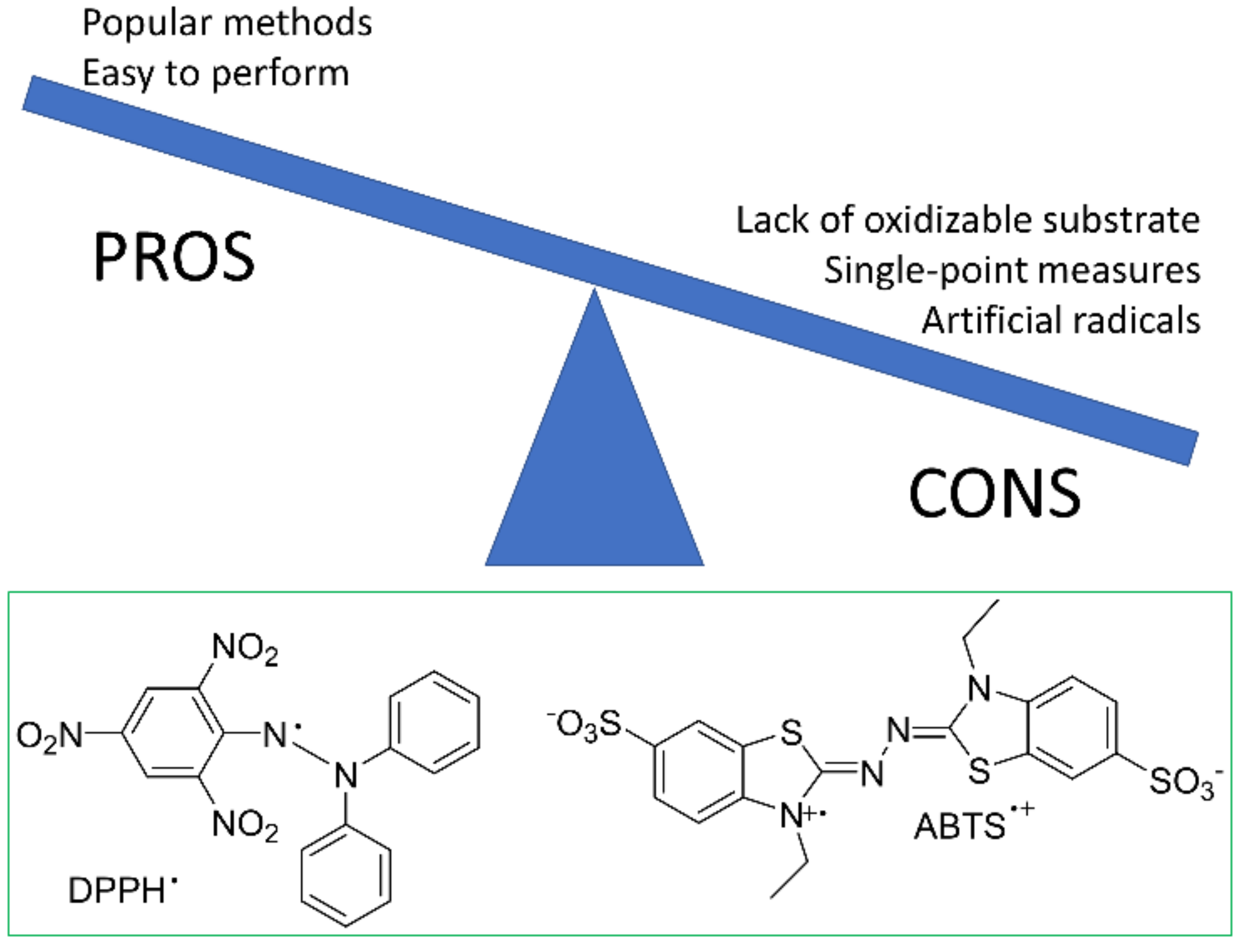
Stable colored radicals such as DPPH• and ABTS•+ (see Figure 3) are believed to be a simple way to measure the antioxidant activity, as their absorption changes upon reaction with reductants including antioxidants [66]. However, these assays have many limitations and their popularity may represent an obstacle to the development of nanoantioxidants, because they prevent researchers to investigate with more accuracy the reactions of biologically relevant radicals. The use of DPPH• and related methods should be avoided especially in the field of new nanomaterials whose chemistry is still to be fully explored and we propose alternative techniques which can be easily implemented to replace these assays. Below, we list the main shortcoming of these techniques.
Artificial radicals. The stable radicals used as probes are chemically very different from the radicals that propagate the autoxidation of real systems, thus they don’t reproduce the kinetic and sometimes neither the stoichiometry of the reaction with ROO•. Moreover, these stable radicals may be quenched also by reductants having no antioxidant activity, such as H2O2 and hydroperoxides [66,67,68,69].
Lack of oxidizable substrate. The absence of a suitable oxidizable substrate makes impossible to know whether the putative antioxidant can interrupt the autoxidation of an organic molecule or not, or even display pro-oxidant effects. The results obtained indicate a “radical trapping power” or a “reducing power” rather than a true chain-breaking activity. The foregoing arguments are valid also for assays based on the reduction of metal ions (i.e., Ferric Reducing Antioxidant Power, FRAP, and CUPric ion Reducing Antioxidant Capaicty, CUPRAC assays) or of different kinds of stable radicals, such as galvinoxyl or dialkylnitroxides [70].
Single-point measurements. Besides these shortcomings that originate from the chemical structures of the stable radicals, other problems derive from single-point measurements of absorbance decay after a fixed time. The time interval employed is usually about 30 min (however, it may change from study to study), that is long enough to allow even low-activity antioxidants to react with the probes. These experiments therefore represent a sort of “titration” of the reducing groups present on a molecule or on a nanomaterial. On the other hand, the study of the kinetic decay of the probe, may provide a more realistic description of the reactivity, but is more difficult than single-point measures because it requires the kinetic analysis of the results, and a specialized instrumentation in case of fast reaction (such as stopped-flow equipment) [70].
Optical interferences. As the DPPH• and related assays are based on spectrophotometric determinations, they may be affected by the scattering of the solution, or by the intense color of the materials that may be superimposed to the absorption maxima of the probes. These problems can be solved by using EPR spectroscopy as a detection method for the probe concentrations, or by separating the materials just before the absorbance reading [70].
4. Autoxidation Mechanism. Why Focus on Peroxyl Radicals?
4.1. Autoxidation
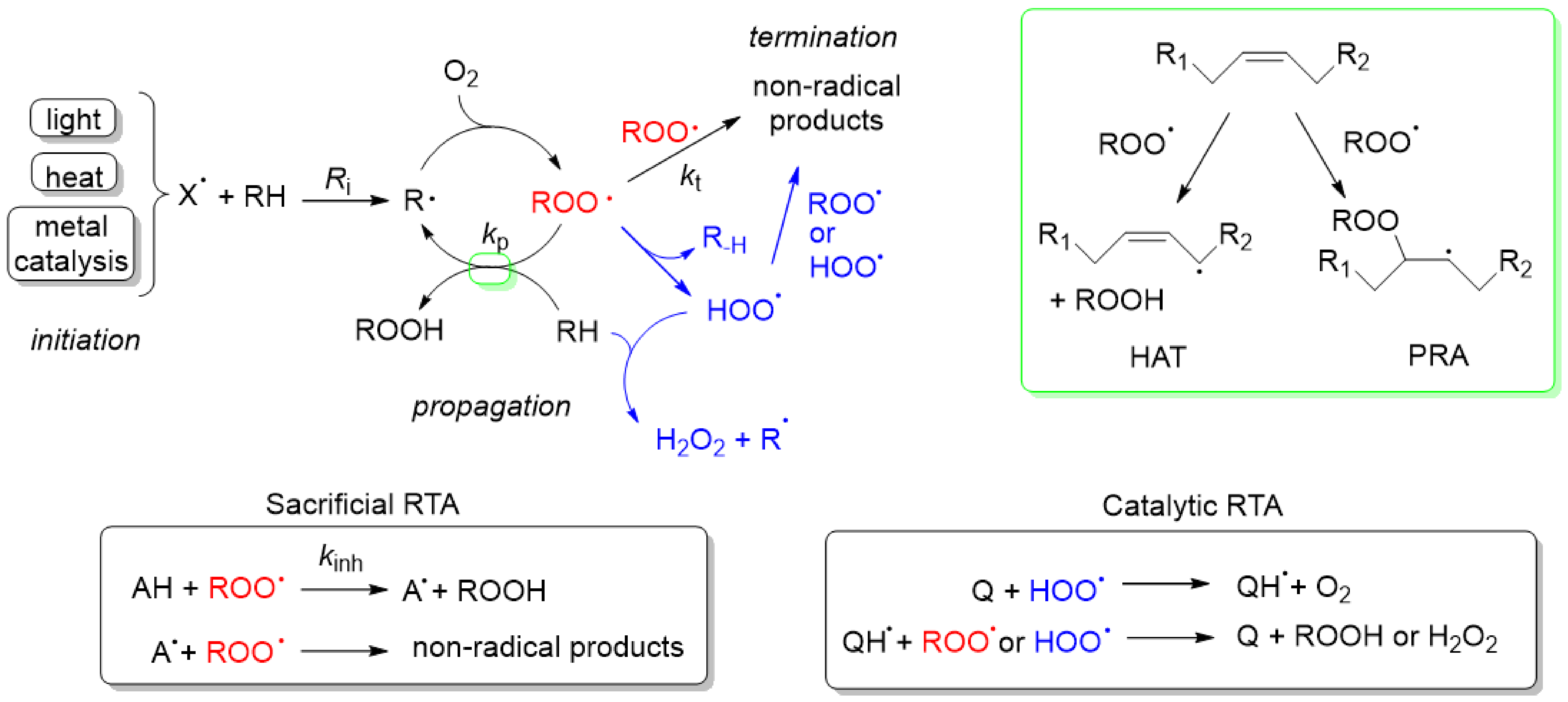
Autoxidation is the spontaneous reaction of organic molecules with O2, occurring through a radical-chain mechanism consisting of the typical initiation, propagation and termination steps depicted in Scheme 1 [1].
Initiation. Many different pathways can lead to the formation of radicals and thus contribute to the initiation step. UV and visible radiations are responsible for initiation in skin and materials exposed to light such as food and plastic, especially in the presence of pigments that may act as photosensitizers [71,72]. Temperature increase facilitates the endothermic reactions leading to radical formation, such as cleavage of weak bonds (such as RO–OR in peroxides) and the hydrogen atom transfer (HAT) from bis-allylic C–H groups to O2 [73]. These processes are important in cooking and long-term storage of unsaturated lipids. Metal catalysis is of paramount importance in water containing multiphasic systems, like living cells, food and cosmetic emulsions. The simultaneous presence of free iron and lipid hydroperoxides triggers cell death trough ferroptosis, and Fe2+ plus H2O2 cause the damage of biological macromolecules through the Fenton reaction [74]. Interfering with these processes is an important strategy to slow-down the autoxidation, for instance by reducing the exposure to light, heat, removing metals by chelation and decomposing H2O2. Some nanomaterials endow their antioxidant ability to one or more of these effects, and not to their ability to trap radicals. For instance, artificial melanin nanoparticles protects from light [75], and certain metal oxides can remove H2O2 behaving like the natural catalase (CAT) [76] or glutathione peroxidase (GpX) enzymes [77].
Propagation. The initiating radical X• reacts with the oxidizable molecules forming a carbon-centred alkyl radical (R•) that in turn reacts with O2 to generate alkylperoxyl radicals (ROO•, see Scheme 1). As the reaction of R• and O2 is very fast, very low levels of O2 are enough to quantitatively transform R• to ROO•. Peroxyl radicals then propagate the oxidative chain by reacting with the substrate via HAT or peroxyl radical addition (PRA) to C=C double bonds, yielding a new alkyl radical (see the box in Scheme 1). The rate constant of the propagation reaction, kp, is the main determinant of the oxidation rate. In the case of pro-aromatic oxidizable substrates (i.e., γ-terpinene) or having alcohol or amine groups, the ROO• radicals can undergo a 1,4-intramolecular HAT [78] leading to the formation of hydroperoxyl radicals (HOO•), which have a specific chemical behaviour that strongly influences the oxidation rate and the efficacy of antioxidants (see blue reactions in Scheme 1).
Termination. In the absence of antioxidants, radicals disappear by the self-recombination of two ROO• radicals, while the role of R• is negligible because the concentration of alkyl radicals, in the presence of O2, is very low. If HOO• is formed, it speeds up termination thanks to the HAT reaction: HOO• + ROO• → ROOH + O2 [79].
4.2. Chain-Breaking or Radical-Trapping Antioxidants (RTA)
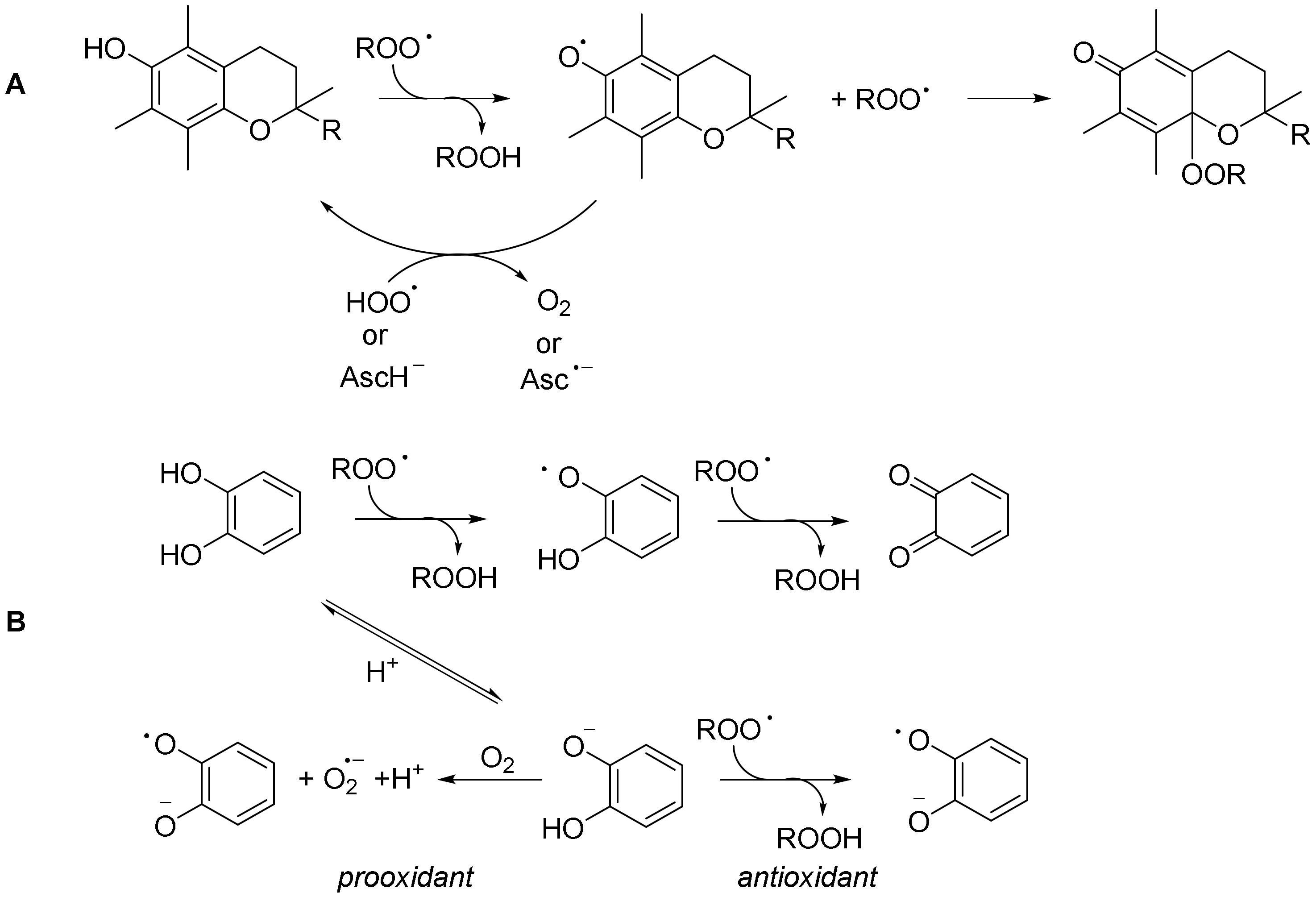
This type of compounds blocks the autoxidation of organic molecules by quenching ROO• radicals and forming stable radicals that don’t propagate the oxidative chain (Scheme 1). The rate constant for the reaction with ROO•, also called the inhibition rate constant, kinh, and the number of ROO• trapped (i.e., the stoichiometric coefficient n) are the two parameters that describe the efficacy of an antioxidant. The great majority of the antioxidants act as sacrificial reductant toward ROO•, so they are consumed during the RTA activity, unless they are reduced back by other sacrificial reductants like in the case of the well-known synergy between α-tocopherol and ascorbate (AscH−) [80], or in the presence of HOO• (see Scheme 2A) [81]. In the presence of HOO•, nitroxides and ortho-quinone [36] containing molecules (indicated with Q in Scheme 1) behave as catalytic antioxidants causing the disappearance of ROO• and HOO• with little antioxidant consumption [82,83]. The reaction between an RTA antioxidant and ROO• can occur through a variety of sequential or concerted proton-coupled electron transfer mechanisms that are important to rationalize the antioxidant effect in any given reaction medium [84,85,86]. In general, antioxidants having a low bond dissociation enthalpy of the O–H or N–H bonds or low redox potential react quickly with ROO• but at the same time are susceptible to react with O2. As the latter reaction causes the depletion of antioxidants over time, a compromise must be achieved between stability under O2 and reactivity toward ROO•. In the case of antioxidants based on a pyridine or pyrimidine nucleus, the reaction with O2 is minimized while conserving a good ROO• trapping [87]. In this regard, the deprotonation of antioxidants (such as ascorbic acid or polyphenols) greatly increases their reaction with ROO•, but also decreases their stability [88,89] (Scheme 2B).
4.3. Why Focus on Peroxyl Radicals?
While a variety of radicals can be transiently formed during the autoxidation of organic substrates (see Table 1), only alkylperoxyl and hydroperoxyl radicals are important for the action of RTA antioxidants.
As mentioned before, alkyl radicals even under low O2 concentrations are promptly converted into ROO•, thus they are not intercepted by RTA. Hydroxyl (HO•) and alkoxyl (RO•) radicals are too reactive toward any organic substrates to be trapped by RTA. Actually, the antioxidant effect of RTA arises from the competition between the reaction of the radical with the substrate and that with RTA, thus if the former reaction is too fast, exceedingly high concentrations of RTA would be needed to stop this reaction [70]. The assays studying the reaction with HO• are therefore not recommended as unique methods to assess the antioxidant activity.
4.4. SOD-like Activity
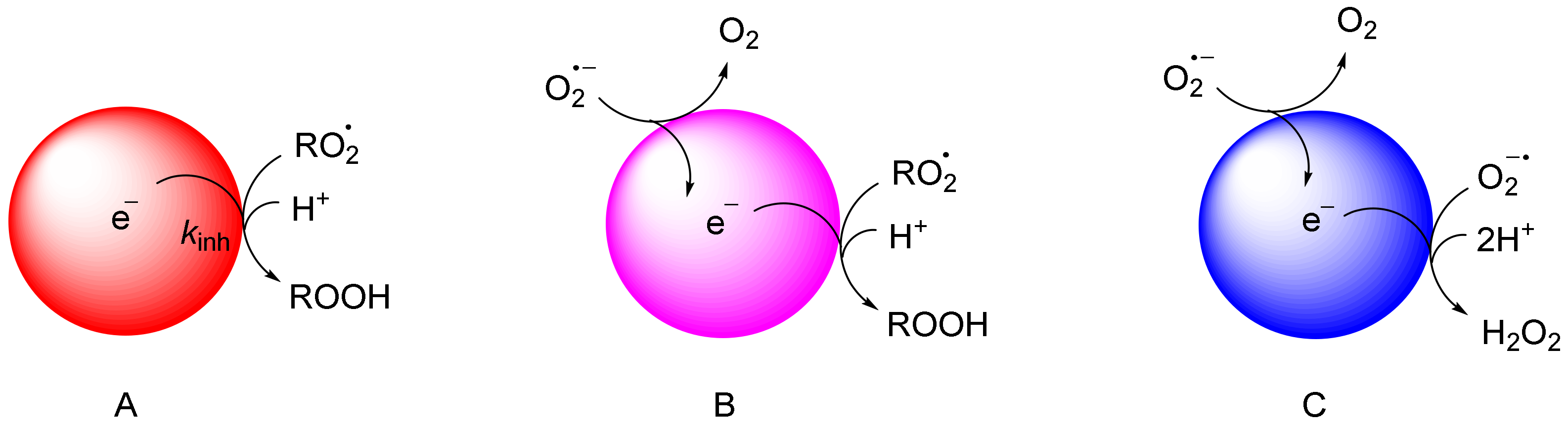
Superoxide (O2•−) is not reactive toward organic substrates and thus it does not propagate the oxidative chain, and it is also unreactive toward most RTA [90]. Nevertheless, O2•− is a precursor of H2O2, that is a non-radical ROS (reactive oxygen species) which influences cellular functions and may originate HO• radicals. Certain nanomaterials, in particular metal oxides (CeO2, Mn3O4, …) have superoxide dismutase (SOD)-like activity because they are cyclically reduced and oxidized by O2•− as shown in Figure 4 [91] and display interesting biological effects [76].
5. How to Study Autoxidations Inhibited by Nanoantioxidants?
The best methods to measure the antioxidant activity of RTA are based on the ability to slow down the autoxidation of an organic substrate, under conditions that should be as similar as possible to those occurring in real systems. For instance, if the antioxidant is intended for food applications, the oxidizable substrate may be represented by polyunsaturated fatty acids or by their esters, as bulk oils or as oil/water emulsions. Instead, if biological applications are sought, liposomes made of unsaturated phospholipids may be used. It should be noticed that many antioxidants (especially polyphenols) [89] become pro-oxidants at high concentrations, thus the dependency of the chain-breaking activity on concentration should be assessed. Autooxidation-based methods are able to detect conditions at which a given antioxidant nanomaterial can act as pro-oxidant. However, although autoxidation studies are a well assessed method, their application to nanoantioxidants is not widespread and thus some guidelines are presented in this section.
5.1. Initiation
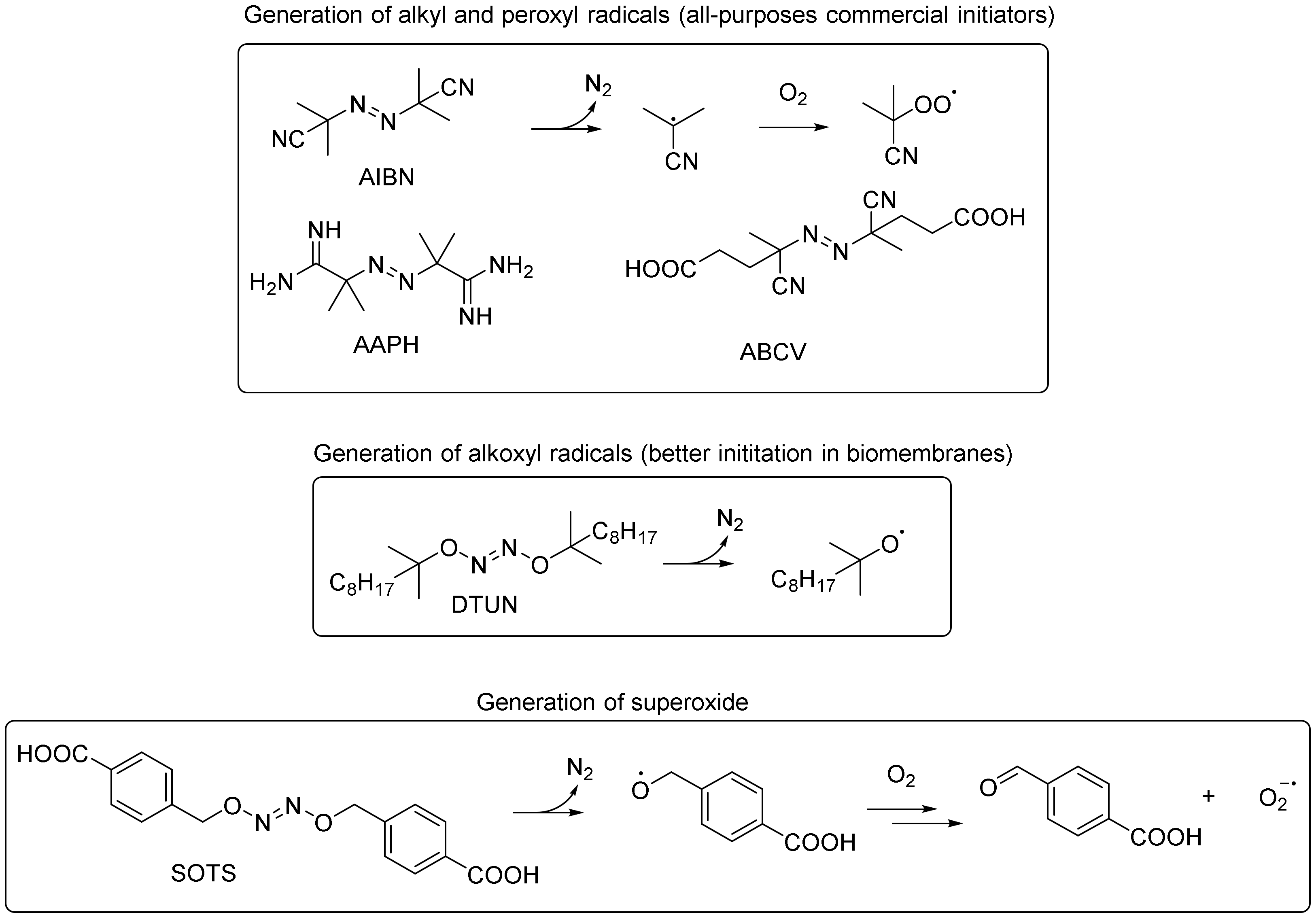
As spontaneous autoxidation at room temperature is a slow process that takes several days to proceed, accelerating strategies are usually employed. In food and biodiesel research, autoxidation is accelerated by rising the temperature to 90–130 °C, such as in the Rancimat and similar OSI (oxygen stability index) methods [92]. Although temperature is a “clean” expedient, as it doesn’t require the addition of initiating compounds, it has many drawbacks, including the volatilization of low-boiling components, that makes difficult the extrapolation of the results to the room temperature. Small amounts of chemical initiators, such as azoinitiators or peroxides, are the most practical way to make autoxidations fast and reproducible at room temperature. Azoinitiators with different solubilities and decomposition rates are commercially available (Figure 5) and can be conveniently used to generate radicals at a constant rate, which is a fundamental prerequisite to perform meaningful autoxidation studies. Initiator consumption is usually negligible at low temperatures, so their concentration can be considered constant during the reaction [93]. Novel initiators have been proposed for specific applications, such as to initiate the autoxidation of liposomial bilayers, or to study the chemistry of superoxide (Figure 5) [94]. Another initiation strategy includes the use of Fe2+ with peroxides (usually H2O2) although in this case a constant initiation rate can hardly be achieved because Fe2+ is quickly consumed, while the level of peroxides increases during the autoxidation [95].
5.2. Oxidizable Substrates and Reaction Medium
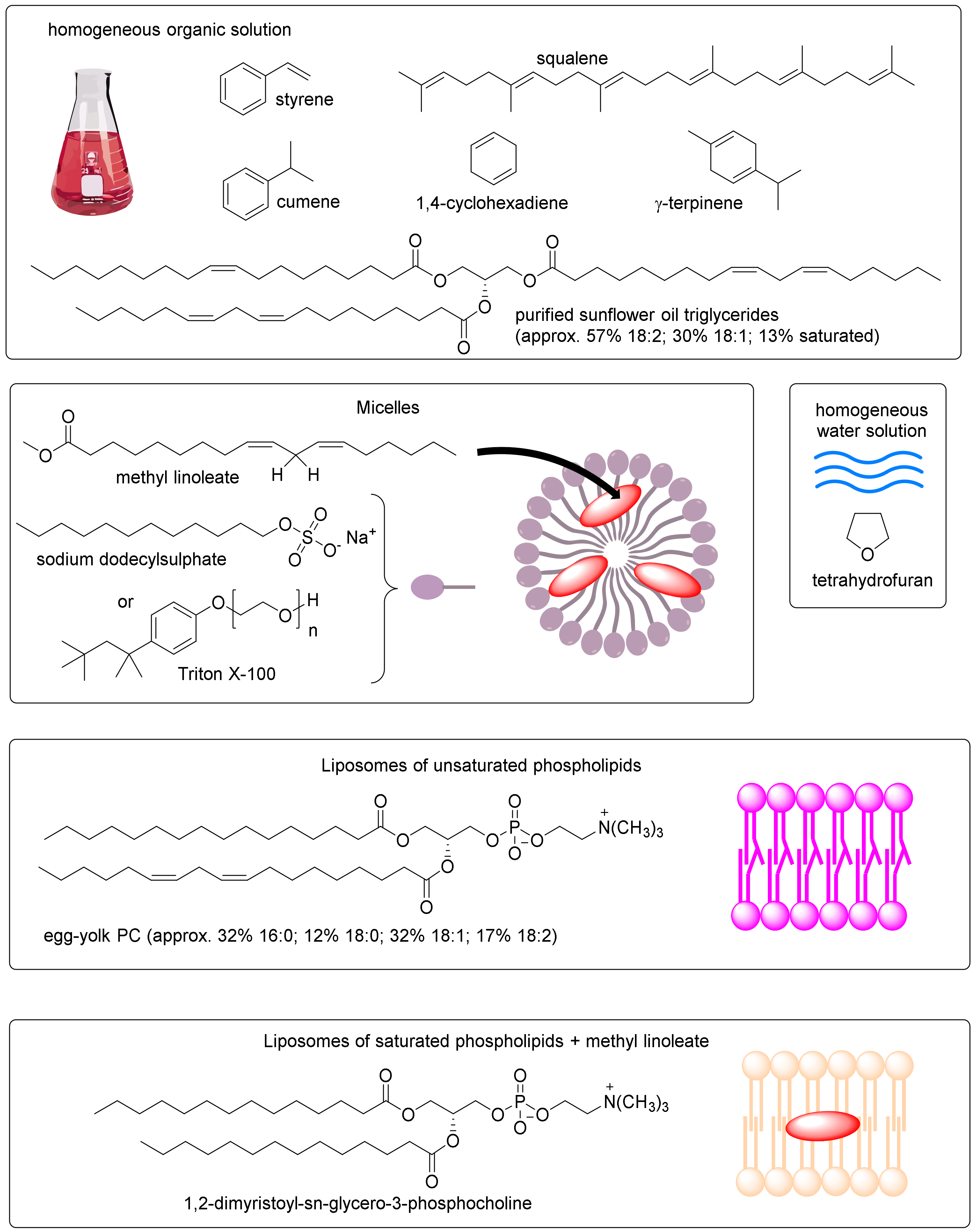
The choice of the oxidizable substrate is also fundamental to obtain meaningful results from autoxidation studies. If quantitative chemico-physical determinations are requested (i.e., measure of kinetic rate constants, see Section 5.5), well-characterized substrates must be chosen. For instance, the propagation and termination rate constants are known (mostly at 30–37 °C) for some organic molecules in homogeneous solution, such as methyl linoleate, styrene, cumene, tetrahydrofuran, sunflower oil triacylglycerides, squalene and para-cymene (see Table 2). Experiments in homogeneous water solution can be performed by using water-soluble oxidizable substrates (such as tetrahydrofuran, THF). Micelles can be used to obtain micro-heterogeneous systems by using neutral or charged surfactants (typically sodium dodecylsulfate [96] or Triton X-100 [97]) and methyl linoleate as substrate. In order to mimic cell membranes, liposomes made of phospholipids containing polyunsaturated fatty acids (such as egg yolk phosphatidylcholine) are the best choice [94]. Another model can be represented by saturated phospholipids incorporating methyl linoleate (see Figure 6) [98].
5.3. How to Study an Autoxidation?
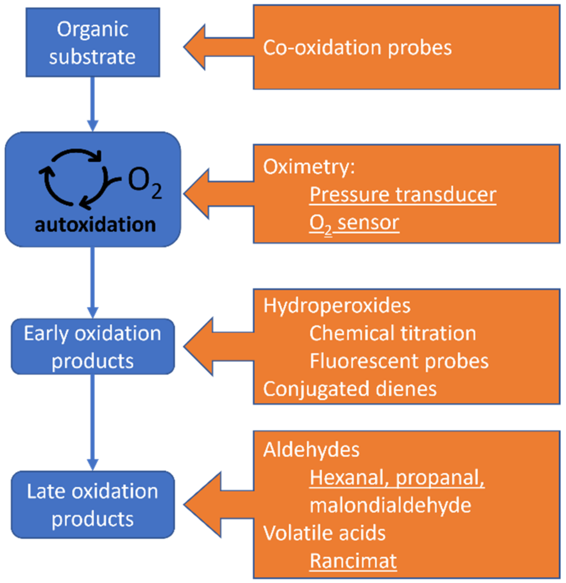
The autoxidation of a given substrate can be studied by the disappearance of reactants or the formation of products, that in turn can be differentiated into early and late products (see Figure 7).
5.3.1. Disappearance of Reactants
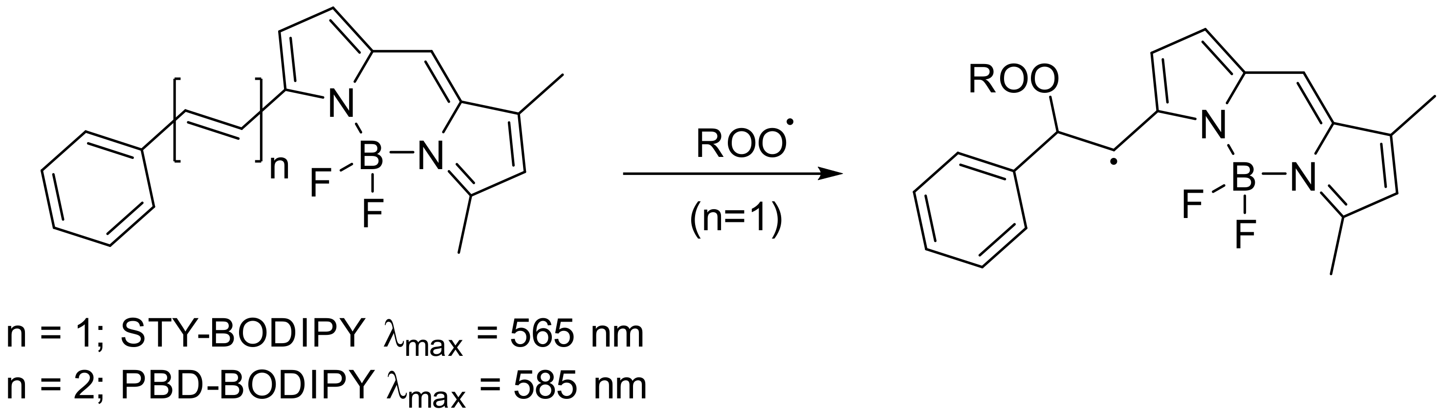
Oxygen consumption is arguably the process that unifies the oxidation of many different substrates, and oximetry is the most versatile method that can be applied to a variety of samples, including nanomaterials, which are not suitable for HPLC analysis or optical spectroscopy. Given their importance, the techniques for measuring O2 during autoxidation are described in detail in the next section. Another method to monitor the disappearance of reactants consists of using probes having an intense absorption (or fluorescence emission) and a chemical structure that resembles that of oxidizable substrates, such as STY-BODIPY or PBD-BODIPY (Scheme 3) [106]. The probes can be co-oxidized with the substrate and followed by UV-vis or fluorescence spectroscopy. The advantage of this method is its high throughput as it can be implemented for use in thermostatic microplate readers.
5.3.2. Formation of Early Products
Hydroperoxides are the most common early products formed during an autoxidation, however it should be emphasized that certain substrates may preferentially form other oxygenated products such as dialkylperoxides or epoxides, and that hydroperoxides are unstable especially at high temperature and in the presence of acids or metals. Hydroperoxides can be determined by iodometric titration, by the ferrous thiocyanate assay or by using fluorescent probes [107]. In the case of the autoxidation of polyunsaturated fatty acids, the hydroperoxides possess conjugated dienes that can be evidenced by spectrophotometry or HPLC-UV-vis detection [108,109]. The analysis of the regiochemistry of the hydroperoxides of linoleic acid or of simpler alkenes also can provide information about the activity of antioxidants (the so called peroxyl-radical clock) [110].
5.3.3. Formation of Late Products
The products that arise from the decomposition of hydroperoxides mainly include aldehydes and acids. Hydroperoxide fragmentation is induced by heat or by the acid-catalysed Hock transposition. The latter reaction can occur during the autoxidation by the traces of acids present in the solvent, or more frequently it is induced by addition of acids during the preparation of the samples [111]. Among the various aldehydes formed by the decomposition of hydroperoxides of polyunsaturated lipids, malondialdehyde is particularly important because its levels are detected by the popular TBARS (thiobarbituric acid reactive substances) method, which is based on the detection of its coloured adduct with thiobarbituric acid. The many limits of the TBARS assay have been reviewed and, due to its low specificity, TBARS method is not recommended for determination of malondialdehyde as one of late product of lipid peroxidation [112]. Low-molecular-weight acids are detected by the Rancimat method, that is based on fluxing an air stream in a heated lipid sample in the presence of the antioxidant, checking the formation of volatile products by bubbling the gases into purified water and measuring the conductivity of this solution [92]. Hexanal and propanal can be conveniently measured in the headspace of the reaction vessel by a gas chromatograph equipped with a solid phase micro extraction (SPME) injector [107].
5.4. Advantages of Oximetry
Most autoxidation methods are not easily applicable to the study of nanoantioxidants. Assays relying on spectrofluorimetry or spectrophotometry may suffer from the light scattering displayed by many nanomaterials, from the intense colors caused by plasmon resonance in metal nanoparticles or from the absorption of highly conjugated materials such as melanins and carbon-based nanomaterials. In this context, the study of the autoxidation by measuring O2 consumption has no limitation respect the use of these kinds of nanoantioxidants, because there is no optical interaction with the sample.
O2 consumption can be measured by using two different approaches: (i) measure of the pressure drop due to O2 consumption, that can be achieved by different kinds of pressure gauges and (ii) direct measure of O2 concentration, either in water or in the headspace, by O2-sensitive probes. Differential pressure transducers measure the small pressure differences between a sample and a reference reaction flask and can be applied to both organic solvents and aqueous solutions. The reference flask contains the same reaction mixture as the sample, but in the presence of a high concentration of an antioxidant, so that it allows to correct the pressure drop observed during the autoxidation of the substrate for the N2 development and O2 consumption from the azo-initiator [93].
The direct measure of O2 concentration comprises optical or electrochemical O2 sensing. The first method is based on the fluorescence quenching, caused by O2, of a fluorescent probe attached on the tip of an optical fiber immersed in the sample or inserted in the headspace. This equipment works best in air and in water but is incompatible with organic solvents [89,113]. A second method suitable for aqueous systems is based on a polarographic Clark electrode, that produces a current that is proportional to the amount of O2 that reaches the electrode tip after crossing a polymeric membrane [114].
5.5. Kinetic Analysis of O2 Consumption Plots
The oxygen consumption during the autoxidation of a generic substrate RH, in the absence of antioxidants, is described by Equation (1), where kp and kt are, respectively, the propagation and termination rate constant for the autoxidation of the oxidizable substrate and Ri is the initiation rate.
−d[O2]/dt = (kp/√2kt) [RH] √Ri + Ri,
Some examples of typical oxidizable substrates are given in Table 2, together with their kp and 2kt values. From the values reported herein, it is evident that the kp constant varies considerably, from very low values for saturated hydrocarbons to 104-fold higher values for polyunsaturated hydrocarbons. As 2kt values with a few notable exceptions (see for instance isopropyl benzene) are in the range 106–107 M−1 s−1, the main determinant of a substrate oxidation is the propagation constant.
The oxygen consumption in the presence of a RTA is described by Equation (2) in the assumption that every ROO• radical is trapped by AH and A•, while the duration of the inhibition period (τ) is given by Equation (3), where n is the stoichiometric coefficient, that is the number of radical trapped by each molecule of antioxidant.
−d[O2]inh/dt = (kp[RH]Ri)/(nkinh[AH]) + Ri,
τ = ([AH]n)/Ri,
Equation (3) provides an easy mean to measure Ri, by using a reference antioxidant with a known n value. For this purpose, usually α-tocopherol and its synthetic derivatives are used, as they trap two radicals (n = 2) (see Scheme 2A).
The effectiveness of an antioxidant is therefore described by two independent parameters: the inhibition rate constant kinh and the number of radical trapped n. From Equation (2) it is evident that the autoxidation of substrates with a high kp is slowed down with more difficulty by antioxidants. A good inhibition is achieved only by antioxidants having a high kinh or that are present in high concentration in the sample. Styrene, with kp of 41 M−1 s−1, is used to measure kinh values in the range 105 –107 M−1 s−1, while cumene (kp = 0.32 M−1 s−1) is better suited for kinh values in the range 103 –105 M−1 s−1. The autoxidation experiments in the presence of weak antioxidants, that provide only a retardation of the O2 consumption, require a specific kinetic treatment as in these cases self-termination (2 ROO• → products) cannot be neglected (see reference [93] for details). As a general rule, autoxidation should be performed in solvents that are not reactive toward peroxyl radicals, such as chlorobenzene and acetonitrile (Figure 8). The presence of traces of acids or bases that could catalyse the reaction of ROO• radicals with antioxidants must be carefully controlled [84]. Moreover, O2 consumption in every part of the autoxidation must be a few times bigger than Ri, to ensure that the trapped ROO• radicals are from the substrate and not from the initiator, and to avoid that kinh value is underestimated.
Equation (2) has been used by our research group on several occasions to determine the kinh of nanoantioxidants, as reported in Table 3, in the case of small antioxidants covalently bound on the surface of nanomaterials, or adsorbed into nanotubes or nanosponges. When a clear inhibition of the autoxidation can be detected, the antioxidant loading can be obtained by the duration of the inhibition time, in the assumption that the stoichiometry of inhibition is the same as that of the parent antioxidant.
5.6. Catalytic Antioxidants
Some antioxidants display a very long inhibition period that goes much beyond the value expected from the concentration of the antioxidant and Ri, indicating an n value larger than 2, that is the typical value for phenolic antioxidants (see Scheme 2). The first explanation is that the antioxidant is alternatively oxidized and reduced by the radicals formed during the autoxidation and thus it facilitates the self-termination of radicals without being consumed. Typically, this effect is observed when the autoxidation of the substrate proceeds through the formation of HOO• or mixed ROO•/HOO• radicals [83]. Recent studies have shown that quinones and quinone-containing nanomaterials such as polydopamine show catalytic antioxidant activity by being cyclically reduced by HOO• and oxidized by HOO• or ROO• [36] (Figure 9). Another example is given by the dialkyl nitroxide TEMPO, which catalytically reacts in organic solvents with ROO• and HOO• by a reductive cycle [82]. To investigate this property, specific substrates able to generate HOO• such as 1,4-cyclohexadiene or γ-terpinene can be used, alone or in mixture with “ordinary” substrates that oxidize through ROO• radicals.
A second explanation for the catalytic antioxidant effect is the regeneration of the oxidized form of the antioxidant by a sacrificial reductant without producing new radicals. This peculiar property has been discovered in the case of nitroxides in water, as in this solvent nitroxides are oxidized by ROO• to the oxoammonium cation, that is reduced back to hydroxylamine by a hydride transfer from the substrate (such as THF) [115].
5.7. Studying Pro-Oxidant Activity
Prooxidant activity is an important aspect of the chemistry of nanomaterial related to their redox properties that is at the basis of their toxicity and antitumoral activity [116]. Interestingly, by performing experiments in the absence of the initiator, oximetry can be used to easily investigate pro-oxidant activity of nanomaterials. For instance, octanethiol-capped gold nanoparticles were found to trigger the autoxidation of an organic substrate (styrene) because the traces of tetra octyl ammonium bromide used as phase transfer agent in the synthesis promoted the homolytic decomposition of organic hydroperoxides (tBuOOH). The rate of generation of radicals by the system Au-NP/tBuOOH could be determined by the standard addition of a reference antioxidant [117].
5.8. Limitations of Oximetry Methods
Despite the advantages listed above, the study of autoxidations by oximetry methods is not widespread. The first reason is that autoxidations are slow reactions and therefore these methods are more time consuming than, for instance, those based on stable radical quenching. The second reason is that measuring O2 consumption requires specialized instrumentation, that is not present in most laboratories, although it is not particularly expensive. For instance, the pressure transducer equipment, used to perform the experiments reported in Figure 8 and Figure 9, is not commercially available, but it can be easily built from its parts at an affordable price for most laboratories [118]. Optical O2 probes, that are suitable for O2 determinations in gas and H2O, [113] have a similar cost.
6. Conclusions
This review highlights the progress made in recent years in the understanding of the mechanism of action of nanomaterials with antioxidant activity, and in the chemical methods used to evaluate their potency. It is demonstrated that inhibited autoxidation experiments provide valuable information about the interaction with the most important radicals involved in the lipid oxidation, namely alkylperoxyl and hydroperoxyl radicals. Autoxidation methods should complement (and possibly replace) the use of assays based on the quenching of stable radicals (such as DPPH• and ABTS•+), because these are artificial radicals that are different from those occurring in real oxidation systems. Based on these considerations a checklist for the rational development of nanoantioxidant can be proposed.
- (1)
- Is the nanomaterial able to stop the autoxidation of a relevant oxidizable substrate?
- (2)
- What is the kinh of the nanoantioxidant?
- (3)
- What is the duration of the inhibition period? Does it behave in a catalytic fashion?
- (4)
- Is the nanoantioxidant stable under the reaction conditions? Is it stable under air? Does it have a pro-oxidant effect the presence of alkyl and hydrogen peroxides?
Of course, for a practical use on nanoantioxidants, this list must be integrated with toxicological and environmental considerations [119], that go beyond the aims of this review [120]. Nevertheless, we believe that the use of these guidelines will inspire the research of novel nanomaterials able to cope with the autoxidation of organic materials in fields spanning from foods to plastic to biological systems.
Author Contributions
Conceptualization, writing and editing, A.B. and R.A. All authors have read and agreed to the published version of the manuscript.
Funding
A.B. acknowledges the Royal Society of Chemistry Research Fund (R19-3106) for support.
Acknowledgments
The Department of Chemistry “G. Ciamician” acknowledges the Fondazione CarisBo for the project#18668 Tecnologie avanzate per il controllo e lo sviluppo di molecole innovative per la salute.
Conflicts of Interest
The authors declare no conflict of interest.
References
- Helberg, J.; Pratt, D.A. Autoxidation vs. antioxidants—The fight for forever. Chem. Soc. Rev. 2021, 50, 7343–7358. [Google Scholar] [CrossRef]
- Valgimigli, L.; Pratt, D.A. Antioxidants in Chemistry and Biology. Encycl. Radic. Chem. Biol. Mater. 2012, 1623–1677. [Google Scholar] [CrossRef]
- Baschieri, A.; Amorati, R.; Benelli, T.; Mazzocchetti, L.; D’Angelo, E.; Valgimigli, L. Enhanced Antioxidant Activity under Biomimetic Settings of Ascorbic Acid Included in Halloysite Nanotubes. Antioxidants 2019, 8, 30. [Google Scholar] [CrossRef] [PubMed] [Green Version]
- Medhe, S.; Bansal, P.; Srivastava, M.M. Enhanced antioxidant activity of gold nanoparticle embedded 3,6-dihydroxyflavone: A combinational study. Appl. Nanosci. 2014, 4, 153–161. [Google Scholar] [CrossRef] [Green Version]
- Viglianisi, C.; Scarlini, A.; Tofani, L.; Menichetti, S.; Baschieri, A.; Amorati, R. Magnetic nanoantioxidants with improved radical-trapping stoichiometry as stabilizers for inhibition of peroxide formation in ethereal solvents. Sci. Rep. 2019, 9, 17219. [Google Scholar] [CrossRef] [PubMed]
- Ferreira, C.A.; Ni, D.; Rosenkrans, Z.T.; Cai, W. Scavenging of reactive oxygen and nitrogen species with nanomaterials. Nano Res 2018, 11, 4955–4984. [Google Scholar] [CrossRef]
- Brindhadevi, K.; Samuel, M.S.; Verma, T.N.; Vasantharaj, S.; Sathiyavimal, S.; Saravanan, M.; Pugazhendhi, A.; Duc, P.A. Zinc oxide nanoparticles (ZnONPs)-induced antioxidants and photocatalytic degradation activity from hybrid grape pulp extract (HGPE). Biocatal. Agric. Biotechnol. 2020, 28, 101730. [Google Scholar] [CrossRef]
- Kokalari, I.; Gassino, R.; Giovannozzi, A.M.; Croin, L.; Gazzano, E.; Bergamaschi, E.; Rossi, A.M.; Perrone, G.; Riganti, C.; Ponti, J.; et al. Pro- and anti-oxidant properties of near-infrared (NIR) light responsive carbon nanoparticles. Free Radic. Biol. Med. 2019, 134, 165–176. [Google Scholar] [CrossRef]
- Mansour Lakourj, M.; Norouzian, R.-S.; Esfandyar, M.; Ghasemi mir, S. Conducting nanocomposites of polypyrrole-co-polyindole doped with carboxylated CNT: Synthesis approach and anticorrosion/antibacterial/antioxidation property. Mater. Sci. Eng. B 2020, 261, 114673. [Google Scholar] [CrossRef]
- Chenthamara, D.; Subramaniam, S.; Ramakrishnan, S.G.; Krishnaswamy, S.; Essa, M.M.; Lin, F.H.; Qoronfleh, M.W. Therapeutic efficacy of nanoparticles and routes of administration. Biomater Res. 2019, 23, 20. [Google Scholar] [CrossRef]
- Flieger, J.; Flieger, W.; Baj, J.; Maciejewski, R. Antioxidants: Classification, Natural Sources, Activity/Capacity Measurements, and Usefulness for the Synthesis of Nanoparticles. Materials 2021, 14, 4135. [Google Scholar] [CrossRef]
- Sharpe, E.; Andreescu, D.; Andreescu, S. Artificial nanoparticle antioxidants. In Oxidative Stress: Diagnostics, Prevention, and Therapy; ACS Publications: Washington, DC, USA, 2011; pp. 235–253. [Google Scholar] [CrossRef]
- Morry, J.; Ngamcherdtrakul, W.; Yantasee, W. Oxidative stress in cancer and fibrosis: Opportunity for therapeutic intervention with antioxidant compounds, enzymes, and nanoparticles. Redox. Biol. 2017, 11, 240–253. [Google Scholar] [CrossRef]
- Khalil, I.; Yehye, W.A.; Etxeberria, A.E.; Alhadi, A.A.; Dezfooli, S.M.; Julkapli, N.B.M.; Basirun, W.J.; Seyfoddin, A. Nanoantioxidants: Recent Trends in Antioxidant Delivery Applications. Antioxidants 2020, 9, 24. [Google Scholar] [CrossRef] [PubMed] [Green Version]
- Lvov, Y.; Wang, W.; Zhang, L.; Fakhrullin, R. Halloysite Clay Nanotubes for Loading and Sustained Release of Functional Compounds. Adv. Mater. 2016, 28, 1227–1250. [Google Scholar] [CrossRef] [PubMed]
- Gastaldi, L.; Ugazio, E.; Sapino, S.; Iliade, P.; Miletto, I.; Berlier, G. Mesoporous silica as a carrier for topical application: The Trolox case study. Phys. Chem. Chem. Phys. 2012, 14, 11318–11326. [Google Scholar] [CrossRef] [PubMed]
- Esch, F.; Fabris, S.; Zhou, L.; Montini, T.; Africh, C.; Fornasiero, P.; Comelli, G.; Rosei, R. Electron localization determines defect formation on ceria substrates. Science 2005, 309, 752–755. [Google Scholar] [CrossRef]
- Wu, H.; Li, F.; Wang, S.; Lu, J.; Li, J.; Du, Y.; Sun, X.; Chen, X.; Gao, J.; Ling, D. Ceria nanocrystals decorated mesoporous silica nanoparticle based ROS-scavenging tissue adhesive for highly efficient regenerative wound healing. Biomaterials 2018, 151, 66–77. [Google Scholar] [CrossRef]
- Tian, Z.; Li, X.; Ma, Y.; Chen, T.; Xu, D.; Wang, B.; Qu, Y.; Gao, Y. Quantitatively Intrinsic Biomimetic Catalytic Activity of Nanocerias as Radical Scavengers and Their Ability against H2O2 and Doxorubicin-Induced Oxidative Stress. ACS Appl. Mater. Interfaces 2017, 9, 23342–23352. [Google Scholar] [CrossRef]
- Zheng, Q.; Fang, Y.; Zeng, L.; Li, X.; Chen, H.; Song, H.; Huang, J.; Shi, S. Cytocompatible cerium oxide-mediated antioxidative stress in inhibiting ocular inflammation-associated corneal neovascularization. J. Mater. Chem. B 2019, 7, 6759–6769. [Google Scholar] [CrossRef]
- Han, S.I.; Lee, S.W.; Cho, M.G.; Yoo, J.M.; Oh, M.H.; Jeong, B.; Kim, D.; Park, O.K.; Kim, J.; Namkoong, E.; et al. Epitaxially Strained CeO2/Mn3O4 Nanocrystals as an Enhanced Antioxidant for Radioprotection. Adv. Mater. 2020, 32, e2001566. [Google Scholar] [CrossRef]
- Valgimigli, L.; Baschieri, A.; Amorati, R. Antioxidant activity of nanomaterials. J. Mater. Chem. B 2018, 6, 2036–2051. [Google Scholar] [CrossRef]
- Duan, H.; Wang, D.; Li, Y. Green chemistry for nanoparticle synthesis. Chem. Soc. Rev. 2015, 44, 5778–5792. [Google Scholar] [CrossRef]
- Gong, W.; Xiang, Z.; Ye, F.; Zhao, G. Composition and structure of an antioxidant acetic acid lignin isolated from shoot shell of bamboo (Dendrocalamus Latiforus). Ind. Crop. Prod. 2016, 91, 340–349. [Google Scholar] [CrossRef]
- Espinoza-Acosta, J.L.; Torres-Chávez, P.I.; Ramírez-Wong, B.; López-Saiz, C.M.; Montaño-Leyva, B. Antioxidant, antimicrobial, and antimutagenic properties of technical lignins and their applications. BioResources 2016, 11, 5452–5481. [Google Scholar] [CrossRef]
- Figueiredo, P.; Lintinen, K.; Hirvonen, J.T.; Kostiainen, M.A.; Santos, H.A. Properties and chemical modifications of lignin: Towards lignin-based nanomaterials for biomedical applications. Prog. Mater. Sci. 2018, 93, 233–269. [Google Scholar] [CrossRef]
- Piccinino, D.; Capecchi, E.; Tomaino, E.; Gabellone, S.; Gigli, V.; Avitabile, D.; Saladino, R. Nano-Structured Lignin as Green Antioxidant and UV Shielding Ingredient for Sunscreen Applications. Antioxidants 2021, 10, 274. [Google Scholar] [CrossRef] [PubMed]
- Yang, W.; Fortunati, E.; Gao, D.; Balestra, G.M.; Giovanale, G.; He, X.; Torre, L.; Kenny, J.M.; Puglia, D. Valorization of Acid Isolated High Yield Lignin Nanoparticles as Innovative Antioxidant/Antimicrobial Organic Materials. ACS Sustain. Chem. Eng. 2018, 6, 3502–3514. [Google Scholar] [CrossRef]
- Zhang, X.; Yang, M.; Yuan, Q.; Cheng, G. Controlled Preparation of Corncob Lignin Nanoparticles and their Size-Dependent Antioxidant Properties: Toward High Value Utilization of Lignin. ACS Sustain. Chem. Eng. 2019, 7, 17166–17174. [Google Scholar] [CrossRef]
- Trevisan, H.; Rezende, C.A. Pure, stable and highly antioxidant lignin nanoparticles from elephant grass. Ind. Crop. Prod. 2020, 145, 112105. [Google Scholar] [CrossRef]
- Liu, Y.; Ai, K.; Ji, X.; Askhatova, D.; Du, R.; Lu, L.; Shi, J. Comprehensive Insights into the Multi-Antioxidative Mechanisms of Melanin Nanoparticles and Their Application To Protect Brain from Injury in Ischemic Stroke. J. Am. Chem. Soc. 2017, 139, 856–862. [Google Scholar] [CrossRef] [PubMed] [Green Version]
- Feng, T.; Ji, W.; Zhang, Y.; Wu, F.; Tang, Q.; Wei, H.; Mao, L.; Zhang, M. Zwitterionic Polydopamine Engineered Interface for In Vivo Sensing with High Biocompatibility. Angew. Chem. Int. Ed. 2020, 59, 23445–23449. [Google Scholar] [CrossRef] [PubMed]
- Yang, P.; Gu, Z.; Zhu, F.; Li, Y. Structural and Functional Tailoring of Melanin-Like Polydopamine Radical Scavengers. CCS Chem. 2020, 2, 128–138. [Google Scholar] [CrossRef]
- Zhao, H.; Zeng, Z.; Liu, L.; Chen, J.; Zhou, H.; Huang, L.; Huang, J.; Xu, H.; Xu, Y.; Chen, Z.; et al. Polydopamine nanoparticles for the treatment of acute inflammation-induced injury. Nanoscale 2018, 10, 6981–6991. [Google Scholar] [CrossRef] [PubMed]
- Liang, Y.; Zhao, X.; Hu, T.; Han, Y.; Guo, B. Mussel-inspired, antibacterial, conductive, antioxidant, injectable composite hydrogel wound dressing to promote the regeneration of infected skin. J. Colloid Interface Sci. 2019, 556, 514–528. [Google Scholar] [CrossRef] [PubMed]
- Guo, Y.; Baschieri, A.; Mollica, F.; Valgimigli, L.; Cedrowski, J.; Litwinienko, G.; Amorati, R. Hydrogen Atom Transfer from HOO• to ortho-Quinones Explains the Antioxidant Activity of Polydopamine. Angew. Chem. Int. Ed. 2021, 60, 15220–15224. [Google Scholar] [CrossRef]
- Mavridi-Printezi, A.; Guernelli, M.; Menichetti, A.; Montalti, M. Bio-Applications of Multifunctional Melanin Nanoparticles: From Nanomedicine to Nanocosmetics. Nanomaterials 2020, 10, 2276. [Google Scholar] [CrossRef]
- Nakatsuka, N.; Hasani-Sadrabadi, M.M.; Cheung, K.M.; Young, T.D.; Bahlakeh, G.; Moshaverinia, A.; Weiss, P.S.; Andrews, A.M. Polyserotonin Nanoparticles as Multifunctional Materials for Biomedical Applications. ACS Nano 2018, 12, 4761–4774. [Google Scholar] [CrossRef]
- Zhou, X.; McCallum, N.C.; Hu, Z.; Cao, W.; Gnanasekaran, K.; Feng, Y.; Stoddart, J.F.; Wang, Z.; Gianneschi, N.C. Artificial Allomelanin Nanoparticles. ACS Nano 2019, 13, 10980–10990. [Google Scholar] [CrossRef]
- Liu, Q.; Zheng, J.; Guan, M.; Fang, X.; Wang, C.; Shu, C. Protective effect of C70-carboxyfullerene against oxidative-induced stress on postmitotic muscle cells. ACS Appl. Mater. Interfaces 2013, 5, 4328–4333. [Google Scholar] [CrossRef]
- Qiao, Y.; Zhang, P.; Wang, C.; Ma, L.; Su, M. Reducing X-Ray Induced Oxidative Damages in Fibroblasts with Graphene Oxide. Nanomaterials 2014, 4, 522–534. [Google Scholar] [CrossRef] [Green Version]
- Shieh, Y.-T.; Wang, W.-W. Radical scavenging efficiencies of modified and microwave-treated multiwalled carbon nanotubes. Carbon 2014, 79, 354–362. [Google Scholar] [CrossRef]
- Huang, Y.; Liu, C.; Pu, F.; Liu, Z.; Ren, J.; Qu, X. A GO-Se nanocomposite as an antioxidant nanozyme for cytoprotection. Chem Commun 2017, 53, 3082–3085. [Google Scholar] [CrossRef]
- Cheng, X.; Ni, X.; Wu, R.; Chong, Y.; Gao, X.; Ge, C.; Yin, J.J. Evaluation of the structure-activity relationship of carbon nanomaterials as antioxidants. Nanomedicine 2018, 13, 733–747. [Google Scholar] [CrossRef]
- Qiu, Y.; Wang, Z.; Owens, A.C.; Kulaots, I.; Chen, Y.; Kane, A.B.; Hurt, R.H. Antioxidant chemistry of graphene-based materials and its role in oxidation protection technology. Nanoscale 2014, 6, 11744–11755. [Google Scholar] [CrossRef] [Green Version]
- Luo, M.; Boudier, A.; Clarot, I.; Maincent, P.; Schneider, R.; Leroy, P. Gold Nanoparticles Grafted by Reduced Glutathione With Thiol Function Preservation. Colloid Interface Sci. Commun. 2016, 14, 8–12. [Google Scholar] [CrossRef]
- Saravani, R.; Sargazi, S.; Saravani, R.; Rabbani, M.; Rahdar, A.; Taboada, P. Newly crocin-coated magnetite nanoparticles induce apoptosis and decrease VEGF expression in breast carcinoma cells. J. Drug Deliv. Sci. Technol. 2020, 60, 101987. [Google Scholar] [CrossRef]
- Deligiannakis, Y.; Sotiriou, G.A.; Pratsinis, S.E. Antioxidant and antiradical SiO2 nanoparticles covalently functionalized with gallic acid. ACS Appl. Mater. Interfaces 2012, 4, 6609–6617. [Google Scholar] [CrossRef] [PubMed]
- Massaro, M.; Amorati, R.; Cavallaro, G.; Guernelli, S.; Lazzara, G.; Milioto, S.; Noto, R.; Poma, P.; Riela, S. Direct chemical grafted curcumin on halloysite nanotubes as dual-responsive prodrug for pharmacological applications. Colloids Surf. B Biointerfaces 2016, 140, 505–513. [Google Scholar] [CrossRef] [PubMed]
- Arriagada, F.; Gunther, G.; Morales, J. Nanoantioxidant-Based Silica Particles as Flavonoid Carrier for Drug Delivery Applications. Pharmaceutics 2020, 12, 302. [Google Scholar] [CrossRef] [Green Version]
- Arriagada, F.; Gunther, G.; Nos, J.; Nonell, S.; Olea-Azar, C.; Morales, J. Antioxidant Nanomaterial Based on Core-Shell Silica Nanospheres with Surface-Bound Caffeic Acid: A Promising Vehicle for Oxidation-Sensitive Drugs. Nanomaterials 2019, 9, 214. [Google Scholar] [CrossRef] [PubMed] [Green Version]
- Massaro, M.; Riela, S.; Guernelli, S.; Parisi, F.; Lazzara, G.; Baschieri, A.; Valgimigli, L.; Amorati, R. A synergic nanoantioxidant based on covalently modified halloysite-trolox nanotubes with intra-lumen loaded quercetin. J. Mater. Chem. B 2016, 4, 2229–2241. [Google Scholar] [CrossRef] [PubMed] [Green Version]
- Yang, T.; Xiao, P.; Zhang, J.; Jia, R.; Nawaz, H.; Chen, Z.; Zhang, J. Multifunctional Cellulose Ester Containing Hindered Phenol Groups with Free-Radical-Scavenging and UV-Resistant Activities. ACS Appl. Mater. Interfaces 2019, 11, 4302–4310. [Google Scholar] [CrossRef] [PubMed]
- Katana, B.; Rouster, P.; Varga, G.; Muráth, S.; Glinel, K.; Jonas, A.M.; Szilagyi, I. Self-Assembly of Protamine Biomacromolecule on Halloysite Nanotubes for Immobilization of Superoxide Dismutase Enzyme. ACS Applied Bio Mater. 2020, 3, 522–530. [Google Scholar] [CrossRef]
- Le, D.; Dilger, M.; Pertici, V.; Diabate, S.; Gigmes, D.; Weiss, C.; Delaittre, G. Ultra-Fast Synthesis of Multivalent Radical Nanoparticles by Ring-Opening Metathesis Polymerization-Induced Self-Assembly. Angew. Chem. Int. Ed. 2019, 58, 4725–4731. [Google Scholar] [CrossRef]
- Genovese, D.; Baschieri, A.; Vona, D.; Baboi, R.E.; Mollica, F.; Prodi, L.; Amorati, R.; Zaccheroni, N. Nitroxides as Building Blocks for Nanoantioxidants. ACS Appl. Mater. Interfaces 2021, 13, 31996–32004. [Google Scholar] [CrossRef]
- Soule, B.P.; Hyodo, F.; Matsumoto, K.; Simone, N.L.; Cook, J.A.; Krishna, M.C.; Mitchell, J.B. The chemistry and biology of nitroxide compounds. Free Radic. Biol. Med. 2007, 42, 1632–1650. [Google Scholar] [CrossRef] [PubMed] [Green Version]
- Shah, S.T.; Yehye, W.A.; Saad, O.; Simarani, K.; Chowdhury, Z.; Alhadi, A.A.; Al-Ani, L.A. Surface Functionalization of Iron Oxide Nanoparticles with Gallic Acid as Potential Antioxidant and Antimicrobial Agents. Nanomaterials 2017, 7, 306. [Google Scholar] [CrossRef]
- Shah, S.T.; Yehye, W.A.; Chowdhury, Z.Z.; Simarani, K. Magnetically directed antioxidant and antimicrobial agent: Synthesis and surface functionalization of magnetite with quercetin. PeerJ 2019, 7, e7651. [Google Scholar] [CrossRef] [Green Version]
- Viglianisi, C.; Di Pilla, V.; Menichetti, S.; Rotello, V.M.; Candiani, G.; Malloggi, C.; Amorati, R. Linking an a-tocopherol derivative to Cobalt(0) nanomagnets: Magnetically responsive antioxidants with superior radical trapping activity and reduced cytotoxicity. Chem.-Eur. J. 2014, 20, 6857–6860. [Google Scholar] [CrossRef] [PubMed] [Green Version]
- Cheng, W.; Compton, R.G. Investigation of single-drug-encapsulating liposomes using the nano-impact method. Angew. Chem. Int. Ed. 2014, 53, 13928–13930. [Google Scholar] [CrossRef]
- Yang, J.H.; Lee, S.Y.; Han, Y.S.; Park, K.C.; Choy, J.H. Efficient transdermal penetration and improved stability of L-ascorbic acid encapsulated in an inorganic nanocapsule. Bull. Korean Chem. Soc. 2003, 24, 499–503. [Google Scholar]
- Fu, Y.; Zhao, D.; Yao, P.; Wang, W.; Zhang, L.; Lvov, Y. Highly aging-resistant elastomers doped with antioxidant-loaded clay nanotubes. ACS Appl. Mater. Interfaces 2015, 7, 8156–8165. [Google Scholar] [CrossRef] [PubMed]
- Massaro, M.; Piana, S.; Colletti, C.G.; Noto, R.; Riela, S.; Baiamonte, C.; Giordano, C.; Pizzolanti, G.; Cavallaro, G.; Milioto, S.; et al. Multicavity halloysite-amphiphilic cyclodextrin hybrids for co-delivery of natural drugs into thyroid cancer cells. J. Mater. Chem. B 2015, 3, 4074–4081. [Google Scholar] [CrossRef] [PubMed] [Green Version]
- Vergaro, V.; Lvov, Y.M.; Leporatti, S. Halloysite clay nanotubes for resveratrol delivery to cancer cells. Macromol. Biosci. 2012, 12, 1265–1271. [Google Scholar] [CrossRef]
- Foti, M.C. Use and Abuse of the DPPH• Radical. J. Agric. Food Chem. 2015, 63, 8765–8776. [Google Scholar] [CrossRef]
- Shah, R.; Farmer, L.A.; Zilka, O.; Van Kessel, A.T.M.; Pratt, D.A. Beyond DPPH: Use of Fluorescence-Enabled Inhibited Autoxidation to Predict Oxidative Cell Death Rescue. Cell Chem Biol 2019, 26, 1594–1607. [Google Scholar] [CrossRef]
- Amorati, R.; Menichetti, S.; Viglianisi, C.; Foti, M.C. Proton-electron transfer pathways in the reactions of peroxyl and DPPH radicals with hydrogen-bonded phenols. Chem. Commun. 2012, 48, 11904–11906. [Google Scholar] [CrossRef]
- Pichla, M.; Bartosz, G.; Pienkowska, N.; Sadowska-Bartosz, I. Possible artefacts of antioxidant assays performed in the presence of nitroxides and nitroxide-containing nanoparticles. Anal. Biochem. 2020, 597, 113698. [Google Scholar] [CrossRef]
- Amorati, R.; Valgimigli, L. Advantages and limitations of common testing methods for antioxidants. Free Radic. Res. 2015, 49, 633–649. [Google Scholar] [CrossRef]
- Kumar, A.; Prasad, A.; Sedlarova, M.; Ksas, B.; Havaux, M.; Pospisil, P. Interplay between antioxidants in response to photooxidative stress in Arabidopsis. Free Radic. Biol. Med. 2020, 160, 894–907. [Google Scholar] [CrossRef]
- Frankel, E.N. Chemistry of extra virgin olive oil: Adulteration, oxidative stability, and antioxidants. J. Agric. Food Chem. 2010, 58, 5991–6006. [Google Scholar] [CrossRef] [PubMed]
- Denisov, E.T.; Khudyakov, I.V. Mechanisms of action and reactivities of the free radicals of inhibitors. Chem. Rev. 2002, 87, 1313–1357. [Google Scholar] [CrossRef]
- Jiang, X.; Stockwell, B.R.; Conrad, M. Ferroptosis: Mechanisms, biology and role in disease. Nat. Rev. Mol. Cell Biol. 2021, 22, 266–282. [Google Scholar] [CrossRef] [PubMed]
- Huang, Y.; Li, Y.; Hu, Z.; Yue, X.; Proetto, M.T.; Jones, Y.; Gianneschi, N.C. Mimicking Melanosomes: Polydopamine Nanoparticles as Artificial Microparasols. ACS Cent. Sci. 2017, 3, 564–569. [Google Scholar] [CrossRef] [PubMed] [Green Version]
- Yu, Z.; Lou, R.; Pan, W.; Li, N.; Tang, B. Nanoenzymes in disease diagnosis and therapy. Chem. Commun. 2020, 56, 15513–15524. [Google Scholar] [CrossRef]
- Feng, W.; Han, X.; Hu, H.; Chang, M.; Ding, L.; Xiang, H.; Chen, Y.; Li, Y. 2D vanadium carbide MXenzyme to alleviate ROS-mediated inflammatory and neurodegenerative diseases. Nat. Commun. 2021, 12, 2203. [Google Scholar] [CrossRef] [PubMed]
- Harrison, K.A.; Haidasz, E.A.; Griesser, M.; Pratt, D.A. Inhibition of hydrocarbon autoxidation by nitroxide-catalyzed cross-dismutation of hydroperoxyl and alkylperoxyl radicals. Chem. Sci. 2018, 9, 6068–6079. [Google Scholar] [CrossRef] [PubMed] [Green Version]
- Foti, M.C.; Ingold, K.U. Mechanism of inhibition of lipid peroxidation by gamma-terpinene, an unusual and potentially useful hydrocarbon antioxidant. J. Agric. Food Chem. 2003, 51, 2758–2765. [Google Scholar] [CrossRef] [Green Version]
- Niki, E. Role of vitamin E as a lipid-soluble peroxyl radical scavenger: In vitro and in vivo evidence. Free Radic. Biol. Med. 2014, 66, 3–12. [Google Scholar] [CrossRef]
- Cedrowski, J.; Litwinienko, G.; Baschieri, A.; Amorati, R. Hydroperoxyl Radicals (HOO• ): Vitamin E Regeneration and H-Bond Effects on the Hydrogen Atom Transfer. Chem.-Eur. J. 2016, 22, 16441–16445. [Google Scholar] [CrossRef]
- Baschieri, A.; Valgimigli, L.; Gabbanini, S.; DiLabio, G.A.; Romero-Montalvo, E.; Amorati, R. Extremely Fast Hydrogen Atom Transfer between Nitroxides and HOO• Radicals and Implication for Catalytic Coantioxidant Systems. J. Am. Chem. Soc. 2018, 140, 10354–10362. [Google Scholar] [CrossRef] [PubMed]
- Denisov, E.T. Cyclic mechanisms of chain termination in the oxidation of organic compounds. Russ. Chem. Rev. 1996, 65, 505–520. [Google Scholar] [CrossRef]
- Valgimigli, L.; Amorati, R.; Petrucci, S.; Pedulli, G.F.; Hu, D.; Hanthorn, J.J.; Pratt, D.A. Unexpected acid catalysis in reactions of peroxyl radicals with phenols. Angew. Chem. Int. Ed. 2009, 48, 8348–8351. [Google Scholar] [CrossRef] [PubMed]
- Amorati, R.; Valgimigli, L.; Pedulli, G.F.; Grabovskiy, S.A.; Kabal´nova, N.N.; Chatgilialoglu, C. Base-promoted reaction of 5-hydroxyuracil derivatives with peroxyl radicals. Org. Lett. 2010, 12, 4130–4133. [Google Scholar] [CrossRef]
- Amorati, R.; Valgimigli, L.; Baschieri, A.; Guo, Y.; Mollica, F.; Menichetti, S.; Lupi, M.; Viglianisi, C. SET and HAT/PCET acid-mediated oxidation processes in helical shaped fused bis-phenothiazines. ChemPhysChem 2021, 22, 1446–1454. [Google Scholar] [CrossRef]
- Ingold, K.U.; Pratt, D.A. Advances in radical-trapping antioxidant chemistry in the 21st century: A kinetics and mechanisms perspective. Chem. Rev. 2014, 114, 9022–9046. [Google Scholar] [CrossRef] [Green Version]
- Mochizuki, M.; Yamazaki, S.-i.; Kano, K.; Ikeda, T. Kinetic analysis and mechanistic aspects of autoxidation of catechins. Biochim. Biophys. Acta Gen. Subj. 2002, 1569, 35–44. [Google Scholar] [CrossRef]
- Guernelli, S.; Cariola, A.; Baschieri, A.; Amorati, R.; Lo Meo, P. Nanosponges for the protection and release of the natural phenolic antioxidants quercetin, curcumin and phenethyl caffeate. Mater. Adv. 2020, 1, 2501–2508. [Google Scholar] [CrossRef]
- Sawyer, D.T.; Valentine, J.S. How super is superoxide? Acc. Chem. Res. 1981, 14, 393–400. [Google Scholar] [CrossRef]
- Agarwal, R.G.; Kim, H.J.; Mayer, J.M. Nanoparticle O-H Bond Dissociation Free Energies from Equilibrium Measurements of Cerium Oxide Colloids. J. Am. Chem. Soc. 2021, 143, 2896–2907. [Google Scholar] [CrossRef] [PubMed]
- Dunn, R.O. Oxidative stability of soybean oil fatty acid methyl esters by oil stability index (OSI). J. Am. Oil Chem. Soc. 2005, 82, 381–387. [Google Scholar] [CrossRef]
- Amorati, R.; Baschieri, A.; Valgimigli, L. Measuring Antioxidant Activity in Bioorganic Samples by the Differential Oxygen Uptake Apparatus: Recent Advances. J. Chem. 2017, 2017, 1–12. [Google Scholar] [CrossRef]
- Poon, J.F.; Zilka, O.; Pratt, D.A. Potent Ferroptosis Inhibitors Can Catalyze the Cross-Dismutation of Phospholipid-Derived Peroxyl Radicals and Hydroperoxyl Radicals. J. Am. Chem. Soc. 2020, 142, 14331–14342. [Google Scholar] [CrossRef] [PubMed]
- Repetto, M.G.; Ferrarotti, N.F.; Boveris, A. The involvement of transition metal ions on iron-dependent lipid peroxidation. Arch. Toxicol. 2010, 84, 255–262. [Google Scholar] [CrossRef]
- Zhou, B.; Wu, L.M.; Yang, L.; Liu, Z.L. Evidence for alpha-tocopherol regeneration reaction of green tea polyphenols in SDS micelles. Free Radic. Biol. Med. 2005, 38, 78–84. [Google Scholar] [CrossRef]
- Kusio, J.; Sitkowska, K.; Konopko, A.; Litwinienko, G. Hydroxycinnamyl Derived BODIPY as a Lipophilic Fluorescence Probe for Peroxyl Radicals. Antioxidants 2020, 9, 88. [Google Scholar] [CrossRef] [PubMed] [Green Version]
- Konopko, A.; Kusio, J.; Litwinienko, G. Antioxidant Activity of Metal Nanoparticles Coated with Tocopherol-Like Residues-The Importance of Studies in Homo- and Heterogeneous Systems. Antioxidants 2019, 9, 5. [Google Scholar] [CrossRef] [Green Version]
- Howard, J.A.; Ingold, K.U. Absolute rate constants for hydrocarbon autoxidation. VI. Alkyl aromatic and olefinic hydrocarbons. Can. J. Chem. 1967, 45, 793–802. [Google Scholar] [CrossRef]
- Amorati, R.; Fumo, M.G.; Menichetti, S.; Mugnaini, V.; Pedulli, G.F. Electronic and hydrogen bonding effects on the chain-breaking activity of sulfur-containing phenolic antioxidants. J. Org. Chem. 2006, 71, 6325–6332. [Google Scholar] [CrossRef] [PubMed] [Green Version]
- Baschieri, A.; Pizzol, R.; Guo, Y.; Amorati, R.; Valgimigli, L. Calibration of Squalene, p-Cymene, and Sunflower Oil as Standard Oxidizable Substrates for Quantitative Antioxidant Testing. J. Agric. Food Chem. 2019, 67, 6902–6910. [Google Scholar] [CrossRef]
- Howard, H.J.; Ingold, K.U. Absolute rate constants for hydrocarbon autoxidation. V. The hydroperoxy radical in chain propagation and termination. Can. J. Chem. 1967, 45, 785–792. [Google Scholar] [CrossRef]
- Barclay, L.R.C.; Baskin, K.A.; Dakin, K.A.; Locke, S.J.; Vinqvist, M.R. The antioxidant activities of phenolic antioxidants in free radical peroxidation of phospholipid membranes. Can. J. Chem. 1990, 68, 2258–2269. [Google Scholar] [CrossRef] [Green Version]
- Barclay, L.R.C.; Locke, S.J.; Macneil, J.M.; Vankessel, J. Quantitative Studies of the Autoxidation of Linoleate Monomers Sequestered in Phosphatidylcholine Bilayers—Absolute Rate Constants in Bilayers. Can. J. Chem. 1985, 63, 2633–2638. [Google Scholar] [CrossRef]
- Amorati, R.; Baschieri, A.; Morroni, G.; Gambino, R.; Valgimigli, L. Peroxyl Radical Reactions in Water Solution: A Gym for Proton-Coupled Electron-Transfer Theories. Chem.-Eur. J. 2016, 22, 7924–7934. [Google Scholar] [CrossRef] [PubMed]
- Haidasz, E.A.; Van Kessel, A.T.; Pratt, D.A. A Continuous Visible Light Spectrophotometric Approach To Accurately Determine the Reactivity of Radical-Trapping Antioxidants. J. Org. Chem. 2016, 81, 737–744. [Google Scholar] [CrossRef]
- Uluata, S.; Durmaz, G.; Julian McClements, D.; Decker, E.A. Comparing DPPP fluorescence and UV based methods to assess oxidation degree of krill oil-in-water emulsions. Food Chem. 2021, 339, 127898. [Google Scholar] [CrossRef]
- Daoud, S.; Bou-Maroun, E.; Waschatko, G.; Cayot, P. Lipid oxidation in oil-in-water emulsions: Iron complexation by buffer ions and transfer on the interface as a possible mechanism. Food Chem. 2021, 342, 128273. [Google Scholar] [CrossRef]
- Amorati, R.; Valgimigli, L.; Diner, P.; Bakhtiari, K.; Saeedi, M.; Engman, L. Multi-faceted reactivity of alkyltellurophenols towards peroxyl radicals: Catalytic antioxidant versus thiol-depletion effect. Chem.-Eur. J. 2013, 19, 7510–7522. [Google Scholar] [CrossRef]
- Roschek, B., Jr.; Tallman, K.A.; Rector, C.L.; Gillmore, J.G.; Pratt, D.A.; Punta, C.; Porter, N.A. Peroxyl radical clocks. J. Org. Chem. 2006, 71, 3527–3532. [Google Scholar] [CrossRef]
- Zielinski, Z.A.; Pratt, D.A. Lipid Peroxidation: Kinetics, Mechanisms, and Products. J. Org. Chem. 2017, 82, 2817–2825. [Google Scholar] [CrossRef]
- Ghani, M.A.; Barril, C.; Bedgood, D.R., Jr.; Prenzler, P.D. Measurement of antioxidant activity with the thiobarbituric acid reactive substances assay. Food Chem. 2017, 230, 195–207. [Google Scholar] [CrossRef]
- Mollica, F.; Lucarini, M.; Passerini, C.; Carati, C.; Pavoni, S.; Bonoldi, L.; Amorati, R. Effect of Antioxidants on High-Temperature Stability of Renewable Bio-Oils Revealed by an Innovative Method for the Determination of Kinetic Parameters of Oxidative Reactions. Antioxidants 2020, 9, 399. [Google Scholar] [CrossRef]
- Grebowski, J.; Konopko, A.; Krokosz, A.; DiLabio, G.A.; Litwinienko, G. Antioxidant activity of highly hydroxylated fullerene C60 and its interactions with the analogue of alpha-tocopherol. Free Radic. Biol. Med. 2020, 160, 734–744. [Google Scholar] [CrossRef]
- Haidasz, E.A.; Meng, D.; Amorati, R.; Baschieri, A.; Ingold, K.U.; Valgimigli, L.; Pratt, D.A. Acid Is Key to the Radical-Trapping Antioxidant Activity of Nitroxides. J. Am. Chem. Soc. 2016, 138, 5290–5298. [Google Scholar] [CrossRef] [Green Version]
- Datta, A.; Mishra, S.; Manna, K.; Saha, K.D.; Mukherjee, S.; Roy, S. Pro-Oxidant Therapeutic Activities of Cerium Oxide Nanoparticles in Colorectal Carcinoma Cells. ACS Omega. 2020, 5, 9714–9723. [Google Scholar] [CrossRef] [Green Version]
- Baschieri, A.; Del Secco, B.; Zaccheroni, N.; Valgimigli, L.; Amorati, R. The Role of Onium Salts in the Pro-Oxidant Effect of Gold Nanoparticles in Lipophilic Environments. Chem.-Eur. J. 2018, 24, 9113–9119. [Google Scholar] [CrossRef] [PubMed]
- Available online: https://site.unibo.it/free-radical-antiox-chem/en/downloads (accessed on 27 September 2021).
- Klaper, R.D. The Known and Unknown about the Environmental Safety of Nanomaterials in Commerce. Small 2020, 16, e2000690. [Google Scholar] [CrossRef]
- Miller, M.R.; Poland, C.A. Nanotoxicology: The Need for a Human Touch? Small 2020, 16, e2001516. [Google Scholar] [CrossRef] [PubMed]
Figure 1.
Nanoparticles with antioxidant activity: “nanoantioxidants”.

Figure 2.
Nanoantioxidants 3 and 4 suspended in THF (inset A) and recovered with a neodymium magnet (inset B), peroxide test strips (QUANTOFIX Peroxide 100, inset C) reveal the absence of peroxide formation in samples containing the nanoantioxidants and the reference BHT. Adapted from Ref [5].
Figure 2.
Nanoantioxidants 3 and 4 suspended in THF (inset A) and recovered with a neodymium magnet (inset B), peroxide test strips (QUANTOFIX Peroxide 100, inset C) reveal the absence of peroxide formation in samples containing the nanoantioxidants and the reference BHT. Adapted from Ref [5].

Figure 3.
Structures of radical compound DPPH• and ABTS•+.

Scheme 1.
General mechanism of inhibited autoxidation. RTA = radical trapping antioxidants; HAT = H-atom transfer; PRA = peroxyl radical addition.
Scheme 1.
General mechanism of inhibited autoxidation. RTA = radical trapping antioxidants; HAT = H-atom transfer; PRA = peroxyl radical addition.

Scheme 2.
Typical mechanism of action of RTA. (A) Reaction of α-tocopherol (R = C16H33) with peroxyl radicals leading to a stoichiometry of 2 radicals trapped by each antioxidant. The mechanism of regeneration by ascorbate (AscH−) or by HOO•, leading to radical trapping stoichiometries larger than 2, is also shown. (B) Reaction of catechols with peroxyl radicals, and effect of the deprotonation of one OH group on the antioxidant/prooxidant behaviour of catechols.

Figure 4.
Similarities and differences between sacrificial (A), catalytic (B) and SOD-like nanoantioxidants (C). The reactions are exemplified for the case of nanoparticles that are only able to donate or accept electron, with protons being released by the solvent. However in many case nanoparticles can host protons on the surface thus displaying a behaviour identical to that of phenols or of quinones.

Figure 5.
Different class of azoinitiators currently used in antioxidant research.

Figure 6.
Oxidizable substrates for inhibited autoxidation studies (PC = phosphatidylcholine).

Figure 7.
Methods (in orange boxes) used to study the rate of autoxidation. The methods that are not based on optical spectroscopy and thus are best suited to study nanoantioxidants are evidenced.
Figure 7.
Methods (in orange boxes) used to study the rate of autoxidation. The methods that are not based on optical spectroscopy and thus are best suited to study nanoantioxidants are evidenced.

Scheme 3.
Probes used to study autoxidation reactions.

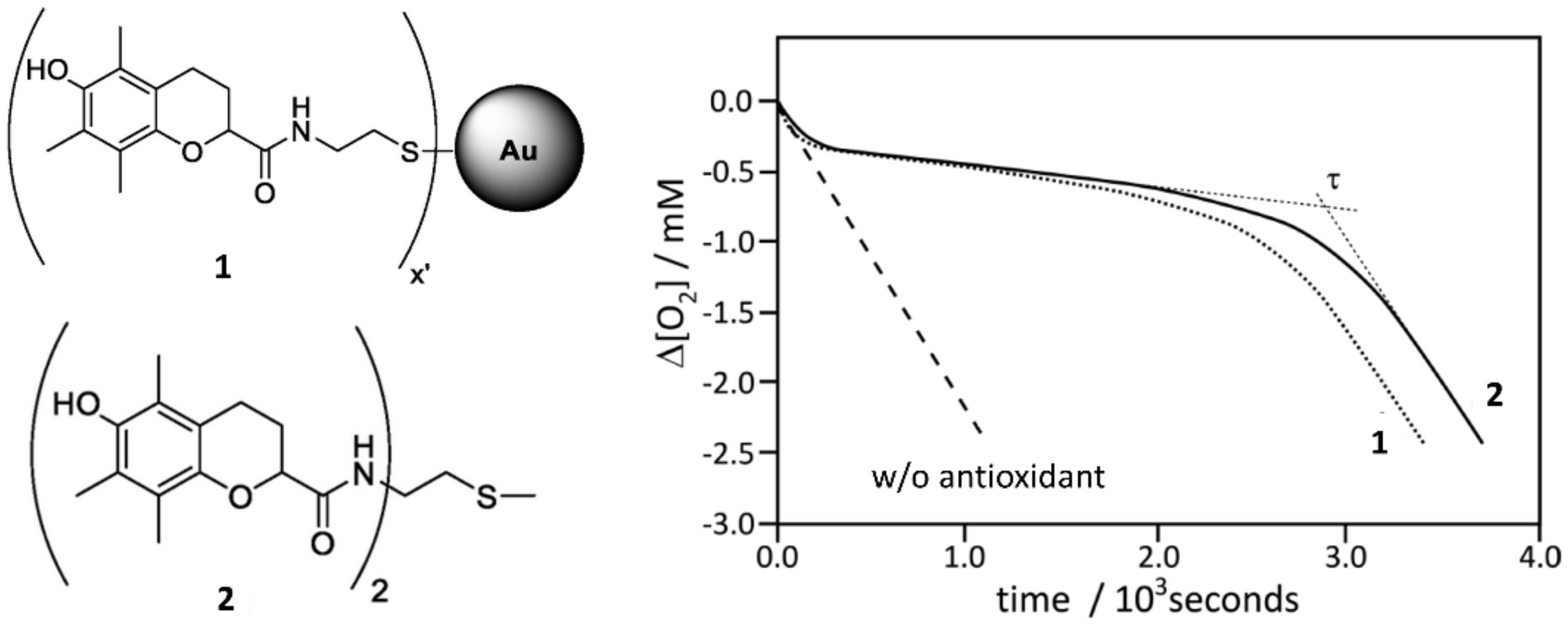
Figure 8.
O2 consumption traces observed during the autoxidation of styrene 4.3 M in chlorobenzene initiated by 50 mM of AIBN in a sample volume 4 mL at 30 °C, inhibited by nanoparticles bearing an analogue of α-tocopherol on the surface (1, 10.3 mg/mL) and by the antioxidant dimer (2, 5 μM). The O2 consumption was measured by a pressure transducer. Adapted from reference [98].

Figure 9.
Catalytic antioxidant activity of polydopamine (PDA). Panel (A): O2 consumption during the autoxidation of styrene (initiator AIBN, 30 °C) without any inhibitor (a), with 1,4-cyclohexadiene (CHD) as source of HOO• radicals (b), with PDA (c), in the presence of both PDA and CHD (d). Panel (B): mechanism explaining the radical trapping activity of PDA showing the reduction of quinone moieties to semiquinone able to trap new radicals by cycling between the quinone/semiquinone/hydroquinone forms. The O2 consumption was measured by a pressure transducer. Adapted from reference. [36].


Table 1.
Probability that radical-trapping antioxidants (RTA) intercept different types of radicals.
Table 1.
Probability that radical-trapping antioxidants (RTA) intercept different types of radicals.
| Radical | Trapped by RTA | Reason | |
|---|---|---|---|
| HO• | Hydroxyl | No | Reaction with organic substrates is too fast |
| RO• | Alkoxyl | No | Reaction with organic substrates is too fast |
| R• | Alkyl | No | Reaction with O2 is diffusion-controlled and [O2] ≈ 10−4–10−3 M |
| ROO• | Alkyl peroxyl | Yes | Reaction with organic substrate is slow |
| HOO• | Hydroperoxyl | Yes | Reaction with organic substrate is slow |
| O2•− | Superoxide | No (Yes) 1 | No reaction with organic substrates nor with most RTA |
1 Superoxide is trapped by SOD and SOD-like antioxidants.
Table 2.
Examples of oxidizable substrates with their kp and 2kt values (M−1 s−1). Temperature 30 °C.
Table 2.
Examples of oxidizable substrates with their kp and 2kt values (M−1 s−1). Temperature 30 °C.
| Substrate | kp | 2kt | Solvent | Reference |
|---|---|---|---|---|
| Styrene | 41 | 4.2 × 10⁷ | PhCl | [99] |
| Cumene | 0.32 | 4.6 × 10⁴ | PhCl | [100] |
| Squalene | 0.68 | 7.4 × 10⁶ | PhCl | [101] |
| 1,4-Cyclohexadiene | 1400 | 1.3 × 10⁹ a | PhCl | [102] |
| p-Cymene | 0.83 | 2.9 × 10⁶ | PhCl | [101] |
| Stripped sunflower oil | 66.9 | 3.5 × 10⁶ | PhCl | [101] |
| Methyl linoleate | 62 | 8.8 × 10⁶ | PhCl | [99] |
| 36 | 3.5 × 10⁵ | Triton X-100 micelles | [103] | |
| 41 | 0.6 × 10⁶ | DMPC b liposomes | [104] | |
| Tetrahydrofuran | 4.8 | 6.6 × 10⁷ | H2O | [105] |
(a) 2kt strongly depends on solvent. (b) 1,2-dimyristoyl-sn-glycero-3-phosphocholine.
Table 3.
Inhibition rate constants (kinh/M−1 s−1) of nanoantioxidants and of related small-molecule antioxidants (kinh’).
Table 3.
Inhibition rate constants (kinh/M−1 s−1) of nanoantioxidants and of related small-molecule antioxidants (kinh’).
| Nanoantioxidant | kinh | kinh’ | Solvent | Reference |
|---|---|---|---|---|
| Trolox-HNT | (1.1 ± 0.1) × 106 | (9.8 ± 0.5) × 105 | Cumene/PhCl | [52] |
| TroloxS-AuNPs | (6.9 ± 0.4) × 105 | (6.9 ± 0.3) × 105 | Styrene/PhCl | [98] |
| CoNPs-Trolox | (5.6 ± 1.5) × 106 | (6.4 ± 1.0) × 105 | Styrene/PhCN | [60] |
| Ascorbic Acid/HNT | (1.4 ± 0.3) × 106 | (1.7 ± 0.2) × 106 | THF/Water | [3] |
| Nitroxides NPs | (1.5 ± 0.4) × 105 | (5.1 ± 1.5) × 106 | THF/Water | [56] |
| Curcumin-HNT | (1.7 ± 0.1) × 104 | (1.8 ± 0.2) × 104 | Cumene/PhCl | [49] |
Publisher’s Note: MDPI stays neutral with regard to jurisdictional claims in published maps and institutional affiliations. |
© 2021 by the authors. Licensee MDPI, Basel, Switzerland. This article is an open access article distributed under the terms and conditions of the Creative Commons Attribution (CC BY) license (https://creativecommons.org/licenses/by/4.0/).
Share and Cite
MDPI and ACS Style
Baschieri, A.; Amorati, R. Methods to Determine Chain-Breaking Antioxidant Activity of Nanomaterials beyond DPPH•. A Review. Antioxidants 2021, 10, 1551. https://0-doi-org.brum.beds.ac.uk/10.3390/antiox10101551
AMA Style
Baschieri A, Amorati R. Methods to Determine Chain-Breaking Antioxidant Activity of Nanomaterials beyond DPPH•. A Review. Antioxidants. 2021; 10(10):1551. https://0-doi-org.brum.beds.ac.uk/10.3390/antiox10101551
Chicago/Turabian StyleBaschieri, Andrea, and Riccardo Amorati. 2021. "Methods to Determine Chain-Breaking Antioxidant Activity of Nanomaterials beyond DPPH•. A Review" Antioxidants 10, no. 10: 1551. https://0-doi-org.brum.beds.ac.uk/10.3390/antiox10101551
Note that from the first issue of 2016, this journal uses article numbers instead of page numbers. See further details here.