
Design and Comprehensive Characterization of Tetramethylpyrazine (TMP) for Targeted Lung Delivery as Inhalation Aerosols in Pulmonary Hypertension (PH): In Vitro Human Lung Cell Culture and In Vivo Efficacy

 ,
,
Abstract
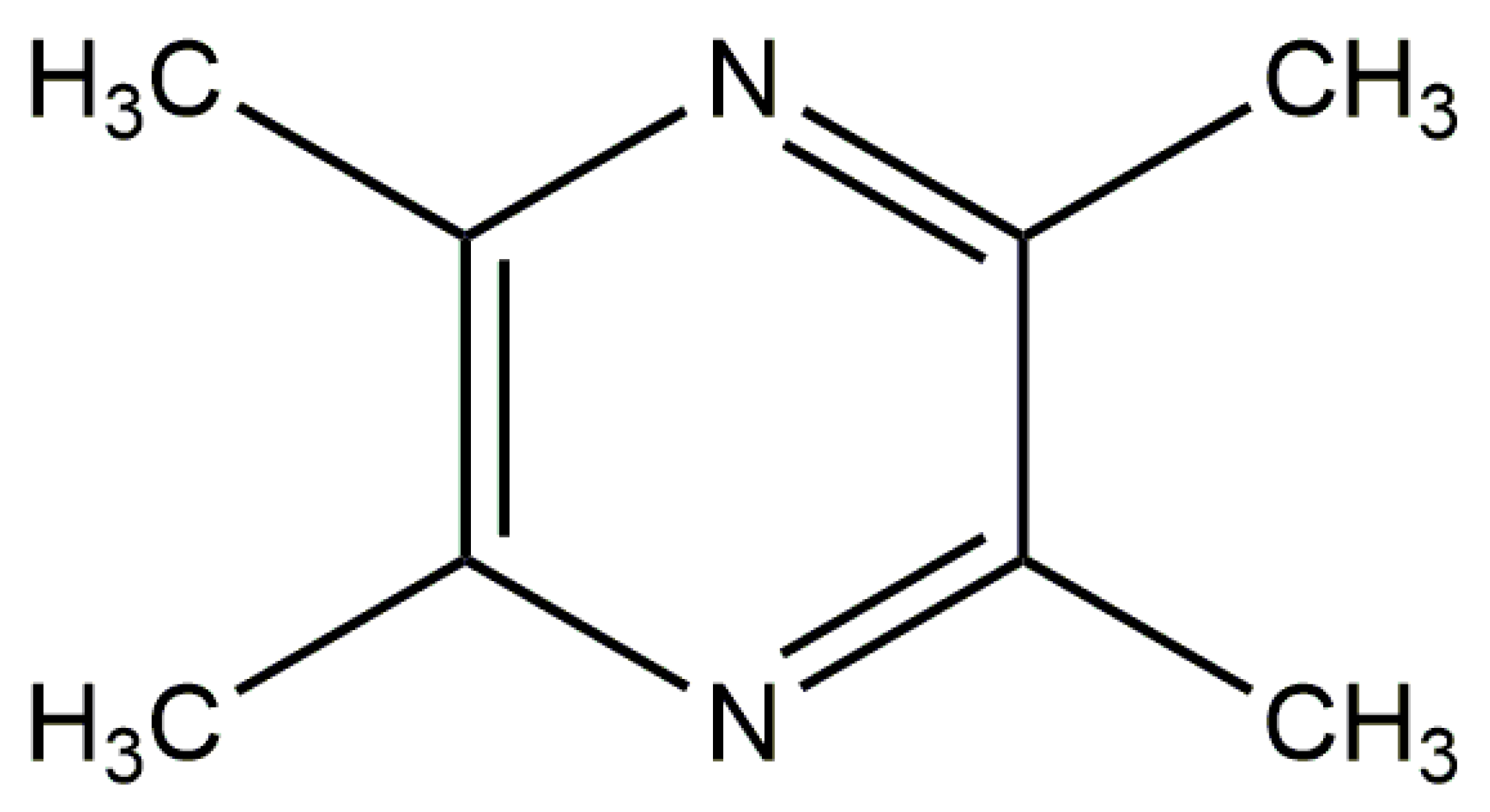
:1. Introduction
2. Experimental: Materials and Methods
2.1. Materials
2.2. Methods
2.2.1. Advanced Closed-Mode Spray Drying from Organic Solution
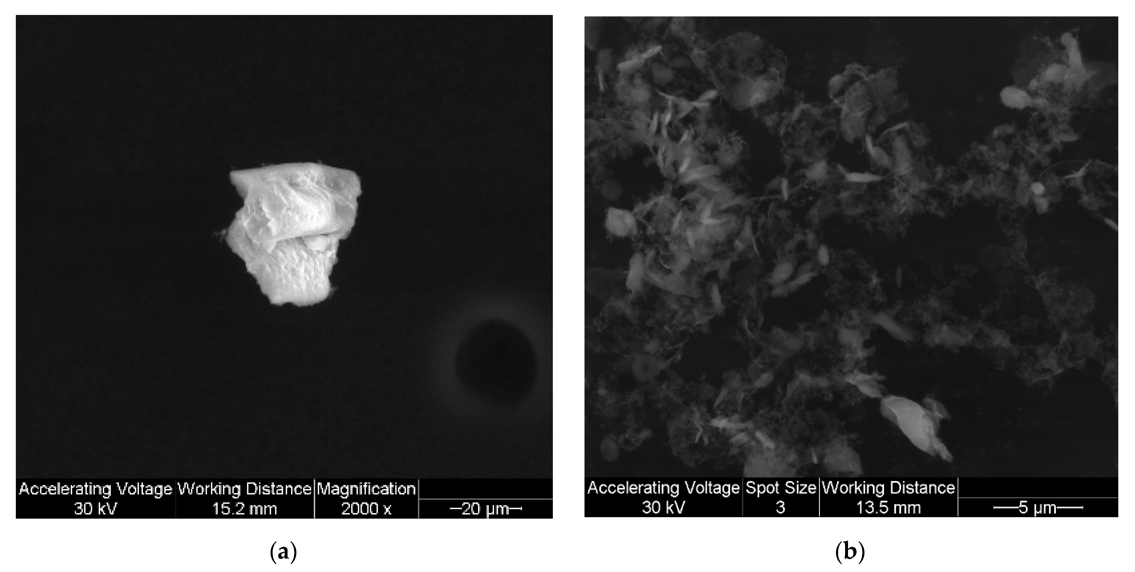
2.2.2. Scanning Electron Microscopy (SEM)
2.2.3. Laser Diffraction Particle Sizing and Size Distribution
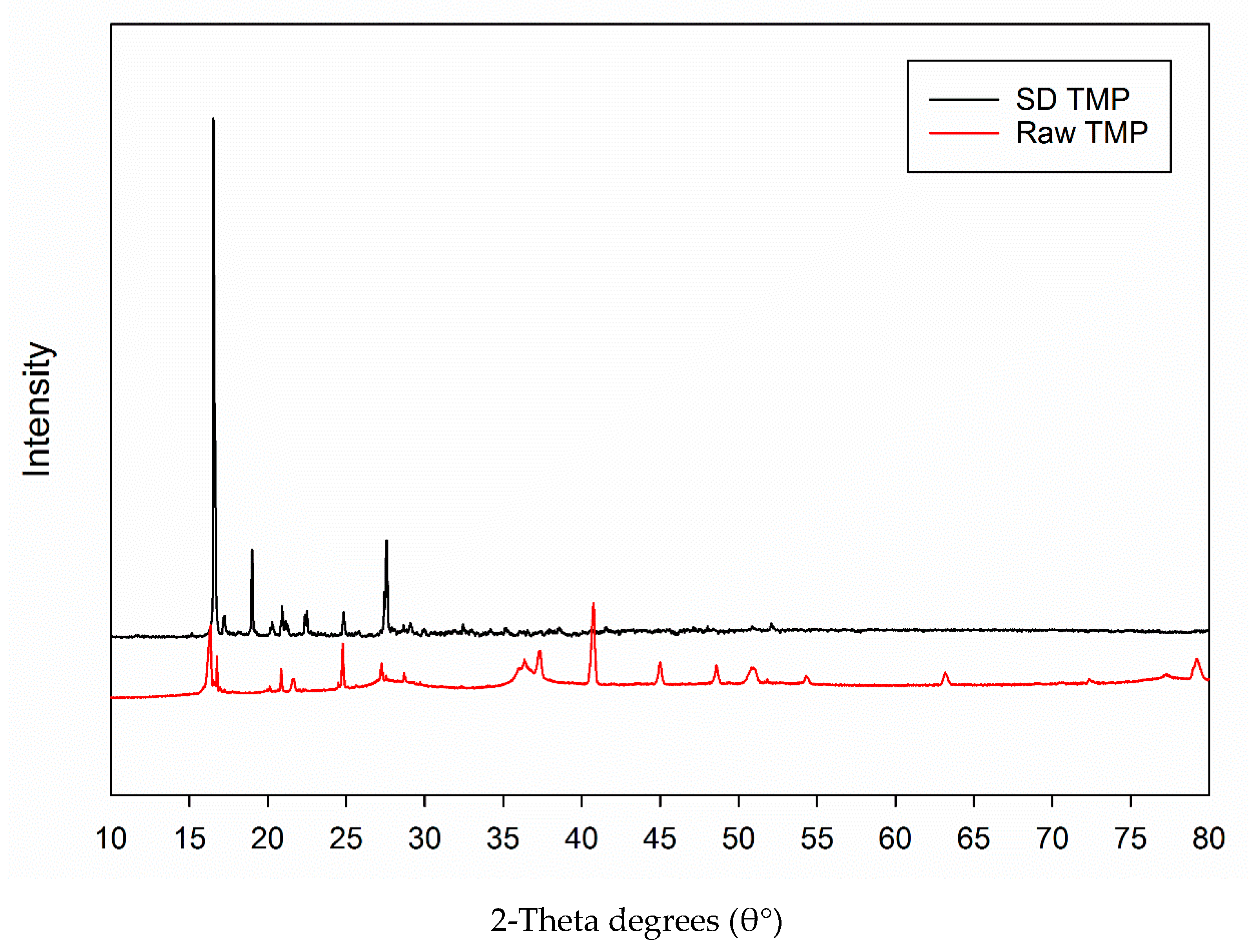
2.2.4. X-ray Powder Diffraction (XRPD)
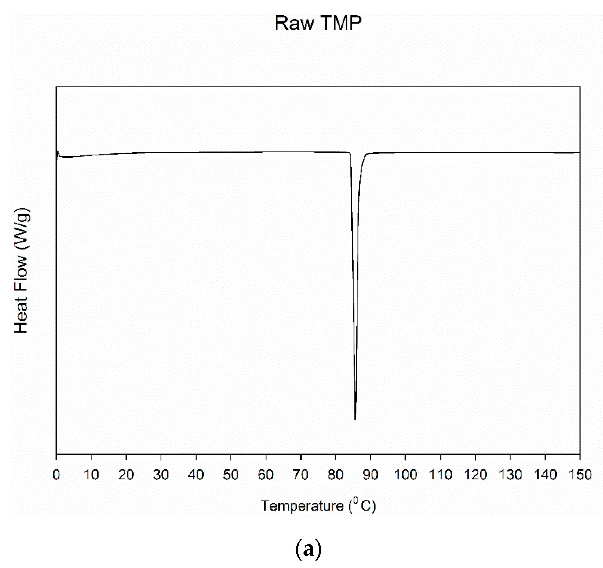
2.2.5. Differential Scanning Calorimetry (DSC)
2.2.6. Hot-Stage Microscopy (HSM) under Cross-Polarizers
2.2.7. Karl Fischer Titration (KFT)
2.2.8. Raman Spectroscopy
2.2.9. In Vitro Aerosol Dispersion Performance
2.2.10. In Vitro Human Cell Viability
2.2.11. In Vitro Transepithelial Electrical Resistance (TEER)
2.2.12. In Vivo Efficacy in PH-Induced Rats
2.2.13. Statistical Analysis
3. Results
3.1. Scanning Electron Microscopy (SEM)
3.2. Laser Diffraction Particle Sizing
3.3. X-ray Powder Diffraction (XRPD)
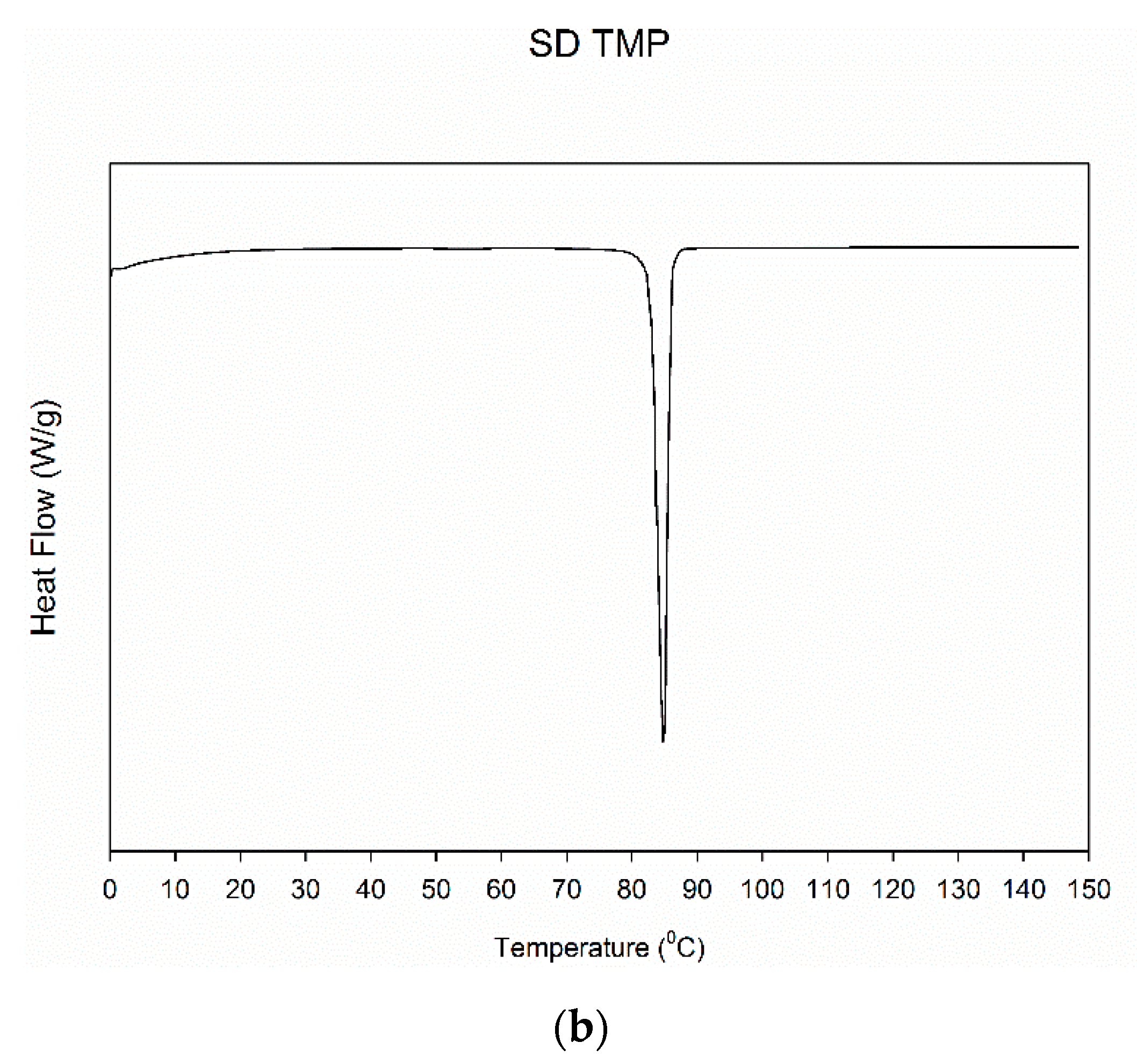
3.4. Differential Scanning Calorimetry (DSC)
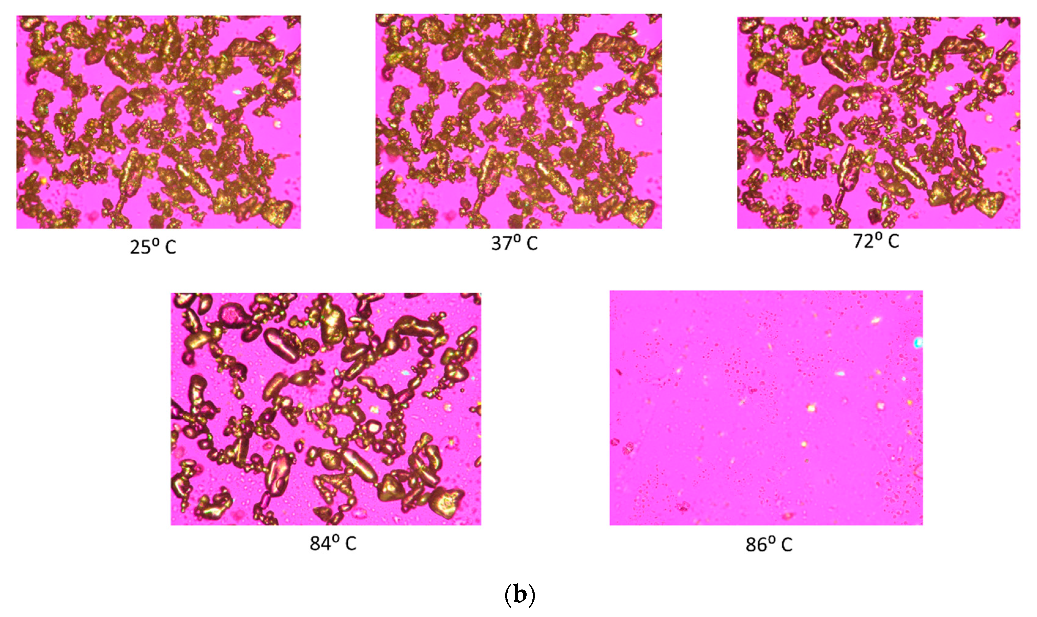
3.5. Hot-Stage Microscopy (HSM) under Cross-Polarizer Lens
3.6. Karl Fischer Titration (KFT)
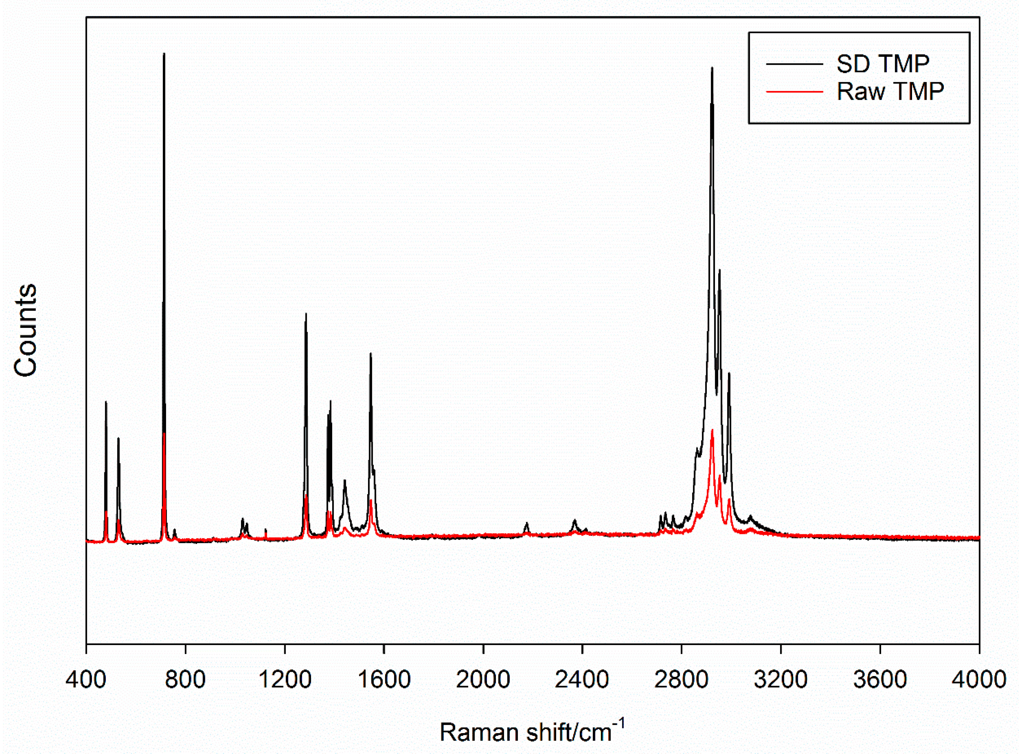
3.7. Raman Spectroscopy
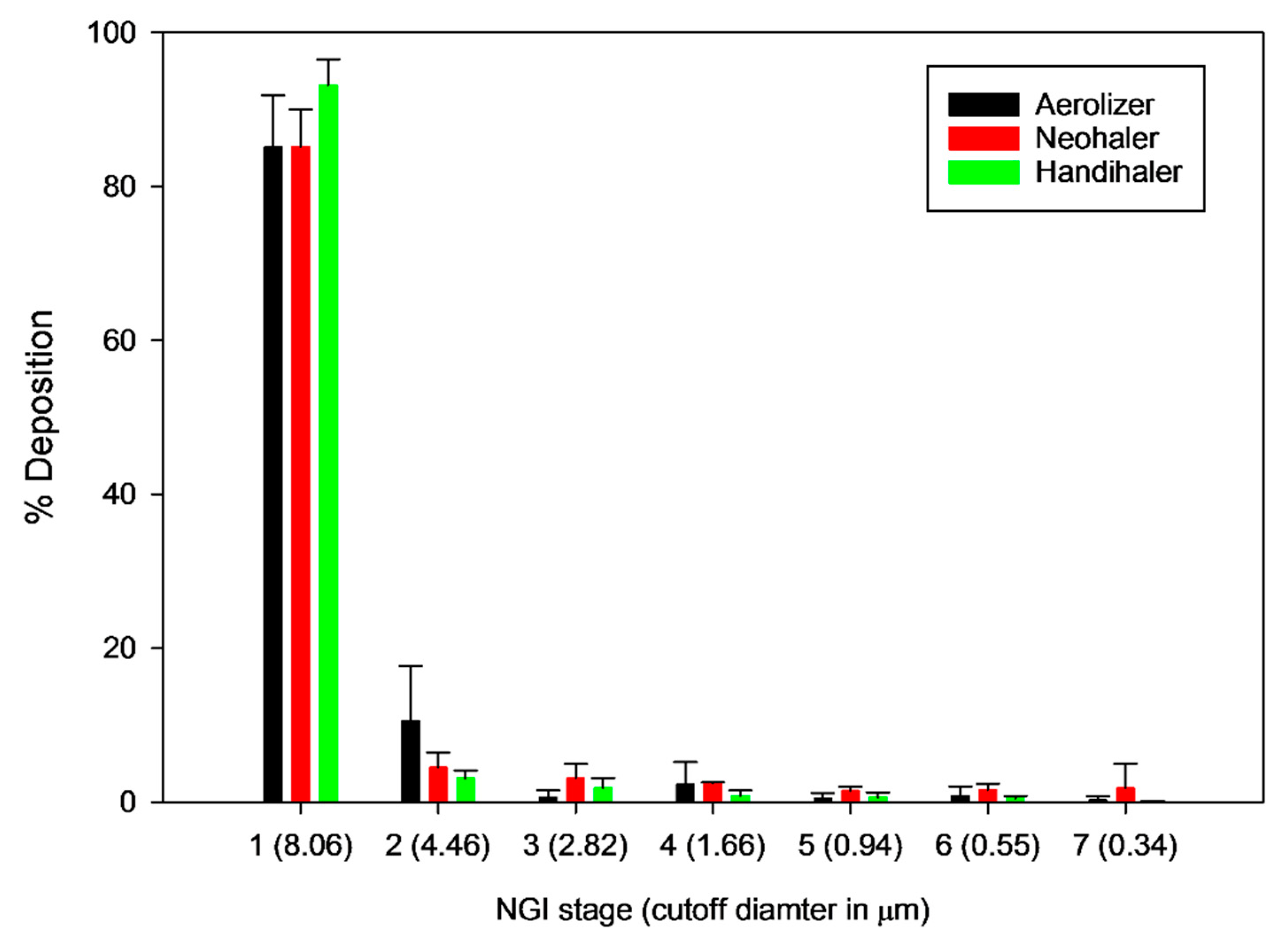
3.8. In Vitro Aerosol Dispersion Performance
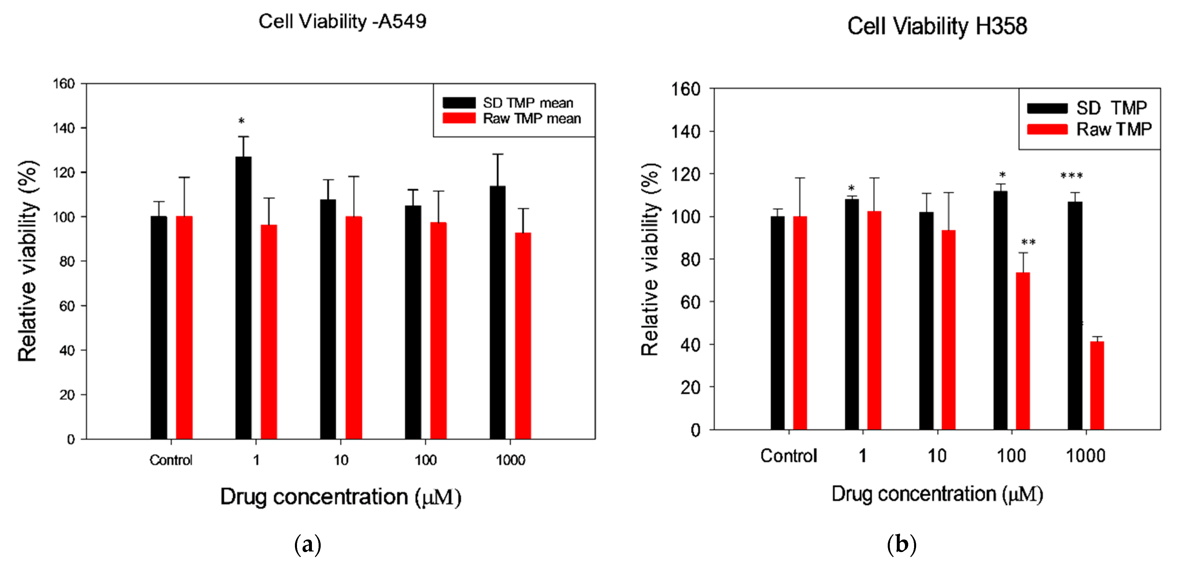
3.9. In Vitro Human Cell Viability
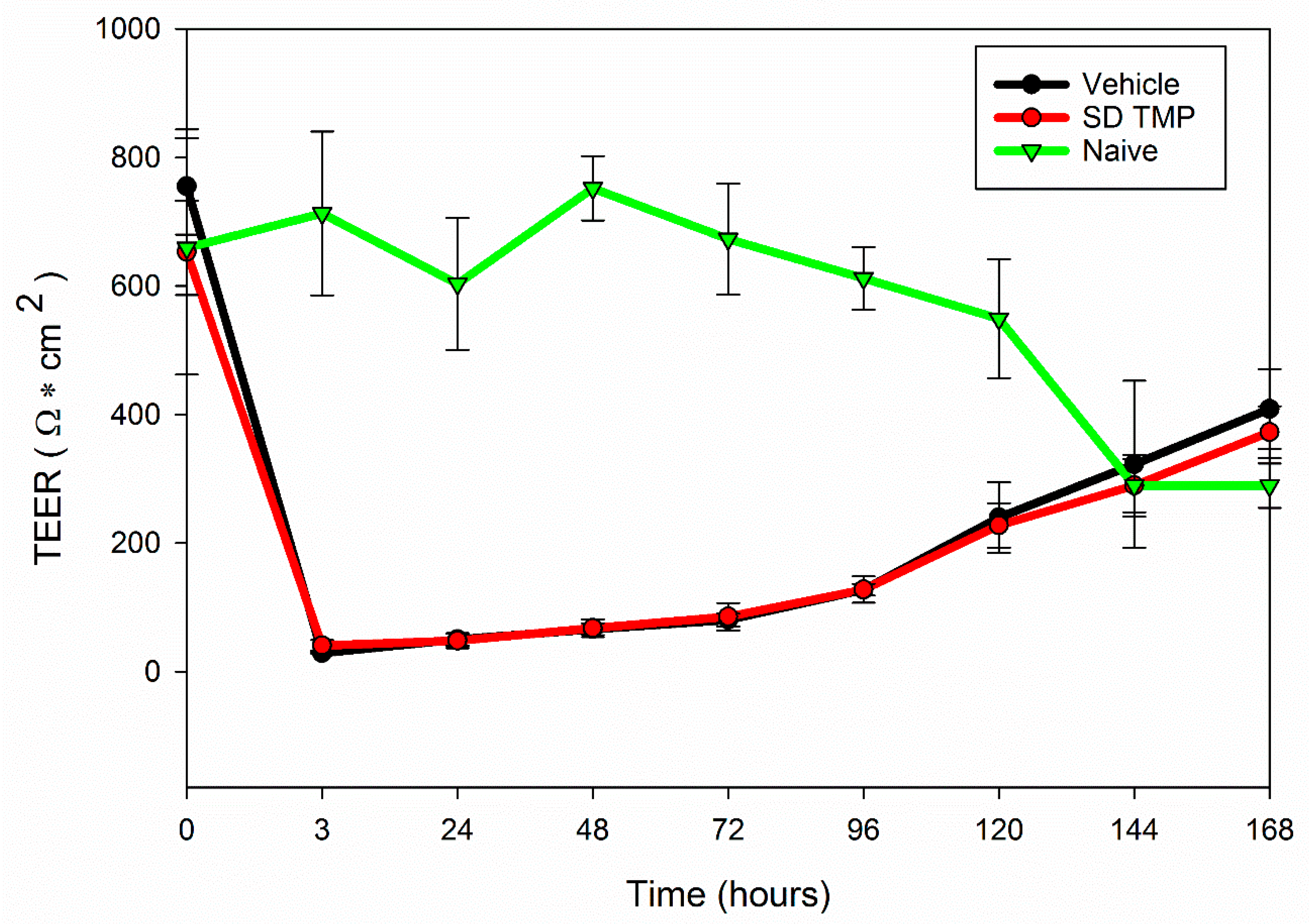
3.10. In Vitro Transepithelial Electrical Resistance (TEER)
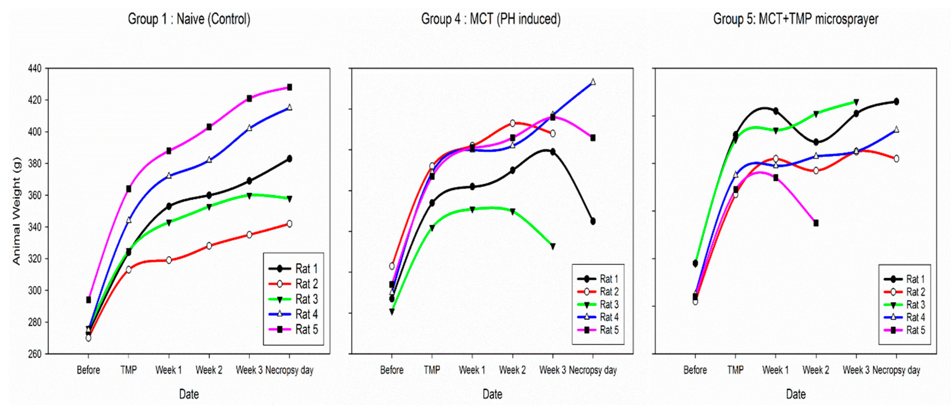
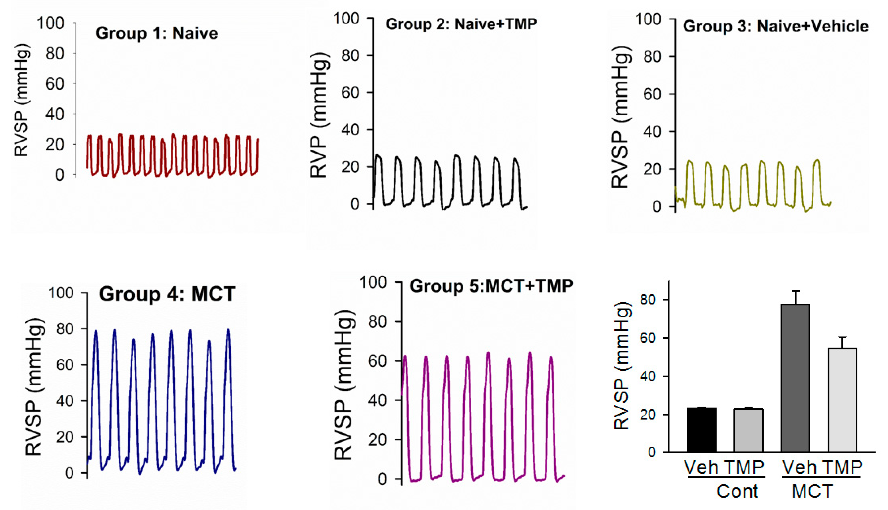
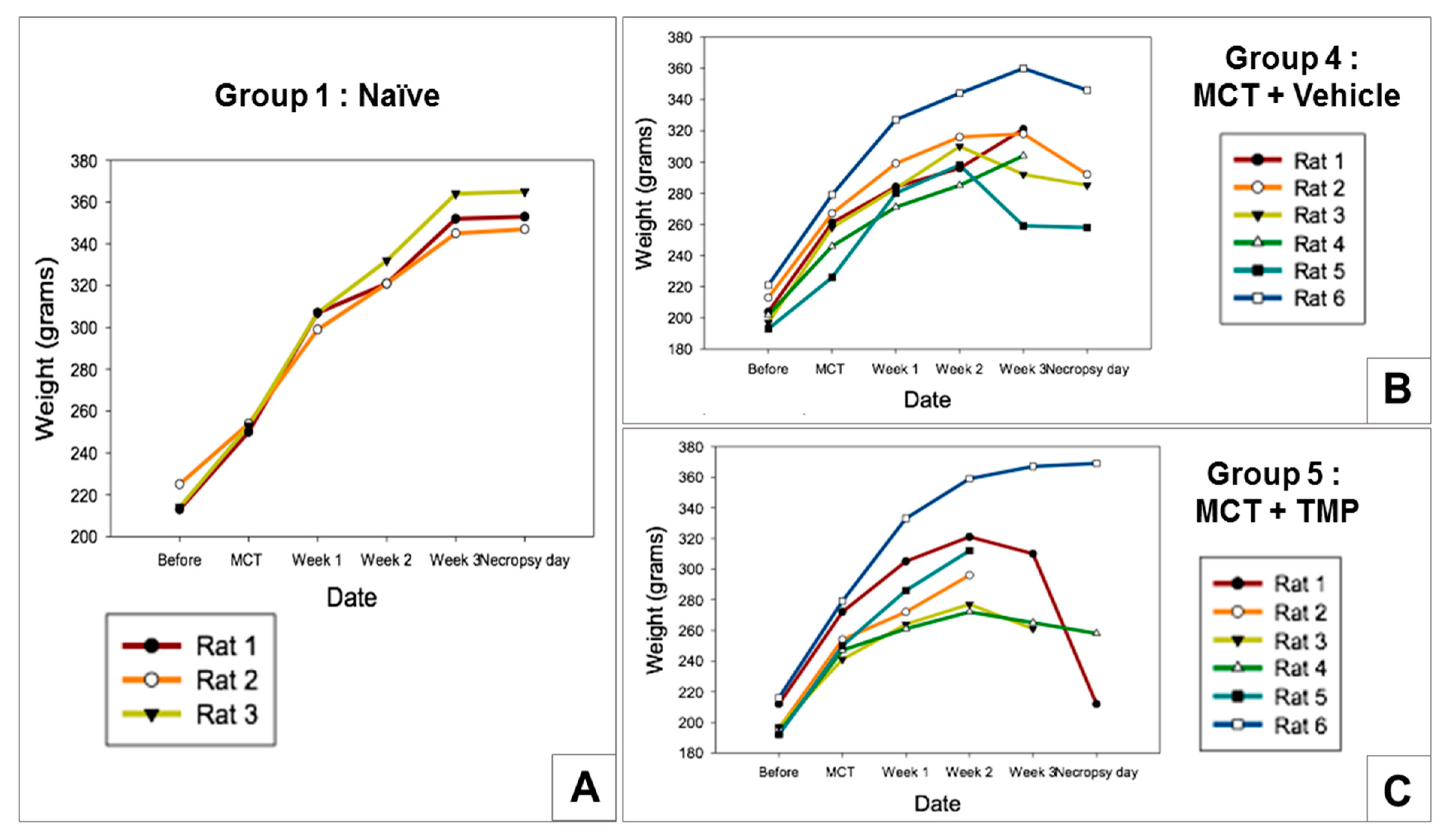
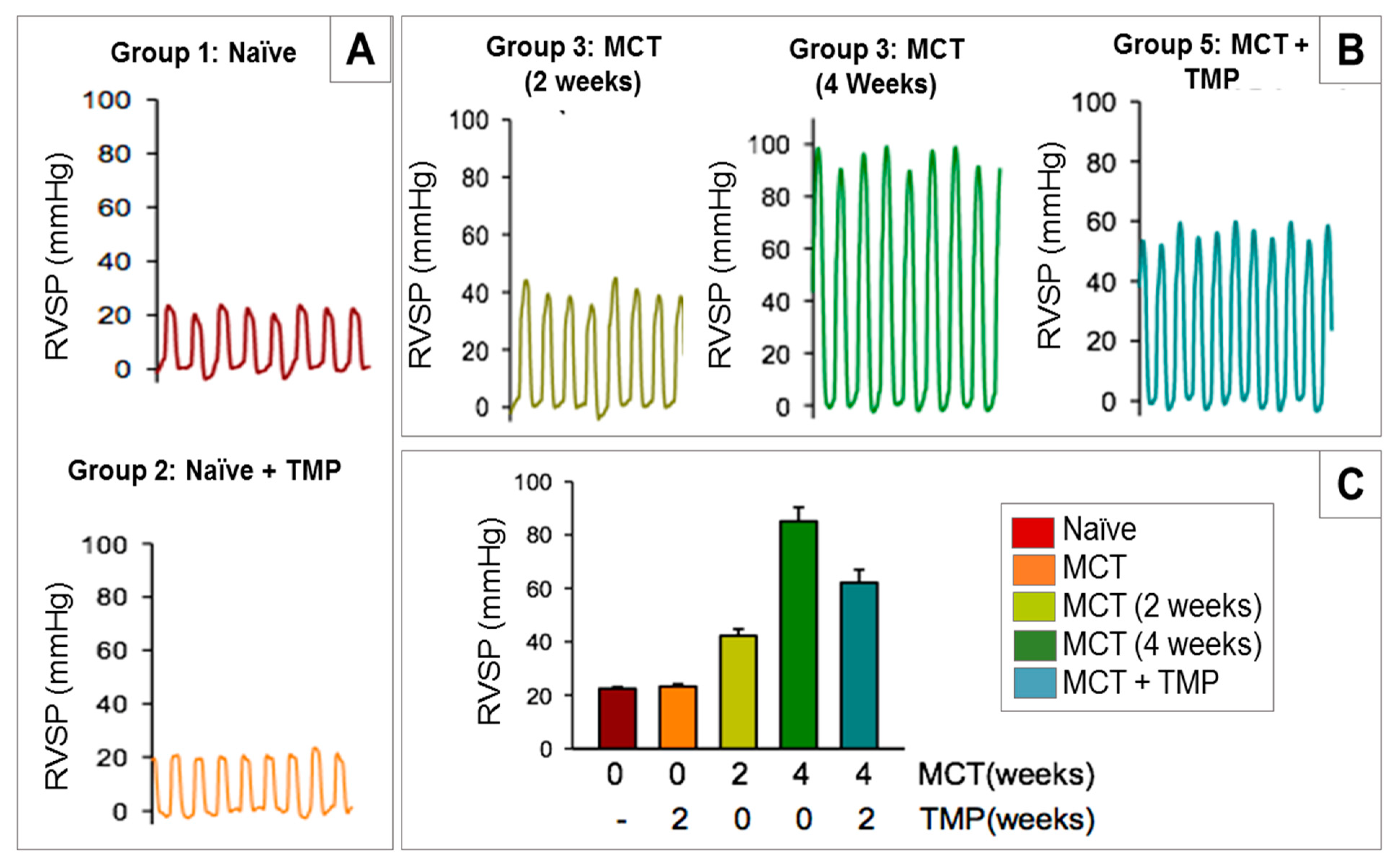
3.11. In Vivo Efficacy Study
4. Discussion
5. Conclusions
Author Contributions
Funding
Institutional Review Board Statement
Informed Consent Statement
Data Availability Statement
Acknowledgments
Conflicts of Interest
References
- Acosta, M.F.; Hayes, D.J.; Fineman, J.R.; Yuan, J.X.-J.; Black, S.M.; Mansour, H.M. Book Chapter 19: Therapeutics in Pulmonary Hypertension. In Inhalation Aerosols: Physical and Biological Basis for Therapy, 3rd ed.; Hickey, A.J., Mansour, H.M., Eds.; CRC Press/Taylor & Francis: London, UK, 2019; pp. 313–322. [Google Scholar]
- Stenmark, K.; Mecham, R. Cellular and molecular mechanisms of pulmonary vascular remodeling. Annu. Rev. Physiol. 1997, 59, 89–144. [Google Scholar] [CrossRef]
- Farber, H.W.; Loscalzo, J. Pulmonary arterial hypertension. N. Engl. J. Med. 2004, 351, 1655–1665. [Google Scholar] [CrossRef]
- Hill, N.S.; Preston, I.R.; Roberts, K.E. Inhaled Therapies for Pulmonary Hypertension. Respir. Care 2015, 60, 794–805. [Google Scholar] [CrossRef] [Green Version]
- Kuhr, F.K.; Smith, K.A.; Song, M.Y.; Levitan, I.; Yuan, J.X. New mechanisms of pulmonary arterial hypertension: Role of Ca2+ signaling. Am. J. Physiol. Heart Circ. Physiol. 2012, 302, H1546–H1562. [Google Scholar] [CrossRef] [Green Version]
- Gessler, T.; Seeger, W.; Schmehl, T. Inhaled prostanoids in the therapy of pulmonary hypertension. J. Aerosol Med. Pulm. Drug Deliv. 2008, 21, 1–12. [Google Scholar] [CrossRef] [PubMed]
- Mansour, H.M. Inhaled Medical Aerosols by Nebulizer Delivery in Pulmonary Hypertension. Pulm. Circ. 2018, 8, 1–2. [Google Scholar] [CrossRef] [Green Version]
- Kwan, C.Y. Plant-derived drugs acting on cellular ca2+ mobilization in vascular smooth muscle: Tetramethylpyrazine and tetrandrine. Stem Cells 1994, 12, 64–67. [Google Scholar] [CrossRef]
- Chang, F.C.; Chen, K.J.; Lin, J.G.; Hong, C.Y.; Huang, Y.T. Effects of tetramethylpyrazine on portal hypertensive rats. J. Pharm. Pharmacol. 1998, 50, 881–884. [Google Scholar] [CrossRef] [PubMed]
- Liu, S.; Cai, Y.; Evans, T.W.; McCormack, D.G.; Barer, G.R.; Barnes, P.J. Ligustrazine is a vasodilator of human pulmonary and bronchial arteries. Eur. J. Pharmacol. 1990, 191, 345–350. [Google Scholar] [CrossRef]
- Cai, Y.; Bee, D.; Barer, G. Pulmonary vasodilator action of ligustrazine, active principle of a traditional Chinese remedy, in rats and ferrets. Proc. Chin. Acad. Med. Sci. Peking Union Med. Coll. 1988, 4, 147–152. [Google Scholar]
- Oddoy, A.; Bee, D.; Emery, C.; Barer, G. Effects of ligustrazine on the pressure/flow relationship in isolated perfused rat lungs. Eur. Respir. J. 1991, 4, 1223–1227. [Google Scholar]
- Li, S.Y.; Jia, Y.H.; Sun, W.G.; Tang, Y.; An, G.S.; Ni, J.H.; Jia, H.T. Stabilization of mitochondrial function by tetramethylpyrazine protects against kainate-induced oxidative lesions in the rat hippocampus. Free Radic. Biol. Med. 2010, 48, 597–608. [Google Scholar] [CrossRef]
- Tan, F.; Fu, W.; Cheng, N.; Meng, D.I.; Gu, Y. Ligustrazine reduces blood-brain barrier permeability in a rat model of focal cerebral ischemia and reperfusion. Exp. Ther. Med. 2015, 9, 1757–1762. [Google Scholar] [CrossRef] [Green Version]
- Wang, Y.; Zhu, H.; Tong, J.; Li, Z. Ligustrazine improves blood circulation by suppressing Platelet activation in a rat model of allergic asthma. Environ. Toxicol. Pharmacol. 2016, 45, 334–339. [Google Scholar] [CrossRef]
- Wei, Y.; Liu, J.; Zhang, H.; Du, X.; Luo, Q.; Sun, J. Ligustrazine Attenuates Inflammation and the Associated Chemokines and Receptors in Ovalbumin-Induced Mouse Asthma Model. Environ. Toxicol. Pharmacol. 2016, 46, 55–61. [Google Scholar] [CrossRef]
- Zhao, S.; Zhang, Y.; Chen, Q.; Dong, S.; Zhang, G.; Li, J. A Modified “Double-Hit” Induced Acute Lung Injury Model in Rats and Protective Effects of Tetramethylpyrazine on the Injury via Rho/ROCK Pathway. Int. J. Clin. Exp. Pathol. 2015, 8, 4581–4588. [Google Scholar] [PubMed]
- Luo, Y.; Xiang, Q.; Wang, Q. Effect of Ligustrazine on Expression of RhoA mRNA, ROCK-II Protein in the Lung and Airway Inflammation of Allergic Asthma Model Mice. Chin. J. Pediatr. 2008, 46, 868–869. [Google Scholar]
- Fan, X.; Wang, E.; He, J.; Zhang, L.; Zeng, X.; Gui, Y.; Sun, Q.; Song, Y.; Yuan, H. Ligustrazine Protects Homocysteine-Induced Apoptosis in Human Umbilical Vein Endothelial Cells by Modulating Mitochondrial Dysfunction. J. Cardiovasc. Transl. Res. 2019, 12, 591–599. [Google Scholar] [CrossRef] [PubMed]
- Shi, J.; Wang, Y.; Luo, G. Ligustrazine Phosphate Ethosomes for Treatment of Alzheimer’s Disease, In Vitro and in Animal Model Studies. AAPS Pharmscitech 2012, 13, 485–492. [Google Scholar] [CrossRef] [PubMed] [Green Version]
- Yan, S.; Yue, Y.; Zeng, L.; Jiang, C.; Li, W.; Li, H.; Qian, Y. Ligustrazine nanoparticles nano spray’s activation on Nrf2/ARE pathway in oxidative stress injury in rats with postoperative abdominal adhesion. Ann. Transl. Med. 2019, 7, 16. [Google Scholar] [CrossRef]
- Liu, M.-W.; Su, M.-X.; Tang, D.-y.; Hao, L.; Xun, X.-H.; Huang, Y.-Q. Ligustrazin increases lung cell autophagy and ameliorates paraquat-induced pulmonary fibrosis by inhibiting PI3K/Akt/mTOR and hedgehog signalling via increasing miR-193a expression. BMC Pulm. Med. 2019, 19, 35. [Google Scholar] [CrossRef] [PubMed]
- Gao, C.; Liu, Y.; Ma, L.; Zhang, X.; Wang, S. Effects of Ligustrazine on pulmonary damage in rats following scald injury. Burns 2012, 38, 743–750. [Google Scholar] [CrossRef]
- Meenach, S.A.; Anderson, K.W.; Hilt, J.Z.; McGarry, R.C.; Mansour, H.M. High-Performing Dry Powder Inhalers of Paclitaxel DPPC/DPPG Lung Surfactant-Mimic Multifunctional Particles in Lung Cancer: Physicochemical Characterization, In Vitro Aerosol Dispersion, and Cellular Studies. AAPS Pharmscitech 2014, 15, 1574–1587. [Google Scholar] [CrossRef] [Green Version]
- Olschewski, H.; Simonneau, G.; Galie, N.; Higenbottam, T.; Naeije, R.; Rubin, L.J.; Nikkho, S.; Speich, R.; Hoeper, M.M.; Behr, J.; et al. Inhaled iloprost for severe pulmonary hypertension. N. Engl. J. Med. 2002, 347, 322–329. [Google Scholar] [CrossRef] [PubMed]
- Tissot, C.; Beghetti, M. Review of inhaled iloprost for the control of pulmonary artery hypertension in children. Vasc. Health Risk Manag. 2009, 5, 325–331. [Google Scholar] [PubMed] [Green Version]
- Mansour, H.M.; Rhee, Y.-S.; Wu, X. Nanomedicine in pulmonary delivery. Int. J. Nanomed. 2009, 4, 299–319. [Google Scholar] [CrossRef] [Green Version]
- Muralidharan, P.; Mallory, E.; Malapit, M.; Hayes, D.; Mansour, H.M. Inhalable PEGylated phospholipid nanocarriers and PEGylated therapeutics for respiratory delivery as aerosolized colloidal dispersions and dry powder inhalers. Pharmaceutics 2014, 6, 333–353. [Google Scholar] [CrossRef] [Green Version]
- Muralidharan, P.; Malapit, M.; Mallory, E.; Hayes, D.; Mansour, H.M. Inhalable nanoparticulate powders for respiratory delivery. Nanomed. Nanotechnol. Biol. Med. 2015, 11, 1189–1199. [Google Scholar] [CrossRef] [Green Version]
- Xu, Z.; Mansour, H.M.; Hickey, A.J. Particle interactions in dry powder inhaler unit processes: A review. J. Adhes. Sci. Technol. 2011, 25, 451–482. [Google Scholar] [CrossRef]
- Stocke, N.A.; Meenach, S.A.; Arnold, S.M.; Mansour, H.M.; Hilt, J.Z. Formulation and characterization of inhalable magnetic nanocomposite microparticles (MnMs) for targeted pulmonary delivery via spray drying. Int. J. Pharm. 2015, 479, 320–328. [Google Scholar] [CrossRef] [Green Version]
- Mansour, H.M.; Rhee, Y.-S.; Park, C.-W.; De Luca, P.P. Lipid nanoparticulate drug delivery and nanomedicine. In Lipids in Nanotechnology; American Oil Chemists Society Press: Chicago, IL, USA, 2011; pp. 221–268. [Google Scholar]
- Muralidharan, P.; Hayes, D.; Black, S.M.; Mansour, H.M. Microparticulate/nanoparticulate powders of a novel Nrf2 activator and an aerosol performance enhancer for pulmonary delivery targeting the lung Nrf2/Keap-1 pathway. Mol. Syst. Des. Eng. 2016, 1, 48–65. [Google Scholar] [CrossRef] [PubMed] [Green Version]
- Li, X.; Mansour, H.M. Physicochemical characterization and water vapor sorption of organic solution advanced spray-dried inhalable trehalose microparticles and nanoparticles for targeted dry powder pulmonary inhalation delivery. AAPS Pharmscitech 2011, 12, 1420–1430. [Google Scholar] [CrossRef] [Green Version]
- Meenach, S.A.; Vogt, F.G.; Anderson, K.W.; Hilt, J.Z.; McGarry, R.C.; Mansour, H.M. Design, physicochemical characterization, and optimization of organic solution advanced spray-dried inhalable dipalmitoylphosphatidylcholine (DPPC) and dipalmitoylphosphatidylethanolamine poly(ethylene glycol) (DPPE-PEG) microparticles and nanoparticles for targeted respiratory nanomedicine delivery as dry powder inhalation aerosols. Int. J. Nanomed. 2013, 8, 275–293. [Google Scholar] [CrossRef] [Green Version]
- Wu, X.; Hayes, D., Jr.; Zwischenberger, J.B.; Kuhn, R.J.; Mansour, H.M. Design and physicochemical characterization of advanced spray-dried tacrolimus multifunctional particles for inhalation. Drug Des. Dev. Ther. 2013, 7, 59–72. [Google Scholar] [CrossRef] [Green Version]
- Wu, X.; Zhang, W.; Hayes, D., Jr.; Mansour, H.M. Physicochemical characterization and aerosol dispersion performance of organic solution advanced spray-dried cyclosporine A multifunctional particles for dry powder inhalation aerosol delivery. Int. J. Nanomed. 2013, 8, 1269–1283. [Google Scholar] [CrossRef] [Green Version]
- Li, X.; Vogt, F.G.; Hayes, D.; Mansour, H.M. Physicochemical characterization and aerosol dispersion performance of organic solution advanced spray-dried microparticulate/nanoparticulate antibiotic dry powders of tobramycin and azithromycin for pulmonary inhalation aerosol delivery. Eur. J. Pharm. Sci. 2014, 52, 191–205. [Google Scholar] [CrossRef]
- Meenach, S.A.; Anderson, K.W.; Hilt, J.Z.; McGarry, R.C.; Mansour, H.M. Characterization and aerosol dispersion performance of advanced spray-dried chemotherapeutic PEGylated phospholipid particles for dry powder inhalation delivery in lung cancer. Eur. J. Pharm. Sci. 2013, 49, 699–711. [Google Scholar] [CrossRef] [PubMed] [Green Version]
- Aerosols, Nasal Sprays, Metered-Dose Inhalers, and Dry Powder Inhalers Monograph. In USP 29-NF 24 The United States Pharmacopoeia and The National Formulary: The Official Compendia of Standards. 29/24; The United States Pharmacopeial Convention, Inc.: Rockville, MD, USA, 2006; pp. 2617–2636.
- Acosta, M.F.; Muralidharan, P.; Meenach, S.A.; Hayes, D.; M-Black, S.; Mansour, H.M. In Vitro Pulmonary Cell Culture in Pharmaceutical Inhalation Aerosol Delivery: 2-D, 3-D, and In Situ Bioimpactor Models. Curr. Pharm. Des. 2016, 22, 2522–2531. [Google Scholar] [CrossRef] [PubMed]
- Meenach, S.A.; Tsoras, A.N.; McGarry, R.C.; Mansour, H.M.; Hilt, J.Z.; Anderson, K.W. Development of three-dimensional lung multicellular spheroids in air- and liquid-interface culture for the evaluation of anticancer therapeutics. Int. J. Oncol. 2016, 48, 1701–1709. [Google Scholar] [CrossRef] [PubMed]
- Zhang, X.; Wei, J.; Ma, P.; Mu, H.; Wang, A.; Zhang, L.; Wu, Z.; Sun, K. Preparation and evaluation of a novel biodegradable long-acting intravitreal implant containing ligustrazine for the treatment of proliferative vitreoretinopathy. J. Pharm. Pharmacol. 2015, 67, 160–169. [Google Scholar] [CrossRef] [PubMed]
- Li, X.; Vogt, F.; Hayes, D.J.; Mansour, H. Design, Characterization, and Aerosol Dispersion Performance Modeling of Advanced Spray-Dried Microparticulate/Nanoparticulate Mannitol Powders for Targeted Pulmonary Delivery as Dry Powder Inhalers. J. Aerosol Med. Pulm. Drug Deliv. 2014, 27, 81–93. [Google Scholar] [CrossRef]
- Lee, H.J.; Kang, J.H.; Lee, H.G.; Kim, D.W.; Rhee, Y.S.; Kim, J.Y.; Park, E.S.; Park, C.W. Preparation and physicochemical characterization of spray-dried and jet-milled microparticles containing bosentan hydrate for dry powder inhalation aerosols. Drug Des. Dev. Ther. 2016, 10, 4017–4030. [Google Scholar] [CrossRef] [PubMed] [Green Version]
- Martinelli, F.; Balducci, A.G.; Kumar, A.; Sonvico, F.; Forbes, B.; Bettini, R.; Buttini, F. Engineered sodium hyaluronate respirable dry powders for pulmonary drug delivery. Int. J. Pharm. 2017, 517, 286–295. [Google Scholar] [CrossRef] [Green Version]
- Hickey, A.J.; Mansour, H.M. Formulation Challenges of Powders for the Delivery of Small Molecular Weight Molecules as Aerosols. Drugs Pharm. Sci. 2003, 126, 835–848. [Google Scholar]
- Park, C.W.; Li, X.; Vogt, F.G.; Hayes, D., Jr.; Zwischenberger, J.B.; Park, E.S.; Mansour, H.M. Advanced spray-dried design, physicochemical characterization, and aerosol dispersion performance of vancomycin and clarithromycin multifunctional controlled release particles for targeted respiratory delivery as dry powder inhalation aerosols. Int. J. Pharm. 2013, 455, 374–392. [Google Scholar] [CrossRef]
- Longest, P.W.; Son, Y.J.; Holbrook, L.; Hindle, M. Aerodynamic factors responsible for the deaggregation of carrier-free drug powders to form micrometer and submicrometer aerosols. Pharm. Res. 2013, 30, 1608–1627. [Google Scholar] [CrossRef] [Green Version]
- Mansour, H.M.; Xu, Z.; Hickey, A.J. Dry powder aerosols generated by standardized entrainment tubes from alternative sugar blends: 3. Trehalose dihydrate and D-mannitol carriers. J. Pharm. Sci. 2010, 99, 3430–3441. [Google Scholar] [CrossRef] [PubMed]
- Xu, Z.; Mansour, H.M.; Mulder, T.; McLean, R.; Langridge, J.; Hickey, A.J. Comparative Dispersion Study of Dry Powder Aerosols of Albuterol Sulfate/Lactose Monohydrate and Cromolyn Sodium/Lactose Monohydrate Delivered by Standardized Entrainment Tubes. In Respiratory Drug Delivery XI; Dalby, R.N., Bryon, P.R., Suman, J.D., Peart, J., Farr, S.J., Eds.; Davis Healthcare International Publishing, LLC: Scotsdale, AZ, USA, 2008; Volume 3, pp. 897–900. [Google Scholar]
- Xu, Z.; Mansour, H.M.; Mulder, T.; McLean, R.; Langridge, J.; Hickey, A.J. Dry powder aerosols generated by standardized entrainment tubes from drug blends with lactose monohydrate: 2. Ipratropium bromide monohydrate and fluticasone propionate. J. Pharm. Sci. 2010, 99, 3415–3429. [Google Scholar] [CrossRef]
- Xu, Z.; Mansour, H.M.; Mulder, T.; McLean, R.; Langridge, J.; Hickey, A.J. Dry powder aerosols generated by standardized entrainment tubes from drug blends with lactose monohydrate: 1. albuterol sulfate and disodium cromoglycate. J. Pharm. Sci. 2010, 99, 3398–3414. [Google Scholar] [CrossRef]
- Xu, Z.; Mansour, H.M.; Mulder, T.; McLean, R.; Langridge, J.; Hickey, A.J. Heterogeneous particle deaggregation and its implication for therapeutic aerosol performance. J. Pharm. Sci. 2010, 99, 3442–3461. [Google Scholar] [CrossRef] [PubMed] [Green Version]
- Muralidharan, P.; Hayes, D., Jr.; Mansour, H.M. Dry powder inhalers in COPD, lung inflammation and pulmonary infections. Expert Opin. Drug Deliv. 2015, 12, 947–962. [Google Scholar] [CrossRef] [PubMed] [Green Version]


| Feed Concentration (%w/v) | Pump Rate (% and mL/min) | Inlet T (°C) | Outlet T (°C) |
|---|---|---|---|
| 1 | 90/27 | 149–152 | 18–25 |
| Physical Properties | Raw TMP | SD TMP |
|---|---|---|
| Particle Sizes | N/A | Dv10 = 4.186 ± 0.701 μm |
| Dv50 = 6.156 ± 1.147 μm | ||
| Dv90 = 14.552 ± 4.928 μm | ||
| (Dv) | Span = 1.684 ± 0.645 μm | |
| Phase Transitions | Tm = 85.93 ± 0.71 °C | Tm = 85.11 ± 0.36 °C |
| (Tm and ΔH) | ΔH = 152.5 ± 5.27 J/g | ΔH = 149.2 ± 7.91 J/g |
| Residual Water Content | 0.633 ± 0.251% w/w | 0.368 ± 0.103% w/w |
| Inhaler Device | Emitted Dose (%) | Fine Particle Dose with Respect to Nominal Dose (mg) Fine Particle Fraction with Respect to Nominal Dose (FPFND) (%) | Fine Particle Dose with Respect to Emitted Dose (mg) Fine Particle Fraction with Respect to Emitted Dose (FPFED) (%) |
|---|---|---|---|
| Handihaler® | 100 ± 2.03 | 3.33 | 37.24 |
| 3.83 ± 0.42 | 8.9 ± 3.87 | ||
| Neohaler® | 88.41 ± 6.70 | 3.48 | 25.19 |
| 4.36 ± 0.65 | 14.87 ± 7.30 | ||
| Aerolizer® | 88.76 ± 7.27 | 1.15 | 8.16 |
| 1.41 ± 0.57 | 14.86 ± 8.0 |
| Control + Vehicle Group (mm Hg) | Control + TMP Group (mm Hg) | MCT Group (mm Hg) | MCT + TMP Group (mm Hg) |
|---|---|---|---|
| 23.6567 | 23.8947 | 94.1697 | 61.1996 |
| 23.7513 | 22.9558 | 60.2783 | 43.1133 |
| 20.6622 | 19.0997 | 78.0645 | 60.1284 |
| 24.0879 | 24.6036 | 78.5773 | N/A |
| 23.6379 | N/A | N/A | N/A |
| Control (mm Hg) | Control + TMP Group (mm Hg) | MCT (2 Weeks) Group (mm Hg) | MCT (4 Weeks) Group (mm Hg) | MCT + TMP Group (mm Hg) |
|---|---|---|---|---|
| 22.5330 | 20.8110 | 39.4080 | 97.6720 | 78.2010 |
| 21.9020 | 24.2410 | 52.0390 | 95.7920 | 70.5310 |
| 20.9950 | 23.8990 | 38.4880 | 90.0360 | 58.6830 |
| 19.3520 | 24.7030 | 41.0640 | 95.1610 | 62.4200 |
| 22.3220 | 26.0550 | 40.3810 | 79.8910 | 43.3890 |
| 23.6890 | 19.6940 | N/A | 78.3780 | 59.8200 |
Publisher’s Note: MDPI stays neutral with regard to jurisdictional claims in published maps and institutional affiliations. |
© 2021 by the authors. Licensee MDPI, Basel, Switzerland. This article is an open access article distributed under the terms and conditions of the Creative Commons Attribution (CC BY) license (http://creativecommons.org/licenses/by/4.0/).
Share and Cite
Muralidharan, P.; Acosta, M.F.; Gomez, A.I.; Grijalva, C.; Tang, H.; Yuan, J.X.-J.; Mansour, H.M. Design and Comprehensive Characterization of Tetramethylpyrazine (TMP) for Targeted Lung Delivery as Inhalation Aerosols in Pulmonary Hypertension (PH): In Vitro Human Lung Cell Culture and In Vivo Efficacy. Antioxidants 2021, 10, 427. https://0-doi-org.brum.beds.ac.uk/10.3390/antiox10030427
Muralidharan P, Acosta MF, Gomez AI, Grijalva C, Tang H, Yuan JX-J, Mansour HM. Design and Comprehensive Characterization of Tetramethylpyrazine (TMP) for Targeted Lung Delivery as Inhalation Aerosols in Pulmonary Hypertension (PH): In Vitro Human Lung Cell Culture and In Vivo Efficacy. Antioxidants. 2021; 10(3):427. https://0-doi-org.brum.beds.ac.uk/10.3390/antiox10030427
Chicago/Turabian StyleMuralidharan, Priya, Maria F. Acosta, Alexan I. Gomez, Carissa Grijalva, Haiyang Tang, Jason X.-J. Yuan, and Heidi M. Mansour. 2021. "Design and Comprehensive Characterization of Tetramethylpyrazine (TMP) for Targeted Lung Delivery as Inhalation Aerosols in Pulmonary Hypertension (PH): In Vitro Human Lung Cell Culture and In Vivo Efficacy" Antioxidants 10, no. 3: 427. https://0-doi-org.brum.beds.ac.uk/10.3390/antiox10030427